abnormalities of lie / presentation
TRANSCRIPT
Page 1 of 24
King Edward Memorial Hospital
Obstetrics & Gynaecology
Contents
Breech presentation ............................................................................. 2
Antenatal management ........................................................................................... 3
External cephalic version ........................................................................................ 4
Elective caesarean section ..................................................................................... 4
Undiagnosed breech presenting in labour .............................................................. 4
Diagnosed breech booked for caesarean presenting in labour ............................... 5
Criteria recommended for a planned vaginal breech term birth .............................. 5
Pre-term breech – Vaginal birth .............................................................................. 5
External cephalic version quick reference guide (QRG) .................... 6
Flow chart for external cephalic version .................................................................. 7
External cephalic version ..................................................................... 8
Background information .......................................................................................... 8
Key points ............................................................................................................... 8
Contra-indications to performing an ECV ................................................................ 8
Procedure ............................................................................................................... 9
Breech presentation: Planned vaginal birth QRG ............................ 11
Breech labour and birth flowchart ......................................................................... 13
Planned vaginal breech birth................................................................................. 14
Unstable lie at or near term ................................................................ 19
Antenatal management ......................................................................................... 19
Birth management options .................................................................................... 20
Birth management for a woman in labour with an unstable lie .............................. 20
Rare presentations ............................................................................. 21
References .......................................................................................... 22
CLINICAL PRACTICE GUIDELINE
Abnormalities of Lie / Presentation This document should be read in conjunction with the Disclaimer

Abnormalities of Lie / Presentation
Page 2 of 24 Obstetrics & Gynaecology
Breech presentation
Background
Breech presentation occurs in 3% to 4% of pregnancies at term.1 The randomised
multicentre Term Breech Trial (TBT) showed that a planned elective caesarean
section (ELUSCS) reduces the risk for adverse perinatal outcomes or serious
maternal morbidity when compared to a planned vaginal breech birth in the short
term.1, 7 Long term follow-up at 2 years has not found neonatal neurological
outcomes or maternal outcomes differing between women who had an ELUSCS
compared to vaginal breech birth.4, 5 A large study conducted in the Netherlands
following the TBT study found that the rapid increase in caesarean section rates
resulted in substantial improvements in perinatal outcomes leading to halving of
perinatal mortality rates, and ever greater reductions in the incidence of perinatal
birth trauma.2, 8 However, the view remains that if the application of strict criteria
before and during labour is met; planned vaginal birth of a singleton breech at term is
a reasonable management option.1, 9
External cephalic version (ECV) from 36 weeks has been shown to decrease the
incidence of breech presentation at term and consequently reduce the ELUSCS
rates.2, 10 It is seen as a safe procedure provided it is performed in a setting where
caesarean section can be performed if necessary. A meta-analysis looking at risk
for performing an ECV indicates that fetal death risk is 1 per 5000 procedures;
pooled complications risk was 6.1%, and risk for requiring caesarean was 0.35%.11
However, a large cohort study found that performing an ECV may carry a higher risk
for caesarean section of 0.5%.1, 12
A recent large multi-centre randomised study found that ECV initiated at 34-35
weeks gestation compared with 37 weeks or more increases the probability of
cephalic presentation at birth, however it does not reduce rate of caesarean
sections, and it may increase the risk rate for preterm birth.13
Key points
1. ELUSCS for a singleton breech at term has been shown to reduce perinatal or neonatal
mortality rates and serious neonatal morbidity rate in the first 6 weeks of life.2, 7
2. Long-term follow-up at 2 years showed neurological infant outcomes do not differ by
planned mode of birth even in the presence of serious short term neonatal morbidity.2
3. ELUSCS is not associated with substantially better or worst outcomes for women 2
years after birth when compared to planned vaginal singleton breech birth at term.4, 5
4. All women with a singleton breech presentation with no contra-indications to the
procedure should be offered an ECV. Success rates for ECV are approximately 40%
in nulliparous women and 60% in multipara women.3

Abnormalities of Lie / Presentation
Page 3 of 24
Obstetrics & Gynaecology
5. A woman attending a low-risk midwifery antenatal clinic, and who is found to have a
breech presentation at 35-36 weeks gestation shall be referred for obstetric medical
review prior to 37 weeks gestation.
6. If breech diagnosis is made after 37 weeks, obstetric review / counselling is required,
and an ultrasound should be performed to assess for fetal or maternal causes of the
malpresentation, and fetal growth / wellbeing.1
7. Careful case selection and labour management in a modern obstetric setting may
achieve a level of safety similar to ELUSCS.2 Planned vaginal singleton breech birth
is an option for women who have no maternal or fetal contra-indications to this
mode of birth. Women who meet the criteria for a planned vaginal breech birth who
develop complications which are contraindications to a planned term breech birth,
must be referred to the team consultant for review and counselling on the day. If after
hours or the consultant is not available the woman must be referred to MFAU/Labour
and Birth Suite, for review by the Senior Registrar.
8. The Consultant / Senior Registrar must have an informed discussion with the woman
(and her support person if available) including options, recommendations and the
possible outcomes.
9. This conversation and the final decision should be clearly documented in the notes by
the medical officer with the appropriate level of seniority undertaking the counselling.
10. The mode of birth for preterm breech presentation is made based according to
individual clinical situations, and the decision is made after discussion with the team
Consultant and the woman.
Antenatal management
Breech presentation may require different options for management:
ECV
Elective caesarean section
Planned breech vaginal birth
Antenatally undiagnosed breech presentation presenting in labour
1. Refer women with a breech presentation between 35-36 weeks gestation for
medical obstetric review as near as possible to 36 weeks gestation.
2. If there are no contra-indications the woman should be offered an ECV1 between
36-37 weeks gestation. An ECV at 34-36 may be performed with Consultant
approval and the woman should be advised of the risk for preterm birth
associated with performing ECV at this gestation. ECV may be attempted after
37 weeks if the diagnosis is delayed, albeit with a lower success rate.
3. Prior to booking an ECV, explanation about the procedure shall be given
including risks, side-effects, and outcomes. Note: An ECV is inappropriate if a
caesarean is indicated for other reasons.1

Abnormalities of Lie / Presentation
Page 4 of 24
Obstetrics & Gynaecology
4. Ultrasound examination should be performed to assess presentation (type of
breech, exclude hyperflexion of the head), placental location, amniotic fluid
volume and to exclude any fetal and uterine anomalies.1
5. The procedure is performed in the Maternal Fetal Assessment Unit (MFAU).
6. Depending on the maternal decision regarding mode of birth, obtain written
consent14:
For a Non-Elective Caesarean on the MR295: ‘Generic consent form’
bearing in mind that it is not possible to confirm the nature of the uterine
incision prior to commencement of surgery, especially in the setting of fetal
malpresentation.
ECV on the MR 295.75: ‘Consent form for External Cephalic Version’
See sections in this document: External Cephalic Version for detailed
information about the procedure and contraindications.
External cephalic version
ECV for uncomplicated term breech presentation should be offered to nulliparous
women from 36 weeks gestation and for multiparous women from 37 weeks
gestation if there are no contra-indications to the procedure.
See:
Sections in this document: External Cephalic Version for detailed information
about the procedure and contraindications and ECV- MFAU – Quick
Reference Guide.
Elective caesarean section
Caesarean section should be booked for women who elect this mode of birth.
A woman whose only indication for CS is breech presentation, should not be
transferred to the theatre suite until the presentation has been confirmed with
bedside ultrasound by a WNHS credentialed practitioner.
Undiagnosed breech presenting in labour
The decision regarding mode of birth will depend on gestation, stage of labour or
imminent birth, maternal and fetal risks, and parental wishes after consultation with
the obstetric team.1 An intrapartum ultrasound should be performed if possible.1
Following counselling and ensuring the criteria are met for a vaginal breech birth, a
woman may choose this option of birth.1 However, it should be stated here that a
woman may choose her method of birth, regardless of risks.
If the diagnosis of breech presentation is made in advanced labour, the lack of
opportunity to assess for contraindications for vaginal breech birth may increase the
risk of adverse perinatal outcomes. However, this risk should be balanced against
the risk of difficult caesarean section at advanced cervical dilatation when decisions
regarding the appropriate mode of birth are made.

Abnormalities of Lie / Presentation
Page 5 of 24 Obstetrics & Gynaecology
Diagnosed breech booked for caesarean presenting in labour
The management plan may be adjusted depending on the gestation, clinical situation
and consultation with the woman and her obstetric team. Proceed to Caesarean
section if breech presentation is verified, only if the woman confirms her request for
this mode of birth.
Criteria recommended for a planned vaginal breech term birth
Pre-term breech – Vaginal birth
The mode of birth is decided by the woman and the Obstetric team following
discussion based on individual circumstances.3
The woman has completed a consent form after counselling regarding risks
and outcomes of a breech birth compared to an elective caesarean
section.1
Availability of a consultant obstetrician trained in breech delivery for the
entire labour process, including arrangements for shift changes & fatigue.
The woman should have a clinically adequate pelvis.1-4
Exclusion of a growth restricted fetus2, 3 or macrosomia2, 4, 5 Estimated fetal
weight is between 2500g and 3800g3, 6
Exclusion of a footling or kneeling breech. The breech should be in the
frank or complete breech position.1
The fetus has a flexed head1, 3
Immediate theatre facilities should be available for caesarean section if
required, including skilled anaesthetic staff & neonatal resuscitation
facilities.1
No previous caesarean section.
No fetal anomaly incompatible with vaginal birth2, 3
Absence of fetal or maternal compromise
Continuous fetal heart rate monitoring during labour.3
Spontaneous onset of labour.
Note: For criteria and management of a vaginal breech birth see sections in
this document: Breech – Vaginal Birth Management and Breech Vaginal
Birth QRG

Abnormalities of Lie / Presentation
Page 6 of 24 Obstetrics & Gynaecology
External cephalic version quick reference guide (QRG) Medical and midwifery staff should be familiar with the contents of the full guideline.
Criteria for referral
A woman with a breech presentation ≥ 36 weeks gestation, who has been
counselled about the procedure has a written maternal consent document in the
medical records.
Prior to the procedure
1. Check a written consent is completed on the MR 295.75
2. Record maternal baseline observations for pulse, respirations and BP.
3. Perform a CTG for 20 minutes, or cease earlier if the CTG meets the definition of normal prior to 20 minutes.
4. Check a formal ultrasound has been performed within 24 hours of the procedure. Ensure the presentation is still breech by use of the real time scanner.
5. Confirm the Medical Officer performing the procedure is available in 30 minutes before administering the prescribed 150mg oral Ranitidine and subcutaneous Terbutaline 0.25mg (250mcg).
6. Following administration of tocolysis monitor the maternal pulse, BP, and the FHR 10 minutely until the ECV is performed.
7. Perform the ECV 30 minutes after tocolysis, or when maternal pulse is >100bpm.
Post procedure - whether successful or not
1. Monitor the FHR by CTG for 40 minutes.
2. Monitor the maternal pulse, BP, vaginal loss, and pain 15 minutely for 30minutes.
3. If the mother is Rhesus negative, obtain blood for a Group and Antibody screen(Kleihauer), then administer Anti-D as required.
4. Discharge the woman home after 1 hour provided:
Maternal observations are normal
There is a normal CTG. See KEMH clinical guideline: O&G: FetalSurveillance: Fetal Heart Rate Monitoring
There are no signs of labour, abnormal vaginal loss, or abdominal pain
The medical team is satisfied with the maternal fetal condition
5. Instruct the woman to contact the hospital, and come in if any of theseabnormalities occur.

Abnormalities of Lie / Presentation
Page 7 of 24 Obstetrics & Gynaecology
Flow chart for external cephalic version
Woman presents to the Maternal Fetal Assessment Unit for ECV
Midwife ensures the woman has a signed consent form
Midwife performs maternal observations and arranges ultrasound assessment if
not done in the last 24 hours
Commence FHR
monitoring via CTG
Inform Obstetric
Registrar
Administer antacid and tocolytic as prescribed.
Obstetrician to perform ECV 30 minutes after tocolysis or when maternal pulse >100
Following ECV (whether successful or not):
• perform a CTG and maternal assessments and
• arrange Kleihauer and anti-D for the Rh negative woman
If the woman elects for
Caesarean section arrange a
date for elective C/S at
39 weeks
If the woman elects for
a trial of vaginal birth
arrange review with
referring team in one
week
If the woman is uncertain
about mode of birth arrange
review with referring team or
clinic at the next available
appointment
Return to routine
antenatal care with
referring team or clinic
within one week
Does
ultrasound reveal
contraindications to
ECV?
Is CTG reactive?
Was ECV
successful?
Is CTG reactive
after 40 mins with no
sinister features?
Arrange for Obstetric Registrar to discuss mode of birth with the woman
Arrange USS for biophysical profile
and Obstetric Registrar review
NO
NO
YES
YES
YES
NO
NO
YES

Abnormalities of Lie / Presentation
Page 8 of 24 Obstetrics & Gynaecology
External cephalic version
Background information
Performing an external cephalic version (ECV) has been shown to reduce the rate of
non-cephalic presentations at term thereby reducing the number of caesarean
sections for breech birth at term.10, 15 Additionally, there is currently insufficient
evidence on the effect of other techniques, such as maternal positioning and
moxibustion, for breech version.16 Spontaneous version rates for nulliparous women
are approximately 8% after 36 weeks gestation, and only 5% after an unsuccessful
ECV. If a successful ECV is done, spontaneous reversion will occur in 5% of cases.
Risk for complications following ECV include abnormal cardiotocograph (CTG)
patterns which may be uncomplicated and transient or pathological17, bleeding which
may be asymptomatic (e.g. fetomaternal transfusion, abruption)17, 18, cord
complications17, 18, ruptured membranes18, and fetal mortality17.
A recent large multi-centre randomised study found that ECV initiated at 34-35
weeks gestation compared with 37 weeks or more increases the probability of
cephalic presentation at birth, however it does not reduce the rate of caesarean
section, and it may increase the risk rate for preterm birth.13
Key points
1. ECV should be offered from 36 weeks gestation for nulliparous women and 37
weeks for multiparous women with uncomplicated breech presentations and no
contra-indications to the procedure.19 ECV is not appropriate if a caesarean is
indicated for other reasons.20
2. The success rates for ECV are approximately 40% in nulliparous women and 60%
in multipara.20
3. Spontaneous reversion to breech presentation after successful ECV occurs in less
than 5% of women.19
4. ECV has low complications rates with approximately 0.5% requiring caesarean
section.20, 21
5. Women who have a successful ECV have a higher risk of requiring a caesarean
section in labour compared to other women.22
6. Tocolysis used to relax uterine muscles increases the success rate of a ECV.15, 19
7. Women who are Rhesus negative will require a blood group and anti-body screen
(Kleihauer) after the ECV is performed, and Anti-D administered.
Contra-indications to performing an ECV
Absolute contra-indications
Where caesarean section (CS) is indicated19, 20 e.g. placenta praevia13,
previous classical CS13

Abnormalities of Lie / Presentation
Page 9 of 24 Obstetrics & Gynaecology
Abnormal CTG19; fetal heart rate abnormalities13
Ruptured membranes13, 19f
Contracted pelvis
Fetal death
Placental abruption13
Relative contra-indications
Small-for-gestational-age fetus with abnormal Doppler parameters19; Fetal
hypoxia20
Pre-eclampsia with proteinuria19; or Antepartum haemorrhage20 in the last
week19
Major fetal anomalies19, 20; Unstable lie19; Multiple pregnancy19, 20
A restrictive nuchal cord20, Hyper-extended head13, 20
Major uterine anomaly13, 19, 20; Scarred uterus19, 20
Oligohydramnios13, 20 or hydramnios13
Procedure
Prior to the procedure
1. Ensure the woman has received counselling about risks, benefits, and
outcomes associated with performing an ECV. The MR 295.75: Consent form
for ECV must be signed before commencing the procedure.
2. Check there are no contra-indications to performing an ECV.
3. A formal ultrasound assessment for fetal presentation, placental location,
amniotic fluid volume and assessment for fetal or uterine anomalies must be
performed 24 hours prior to the procedure.
4. Perform a CTG for 20 minutes (or less if a normal trace is obtained in a
shorter time) prior to the procedure.
5. Complete a portable ultrasound prior to commencing preparation for the
procedure to ensure the fetus is still in the breech presentation.
6. Perform baseline maternal observations of pulse, respirations, and blood
pressure (BP). Then monitor the maternal pulse, BP, and fetal heart rate
(FHR) every 10 minutes after tocolysis is given until the ECV commences.
7. Arrange written orders for oral Ranitidine 150mg and subcutaneous
Terbutaline 0.25mg (250mcg).
8. Ensure the Obstetrician or Medical Officer performing the procedure will be
available to perform the procedure in 30 minutes time before administering
the prescribed anti-emetic and tocolytic.
9. Commence the ECV 30 minutes after tocolysis, or when the maternal pulse is
>100bpm.

Abnormalities of Lie / Presentation
Page 10 of 24
Obstetrics & Gynaecology
Procedure
1. Ensure the woman has emptied her bladder.
2. Position the woman in a recumbent position (a wedge placed under her
buttocks).
3. Lubricate the maternal abdomen using mineral oil, ultrasonic gel, or talcum
powder. This decreases friction which may reduce maternal discomfort.
4. Place your hands between the fetal breech and the maternal symphysis
pubis.
5. Dislodge the breech from the maternal pelvis.
6. After the breech is dislodged, guide the fetal head in a forward or backward
roll toward the maternal pelvis while simultaneously guiding the breech
towards the fundus.
7. If the forward roll is unsuccessful an alternative approach, the backward flip
can be attempted.
8. Abandon the procedure if:
attempts at a forward roll or a backward flip are unsuccessful
more than 5 minutes of uterine pressure is required
there is maternal intolerance to the procedure
there is evidence of an abnormal FHR using sonography.
Post procedure
Regardless of whether the ECV is successful or not:
1. Monitor the FHR by CTG for 40 minutes. A normal CTG must be achieved
prior to discharge.
2. Monitor and record the maternal pulse, BP, and vaginal loss 15 minutely for
30 minutes.
3. Obtain a blood group and antibody screen sample for a Kleihauer test and
arrange prophylactic Anti-D administration if the maternal blood group is
Rhesus negative.
4. Women may be discharged home after 1 hour provided:
The maternal observations are normal
The CTG is normal
The obstetric team is satisfied with the fetal and maternal condition.
5. Instruct the woman to phone or return to the hospital if any of the following
occur:
Vaginal bleeding
Rupture of membranes
Commencement of labour

Abnormalities of Lie / Presentation
Page 11 of 24
Obstetrics & Gynaecology
Change in pattern or decreased fetal movements
Abnormal abdominal pain.
6. Ensure an antenatal clinic appointment is made for obstetric medical review in
1 week to assess for spontaneous reversion.
Breech presentation: Planned vaginal birth QRG
This QRG is to be read in conjunction with the full details in this document. Medical
and midwifery staff should be familiar with the contents of the full guideline.
NB:
Women planning a breech birth who develop complications which are
contraindications to a planned term breech birth must be referred for review by the
team consultant. If the consultant is unavailable or after hours, the woman must be
reviewed in MFAU / Labour and Birth Suite by the Senior Registrar.
The Consultant / Senior Registrar must have an informed discussion with the woman
(and her support person if available) including options, recommendations and the
possible outcomes.
This conversation and the final decision should be clearly documented in the notes
by the medical officer with the appropriate level of seniority undertaking the
counselling.
NOTE 1: DEFINITION OF UNCOMPLICATED BREECH
Flexed or extended legs
37-42 weeks gestation
No evidence of cephalopelvic disproportion (CPD)1
Clinical estimation of fetus >2.5kg and < 3.8kg
Well flexed head1
No anticipated mechanical difficulty
NOTE 2: PROGRESS OF LABOUR
Cervical dilatation of 1cm per hour regardless of parity
In second stage – should be progressive descent of the buttocks through the
pelvis, and the breech should be on the pelvic floor within 1 hour of full
dilatation, even in the absence of active pushing.

Abnormalities of Lie / Presentation
Page 12 of 24
Obstetrics & Gynaecology
NOTE 3: LABOUR RECOMMENDATIONS
Inform the Obstetric Consultant at the onset of labour, and when the woman’s
cervix is fully dilated.
Notify the paediatrician at the onset of labour, and arrange also to be present
at the birth.
Continuous fetal monitoring1
Availability of facilities to perform a caesarean section.1
Arrange additional equipment – e.g. breech towel, lithotomy stirrups.
Confirm the cervix is fully dilated prior to pushing to ensure the woman does
not have a premature urge to push.
When fully dilated the women should not be encouraged to actively push until
she has a strong urge to do so, or the buttocks are on view.
Birth should be imminent after 1 hour of active pushing in a nullipara, and
after ½ hour of active pushing for a multipara.
Consider urinary catheterisation prior to birth.
Controlled and gentle birth of the neonate’s head:
Maurice Smellie-Veit grip (or adaptations for active birth positions)
Forceps to the after-coming head
No breech extraction
Do not administer third stage oxytocic until after the breech birth is completed i.e.
until the head is delivered.

Abnormalities of Lie / Presentation
Page 13 of 24
Obstetrics & Gynaecology
Breech labour and birth flowchart
Woman in labour with uncomplicated breech presentation
Note 1
Obstetric registrar or above informed and management plan
discussed
Known breech?
Advanced
labour/birth
imminent?
Note 3
Discuss with
Consultant
Obstetrician re further
management
Planned vaginal
birth?
Satisfactory progress?
Note 2
• Review contraindications
• Discuss options
• Informed choice and
consent
Note 3
NO
NO
NO
NO
YES
Caesarean section birth with
US prior to transfer to theatre
Vaginal birth with
consultant present
Note 3
YES
YES
Continue with
documented birth
plan
Fully
dilated, breech on
perineum?
YES
NO YES

Abnormalities of Lie / Presentation
Page 14 of 24
Obstetrics & Gynaecology
Planned vaginal breech birth
Background information
Vaginal breech birth can be associated with a higher risk of perinatal mortality and
short-term neonatal morbidity compared to birth by elective caesarean section23,
however study of long term follow-up at 2 years found that the neonatal neurological
outcomes did not differ between either mode of birth even in the presence of serious
short-term morbidity.2
Complications of vaginal breech birth include Erb’s palsy, fractures to the clavicle,
humerus or femur, and dislocation of the hips or shoulders. Trauma to the
abdominal structures may occur if the fetal abdomen is grasped incorrectly, some
bruising may be noted especially to male genitalia,24 and other complications such
as cerebral haemorrhage or fractures, or spinal cord injury are additional risks.23
Key points
1. Planned term vaginal breech birth is a reasonable option provided there are no
fetal or maternal contra-indications and the strict criteria is followed. The
presence of an Obstetrician competent in breech birth and facilities for
immediate caesarean section are required25.
2. Women planning a breech birth who develop complications which are
contraindications to a planned breech birth must be referred for review by the
team consultant. If the consultant is unavailable or after hours, the woman must
be reviewed in MFAU / Labour and Birth Suite by the Senior Registrar. The
Consultant / Senior Registrar must have discussions with the woman and the
junior medical staff.
3. The Consultant / Senior Registrar must have an informed discussion with the
woman (and her support person if available) including options, recommendations
and the possible outcomes.
4. This conversation and the final decision should be clearly documented in the
notes by the medical officer with the appropriate level of seniority undertaking
the counselling.
5. The Consultant Obstetrician is informed at the onset of labour, when the
woman’s cervix is fully dilated, and if there are concerns with maternal-fetal
wellbeing or labour progress
6. The paediatric team is informed at the onset of labour, and should be present for
the birth as per KEMH Clinical Guideline, O&G: Labour & Birth: Paediatric Team
Attendance for ‘At Risk’ Births- LBS QRG
7. Clinical pelvic examination should be performed to assess pelvic adequacy when
assessing suitability for vaginal breech birth.
8. Induction of labour is not recommended and is considered non-standard
management2.

Abnormalities of Lie / Presentation
Page 15 of 24
Obstetrics & Gynaecology
9. Augmentation of labour is not recommended but may be appropriate in the
presence of uterine dystocia provided the consultant obstetrician is confident
there is no fetopelvic disproportion2, 23.
10. Continuous CTG monitoring during labour should be performed25.
11. A vaginal examination should be performed if rupture of membranes occurs2, or to
confirm full dilatation prior to a woman pushing, ensuring she does not have a
premature urge to push.
12. Cervical dilatation during active labour should occur at a rate of at least 1cm per hour.
13. The woman should not be encouraged to actively push until the breech has
reached the pelvic floor and she has a strong urge, or the buttocks are on view.
14. If birth is not imminent after 1 hour of active pushing for a nullipara woman, or ½ hour
for a multipara woman, a caesarean section should be initiated.
15. Breech extraction is not recommended during the breech birth of a singleton fetus.
16. Third stage oxytocic should not be administered until the fetal head is delivered.
Definition of an uncomplicated breech presentation
Flexed or extended fetal legs.
37-42 weeks (women should be advised of risks associated with prolonged
pregnancy).
No evidence of cephalopelvic disproportion (CPD).
Clinical estimation of the fetus >2.5kg or < 3.8kg.
Well flexed head.
No anticipated pelvic obstruction to birth.
On admission – management for women in labour
1. Confirm the fetal presentation as flexed or extended breech of uncomplicated
term breech and exclude contra-indications for vaginal breech birth by
ultrasound.
2. Inform the Consultant Obstetrician.
3. Notify the paediatric team.
4. Commence CTG for continuous fetal heart rate monitoring.
5. Perform a digital vaginal examination to assess progress, and exclude cord
presentation / prolapse.
6. Collect blood for a group and hold.
See the following two pages for planned vaginal breech management in the first stage and second stage of labour.
Abnormalities of Lie / Presentation
Page 16 of 24
Obstetrics & Gynaecology
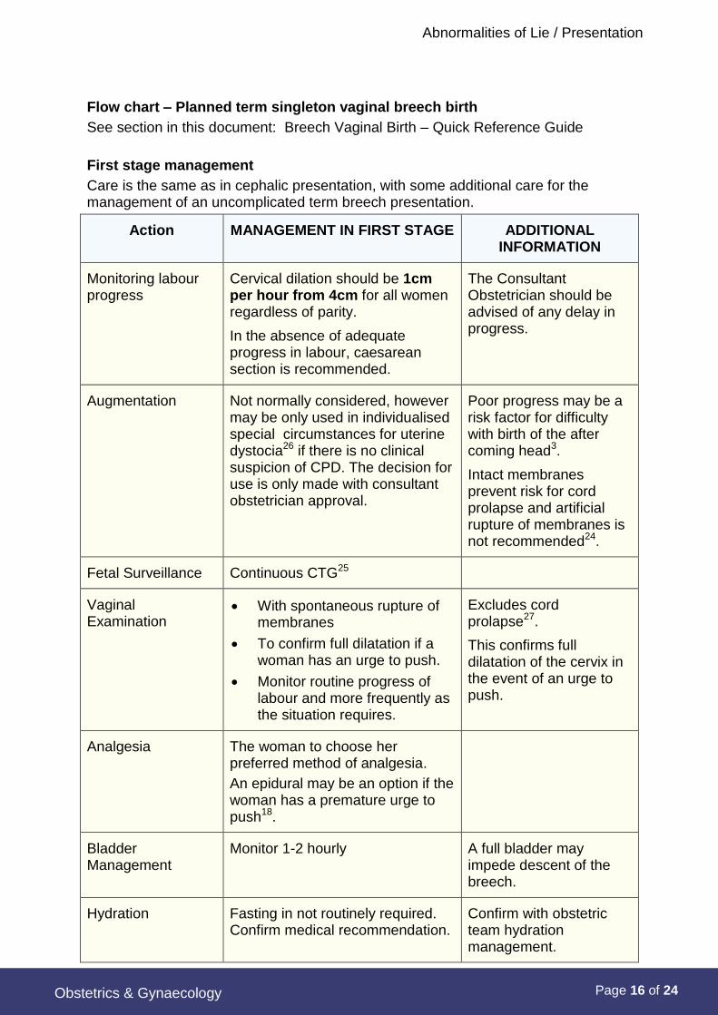
Flow chart – Planned term singleton vaginal breech birth
See section in this document: Breech Vaginal Birth – Quick Reference Guide
First stage management
Care is the same as in cephalic presentation, with some additional care for the management of an uncomplicated term breech presentation.
Action MANAGEMENT IN FIRST STAGE ADDITIONAL INFORMATION
Monitoring labour progress
Cervical dilation should be 1cm per hour from 4cm for all women regardless of parity.
In the absence of adequate progress in labour, caesarean section is recommended.
The Consultant Obstetrician should be advised of any delay in progress.
Augmentation Not normally considered, however may be only used in individualised special circumstances for uterine dystocia26 if there is no clinical suspicion of CPD. The decision for use is only made with consultant obstetrician approval.
Poor progress may be a risk factor for difficulty with birth of the after coming head3.
Intact membranes prevent risk for cord prolapse and artificial rupture of membranes is not recommended24.
Fetal Surveillance Continuous CTG25
Vaginal Examination
With spontaneous rupture of membranes
To confirm full dilatation if a woman has an urge to push.
Monitor routine progress of labour and more frequently as the situation requires.
Excludes cord prolapse27.
This confirms full dilatation of the cervix in the event of an urge to push.
Analgesia The woman to choose her preferred method of analgesia.
An epidural may be an option if the woman has a premature urge to push18.
Bladder Management
Monitor 1-2 hourly A full bladder may impede descent of the breech.
Hydration Fasting in not routinely required. Confirm medical recommendation.
Confirm with obstetric team hydration management.

Abnormalities of Lie / Presentation
Page 17 of 24
Obstetrics & Gynaecology
Action MANAGEMENT IN FIRST STAGE ADDITIONAL INFORMATION
Maternal Positioning
An upright position can be encouraged18.
An upright position may aid the descent of the breech27.
Additional Equipment
Availability of the real time ultrasound machine
Medical notifications
Notify the Consultant Obstetrician:
At full dilatation
If poor progress of labour
If concerns of maternal-fetal wellbeing
Second Stage Management
Action MANAGEMENT IN SECOND STAGE
ADDITIONAL INFORMATION
Confirm second stage
Perform a vaginal examination to confirm full dilatation prior to pushing.
Confirms that the woman is able to push if she has the urge.
Pushing Encourage active pushing when the woman has a strong urge, or the buttocks are on view.
Monitoring progress Birth should be imminent after one hour of active pushing in a nullipara, and after ½ hour for a multipara.
In the absence of adequate progress in second stage, caesarean section is recommended3.
The consultant obstetrician should be immediately notified of any delay in progress.
Fetal Surveillance Continuous CTG25
Bladder Management
Consider urinary catheterisation prior to birth if the bladder is not emptied.
Position for birth Dorsal or lithotomy Following maternal consent the practitioner should utilise the maternal position with which they are familiar3.

Abnormalities of Lie / Presentation
Page 18 of 24
Obstetrics & Gynaecology
Action MANAGEMENT IN SECOND STAGE
ADDITIONAL INFORMATION
Equipment Breech towel (warmed)
Lithotomy stirrups if necessary
Neville-Barnes’ and Wrigley’s Forceps immediately available
Analgesia As indicated.
Episiotomy Not routine – should be performed when indicated to facilitate birth3.
Birth principles No breech extraction
Traction/ fetal breech manoeuvres on breech are to be avoided unless necessary to expedite birth of a partially expelled fetus in a timely fashion.
Gentle suprapubic pressure may aid flexion of the head3.
Do not handle / manipulate the cord.
Extended arms may be delivered by the Løvset manoeuvre.2, 18 Nuchal arms may be reduced with reverse Løvsets.
Aftercoming head may be delivered spontaneously, with forceps, or by the Mariceau-Smellie-Veit manoeuvre.
A small towel wrapped around the fetal hips is useful.
Can cause extension of the head and nuchal displacement of the arms.27
May cause spasm of the cord18.
Rapid birth of the head can cause sudden compression and risk for tentorium cerebelli tear.24
Preserves warmth and provides a grip on the skin.
Paediatrician Contact the paediatric team to be present for the birth.
Oxytocin for 3rd stage
Withhold until the head is born.

Abnormalities of Lie / Presentation
Page 19 of 24
Obstetrics & Gynaecology
Unstable lie at or near term
Background
An unstable lie is when the fetal presentation repeatedly changes beyond 36 weeks
gestation.28 It is more common in parous women. Maternal causes include high
parity, placenta praevia, pelvic contracture, uterine malformations,28 pelvic tumours,
and a distended maternal urinary bladder. Fetal causes of unstable lie include
polyhydramnios,28 oligohydramnios, multiple pregnancy28, fetal macrosomia, and
fetal abnormalities (e.g. hydrocephaly, abdominal distension, fetal death28).29
If the membranes rupture when there is an unstable lie, regardless of whether the
woman is contracting there is significant risk for cord prolapse, especially if the lie is
oblique or transverse, or if the presenting part is high above the pelvic inlet. If lie is
not longitudinal when labour commences a compound presentation may result, or
the pelvis may remain empty which can lead to fetal distress and other
complications.29
Key points
1. The Obstetric Team Consultant shall be advised of all women with an unstable
lie at or near term.
2. A management plan shall be formulated and documented on the ‘MR004
Obstetric Special Instruction Sheet’.
Antenatal management
1. If a woman is attending a low risk midwifery antenatal clinic and is found to
have an unstable lie at term the midwife shall contact the team
Consultant/Senior Registrar to discuss management. The next antenatal
appointment needs to be with an obstetric medical antenatal team.
2. Investigate for causes of unstable lie. Ultrasound assessment may be
required.28, 30
3. Conduct clinical assessment for the size of the fetus and the pelvis.
Ultrasound assessment may be required in addition.
4. Formulate a plan for the mode of birth, and document on the MR004 Obstetric
Special Instruction Sheet.
5. Advise the woman to contact the hospital if she commences labour, or has
spontaneous rupture of membranes (SROM).
6. Inform the woman about risks of cord prolapse and management if this occurs
at home or in the hospital.28
7. Provide written advice for the woman (to be given to the St. Johns Ambulance
crew) describing management in the event of spontaneous rupture of
membranes.

Abnormalities of Lie / Presentation
Page 20 of 24
Obstetrics & Gynaecology
Birth management options
After discussion with the woman who has an unstable lie, one of the 3 birth options
should be decided:
Elective Caesarean Section
Expectant management – if no contraindications, await onset of labour
Active management – perform external version of the fetus to longitudinal lie
and then commence an induction of labour.
1. If a woman lives a long distance from the hospital, admission28 at 38-39 weeks
gestation – allows daily observation of lie and presentation and availability of
immediate assistance should SROM, cord prolapse, fetal distress, or labour
occur.29, 30
2. If spontaneous resolution to a longitudinal cephalic lie eventuates management
options include:
a presentation which remains cephalic for 48 hours may be discharged
home after review by the team Consultant and await spontaneous labour29
induce labour following team Consultant review.29
3. If the lie remains unstable, a stabilising induction may be an option28, 30 after review by the team Consultant.
Birth management for a woman in labour with an unstable lie
On admission
Perform a palpation.
Auscultate the fetal heart rate
Assess for SROM
Inform the obstetric medical team including the Senior Registrar
Labour management
ECV may be performed in early labour provided there are no contra-
indications. A stabilising / controlled artificial rupture of the membranes
(ARM) may then be performed.28 Note: Prior to controlled ARM, the woman
should have an empty rectum and bladder, as these can interfere with the
descent of the presenting part.28
Assess the presentation, lie and descent of the fetus frequently28 until the
presenting part is well into the pelvis.
If SROM occurs perform a vaginal examination (VE) to exclude cord prolapse
or malpresentation.
Conduct continuous fetal heart rate monitoring in labour.

Abnormalities of Lie / Presentation
Page 21 of 24
Obstetrics & Gynaecology
Obtain intravenous access and take blood for a full blood count, group and
hold- the woman is at increased risk for caesarean section, and possible post-
partum haemorrhage particularly if polyhydramnios is present.
Rare presentations
Aim
To provide guidance on the appropriate consultation and management of
malpresentations at KEMH.
Key points
1. Rare presentations (malpresentations) of the fetus include the following
Face presentation
Brow presentation
Compound presentation
Shoulder presentation
Oblique lie
2. Breech presentation- a consultant is to attend any viable vaginal breech birth.
See Breech Presentation above.
3. If a woman is suspected to have a malpresentation antenatally, it must be
discussed with the team consultant.
4. Management of unstable lie at term shall be discussed with the team consultant.
5. The team obstetrician, senior registrar and registrar must be notified immediately
of all malpresentations that present in labour.28
6. All malpresentations presenting in labour must be reviewed by a consultant

Abnormalities of Lie / Presentation
Page 22 of 24
Obstetrics & Gynaecology
References
1. RANZCOG. C-Obs 11: College statement: Management of breech presentation at term2013. Available from: https://www.ranzcog.edu.au/college-statements-guidelines.html#obstetrics.
2. Society of Obstetricians and Gynaecologist of Canada. Vaginal delivery of breech presentation. Journal of Obstetric Gynaecology of Canada. 2009 (June):557-66.
3. Royal college of Obstetricians and Gynaecologists (RCOG). Management of Breech Presentation (Green-top Guideline No. 20b) 2017 [Available from: https://www.rcog.org.uk/en/guidelines-research-services/guidelines/gtg20b/.
4. Whyte H, Hannah ME, Saigal S, Hannah WJ, Hewson S, Amankwah K, et al. Outcomes of children at 2 years after planned cesarean birth versus planned vaginal birth for breech presentation at term: The international randomized term breech trial. Am J Obstet Gynecol. 2004;191(3):864-71. Available from: http://www.ncbi.nlm.nih.gov/pubmed/15467555.
5. Hannah ME, Whyte H, Hannah WJ, Hewson S, Amankwah K, Cheng M, et al. Maternal outcomes at 2 years after planned cesarean section versus planned vaginal birth for breech presentation at term: The international randomized Term Breech Trial. Am J Obstet Gynecol. 2004;191(3):917-27. Available from: http://www.ncbi.nlm.nih.gov/pubmed/15467565.
6. Taillefer C, Dube J. Single breech at term: Two continents, two approaches. JOGC. 2010 (March):238-43.
7. Hannah ME, Hannah WJ, Hewson SA, Hodnett ED, Saigal S, Willan AR. Planned caesarean section versus planned vaginal birth for breech presentation at term: a randomised multicentre trial. Term Breech Trial Collaborative Group. Lancet. 2000;356(9239):1375-83. Available from: http://www.ncbi.nlm.nih.gov/pubmed/11052579.
8. Rietberg CC, Elferink-Stinkens PM, Visser GHA. The effect of the term breech trial on medical intervention behaviour and neonatal outcome in the Netherlands: An analysis of 35,453 term breech infants. BJOG: an International Journal of Obstetrics and Gynaecology. 2005;112:205-9.
9. Azria E, Le Meaux JP, Khoshnood B, Alexander S, Subtil D, Goffinet F, et al. Factors associated with adverse perinatal outcomes for term breech fetuses with planned vaginal delivery. Am J Obstet Gynecol. 2012;207(4):285 e1-9. Available from: http://www.ncbi.nlm.nih.gov/pubmed/23021690.
10. Hofmeyr GJ, Kulier R. External cephalic version for breech presentation at term (Review). Cochrane Database of Systematic Reviews. 2012 (10). Available from: http://onlinelibrary.wiley.com/doi/10.1002/14651858.CD000083.pub2/pdf.
11. Grootscholten K, Kok M, Oei G, et al. External Cephalic Version-Related Risks A Meta-analysis. Obstetrics & Gynecology. 2008;112(5):1143-51.
12. Collins S, Ellaway P, Harrington D, et al. The complications of external cephalic version: results from 805 consecutive attempts. BJOG: An International Journal of Obstetrics and Gynaecology. 2007;114:636-38.
13. Hutton EK, Hannah ME, Ross SJ. The Early External Cephalic Version (ECV) 2 Trial: An international multicentre randomised controlled trial of timing of ECV for breech pregnancies. BJOG: an International Journal of Obstetrics and Gynaecology. 2011;118:564-77.
14. Department of Health Western Australia. Consent to treatment policy for the Western Australian Health System 20112011. Available from: http://www.health.wa.gov.au/circularsnew/attachments/564.pdf.
15. Cluver C, Gyte GM, Sinclair M, Dowswell T, Hofmeyr GJ. Interventions for helping to turn term breech babies to head first presentation when using external cephalic version. Cochrane Database Syst Rev. 2015;2. Available from: http://onlinelibrary.wiley.com/doi/10.1002/14651858.CD000184.pub4/pdf.
16. Hofmeyr GJ, Kulier R. Cephalic version by postural management for breech presentation. Cochrane Database Syst Rev. 2012;10. Available from: http://www.ncbi.nlm.nih.gov/pubmed/23076882.
17. Collaris RJ, Oei SG. External cephalic version: A safe procedure? A systematic review of version-related risks. Acta Obstetrics and Gynecology Scandanavia. 2004;83:511-8.
18. Coates T. Malpositions of the occiput and malpresentations. In: Fraser DM, Cooper MA, editors. Myles Textbook for Midwives. 15th ed. Sydney: Churchill Livingstone; 2009. p. 573-605.
19. Royal College of Obstetricians and Gynaecologists. External cephalic version and reducing the

Abnormalities of Lie / Presentation
Page 23 of 24 Obstetrics & Gynaecology
incidence of breech presentation. Green-top Guideline No 20a. 2006.
20. The Royal Australian and New Zealand College of Obstetricians and Gynaecologists. C-Obs 11:College statement: Management of breech presentation at term2013. Available from:https://www.ranzcog.edu.au/college-statements-guidelines.html#obstetrics.
21. Collins S, Ellaway P, Harrington D, Pandit M, Impey LW. The complications of external cephalicversion: Results from 805 consecutive attempts. BJOG. 2007;114(5):636-8. Available from:http://onlinelibrary.wiley.com/doi/10.1111/j.1471-0528.2007.01271.x/pdf.
22. de Hundt M, Velzel J, de Groot CJ, Mol BW, Kok M. Mode of delivery after successful external cephalicversion: A systematic review and meta-analysis. Obstet Gynecol. 2014;123(6):1327-34. Availablefrom: http://www.ncbi.nlm.nih.gov/pubmed/24807332.
23. Hannah ME, Hannah WJ, Hewson SA, et al. Planned caesarean section versus planned vaginal birthfor breech presentation at term: A randomised multicentre trial. The Lancet. 2000;356:1375-83.
24. Thorogood C, Donaldson C. Disturbances in the rhythm of labour. Midwifery preparation for practice.2nd ed. Sydney: Churchill Livingstones; 2010. p. 819-61.
25. Royal Australian and New Zealand College of Obstetricians and Gynaecologists (RANZCOG). CollegeStatement C Obs-11: Management of breech presentation at term 2016 [Available from:https://www.ranzcog.edu.au/RANZCOG_SITE/media/RANZCOG-MEDIA/Women%27s%20Health/Statement%20and%20guidelines/Clinical-Obstetrics/Management-of-breech-presentation-at-term-(C-Obs-11)-Review-July-2016.pdf?ext=.pdf.
26. Society of Obstetrics and Gynecology of Canada. SGOC Clinical Practice Guideline No. 256.Substance Use in Pregnancy. JOGC. 2011 (April ):367-84.
27. Lewis P. Malpositions and malpresentations. In: Macdonald S, Magill-Cuerden J, editors. Mayes'Midwifery. 14th ed. Sydney: Bailliere Tindall; 2011. p. 869-98.
28. Coates T. Malpositions of the occiput and malpresentations. In: Marshall J, Raynor M, editors. Mylestextbook for midwives. 16th ed. Edinburgh: Churchill Livingstone Elsevier; 2014. p. 435-54.
29. Mackenzie IZ. Unstable lie, malpresentations and malpositions. In: James D, Steer PJ, Weiner CP, etal, editors. High Risk Pregnancy Management Options. 4th ed. Nottingham: Elsevier Saunders; 2011.p. 1123-37.
30. Baskett T, Calder A. Malpresentations. In: Baskett TF, Calder AA, Arulkumaran S, editors. Munro Kerr'soperative obstetrics 12th ed. Edinburgh: Elsevier; 2014. p. 116-22.
Related policies
WA Health Consent to Treatment Policy 2016
Related WNHS policies, procedures and guidelines
Form: MR 295.75: Consent form for ECV
KEMH O&G Clinical Guidelines:
[Restricted Area Guideline]: Induction of Labour: Artificial Rupture of the Membranes (ARM)
[access via Health point intranet]
Fetal Surveillance: Fetal Heart Rate Monitoring

Abnormalities of Lie / Presentation
Page 24 of 24 Obstetrics & Gynaecology
Keywords: breech, external cephalic version, ELUSCS, obstetric ultrasound, undiagnosed breech, planned breech birth, abnormal lie, unstable lie, transverse lie, oblique lie, high presenting part, polyhydramnios, fetal presentation, face presentation, brow presentation, rare presentation, compound presentation, shoulder presentation, oblique lie, unstable lie, breech presentation, ECV, external cephalic version, QRG, vaginal breech, presenting part
Document owner: OGID
Author / Reviewer: Head of Department- Obstetrics
July 2018: Evidence on this topic was reviewed and overall guidance remains unchanged. Minor changes and formatting have been made.
Date first issued: July 2018 Version 2
Supersedes: History: In July 2018 amalgamated seven individual guidelines on abnormalities of lie/presentation dating from March 2001.
Supersedes:
1. Breech Presentation (dated Feb 2018)
2. Breech Presentation (Uncomplicated Term) - Planned Vaginal Birth (dated May2017)
3. Breech (Uncomplicated Term) Vaginal Birth QRG (dated Feb 2018)
4. External Cephalic Version (ECV) (dated April 2015)
5. ECV: MFAU QRG (dated April 2015)
6. Rare Presentations (dated April 2015)
7. Unstable Lie at or Near Term (dated April 2015)
Reviewed: July 2018; (amended Oct 2018; April 2021 (v2))Next review date: July 2021
Endorsed by: MSMSC Date: 24/7/2018
NSQHS Standards (v2) applicable:
1 Governance, 4 Medication Safety; 8 Recognising & Responding to Acute Deterioration
Printed or personally saved electronic copies of this document are considered uncontrolled.
Access the current version from the WNHS website.