aberrantly hydroxymethylated differentially expressed ... fileosteoarthritis (oa), a common...
TRANSCRIPT

Submitted 11 October 2018Accepted 10 January 2019Published 25 February 2019
Corresponding authorBing Shen, [email protected]
Academic editorShi-Cong Tao
Additional Information andDeclarations can be found onpage 13
DOI 10.7717/peerj.6425
Copyright2019 Fang et al.
Distributed underCreative Commons CC-BY 4.0
OPEN ACCESS
Aberrantly hydroxymethylated differentiallyexpressed genes and the associatedprotein pathways in osteoarthritisYang Fang1, Pingping Wang1, Lin Xia1, Suwen Bai1, Yonggang Shen2, Qing Li3,Yang Wang1, Jinhang Zhu1, Juan Du1 and Bing Shen1
1 School of Basic Medical Sciences, Anhui Medical University, Hefei, Anhui, China2Nursing Faculty, Anhui Health College, Chizhou, Anhui, China3Central Laboratory of Medical Research Center, Anhui Provincial Hospital, Hefei, Anhui, China
ABSTRACTBackground. The elderly population is at risk of osteoarthritis (OA), a common,multifactorial, degenerative joint disease. Environmental, genetic, and epigenetic (suchas DNA hydroxymethylation) factors may be involved in the etiology, development,and pathogenesis of OA. Here, comprehensive bioinformatic analyses were used toidentify aberrantly hydroxymethylated differentially expressed genes and pathways inosteoarthritis to determine the underlying molecular mechanisms of osteoarthritis andsusceptibility-related genes for osteoarthritis inheritance.Methods. Gene expressionmicroarray data,mRNA expression profile data, and awholegenome 5hmC dataset were obtained from the Gene Expression Omnibus repository.Differentially expressed genes with abnormal hydroxymethylation were identified byMATCH function. Gene ontology and Kyoto Encyclopedia of Genes and Genomes(KEGG) pathway enrichment analyses of the genes differentially expressed in OA wereperformed using Metascape and the KOBAS online tool, respectively. The protein–protein interaction network was built using STRING and visualized in Cytoscape, andthe modular analysis of the network was performed using the Molecular ComplexDetection app.Results. In total, 104 hyperhydroxymethylated highly expressed genes and 14 hypohy-droxymethylated genes with low expression were identified. Gene ontology analyses in-dicated that the biological functions of hyperhydroxymethylated highly expressed genesincluded skeletal system development, ossification, and bone development; KEGGpathway analysis showed enrichment in protein digestion and absorption, extracellularmatrix–receptor interaction, and focal adhesion. The top 10 hub genes in the protein–protein interaction network were COL1A1, COL1A2, COL2A1, COL3A1, COL5A1,COL5A2, COL6A1, COL8A1, COL11A1, and COL24A1. All the aforementioned resultsare consistent with changes observed in OA.Conclusion. After comprehensive bioinformatics analysis, we found aberrantly hydrox-ymethylated differentially expressed genes and pathways in OA. The top 10 hub genesmay be useful hydroxymethylation analysis biomarkers to provide more accurate OAdiagnoses and target genes for treatment of OA.
Subjects Bioinformatics, Genetics, Orthopedics, Data Mining and Machine LearningKeywords DNA hydroxymethylation, Expression profile, Bioinformatics, Cartilage, Collagen
How to cite this article Fang Y, Wang P, Xia L, Bai S, Shen Y, Li Q, Wang Y, Zhu J, Du J, Shen B. 2019. Aberrantly hydroxymethylateddifferentially expressed genes and the associated protein pathways in osteoarthritis. PeerJ 7:e6425 http://doi.org/10.7717/peerj.6425

INTRODUCTIONOsteoarthritis (OA), a common degenerative joint disease, is associated with biochemical,metabolic, and morphological changes of the tissues, mainly in articular cartilage (Liao etal., 2015). Articular cartilage is composed and maintained primarily by chondrocytes andthe extracellular matrix (ECM). Under normal conditions, chondrocytes are responsiblefor maintaining a balance between anabolic and catabolic factors while the ECM of thecartilage goes through continuous remodeling to maintain homeostasis (Ni et al., 2015).In OA, chondrocytes morphology and function and cartilage homeostasis changes causesoverall matrix degradation (Blanco, Rego & Ruiz-Romero, 2011).
Epigenetics investigates the heritable changes in gene expressionwhen theDNA sequenceis unchanged (Bird, 2002). Inmost diseases, epigenetic alterations include, but is not limitedto, DNAmethylation, histone modification, etc. An oxidative product of 5-methylcytosine,5-hydroxymethylcytosine (5hmC), is an intermediate in the active DNA demethylationpathway. Increasing evidence shows that 5hmC is not only an intermediate for DNAdemethylation but also exists as an independent, stable epigenetic marker to influence geneexpression (Bachman et al., 2014; Karlsson et al., 2010; Wu et al., 2011). In patients withOA, the global 5hmC levels was significantly increased compared to normal chondrocytesand associated with key OA genes expression (Taylor et al., 2015). Therefore, 5hmCmay beimportantly involved in the development of OA and potentially diagnostic and therapeutictargets (Taylor et al., 2014).
Microarray systems and high-throughput sequencing are efficient tools for examininggene expression and genetic or epigenetic variation and for identifying biomarkers inclinical studies (Soon, Hariharan & Snyder, 2013). However, many previous studies haveused only one of these methods, making it difficult to verify the key genes and pathwaysinvolved in multiple cellular processes and biological functions. Overlapping several typesof related data using bioinformatics analysis of databases may provide more reliableand accurate results (Liu et al., 2017a). Although numerous studies examining OA geneexpression profiles and methylation profiles have used bioinformatics analyses to screenhundreds of differentially expressed genes (DEGs) that may be involved in the developmentof OA (Chou et al., 2013; Fisch et al., 2018), few of these studies have focused on abnormalhydroxymethylation (Taylor et al., 2015). In the present study, data from a gene expressionmicroarray (GSE51588), mRNA high-throughput sequencing (GSE114007), and high-throughput genome-wide 5hmC analyses (GSE64393) were integrated and analyzed usingseveral bioinformatics tools. Aberrantly hydroxymethylated DEGs and their associatedprotein pathways were identified in OA to construct a protein–protein interaction (PPI)network and to determine hub genes. The findings using such an integrated approach havethe potential not only to identify aberrantly hydroxymethylated genes and their proteinpathways in OA but also to elucidate the underlyingmolecular mechanisms that coordinatethe occurrence of OA, potentially opening therapeutic avenues for the epigenetic regulationof OA.
Fang et al. (2019), PeerJ, DOI 10.7717/peerj.6425 2/17

Table 1 The information of samples in GSE51588, GSE64393 and GSE114007 datasets.
Item GSE51588 Dataset GSE64393 Dataset GSE114007 Dataset
Normal Osteoarthritis Normal Osteoarthritis Normal Osteoarthritis
Gender F: 6; M: 3 F: 22; M: 18 F: 1; M: 1 F: 1; M: 3 F: 13; M: 5 F:12 ; M: 8Age 38.4 ± 4.01 69.675 ± 1.40 30.5 ± 2.02 68.25 ± 3.15 36.6 ± 3.17 66.2 ± 1.64Tissue Subchondral bone from lateral or
medial tibial plateauChondrocytes Knee articular cartilage
Nucleotide Total RNA Total RNA Total RNA Total RNA Genomic DNA Genomic DNA
MATERIAL AND METHODSMicroarray analysis and high-throughput sequencingGene expression profiling datasets (GSE51588, GSE114007) and a gene methylolationanalysis dataset (GSE64393) were obtained from the Gene Expression Omnibusrepository (https://www.ncbi.nlm.nih.gov/geo/) at the National Center for BiotechnologyInformation. In total, subchondral bone obtained from 40 OA patients and 10 healthydonors were examined using the GeneChip expression profiling dataset GSE51588(platform: GPL13497, Agilent-026652 Whole Human Genome Microarray 4 × 44 K v2[Probe Name version]), and cartilage tissues obtained from 20 OA patients and 18 healthydonors were examined using the high-throughput sequencing mRNA expression profiledataset GSE114007 (platform: Illumina HiSeq 2000 [Homo sapiens ]; Illumina NextSeq 500[Homo sapiens]). The genome-wide 5hmC profile dataset GSE64393 (platform: GPL11154Illumina HiSeq 2000 [Homo sapiens]) included four human articular chondrocytes withOA and four total genomic DNA 5hmc datasets for articular chondrocytes without OA.The information of all samples was shown in Table 1.
Data processingThe data were processed according to the flowchart (Fig. 1). The downloaded platformsand a series of matrix files were converted using R (v3.4.3) programming language andannotation software packages. The probe name was converted to the gene symbol of thecorresponding gene and saved in a TXT file. Expression microarray datasets were analyzedusing the Limma (v3.26.8) software package with default settings, and expression profiledata was analyzed with the DESeq2 (v1.22.1) package with default settings (Varet et al.,2016). The whole genome 5hmC analysis result was obtained from the supplementary file ofdataset of GSE64393. The selection criteria for DEGs and differentially hydroxymethylatedgenes were those with P values <0.05 and an absolute log2 (fold change) >1.
Data integrationThe MATCH function was used to find overlapping DEGs in the two gene expressionprofile datasets (GSE114007 and GSE51588), and those intersecting genes that wereeither upregulated or downregulated were identified (Fig. 1). In addition, the differentiallyhydroxymethylated geneswere superimposed on the gene hydroxymethylationprofile in theGSE64393 dataset. Finally, genes that were both hyperhydroxymethylated and upregulated
Fang et al. (2019), PeerJ, DOI 10.7717/peerj.6425 3/17

GSE51588 GSE114007 GSE64393
GEO/NCBI
DEGs or differentially hydroxymethylated gene
Hypohydroxymethylated highly expressed genesHyperhydroxymethylated lowly expressed genes
GO ontology enrichment analyses
KEGG pathway
enrichment analyses
PPI network , code module
and hup genes
Limma DESeq2
MATCH function
STRING, CytoscapeKOBASMetascape
Figure 1 Flowchart of data analysis procedure.Full-size DOI: 10.7717/peerj.6425/fig-1
were identified, and those that were both hypohydroxymethylated and downregulatedwere identified.
Gene ontology and KEGG pathway enrichment analysesMetascape, a web-based resource for gene annotation, visualization, and integrationdiscovery (http://metascape.org) was used to perform functional and pathway enrichmentanalyses (Soonthornvacharin et al., 2017). Gene ontology (GO) analysis was performedusing Metascape (Fig. 1). Kyoto Encyclopedia of Genes and Genomes (KEGG) pathwayanalysis of these aberrantly hydroxymethylated DEGs was performed using the KOBAS
Fang et al. (2019), PeerJ, DOI 10.7717/peerj.6425 4/17

online analysis database (http://kobas.cbi.pku.edu.cn/) (Fig. 1). Values of P < 0.05 wereconsidered statistically significant (Xie et al., 2011).
PPI network construction and module analysisPPI analysis may reveal the general organizational principles of functional cellular networksand provide new insights into protein function. The Search Tool for the Retrieval ofInteracting Genes (STRING; http://string.embl.de/) provides information on the functionalrelationship between proteins (Fig. 1) (Von Mering et al., 2003). The PPI network associatedwith the respective aberrantly hydroxymethylated DEGs was constructed to predict theinteraction of selected genes. Cytoscape (http://www.cytoscape.org/) is widely used tointegrate biomolecular interaction networks with models to construct PPI networks ofaberrantly hydroxymethylated DEGs (Fig. 1) (Lim et al., 2006). The Molecular ComplexDetection (MCODE) app in Cytoscape was used to screen modules in the PPI network(Cao et al., 2018). Topology analysis was used to analyze the connectivity of the nodes inthe PPI network to obtain a higher degree of key nodes (central proteins) (He & Zhang,2006). The top 10 hub genes were selected for further analysis. Functional enrichmentanalysis of each module was performed using Metascape, with a significance threshold ofP < 0.05.
RESULTSIdentification of aberrantly hydroxymethylated DEGs in OAUsing Limma software to analyze the microarray GSE51588 system, we obtained 1,109significantly upregulated DEGs and 693 significantly downregulated DEGs (Fig. 2A).Using the DESeq2 package to analyze the high-throughput sequencing mRNA expressionprofile dataset GSE114007, we obtained 1,769 significantly upregulated DEGs and1,052 significantly downregulated DEGs (Fig. 2B). We then identified those DEGscontained in both gene expression profiles, finding 223 overlapping upregulated genesand 97 overlapping downregulated genes (Fig. 3). An analysis of the high-throughputhydroxymethylation dataset GSE64393 showed 7,262 hyperhydroxymethylated genesand 4,731 hypohydroxymethylated genes (Fig. 2C). We isolated the overlap of 7,262hyperhydroxymethylated genes with the 223 upregulated genes. Then, a total of 104hyperhydroxymethylated highly expressed genes were obtained. We overlapped the4,731 hypohydroxymethylated genes and the 97 downregulated genes to identify 14hypohydroxymethylated genes with low expression (Fig. 3). The heat maps for theexpression of the aberrantly hydroxymethylated DEGs are shown in Fig. 4.
GO functional enrichment analysisThe top 10 significantly enriched GO terms, as determined using Metascape, areillustrated in Fig. 5. Hyperhydroxymethylated highly expressed genes were enriched inthe biological processes of skeletal system development, ossification, bone development,blood vessel development, response to growth factor, and osteoblast differentiation. Inthe molecular function GO category, these genes showed enrichment in ECM structuralconstituent, collagen binding, calcium ion binding, ECM structural constituent conferring
Fang et al. (2019), PeerJ, DOI 10.7717/peerj.6425 5/17

Figure 2 Differential gene expression and differential gene hydroxymethylation. (A) GSE51588 mi-croarray dataset. (B) GSE114007 mRNA expression profile dataset. (C) GSE64393 high-throughput hy-droxymethylation dataset. Red indicates upregulation, green indicates downregulation, and blue indicatesno significant change in gene expression or hydroxymethylation based on the criteria of an absolute log2(fold change) >1 and P < 0.05.
Full-size DOI: 10.7717/peerj.6425/fig-2
Figure 3 Identification of aberrantly hydroxymethylated differentially expressed genes in the geneexpression datasets (GSE51588, GSE114007) and the gene hydroxymethylation dataset (GSE64393).(A) Hyperhydroxymethylation and upregulated genes; (B) hypohydroxymethylation and downregulatedgenes.
Full-size DOI: 10.7717/peerj.6425/fig-3
compression resistance, and ECM binding. The cell component GO category showedenrichment predominantly for the ECM, basement membrane, and synapse, indicatingthat hyperhydroxymethylated highly expressed genes may play key roles in the formationof the ECM and cartilage. For genes with hypohydroxymethylation and low expression,our Metascape findings indicated enrichments in the GO biological process category for
Fang et al. (2019), PeerJ, DOI 10.7717/peerj.6425 6/17
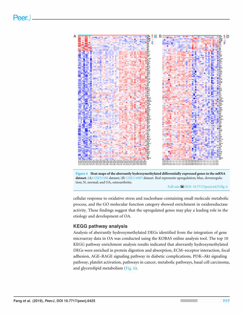
Figure 4 Heat maps of the aberrantly hydroxymethylated differentially expressed genes in the mRNAdataset. (A) GSE51588 dataset; (B) GSE114007 dataset. Red represents upregulation; blue, downregula-tion; N, normal; and OA, osteoarthritis.
Full-size DOI: 10.7717/peerj.6425/fig-4
cellular response to oxidative stress and nucleobase-containing small molecule metabolicprocess, and the GO molecular function category showed enrichment in oxidoreductaseactivity. These findings suggest that the upregulated genes may play a leading role in theetiology and development of OA.
KEGG pathway analysisAnalysis of aberrantly hydroxymethylated DEGs identified from the integration of genemicroarray data in OA was conducted using the KOBAS online analysis tool. The top 10KEGG pathway enrichment analysis results indicated that aberrantly hydroxymethylatedDEGs were enriched in protein digestion and absorption, ECM–receptor interaction, focaladhesion, AGE–RAGE signaling pathway in diabetic complications, PI3K–Akt signalingpathway, platelet activation, pathways in cancer, metabolic pathways, basal cell carcinoma,and glycerolipid metabolism (Fig. 6).
Fang et al. (2019), PeerJ, DOI 10.7717/peerj.6425 7/17

Figure 5 Enrichment of the gene ontology (GO) functional terms for the hyperhydroxymethylatedhighly expressed genes.
Full-size DOI: 10.7717/peerj.6425/fig-5
PPI network construction, module analysis, and hub gene selectionThe PPI network was created using the STRING website, and the function modules wereanalyzed using MCODE in Cytoscape. For the aberrantly hydroxymethylated DEGs, thePPI network is shown in Fig. 7A, and threemodules are displayed in Fig. 7B. Important coremodules showed collagen trimer, complex of collagen trimers, ECM structural constituent,glycosaminoglycan biosynthesis-keratan sulfate, and oxidoreductase activity. As showedin Fig. 8, the 10 genes with the most substantial interactions were COL1A1, COL1A2,COL2A1, COL3A1, COL5A1, COL5A2, COL6A1, COL8A1, COL11A1, and COL24A1, andall 10 core genes were hyperhydroxymethylated highly expressed genes.
Fang et al. (2019), PeerJ, DOI 10.7717/peerj.6425 8/17

Figure 6 Enrichment of the aberrantly hydroxymethylated differentially expressed genes in Kyoto En-cyclopedia of Genes and Genomes pathway analysis.
Full-size DOI: 10.7717/peerj.6425/fig-6
DISCUSSIONAlthough OA affects approximately 40% of the elderly population, it is a growing publichealth problem worldwide because the genetic and molecular mechanisms underlyingits occurrence and development remain unclear (Balakrishnan et al., 2014; Chapman& Valdes, 2012). Understanding the mechanisms underpinning OA development andprogression will greatly contribute to the diagnosis, treatment, and prognosis of patientswith this disease. Gene chip and high-throughput sequencing technologies can detect theexpression levels of tens of millions of genes in humans and have been widely used to studythe molecular mechanisms of various diseases, predict potential therapeutic targets, andfind biomarkers (Dong et al., 2018; Liu et al., 2017a). Despite the numerous basic studiesreporting on OA in recent years, the molecular mechanisms, early diagnosis, and epigenetic
Fang et al. (2019), PeerJ, DOI 10.7717/peerj.6425 9/17

Figure 7 Protein–protein interaction network. Protein–protein interaction network (A) and the topthree modules (B).
Full-size DOI: 10.7717/peerj.6425/fig-7
Figure 8 The top 10 hub genes. Blue indicates genes with low scores. Red indicates genes with highscores. Yellow and orange indicate genes with middle scores, increasing respectively.
Full-size DOI: 10.7717/peerj.6425/fig-8
Fang et al. (2019), PeerJ, DOI 10.7717/peerj.6425 10/17

mechanisms of OA have not been elucidated because most of these studies have focusedon the gene expression or pure methylation from a single cohort (Ren et al., 2018).
By contrast, the present bioinformatics study analyzed the mRNA expressionprofiles obtained from GeneChip data, transcriptome sequencing data, and a set ofexpression profile datasets from different groups of whole genome 5hmC data. UsingMATCH analysis, we identified 118 aberrantly hydroxymethylated DEGs, including 104hyperhydroxymethylated highly expression genes and 14 hypohydroxymethylated geneswith low expression levels. The 118 aberrantly hydroxymethylated DEGs were categorizedusing the three GO functional annotations molecular functions, biological processes,and cellular components. DEG enrichment was determined using KEGG signal pathwayanalysis to construct a PPI of proteins that were encoded by these DEGs and to screen forthe 10 most closely related genes.
Our results indicated that the aberrantly hydroxymethylated highly expressed DEGsin OA were involved mainly in the biological processes of skeletal system development,ossification, bone development, blood vessel development, response to growth factor,and osteoblast differentiation. These genes showed enrichment of molecular functionsfor ECM structural constituents, collagen binding, calcium ion binding, ECM structuralconstituent conferring compression resistance, and ECM binding. The cellular componentenrichment for these genes was predominantly in the ECM, basement membrane, andsynapse. These enriched functions and components are closely related to the growth ofcartilage cells and the production of the cartilage that composes the ECM and are consistentwith changes of the ECM and cartilage in patients with OA. In humans, the ECM consistsmainly of proteins (especially collagen) and glycosaminoglycans (mainly proteoglycans)and is secreted by nearby cells. The ECM structural constituent, collagen binding, calciumion binding, ECM structural constituent conferring compression resistance, ECM binding,ECM, basement membrane, and synapse are also closely related to the growth of cartilagecells as well as to the development and remodeling of cartilage. Our findings for theseenrichments in OA suggests that collagen changes may play an important part in thedevelopment of OA. The hypohydroxymethylated genes with low expression levels wereenriched in the cellular response to oxidative stress, nucleobase-containing small moleculemetabolic processes, and oxidoreductase activity. The results of our KEGG signalingpathway analysis showed enrichments in protein digestion and absorption, ECM–receptorinteraction. Thus, the results of our GO and KEGG enrichment analyses indicated thataberrantly hydroxymethylated DEGs are involved in numerous pathways associated withcartilage development and composition, consistent with the notion that these aberrantlyhydroxymethylated DEGs play important roles in the occurrence and development of OA.
Our PPI network analyses of these aberrantly hydroxymethylated DEGs suggestedthat collagen trimer, complex of collagen trimers, ECM structural constituent,glycosaminoglycan biosynthesis-keratan sulfate, and oxidoreductase activity may beinvolved in OA development. These modules are closely related to the composition andhomeostasis of cartilage and, again, demonstrate that collagen plays an important role inOA. We identified the top 10 hub genes in the PPI; the proteins encoded by these genes arethe key nodes in the PPI network. We found that they were all highly methylolated, highly
Fang et al. (2019), PeerJ, DOI 10.7717/peerj.6425 11/17
expressed genes encoding collagen and primarily involved in collagen trimer, collagentrimer complexes, and receptor tyrosine kinase signaling.
In the 10 core genes, increased mRNA expression levels of COL1A1, COL1A2, COL2A1,COL3A1, COL5A2 genes have been reported in OA (Bay-Jensen et al., 2018; Jin et al.,2009; Karlsson et al., 2010; Hermansson et al., 2004). Studies have shown that increasedsecretion of type I collagen encoded by COL1A1 and COL1A2 homotrimers in OA caninduce stenosis and unorganized collagen fibers in subchondral bone (Clements et al., 2009;Couchourel et al., 2009; Jin et al., 2009; Steinberg et al., 2017). Type II collagen encoded byCOL2A1, which is specific for cartilage tissue and is important for normal embryonicdevelopment of the bone, linear growth, and cartilage resistance to compression (Chen etal., 2016). Type III collagen encoded by COL3A1, together with type I collagen, providesa structural framework for the synovium surrounding the synovial joint (Hu et al., 2017;Reichert et al., 2018). Upregulation of type III in OA leads to thickening of the synovialmembrane and has potential as a biomarker for OA (Bay-Jensen et al., 2018). Type Vcollagen encoded by COL5A1 and COL5A2, as a member of group I collagen, is presentin tissues containing type I collagen, and appears to regulate the assembly of shaped fiberscomposed of type I and type V collagen (Colombi et al., 2017;Willard et al., 2018). Type XIcollagen encoded by COL11A1 plays an important role in fiber formation by controllingthe lateral growth of collagen II fibrils, since type XI collagen has previously been reportedin the core of type II collagen and is believed to be useful in the stability of type II collagen(Liu et al., 2017b). The close interaction between type II and type XI collagen, which affectseither of these mutations, may result in similar instability of cartilage tissue. Type XXIVcollagen encoded by COL24A1, which may regulate the formation of type I collagen fibersduring fetal development (Koch et al., 2003). The pathways associated with COL24A1and the GO annotation linked with this gene include collagen chain trimerization, andECM structural composition (Koch et al., 2003). These collagens have been reported to beclosely related to cartilage composition and structural stability. Our results showed thatthe hydroxymethylation of these genes encoding collagens were significantly increased.Therefore, we believe that collagens may importantly involved in the development of OA.
CONCLUSIONSUsing a combination of bioinformatics analysis of gene expression and of altered DNAhydroxymethylation, this study identified numerous aberrantly hydroxymethylated DEGsand their associated protein pathways in OA. These findings may help to identify the keygenes and molecular mechanisms associated with OA initiation and development. Thetop 10 hub genes, COL1A1, COL1A2, COL2A1, COL3A1, COL5A1, COL5A2, COL6A1,COL8A1, COL11A1, and COL24A1, may be candidate biomarkers of hydroxymethylationabnormalities in OA that may be considered for more accurate diagnosis and treatmentof OA. Compared with previous studies, the present study may provide more reliable andaccurate screening results through our use of overlapping sets of related data. Althoughwe have identified some candidate genes, further molecular experiments are still needed todiscover other genes associated with OA.
Fang et al. (2019), PeerJ, DOI 10.7717/peerj.6425 12/17

ADDITIONAL INFORMATION AND DECLARATIONS
FundingThis work was supported by grants from the National Natural Science Foundation ofChina (Grant No. U1732157, 81570403, 81472018), the Anhui Provincial Natural ScienceFoundation (Grant No. 1708085MH187), the Natural Science Foundation of AnhuiProvince Department of Education (Grant No. KJ2018A0974); Anhui Medical Universityfor Scientific Research (BSKY XJ201607), the Outstanding Young Investigator of AnhuiMedical University, and the Science and Technology Research Project of Anhui Province(Grant No. 1501041147). The funders had no role in study design, data collection andanalysis, decision to publish, or preparation of the manuscript.
Grant DisclosuresThe following grant information was disclosed by the authors:National Natural Science Foundation of China: U1732157, 81570403, 81472018.Anhui Provincial Natural Science Foundation: 1708085MH187.Natural Science Foundation of Anhui Province Department of Education: KJ2018A0974.Anhui Medical University for Scientific Research: BSKY XJ201607.Outstanding Young Investigator of Anhui Medical University.Science and Technology Research Project of Anhui Province: 1501041147.
Competing InterestsThe authors report no conflicts of interest in this work.
Author Contributions• Yang Fang conceived and designed the experiments, performed the experiments,analyzed the data, prepared figures and/or tables, authored or reviewed drafts of thepaper, approved the final draft.
• PingpingWang and YangWang performed the experiments, analyzed the data, approvedthe final draft.
• LinXia prepared figures and/or tables, authored or reviewed drafts of the paper, approvedthe final draft.
• Suwen Bai analyzed the data, approved the final draft.• Yonggang Shen and Jinhang Zhu conceived and designed the experiments, analyzed thedata, approved the final draft.
• Qing Li prepared figures and/or tables.• Juan Du conceived and designed the experiments, contributed reagents/materials/anal-ysis tools, authored or reviewed drafts of the paper, approved the final draft.
• Bing Shen conceived and designed the experiments, contributed reagents/materials/-analysis tools, prepared figures and/or tables, authored or reviewed drafts of the paper,approved the final draft.
Data AvailabilityThe following information was supplied regarding data availability:
Fang et al. (2019), PeerJ, DOI 10.7717/peerj.6425 13/17

Gene expression profiling datasets (GSE51588, GSE114007) and a gene methylationanalysis data set (GSE64393) were obtained from the Gene Expression Omnibusrepository (https://www.ncbi.nlm.nih.gov/geo/) at the National Center for BiotechnologyInformation.
Supplemental InformationSupplemental information for this article can be found online at http://dx.doi.org/10.7717/peerj.6425#supplemental-information.
REFERENCESBachmanM, Uribe-Lewis S, Yang X,WilliamsM,Murrell A, Balasubramanian S. 2014.
5-Hydroxymethylcytosine is a predominantly stable DNA modification. NatureChemistry 6:1049–1055 DOI 10.1038/nchem.2064.
Balakrishnan L, Nirujogi RS, Ahmad S, Bhattacharjee M, Manda SS, Renuse S, KelkarDS, Subbannayya Y, Raju R, Goel R, Thomas JK, Kaur N, DhillonM, Tankala SG,Jois R, Vasdev V, Ramachandra Y, Sahasrabuddhe NA, Prasad Ts K, Mohan S,Gowda H, Shankar S, Pandey A. 2014. Proteomic analysis of human osteoarthritissynovial fluid. Clinical Proteomics 11:Article 6 DOI 10.1186/1559-0275-11-6.
Bay-Jensen AC, Kjelgaard-Petersen CF, Petersen KK, Arendt-Nielsen L, QuasnichkaHL, Mobasheri A, Karsdal MA, Leeming DJ. 2018. Aggrecanase degradation of typeIII collagen is associated with clinical knee pain. Clinical Biochemistry 58:37–43DOI 10.1016/j.clinbiochem.2018.04.022.
Bird A. 2002. DNA methylation patterns and epigenetic memory. Genes and Development16:6–21 DOI 10.1101/gad.947102.
Blanco FJ, Rego I, Ruiz-Romero C. 2011. The role of mitochondria in osteoarthritis.Nature Reviews Rheumatology 7:161–169 DOI 10.1038/nrrheum.2010.213.
Cao L, Chen Y, ZhangM, Xu DQ, Liu Y, Liu T, Liu SX,Wang P. 2018. Identificationof hub genes and potential molecular mechanisms in gastric cancer by integratedbioinformatics analysis. PeerJ 6:e5180 DOI 10.7717/peerj.5180.
Chapman K, Valdes AM. 2012. Genetic factors in OA pathogenesis. Bone 51:258–264DOI 10.1016/j.bone.2011.11.026.
Chen X, Guo J, Cai T, Zhang F, Pan S, Zhang L,Wang S, Zhou F, Diao Y, Zhao Y, ChenZ, Liu X, Chen Z, Liu Z, Sun Y, Du J. 2016. Targeted next-generation sequencingreveals multiple deleterious variants in OPLL-associated genes. Scientific Reports6:26962 DOI 10.1038/srep26962.
Chou CH,Wu CC, Song IW, Chuang HP, Lu LS, Chang JH, Kuo SY, Lee CH,WuJY, Chen YT, Kraus VB, Lee MT. 2013. Genome-wide expression profiles ofsubchondral bone in osteoarthritis. Arthritis Research & Therapy 15:Article R190DOI 10.1186/ar4380.
Clements DN, Fitzpatrick N, Carter SD, Day PJ. 2009. Cartilage gene expressioncorrelates with radiographic severity of canine elbow osteoarthritis. The VeterinaryJournal 179:211–218 DOI 10.1016/j.tvjl.2007.08.027.
Fang et al. (2019), PeerJ, DOI 10.7717/peerj.6425 14/17

Colombi M, Dordoni C, Venturini M, Ciaccio C, Morlino S, Chiarelli N, ZancaA, Calzavara-Pinton P, Zoppi N, Castori M, Ritelli M. 2017. Spectrum of mu-cocutaneous, ocular and facial features and delineation of novel presentationsin 62 classical Ehlers-Danlos syndrome patients. Clinical Genetics 92:624–631DOI 10.1111/cge.13052.
Couchourel D, Aubry I, Delalandre A, LavigneM,Martel-Pelletier J, Pelletier JP,Lajeunesse D. 2009. Altered mineralization of human osteoarthritic osteoblastsis attributable to abnormal type I collagen production. Arthtitis and Rheumatism60:1438–1450 DOI 10.1002/art.24489.
Dong Z,Wang J, Zhan T, Xu S. 2018. Identification of prognostic risk factors foresophageal adenocarcinoma using bioinformatics analysis. OncoTargets and Therapy11:4327–4337 DOI 10.2147/OTT.S156716.
Fisch KM, Gamini R, Alvarez-Garcia O, Akagi R, Saito M, Muramatsu Y, Sasho T,Koziol JA, Su AI, Lotz MK. 2018. Identification of transcription factors responsiblefor dysregulated networks in human osteoarthritis cartilage by global gene expressionanalysis. Osteoarthritis and Cartilage DOI 10.1016/j.joca.2018.07.012.
He X, Zhang J. 2006.Why do hubs tend to be essential in protein networks? PLOSGenetics 2:e88 DOI 10.1371/journal.pgen.0020088.
HermanssonM, Sawaji Y, BoltonM, Alexander S, Wallace A, Begum S,Wait R, Saklat-vala J. 2004. Proteomic analysis of articular cartilage shows increased type II collagensynthesis in osteoarthritis and expression of inhibin betaA (activin A), a regulatorymolecule for chondrocytes. Journal of Biological Chemistry 279:43514–43521DOI 10.1074/jbc.M407041200.
Hu Y,Wu R, Li H, Gu Y,WeiW. 2017. Expression and significance of metalloproteinaseand collagen in vaginal wall tissues of patients with pelvic organ prolapse. Annals ofClinical and Laboratory Science 47:698–705.
Jin H, van’t Hof RJ, Albagha OM, Ralston SH. 2009. Promoter and intron 1 poly-morphisms of COL1A1 interact to regulate transcription and susceptibility toosteoporosis. Human Molecular Genetics 18:2729–2738 DOI 10.1093/hmg/ddp205.
Karlsson C, Dehne T, Lindahl A, Brittberg M, Pruss A, Sittinger M, Ringe J. 2010.Genome-wide expression profiling reveals new candidate genes associated withosteoarthritis. Osteoarthritis Cartilage 18:581–592 DOI 10.1016/j.joca.2009.12.002.
KochM, Laub F, Zhou P, Hahn RA, Tanaka S, Burgeson RE, Gerecke DR, Ramirez F,GordonMK. 2003. Collagen XXIV, a vertebrate fibrillar collagen with structural fea-tures of invertebrate collagens: selective expression in developing cornea and bone.Journal of Biological Chemistry 278:43236–43244 DOI 10.1074/jbc.M302112200.
LiaoW, Li Z, Zhang H, Li J, Wang K, Yang Y. 2015. Proteomic analysis of synovialfluid as an analytical tool to detect candidate biomarkers for knee osteoarthritis.International Journal of Clinical and Experimental Pathology 8:9975–9989.
Lim J, Hao T, Shaw C, Patel AJ, Szabo G, Rual JF, Fisk CJ, Li N, Smolyar A, Hill DE,Barabasi AL, Vidal M, Zoghbi HY. 2006. A protein-protein interaction networkfor human inherited ataxias and disorders of Purkinje cell degeneration. Cell125:801–814 DOI 10.1016/j.cell.2006.03.032.
Fang et al. (2019), PeerJ, DOI 10.7717/peerj.6425 15/17

Liu J, Li H, Sun L,Wang Z, Xing C, Yuan Y. 2017a. Aberrantly methylated-differentiallyexpressed genes and pathways in colorectal cancer. Cancer Cell International17:Article 75 DOI 10.1186/s12935-017-0444-4.
LiuW, Sun G, Guo L,Wang L, FanW, LangM, Chen D, Yi X. 2017b. A genetic variantin COL11A1 is functionally associated with lumbar disc herniation in Chinesepopulation. Journal of Genetics 96:867–872 DOI 10.1007/s12041-017-0874-8.
Ni J, Yuan XM, Yao Q, Peng LB. 2015. OSM is overexpressed in knee osteoarthritis andNotch signaling is involved in the effects of OSM on MC3T3-E1 cell proliferationand differentiation. International Journal of Molecular Medicine 35:1755–1760DOI 10.3892/ijmm.2015.2168.
Reichert MC, Kupcinskas J, KrawczykM, Jungst C, Casper M, Grunhage F, AppenrodtB, Zimmer V,Weber SN, Tamelis A, Lukosiene JI, Pauziene N, Kiudelis G, JonaitisL, SchrammC, Goeser T, Schulz A, Malinowski M, GlanemannM, KupcinskasL, Lammert F. 2018. A variant of COL3A1 (rs3134646) is associated with risk ofdeveloping diverticulosis in white men. Diseases of the Colon and Rectum 61:604–611DOI 10.1097/DCR.0000000000001001.
Ren YM, Zhao X, Yang T, Duan YH, Sun YB, ZhaoWJ, TianMQ. 2018. Explor-ing the key genes and pathways of osteoarthritis in knee cartilage in a ratmodel using gene expression profiling. Yonsei Medical Journal 59:760–768DOI 10.3349/ymj.2018.59.6.760.
SoonWW, HariharanM, Snyder MP. 2013.High-throughput sequencing for biologyand medicine.Molecular Systems Biology 9:Article 640 DOI 10.1038/msb.2012.61.
Soonthornvacharin S, Rodriguez-Frandsen A, Zhou Y, Galvez F, Huffmaster NJ,Tripathi S, Balasubramaniam VR, Inoue A, De Castro E, Moulton H, Stein DA,Sanchez-Aparicio MT, De Jesus PD, Nguyen Q, Konig R, Krogan NJ, Garcia-SastreA, Yoh SM, Chanda SK. 2017. Systems-based analysis of RIG-I-dependent signallingidentifies KHSRP as an inhibitor of RIG-I receptor activation. Nature Microbiology2:Article 17022 DOI 10.1038/nmicrobiol.2017.22.
Steinberg J, Ritchie GRS, Roumeliotis TI, Jayasuriya RL, ClarkMJ, Brooks RA,Binch ALA, Shah KM, Coyle R, PardoM, LeMaitre CL, Ramos YFM, NelissenR, Meulenbelt I, McCaskie AW, Choudhary JS, Wilkinson JM, Zeggini E. 2017.Integrative epigenomics, transcriptomics and proteomics of patient chondrocytesreveal genes and pathways involved in osteoarthritis. Scientific Reports 7:8935DOI 10.1038/s41598-017-09335-6.
Taylor SE, Li YH,WongWH, Bhutani N. 2015. Genome-wide mapping of DNA hy-droxymethylation in osteoarthritic chondrocytes. Arthritis Rheumatol 67:2129–2140DOI 10.1002/art.39179.
Taylor SE, Smeriglio P, Dhulipala L, RathM, Bhutani N. 2014. A global increase in 5-hydroxymethylcytosine levels marks osteoarthritic chondrocytes. Arthritis Rheumatol66:90–100 DOI 10.1002/art.38200.
Varet H, Brillet-Guéguen L, Coppée JY, Dillies MA. 2016. SARTools: a DESeq2- andEdgeR-Based R pipeline for comprehensive differential analysis of RNA-Seq data.PLOS ONE 11(6):e0157022 DOI 10.1371/journal.pone.0157022.
Fang et al. (2019), PeerJ, DOI 10.7717/peerj.6425 16/17

VonMering C, HuynenM, Jaeggi D, Schmidt S, Bork P, Snel B. 2003. STRING: adatabase of predicted functional associations between proteins. Nucleic AcidsResearch 31:258–261 DOI 10.1093/nar/gkg034.
Willard K, Mannion S, Saunders CJ, Collins M, September AV. 2018. The interactionof polymorphisms in extracellular matrix genes and underlying miRNA motifs thatmodulate susceptibility to anterior cruciate ligament rupture. Journal of Science andMedicine in Sport 21:22–28 DOI 10.1016/j.jsams.2017.08.017.
WuH, D’Alessio AC, Ito S, Wang Z, Cui K, Zhao K, Sun YE, Zhang Y. 2011. Genome-wide analysis of 5-hydroxymethylcytosine distribution reveals its dual function intranscriptional regulation in mouse embryonic stem cells. Genes and Development25:679–684 DOI 10.1101/gad.2036011.
Xie C, Mao X, Huang J, Ding Y,Wu J, Dong S, Kong L, Gao G, Li CY,Wei L. 2011.KOBAS 2.0: a web server for annotation and identification of enriched pathways anddiseases. Nucleic Acids Research 39:W316–W322 DOI 10.1093/nar/gkr483.
Fang et al. (2019), PeerJ, DOI 10.7717/peerj.6425 17/17