aberrant hippocampal neurogenesis contributes to epilepsy ... · article received 22 dec 2014 |...
TRANSCRIPT

ARTICLE
Received 22 Dec 2014 | Accepted 10 Feb 2015 | Published 26 Mar 2015
Aberrant hippocampal neurogenesis contributes toepilepsy and associated cognitive declineKyung-Ok Cho1,2, Zane R. Lybrand1,*, Naoki Ito3,4,*, Rebecca Brulet1, Farrah Tafacory1, Ling Zhang1, Levi Good5,
Kerstin Ure6, Steven G. Kernie7, Shari G. Birnbaum3, Helen E. Scharfman8, Amelia J. Eisch3 & Jenny Hsieh1
Acute seizures after a severe brain insult can often lead to epilepsy and cognitive impairment.
Aberrant hippocampal neurogenesis follows the insult but the role of adult-generated neurons
in the development of chronic seizures or associated cognitive deficits remains to be
determined. Here we show that the ablation of adult neurogenesis before pilocarpine-induced
acute seizures in mice leads to a reduction in chronic seizure frequency. We also show that
ablation of neurogenesis normalizes epilepsy-associated cognitive deficits. Remarkably, the
effect of ablating adult neurogenesis before acute seizures is long lasting as it suppresses
chronic seizure frequency for nearly 1 year. These findings establish a key role of neurogenesis
in chronic seizure development and associated memory impairment and suggest that
targeting aberrant hippocampal neurogenesis may reduce recurrent seizures and restore
cognitive function following a pro-epileptic brain insult.
DOI: 10.1038/ncomms7606 OPEN
1 Department of Molecular Biology and Hamon Center for Regenerative Science and Medicine, UT Southwestern Medical Center, Dallas, Texas 75390, USA.2 Department of Pharmacology, School of Medicine, The Catholic University of Korea, Seoul 137-701, South Korea. 3 Department of Psychiatry, UTSouthwestern Medical Center, Dallas, Texas 75390, USA. 4 Department of Clinical Research, Oriental Medicine Research Center, Kitasato University, Tokyo108-8641, Japan. 5 Department of Neurology & Neurotherapeutics, UT Southwestern Medical Center, Dallas, Texas 75390, USA. 6 Jan and Dan DuncanNeurological Research Institute at Texas Children’s Hospital and Baylor College of Medicine, Houston, Texas 77030, USA. 7 Department of Pediatrics,Columbia University, New York, New York 10027, USA. 8 The Nathan Kline Institute for Psychiatric Research and NYU Langone Medical Center, Orangeburg,New York 10962, USA. * These authors equally contributed to this work. Correspondence and requests for materials should be addressed to J.H.(email: [email protected]).
NATURE COMMUNICATIONS | 6:6606 | DOI: 10.1038/ncomms7606 | www.nature.com/naturecommunications 1
& 2015 Macmillan Publishers Limited. All rights reserved.

Epilepsy is a group of neurological disorders identified byunprovoked recurrent seizures1. Temporal lobe epilepsy(TLE) is the most common type of adult epilepsy and is
characterized by seizures that initiate locally and spreadthroughout the entire brain1,2. It is a devastating disease, stillwithout cure and often without effective treatment2. The reasonwhy most current drug treatments do not stop the disease, butmerely control convulsive seizures, may be due to a limitedunderstanding of the basic mechanisms underlying epilepsydevelopment (‘epileptogenesis’)1. Notably, acquired epilepsies dueto stroke, head trauma, brain tumours, febrile seizures or statusepilepticus arise after several months to years3. This long latencyprovides a possible window for therapeutic intervention. Duringthis latent period before the onset of spontaneous recurrentseizures (SRS), a variety of cellular changes can occur in thehippocampus, that is, neuronal loss, reactive gliosis, inflammationand neurogenesis3. While each factor has been intenselyinvestigated, it is not clearly known which of these alterationsare critically important for development of the disease1,3.
Hippocampal neurogenesis occurs throughout life in a widevariety of mammalian species, including humans and non-humanprimates4,5. After most physiological and pathophysiologicalstimuli, adult hippocampal neurogenesis appears to be necessaryand beneficial5–9, thus providing evidence for its therapeuticpotential. In contrast, epileptic seizures lead to aberranthippocampal neurogenesis, including increased proliferation ofneural progenitors, production of ectopic granule cells (EGCs),mossy fibre sprouting (MFS), neuronal hypertrophy andpersistence of hilar basal dendrites on adult-generatedgranule neurons10–12. Despite these observations, directevidence for the role of aberrant neurogenesis in epilepsy islacking. Past reports using nonspecific pharmacological agentssuggest that inhibiting adult hippocampal neurogenesis afteracute seizures leads to reduced seizures13,14, while other studiesindicate that blocking adult neurogenesis via low-dose irradiationdoes not alter the stepwise progression of kindling15 or evenslightly accelerated it16. Studies focusing on the manipulation ofMFS showed that rapamycin treatment successfully inhibitedMFS but produced controversial results in regards to thedevelopment of epilepsy17,18. In addition, granule neuronsarising after an epileptic stimuli display variable levels ofexcitability19–25. Hilar EGCs receive more excitatory input andincrease hippocampal excitability20–23, whereas adult-generatedneurons in the granule cell layer are reported either to receiveexcessive excitatory input19,25 or show decreased excitability24. Inspite of these mixed findings, a recent study showed thatconditional deletion of phosphatase and tensin homologue inas little as 9% of postnatally generated granule neurons wassufficient to cause spontaneous seizures in mice12. However, itremains unclear whether adult-generated neurons play anessential or contributory role in the development of epilepsy.Therefore, it is necessary to define the function of adultneurogenesis in epileptogenesis to develop therapeuticinterventions.
In addition to epileptogenesis, epilepsy-associated co-morbid-ities such as cognitive deficits often manifest in epilepsy patients26.Since adult hippocampal neurogenesis plays a crucial role inlearning and memory5,9, seizure-induced neurogenesis may be atarget to treat memory impairment in epilepsy. For instance,pharmacological modulation of seizure-induced hippocampalneurogenesis by valproic acid (VPA) or endoneuraminidaserestored hippocampal-dependent memory function27,28. Whilethese studies are consistent with the involvement of adult-generated neurons in epilepsy-associated cognitive function, thesedrugs may also have other cellular targets that contribute to thebehavioural changes in epileptic animals. Thus, more selective
manipulation of adult neurogenesis is warranted to link adult-generated neurons with epilepsy-associated cognitive function.
Here using the pilocarpine mouse model of TLE, we sought todefine the role of aberrant neurogenesis in epilepsy and associatedcognitive function. Using a genetic approach to induciblysuppress adult neurogenesis, we show that the ablation ofneurogenesis before acute seizures reduces chronic seizurefrequency, but does not completely impede epilepsy development.Even with near-complete ablation of neurogenesis pre- andpost-acute seizures, there are still recurrent seizures suggestingadult neurogenesis contributes to epilepsy, but is not strictlyrequired. Importantly, mice lacking aberrant neurogenesis shownormal hippocampal-dependent novel object (NO) locationrecognition. Finally, the frequency of spontaneous seizurescontinues to be suppressed by a single ablation of neurogenesiseven when evaluated at nearly 1 year after pilocarpine injection,reinforcing our hypothesis of the pro-epileptogenic role ofaberrant neurogenesis and making it a promising target forpermanently reducing seizures. These findings highlight themajor role of adult neurogenesis in TLE and associated cognitivedecline. These results also support the idea that adult neurogen-esis is one of the many contributing factors to TLE.
ResultsGenetic ablation of adult-generated granule neurons. Toinvestigate the role of aberrant neurogenesis in the generationof recurrent seizures, we took advantage of Nestin-d-HSV-thymidine kinase-EGFP (Nestin-TK) transgenic mice to achieveablation of adult hippocampal neurogenesis. This genetic modelselectively ablates dividing neural stem/progenitors on ganciclovir(GCV) administration without affecting glial and endothelialcells29,30, giving us specificity superior to brain irradiation oranti-mitotic agents13,15. With 4 weeks of GCV treatment, weconfirmed that NeuroD/doublecortin (DCX)-positive late-stageprogenitors and neuroblasts were absent and the total numberof DCX-expressing cells was decreased by more than 98% inthe dentate gyrus (Fig. 1a–c, Supplementary Fig. 1). Moreover,we found no difference in the number of Nestin-TK greenfluorescent protein (GFP)/glial fibrillary acidic protein (GFAP)-positive hippocampal neural stem cells between vehicle (Veh)-and GCV-treated groups, consistent with previous literature7,30
(Fig. 1d,e). Taken together, these data confirm successful ablationof adult-generated neurons within the hippocampus.
Ablation of adult hippocampal neurogenesis reduces SRS. Toexamine the role of aberrant hippocampal neurogenesis inepilepsy, Nestin-TK mice were treated with Veh or GCV for 4weeks, at which point they were injected with pilocarpine toestablish chronic epilepsy (Fig. 2a). We then assessed the func-tional impact of ablation of neurogenesis on SRS 5 weeks afterpilocarpine using continuous video/electroencephalogram (EEG)monitoring for 2 weeks. Two cortical epidural and twohippocampal depth electrodes were implanted 4 weeks after acuteseizures and the number of generalized seizures, defined assimultaneous seizure activity in all four channels, was determinedfor 2 weeks in freely moving mice (Fig. 2b–e). Ablation ofneurogenesis resulted in an B40% reduction in SRS frequency,although SRS duration for each seizure was unaffected (Fig. 2f),suggesting that neurons born before acute seizure activitycontribute to the development of recurrent seizures, but are notessential for epilepsy development.
Next we examined the effects of ablated neurogenesis on acuteseizures to exclude the possibility that a less severe period ofinitial seizures in the GCV/Pilo group contributed to a reductionin SRS. We recorded EEG seizure activity from 1 h before to 3
ARTICLE NATURE COMMUNICATIONS | DOI: 10.1038/ncomms7606
2 NATURE COMMUNICATIONS | 6:6606 | DOI: 10.1038/ncomms7606 | www.nature.com/naturecommunications
& 2015 Macmillan Publishers Limited. All rights reserved.

days after pilocarpine injection (Fig. 3a). Nestin-TK mice in bothVeh/Pilo and GCV/Pilo groups showed a similar onset andprogression of acute seizure activity from discrete events tocontinuous seizures, with no difference in time to the first EEGseizure or to status epilepticus (Fig. 3b–f). Consistent with nodifference in acute seizure severity between the two groups, wealso stained Fluoro-Jade C (FJC), a marker for degeneratingneurons, for histologic confirmation of neuronal cell death afteracute seizures. We found the number of FJC-positive cells in thehilus and the CA3 subregion of the hippocampus was comparablebetween Veh/Pilo and GCV/Pilo groups at 3 days afterpilocarpine injection (Fig. 3g,h).
As an additional control experiment, we examined thepotential side effects of GCV by counting DCX-expressing cells
in non-transgenic control mice without thymidine kinase(Fig. 4a–c). GCV administration had no effects on DCX-expressing cell number in the dentate gyrus, SRS frequency orSRS duration in non-transgenic control mice (Fig. 4b–e).Together these results demonstrate that adult-generated neuronspromote the development of epilepsy.
Ablating adult neurogenesis alleviates aberrant neurogenesis.We next asked whether the reduced seizure frequency afterablation of adult neurogenesis in our study was due to changes inadult-generated neurons, such as the formation of EGCsand MFS. First, we assessed the number of DCX-expressingneuroblasts and immature neurons in the subgranular dentate
GCV or Veh
Nestin-TK
5,000
15,000
10,000
20,000NS*
GFAP
5,000
4,000
3,000
2,000
1,000
00Veh Veh
MergeNestin-GFP
Num
ber
of G
FP
/GFA
P+
cel
ls
Num
ber
of N
euro
D/D
CX
+ c
ells
GCV GCV
Perfusion
4 Weeks
GCVVehNeuroD DCX
Figure 1 | Genetic ablation of adult-born granule neurons. (a) Time line showing the experimental design. (b) Representative confocal images from three
independent experiments showing dentate gyrus DCX immunostaining in mice treated with either Veh or GCV for 4 weeks. Scale bar, 50 mm. Inset shows
the cells co-localized with NeuroD and DCX. Scale bar, 10mm. (c) A graph showing the number of NeuroD/DCX-expressing newborn neurons in the
dentate gyrus in Veh (n¼4) and GCV group (n¼ 5). Mann–Whitney U-test, P¼0.016, Uo0.001. (d) Representative confocal images of hippocampal
neural stem cells co-expressing Nestin-GFP and GFAP out of three independent experiments. Arrows indicate representative merged cells. Scale bar,
20mm. (e) A graph showing the number of GFP/GFAP-positive neural stem cells in Veh and GCV groups (n¼ 6 per group). Student’s t-test, P¼0.976,
t(10)¼0.031. Data presented as mean±s.e.m. *Po0.05. NS, not significant.
NATURE COMMUNICATIONS | DOI: 10.1038/ncomms7606 ARTICLE
NATURE COMMUNICATIONS | 6:6606 | DOI: 10.1038/ncomms7606 | www.nature.com/naturecommunications 3
& 2015 Macmillan Publishers Limited. All rights reserved.

gyrus and in the dentate hilus of Veh- or GCV-treated Nestin-TKmice. At 6 weeks after pilocarpine injection, when SRS wereobserved in all animals, animals that had ablation of neurogenesisby GCV administration (GCV/Pilo) had fewer DCX-expressingcells in the subgranular dentate gyrus and the hilus, comparedwith the group with no ablation of neurogenesis (Veh/Pilo;Fig. 5a–c). Furthermore, we found that Prox1-expressing hilarEGCs were significantly decreased in the GCV/Pilo group(Fig. 5d,e). However, MFS stained by zinc transporter-3 did notsignificantly differ between the Veh/Pilo and GCV/Pilo groups(Supplementary Fig. 2).
To begin to gain insight into important molecular regulators ofaberrant neurogenesis, we found that NeuroD-expressing cellswere increased in the dentate gyrus and the hilus after kainic acid(KA) induced seizures, compared with sham (SupplementaryFig. 3a–c). Next we asked whether NeuroD is required for theformation of seizure-induced neurons. To accomplish this, weutilized Nestin-CreERT2;NeuroDloxP/loxP;Rosa26(R26R)-YFP(cKO) mice and Nestin-CreERT2;NeuroDþ /þ ;R26R-YFP (wild-type, WT) mice to delete NeuroD in nestin-expressing stem cellsand their progeny after tamoxifen injection31. Three weeksfollowing KA injection, we found that proliferating neuroblastsidentified by triple labelling with YFP/DCX/Ki67 were decreasedin NeuroD cKO mice, compared with WT littermates(Supplementary Fig. 3d–f). Moreover, NeuroD deletion beforeacute seizures led to the reduction of Prox1-positive granule cellsin the granule cell layer and the hilus (Supplementary Fig. 3g,h),suggesting that targeting NeuroD can be an alternative approachof eliminating adult neurogenesis. Together our findings indicatethat ablation of adult neurogenesis using two distinct approachescan decrease seizure-induced aberrant neurogenesis.
SRS persisted after near-complete ablation of neurogenesis.The reason why ablation of neurogenesis before acute seizuresonly reduced chronic seizure frequency, but did not completelyprevent epilepsy, could be explained by neurons generated afterseizures or other factors. To discriminate between these twopossibilities, we treated Nestin-TK mice with GCV before andafter pilocarpine to achieve near-complete ablation of neuro-genesis (Fig. 6a). We found that after two rounds of GCVtreatment (GCV/Pilo/GCV), DCX-expressing cells in the sub-granular dentate gyrus and the hilus were fewer, compared withthe group with no ablation of neurogenesis (Veh/Pilo/Veh;Fig. 6b,c). Furthermore, we found that Prox1-expressing hilarEGCs were significantly decreased in GCV/Pilo/GCV group(Fig. 6d,e). However, MFS stained by zinc transporter-3 still didnot significantly differ between Veh/Pilo and GCV/Pilo groups,similar to ablation of neurogenesis before acute seizures(Supplementary Fig. 4). Unexpectedly, we found no significant
RHLH
RO**
*
*
LF
GCV or Veh
–4 WeeksNestin-TK
(1) (2) (3)
4 Weeks
Implant EEG
7 Weeks
Perfusion
LF
5 s
1 s
1 s
1 s
RH
RO
LH
LF
RH
RO
LH
LF
RH
RO
LH
LF
RH
RO
4
3
2
1
0 0
20
40
SR
S d
urat
ion
(s)
SR
S fr
eque
ncy
per
day 60
LH
5 Weeks
Pilo
*
NS
Veh/Pilo
Veh/Pilo
GCV/Pilo
GCV/Pilo
Figure 2 | Ablation of neurogenesis reduces spontaneous seizures. (a)
Time line showing the experimental design. (b) Two subdural screws and
two bipolar hippocampal in-depth electrodes were implanted to record EEG.
LF, left frontal screw; LH, left hippocampal depth electrode; RH, right
hippocampal depth electrode; RO, right occipital screw. (c) Video/EEG
monitoring was performed on a freely moving mouse with a tethered
system. (d) A representative Nissl image from four independent
experiments showing a hippocampal electrode track (arrows). Scale bar,
500mm. (e) A representative EEG trace from eight independent
experiments showing generalized seizure activity. Details are presented as
initial (1), middle (2) and end sections (3). (f) Graphs showing the
frequency and duration of SRS of GCV-treated mice (n¼ 18) and Veh-
treated mice (n¼ 15). Student’s t-test, P¼0.037, t(31)¼ 2.185 for the left
graph; Student’s t-test, P¼0.875, t(31)¼ �0.159 for the right graph. Data
presented as mean±s.e.m. *Po0.05. NS, not significant.
ARTICLE NATURE COMMUNICATIONS | DOI: 10.1038/ncomms7606
4 NATURE COMMUNICATIONS | 6:6606 | DOI: 10.1038/ncomms7606 | www.nature.com/naturecommunications
& 2015 Macmillan Publishers Limited. All rights reserved.

difference in chronic seizures between the Veh/Pilo/Veh andGCV/Pilo/GCV groups, although there was a decreasing trend inthe SRS frequency in the GCV/Pilo/GCV group (Fig. 6f,g).
As reactive astrocytes can express nestin32, we further examinedif reactive astrocytes born after acute seizures could express Nestin-TK GFP and be ablated by the second round of GCV treatment(Fig. 6h). To test this hypothesis, we injected 5-bromo-20-deoxyuridine (BrdU) after pilocarpine to label proliferatingastrocytes and examined Nestin-TK GFP/GFAP/BrdU-expressingcells in the dentate gyrus. Indeed, at 6 weeks after pilocarpine, thenumber of Nestin-TK GFP/GFAP/BrdU-expressing cells in thehilus was significantly reduced in the GCV/Pilo/GCV group,whereas proliferating astrocytes not expressing Nestin-TK GFP(GFAP/BrdU-expressing cells) were not altered by GCV treatmentafter pilocarpine injection (Fig. 6i,j). These data indicate that GCVtreatment after acute seizures can ablate a subpopulation ofreactive astrocytes expressing Nestin-TK GFP, in addition toseizure-born neurons. As a result, the reason why suppressiveeffects on SRS were no longer observed after two rounds of GCVtreatment could be due to the inhibition of seizures by ablatingneurogenesis by one round of GCV and pro-excitatory effects ofablating astrocytes by the second round of GCV.
Ablating neurogenesis rescues cognitive decline in epilepsy. Inaddition to the contribution of aberrant hippocampal neurogen-esis to the formation of spontaneous seizures, abnormal neuronsmay also impair learning and memory, which is one of the mostpervasive of epilepsy-related comorbidities33. We subjectedNestin-TK mice to a novel location (NL) recognition test, ahippocampus-dependent memory task34. Compared with thesham group, epileptic mice (Pilo) showed no preference for theNL, indicating impaired hippocampus-dependent memoryfunction in chronic epilepsy (Fig. 7a,c). As expected, Veh/Pilo-treated mice showed a similar preference ratio as the Pilo group,whereas GCV/Pilo-treated Nestin-TK mice recognized the NL ofan object, at a level similar to the sham group (Fig. 7b,d).
In the NO recognition task, a hippocampus-independentmemory test35, all groups showed a preference for the newobject relative to the familiar one (Fig. 7e,f), consistent with theidea that the rescued cognitive function observed in the NL test isrelated to the reduction of aberrant new neurons in thehippocampus. Importantly, we confirmed similar levels oflocomotor activity in the open-field test between the groups(Supplementary Fig. 5). Taken together, these data indicate thatablation of neurogenesis in the pilocarpine model of epilepsyrescues hippocampal spatial memory impairment associated withchronic seizures.
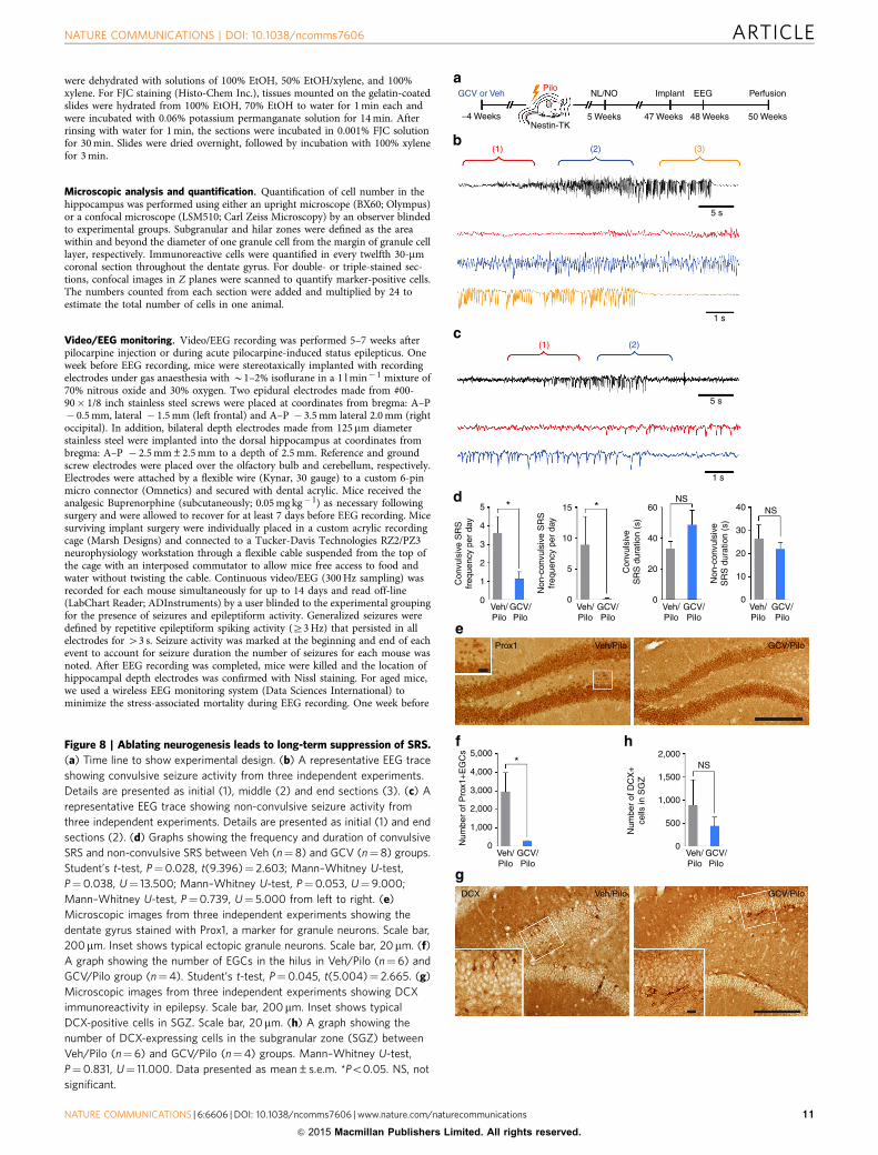
Ablating neurogenesis leads to long-term suppression of SRS.To examine whether ablated neurogenesis has a persistent effecton SRS frequency, we allowed the NL/NO-tested mice to age forB1 year after pilocarpine injection and performed EEG mon-itoring for 2 weeks (Fig. 8a). To minimize stress-associatedmortality in aged mice, we implanted epidural cortical screws andused a wireless EEG monitoring system instead of hippocampaldepth electrodes and a tethered system used previously foryoung animals. Since there was only one channel to monitorrecurrent seizures, we classified SRS as either convulsive (Fig. 8b,Supplementary Video 1) or non-convulsive (Fig. 8c,Supplementary Video 2) with video confirmation. Interestingly,the frequency of both convulsive and non-convulsive SRSwas significantly lower in GCV-treated mice than Veh-treatedanimals, with no difference in SRS duration for each seizure(Fig. 8d), consistent with our data of young animals. Moreover,the number of hilar EGCs was markedly decreased by ablated
neurogenesis (Fig. 8e,f), whereas the subgranular neurogenesisexamined by DCX immunohistochemistry was comparablebetween Veh and GCV groups (although both groupsshowed reduced number and altered morphology of DCXcells characteristic of aged mice36,37), suggesting the recovery ofsubgranular neurogenesis after a single round of GCV-inducedablation of neurogenesis (Fig. 8g,h). Taken together, ablationof aberrant neurogenesis before acute seizures can permanentlyreduce the number of chronic seizures, thus reinforcingthe therapeutic relevance of aberrant neurogenesis as a targetfor epilepsy.
DiscussionDefining the cellular and molecular changes after an initialseizure episode responsible for the formation of chronic seizuresis paramount for the development of effective anti-epileptogenicstrategies. Our data show that blocking adult neurogenesis beforeacute seizures reduced SRS, whereas the ablation of neurogenesispre- and post-acute seizures did not decrease the frequency ofrecurrent seizures. However, we also found that two rounds ofGCV treatment killed proliferating reactive astrocytes togetherwith seizure-generated granule neurons, so the question regardingthe role of post-seizure neurogenesis in epileptogenesis is stillopen. In addition, we showed that ablation of neurogenesis beforeacute seizures alleviated epilepsy-associated hippocampal mem-ory deficits. We also found that NeuroD is required for seizure-generated neurons, although future work is needed to determinethe functional consequences of deleting NeuroD in chronicseizures or associated cognitive function. Finally, a single ablationof adult neurogenesis showed a long-lasting effect on SRS byreducing seizure frequency, even at a late chronic stage of thedisease, further emphasizing the contributory role of aberrantneurogenesis in epileptogenesis.
A major goal in developing specific interventions forintractable epilepsy is to identify the cellular culprit(s) thatcontribute to neural circuits underlying seizure formation. Wefound fewer hilar EGCs as a result of ablating neural progenitors,which correlated with the reduction of SRS frequency in bothyoung and aged animals. Hilar EGCs receive more excitatoryinputs from mossy fibres than cells in the granule cell layer23,38.Thus, hilar EGCs can create more excitatory circuitry in thedentate gyrus and CA3 subregion of the hippocampus andfunction as hyperexcitable hubs, causing recurrent seizures39.Computational work further supported this idea, showing thatthe addition of a few granule cell hubs remarkably increased theexcitability of the network and lowered the threshold for seizureinitiation20,40, possibly leading to increased SRS frequency. Wethus speculate that the reduction of hilar EGCs in the networkthrough the ablation of neurogenesis could attenuate theformation of aberrant excitatory circuits in the hippocampus,and increase the threshold for seizure initiation, therebydecreasing SRS frequency. Indeed, a less-specific approachshowed that decreasing hilar EGCs decreases seizure frequencyin an earlier study13. However, it is possible that other features ofseizure-induced aberrant neurogenesis including hilar basaldendrites and cellular hypertrophy may also contribute to thedevelopment of epilepsy.
Another common pathological finding in TLE is MFS10,17,18.Prior reports indicate that only adult-generated granule neuronsdevelop MFS41,42; thus, it is surprising that we did not observe adifference in MFS after the ablation of neurogenesis. It is possiblethat non-ablated neurons (either still dividing or post mitotic atthe time of pilocarpine) after 4 weeks of GCV treatment canundergo changes in MFS, possibly compensating for the loss ofMFS from ablated adult-generated neurons. Alternatively,
NATURE COMMUNICATIONS | DOI: 10.1038/ncomms7606 ARTICLE
NATURE COMMUNICATIONS | 6:6606 | DOI: 10.1038/ncomms7606 | www.nature.com/naturecommunications 5
& 2015 Macmillan Publishers Limited. All rights reserved.

seizure-born neurons may increase MFS only in the GCV group,perhaps in response to ablation of neurogenesis. However,regardless of the detailed cell type(s) that contribute to MFS,our results showing no change in MFS while SRS is reduced,support the notion that MFS does not influence chronicseizures12,18.
It is surprising that we did not observe a reduction of chronicseizures after near-complete ablation of neurogenesis, which is incontrast to the single round of GCV treatment before acuteseizures. We speculate that there could be two possible reasons.First, two rounds of GCV treatment not only decreased aberrantneurogenesis but also deleted a population of proliferatingastrocytes, due to seizure-induced expression of Nestin-TK inreactive astrocytes. Since astrocytes have been shown to influenceexcitability greatly, removing this population could potentiallyresult in unexpected changes in SRS. Second, a population ofnewborn neurons that can suppress chronic seizures was ablatedby two rounds of GCV treatment. Consistent with the firstpossibility, we found two rounds of GCV treatment ablatedseizure-generated neurons and proliferating reactive astrocytes.Considering the uncertain role of astrocytes in epilepsy develop-ment43, ablation of reactive astrocytes might explain why thesuppressing effect of ablated neurogenesis on chronic seizures wasnot detected. Clearly, better tools are required to dissect the solecontribution of seizure-generated neurons in epileptogenesis.
Another possibility to consider is that adult-generatedneurons after seizures may be heterogeneous44. Seizureactivity stimulates the generation of adult-generated neuronsmigrating into the granule cell layer as well as ectopicallyinto the hilus. Moreover, even in the granule cell layer,seizure-born neurons are morphologically heterogeneous interms of spine density and number44, possibly contributingto a mixture of hyper- and hypoexcitable cells. Consistent withthe idea that functionally heterogeneous populations ofadult-born neurons exist and new neurons may also behypoexcitable, emerging data indicate that newborn neuronsmay serve as an inhibitory gate to prevent the propagation ofseizure activity entering the hippocampus45. Furthermore, wecannot assume that EGCs are always pathological, as they can befound in small numbers in the normal hippocampus20,46–48.Thus, a future challenge will be to identify unique tools thatdistinguish between aberrant and normal (or beneficial) adult-generated neurons.
We also show that the ablation of neurogenesis improvesepilepsy-associated cognitive decline. Cognitive decline observedin TLE patients includes problems with memory, executivefunction and low levels of intelligence49. Among them, memoryimpairment is the most common problem, which is notsurprising considering that the temporal lobe is required formemory formation49. Because epileptic seizures may produceadult-generated neurons that contribute to the formation ofaberrant hippocampal circuits and disrupt the normalnetwork22,38,50, seizure-induced abnormal granule cells may
cause epilepsy-associated learning and memory deficits.Consistent with this idea, we previously discovered that histonedeacetylase inhibitors such as VPA potently suppressed seizure-induced neurogenesis and restored hippocampal-dependentmemory function27. Since VPA is reported to have additionaltargets51, we further confirmed this hypothesis using a geneticablation model to address the role of aberrant neurogenesis inepilepsy-associated memory decline. Ablation of adultneurogenesis before acute seizures successfully normalizedhippocampal-dependent memory deficit observed in non-ablated epileptic mice. Currently, we lack specific tools tocompletely dissociate the contribution of aberrant newneurons—from the seizures themselves—to spatial memorydysfunction. Thus, although both groups experienced fewerthan three SRS per day (on average) in the pilocarpine model,it is possible that spatial memories may still be disrupted.However, we believe that this is the first report to demonstrate thelinks between seizure-induced aberrant hippocampalneurogenesis and cognitive decline associated with epilepsy.
Since epileptogenesis is a complex process involving multiplebrain regions and cell types, it is essential to identify which brainregions and circuit elements are critically involved in seizuregeneration. We showed that the ablation of aberrant hippocampalneurogenesis only reduced SRS frequency, but did not prevent theoccurrence of spontaneous seizures. Since the pilocarpine modelaffects not only the hippocampus, but also extrahippocampalregions such as the piriform and entorhinal cortices, amygdalaand thalamus52, it is possible that the remaining seizures stillobserved after ablation of neurogenesis might be chronic seizuresinvolving extrahippocampal regions. However, current animalmodels of epilepsy such as intrahippocampal KA injection are notideal to address this question, since there is destruction of theneurogenic niche in the injected hippocampus53,54. Thus, it willbe interesting to examine whether aberrant neurogenesis canprevent the occurrence of hippocampus-associated chronicseizures with a model that can specifically manipulatehippocampal circuitry without affecting other brain regions inthe future. In this manner, we may be able to address thecontribution of new dentate neurons to the initiation of seizures.A future goal is to confirm our work in a hippocampus-originatedmodel of chronic epilepsy where the ability to promoteneurogenesis is not compromised.
Finally, as epilepsy is typically a life-long disease withdebilitating recurrent seizures, it is crucial to develop long-lasting modalities to prevent, or at least alleviate, chronic seizures.To achieve this goal, two possible approaches are targetingthe latent period before the establishment of epilepsy or thechronic stage of epilepsy when spontaneous seizures arealready observed, hoping to permanently alter the naturalcourse of epilepsy. In this paper, we provide evidence thatblocking hippocampal neurogenesis before acute seizuresredirects the epileptogenic course to persistently mitigatethe frequency of spontaneous seizures even during aging.
Figure 3 | Ablating neurogenesis does not affect acute seizure severity (a) Experimental time line. After 4 weeks of GCV or Veh treatment, video/EEG
was recorded from 1 h before pilocarpine (Pilo) injection to 3 days after acute seizures. (b–d) Representative EEG traces from two independent
experiments showing EEG stage 1, stage 2 and stage 3, respectively. (e) A graph showing time to the first EEG seizure, which was not different between
Veh/Pilo (n¼ 10) and GCV/Pilo groups (n¼ 13). Mann–Whitney U-test, P¼0.457, U¼ 53.000. (f) A graph showing time to status epilepticus (SE)
between Veh- and GCV-treated groups (n¼ 10 for Veh/Pilo, n¼ 13 for GCV/Pilo). Mann–Whitney U-test, P¼0.154, U¼42.000. (g) Representative
microscopic images from four independent experiments showing degenerating neurons labelled with FJC after acute seizures. FJC-positive cells were
observed in the hilus and the CA3 subregion of the hippocampus in both Veh/Pilo and GCV/Pilo groups. Scale bar, 100 mm. Insets show typical FJC-positive
cells in the hilus and the CA3 subregion. Scale bar, 20mm. (h) A graph showing the number of FJC-positive cells in the hilus and the CA3 subregion of the
hippocampus between Veh/Pilo (n¼4) and GCV/Pilo (n¼ 5) groups. Mann–Whitney U-test, P¼ 1.000, U¼ 10.000 for the hilar analysis; Mann–Whitney
U-test, P¼0.327, U¼ 6.000 for the CA3 analysis. Data presented as mean±s.e.m. NS, not significant. LF, left frontal; LH, left hippocampal; RH, right
hippocampal; RO, right occipital.
ARTICLE NATURE COMMUNICATIONS | DOI: 10.1038/ncomms7606
6 NATURE COMMUNICATIONS | 6:6606 | DOI: 10.1038/ncomms7606 | www.nature.com/naturecommunications
& 2015 Macmillan Publishers Limited. All rights reserved.

GCV or Veh
–4 Weeks –1 Week
Implant
EEG
Perfusion
3dNestin-TK
1st EEG seizure activity - discrete event ~9 min after Pilo (EEG stage 1)
Waxing and waning EEG seizure activity ~25 min after Pilo (EEG stage 2)
Continuous EEG seizure activity ~30 min after Pilo (EEG stage 3)
LF
LH
RH
RO
LF
LH
RH
RO
LF
LH
RH
RO
LF
LH
RH
RO
LF
LH
RH
RO
LF
LH
RH
RO
50 NS NS NS
NS
Veh/Pilo
Veh/Pilo
GCV/Pilo
Veh/Pilo
GCV/Pilo
Veh/Pilo
GCV/Pilo
Hilus
Hilu
s
GCV/PiloVeh/PiloFJC
CA3
CA
3
GCV/Pilo
4040
60
30
2020
Tim
e to
SE
ons
et (
min
)
Num
ber
of F
JC+
cel
ls
Tim
e to
firs
t ele
ctro
grap
hic
seiz
ure
(min
)
10
0 0
Pilo
20,000
10,000
15,000
5,000
0
NATURE COMMUNICATIONS | DOI: 10.1038/ncomms7606 ARTICLE
NATURE COMMUNICATIONS | 6:6606 | DOI: 10.1038/ncomms7606 | www.nature.com/naturecommunications 7
& 2015 Macmillan Publishers Limited. All rights reserved.

Since a single ablation of adult neurogenesis wassufficient to produce long-lasting suppressive effects onrecurrent seizures at a late chronic stage of epilepsy, ourfinding can greatly strengthen the therapeutic impact forreducing seizures throughout life. Now with knowledge from
GCV or Veh
GCV or Veh
Non-TG CONT
Non-TG CONT4 Weeks 5 Weeks
EEGImplantPilo
DCX
40,000
30,000
20,000
Veh GCV GCV/Pilo
GCV/Pilo
Veh/Pilo
Veh/Pilo
NS
Num
ber
of D
CX
+ce
lls in
SG
Z
SR
S fr
eque
ncy
per
day
SR
S d
urat
ion
(s)
10,000
0
4
60
40
20
5
3
2
1
0 0
Veh GCV
Perfusion
Perfusion
4 Weeks
–4 Weeks 7 Weeks
NSNS
Figure 4 | No off-target effects of GCV administration. (a) Experimental
time line. Non-transgenic control mice without thymidine kinase (Non-TG
CONT) were administered GCV or Veh for 4 weeks. (b) Representative
microscopic images from four independent experiments showing DCX
immunoreactivity between Veh and GCV groups. Scale bar, 100mm. Inset
shows typical DCX-positive cells. Scale bar, 20mm. (c) A graph showing the
number of DCX-expressing cells in the subgranular zone (SGZ) between
Veh and GCV groups (n¼ 3 per group). Student’s t-test, P¼0.818,
t(4)¼ �0.246. (d) Time line to show experimental design. (e) Graphs
showing the frequency and duration of SRS between Veh/Pilo (n¼8) and
GCV/Pilo groups (n¼ 6). Student’s t-test, P¼0.803, t(12)¼ �0.255 for
the left graph; Student’s t-test, P¼0.664, t(6.243)¼ �0.456 for the right
graph. Data presented as mean±s.e.m. NS, not significant.
20,000
2,000
2,0001,000
10,000
3,000
4,000
4,000
6,000
*8,000
*15,000
Num
ber
of D
CX
+ce
lls in
SG
Z
Num
ber
of D
CX
+ce
lls in
the
hilu
s
Num
ber
of P
rox1
+ E
GC
s
5,000
*
0 0 0Veh/Pilo
Veh/Pilo
Veh/PiloProx1
Veh/Pilo
GCV/Pilo
GCV/Pilo
GCV/Pilo
GCV/Pilo
Veh/PiloDCX
–4 Weeks
GCV or VehPilo
Nestin-TK6 Weeks
Perfusion
GCV/Pilo
Figure 5 | Ablating adult neurogenesis reduces aberrant neurogenesis.
(a) Time line for histologic analysis. (b) Representative microscopic images
from three independent experiments showing DCX immunoreactivity at 6
weeks after pilocarpine (Pilo) injection. Scale bar is 100mm. Inset shows
typical DCX-positive cells after pilocarpine. Scale bar is 20mm. (c) A graph
showing the number of DCX-expressing cells in the SGZ and the hilus in
Veh/Pilo (n¼ 8) and GCV/Pilo group (n¼ 7). Student’s t-test, P¼0.003,
t(8.569)¼4.156 for the left graph; Student’s t-test, P¼0.020,
t(7.137)¼ 2.971 for the right graph. (d) Representative microscopic images
from three independent experiments showing the dentate gyrus stained with
Prox1, a marker for granule neurons. Scale bar, 100mm. Inset shows a typical
ectopic granule neuron. Scale bar, 20mm. (e) A graph showing the number of
EGCs in the hilus of Veh/Pilo (n¼8) and GCV/Pilo group (n¼8). Student’s
t-test, P¼0.012, t(9.871)¼ 3.056. Data presented as mean±s.e.m.
*Po0.05.
Figure 6 | Seizures persist after near-complete ablation of neurogenesis. (a) Experimental design. (b) Microscopic images from three experiments
showing DCX immunoreactivity at 6 weeks after pilocarpine (Pilo) injection. Scale bar, 200mm. Inset shows typical DCX-positive cells in epilepsy. Scale
bar, 20mm. (c) Graphs showing the number of DCX-expressing cells in subgranular zone (SGZ) and the hilus in Veh/Pilo/Veh (n¼ 11) and GCV/Pilo/GCV
group (n¼ 12). Mann–Whitney U-test, Po0.001, U¼ 3.000 for the left graph; Mann–Whitney U-test, Po0.001, U¼8.000 for the right graph.
(d) Microscopic images from three experiments showing the dentate gyrus stained with Prox1, a marker for granule neurons. Scale bar, 100 mm. Inset
shows typical EGCs in epilepsy. Scale bar, 20mm. (e) A graph showing the number of EGCs in the hilus of Veh/Pilo/Veh (n¼ 11) and GCV/Pilo/GCV group
(n¼ 12). Mann–Whitney U-test, Po0.001, U¼8.000. (f) Experimental design. (g) Graphs showing SRS frequency and duration between Veh/Pilo/Veh
(n¼ 24) and GCV/Pilo/GCV groups (n¼ 22). Mann–Whitney U-test, P¼0.092, U¼ 187.500 for the left graph; Student’s t-test, P¼0.404,
t(41)¼ �0.843 for the right graph. (h) Experimental design. (i) Confocal images from three experiments showing representative Nestin-TK GFP/GFAP/
BrdU- (arrows) and GFAP/BrdU-immunoreactive cells (arrowheads) in the hilus. Scale bar, 20mm. (j) Graphs showing the number of proliferating reactive
astrocytes expressing Nestin-TK GFP, examined by Nestin-TK GFP/GFAP/BrdUþ cells and proliferating astrocytes not expressing Nestin-TK GFP, labelled
as GFAP/BrdU staining, between Veh/Pilo/Veh (n¼ 5) and GCV/Pilo/GCV group (n¼ 6). Student’s t-test, P¼0.049, t(9)¼ 2.281 for the left graph;
Student’s t-test, P¼0.270, t(9)¼ 1.174 for the right graph. Data presented as mean±s.e.m. *Po0.05. NS, not significant.
ARTICLE NATURE COMMUNICATIONS | DOI: 10.1038/ncomms7606
8 NATURE COMMUNICATIONS | 6:6606 | DOI: 10.1038/ncomms7606 | www.nature.com/naturecommunications
& 2015 Macmillan Publishers Limited. All rights reserved.

our work that seizure-induced neurons contribute torecurrent seizures and associated memory impairment, we maybe able to develop more effective treatment strategiesfor intractable epilepsy by targeting aberrant hippocampal
neurogenesis. Our studies also provide a cautionary noteregarding neuroregenerative approaches whereby induction ofaberrant neurogenesis may exacerbate rather than mitigatedisease symptoms.
GCV or Veh
GCV or VehPilo
–4 WeeksNestin-TK
DCX Veh/Pilo/Veh GCV/Pilo/GCV
Prox1
2,500
2,000
1,000
1,000
2,000
3,000
4,000
No.
of D
CX
+ c
ells
in h
ilus
No.
of D
CX
+ c
ells
in S
GZ
Num
ber
of P
rox1
+ E
GC
s
1,500
500
0Veh/Pilo/Veh
Veh/Pilo/Veh
GCV/Pilo/GCV
GCV/Pilo/GCV
Veh/Pilo/Veh
GCV/Pilo/GCV
Veh/Pilo/Veh
GCV/Pilo/GCV
10,000
15,000 **
5,000
0
0
10
20
30
40
50S
RS
dur
atio
n (s
)
SR
S fr
eque
ncy
per
day
Veh/Pilo/Veh
GCV/Pilo/GCV
NSNS
0
1
2
3
4
0
Veh/Pilo/Veh GCV/Pilo/GCV
6 Weeks
Perfusion
GCV or Veh
GCV or Veh Pilo
–4 Weeks 4 Weeks 5 WeeksNestin-TK
7 Weeks
PerfusionEEGImplant
GCV or Veh
GCV or VehPilo
–4 Weeks 1d 3d2dNestin-TK
Nestin-GFP GFAP BrdU Merge
6 Weeks
PerfusionBrdU
*
1,000
2,000
3,000
4,000
Num
ber
of G
FAP
/Brd
U+
cells
in th
e hi
lus
Num
ber
ofG
FP
/GFA
P/B
rdU
+ce
lls in
the
hilu
s
Veh/Pilo/Veh
GCV/Pilo/GCV
0
*
1,000
2,000
3,000
4,000
Veh/Pilo/Veh
GCV/Pilo/GCV
0
NS
NATURE COMMUNICATIONS | DOI: 10.1038/ncomms7606 ARTICLE
NATURE COMMUNICATIONS | 6:6606 | DOI: 10.1038/ncomms7606 | www.nature.com/naturecommunications 9
& 2015 Macmillan Publishers Limited. All rights reserved.

MethodsMice. All mice (C57BL/6 background, male and female) were bred andhoused in the animal facility with a 12-h light, 12-h dark cycle with no more thanfive mice per cage, unless stated otherwise. Nestin-d-HSV-thymidine kinase-EGFP transgenic (Nestin-TK) mice30 were genotyped by PCR using genomic DNAand primers for Nestin-TK (50-GCC TTG ACC AGG GTG AGA TA-30 , 50-ATGCTG CCC ATA AGG TAT CG-30) and GFP (50-GAG CTG GAC GGC GAC GTAAAC-30 , 50-CGT TGT GGC TGT TGT TAG TTG TAC-30). Nestin-TK micewere crossed with C57BL/6 obtained from Harlan Laboratories to generatetransgenic and WT mice. Male and female mice at 6 weeks of age were insertedwith an osmotic mini pump (Model 2004; Alzet) filled with either GCV(subcutaneously) at 150 mg kg� 1 per day or distilled water, which was removed4 weeks later. All the experiments were performed in compliance with theanimal care guidelines issued by the National Institutes of Health and by theInstitutional Animal Use and Care Committee at University of Texas SouthwesternMedical Center.
Chemoconvulsant models of TLE. Nestin-TK mice at 10 weeks of age wereadministered scopolamine methyl nitrate (intraperitoneally (i.p.); 2 mg kg� 1;Sigma-Aldrich S2250) and terbutaline hemisulfate salt (i.p.; 2 mg kg� 1; Sigma-Aldrich T2528) to block peripheral effects of pilocarpine and dilate the respiratorytract, respectively. Thirty min later, pilocarpine hydrochloride (i.p.; Sigma-AldrichP6503) at 245 mg kg� 1 for males and 270 mg kg� 1 for females was injected, andmice were placed in an incubator maintained at 31 �C (ThermoCare). Acute sei-zures were behaviourally monitored using a modified Racine’s scale55 (stage 1,mouth and facial movement; stage 2, head nodding; stage 3, forelimb clonus; stage4, rearing with forelimb clonus; stage 5, rearing and falling with forelimb clonus).Once status epilepticus began (defined by continuous tonic clonic convulsiveseizures), mice were placed at room temperature for 3 h and returned to theincubator after seizure activity was reduced with diazepam (10 mg kg� 1; Sigma-Aldrich D0899). Only mice showing status epilepticus were selected for furtherprocessing and osmotic mini pumps were removed. Mice were administered 5%dextrose solution (i.p.; 1 ml) and saline (i.p.; 1 ml) to facilitate their recovery. At2 days after pilocarpine injection, mice were returned to their cages and randomlyassigned to the experiments for histology, video/EEG and behavioural tests. Onecohort of animals were injected with BrdU (i.p.; 150 mg kg� 1; Sigma-AldrichB5002) once a day on days 1–3 after pilocarpine to label proliferating cells afteracute seizures.
Immunohistochemistry. Mice were anaesthetized and perfused transcardially withcold 4% paraformaldehyde (PFA) in 0.1 M PBS. Brains were removed and postfixedin 4% PFA overnight, then cryoprotected in 30% sucrose in 0.1 M PBS. Brains werebisected and half-brains were coronally sectioned 30 mm thick on a freezingmicrotome. Immunohistochemistry was performed with either tissue mounted oncharged slides or free-floating tissue sections. Slides underwent antigen retrievalusing 0.01 M citric acid, pH 6.0 at 100 �C for 15 min, followed by 12 min in TBS atroom temperature. Staining with free-floating tissue sections was the same exceptfor the antigen retrieval step that was omitted. For Tyramide-Plus signal amplifi-cation31, we removed endogenous peroxidase activity by incubating sections with0.3% H2O2 for 30 min at room temperature. Nonspecific binding was blocked with3% normal donkey serum and 0.3% Triton-X-100 in TBS for 1 h at roomtemperature. Primary antibodies in this study were chosen based on the validationresults by the manufacturer: goat anti-NeuroD (1:1,000, Santa Cruz Biotechnologysc-1084), chicken anti-GFP (1:8,000, Aves Lab GFP-1020), goat anti-DCX (1:2,000for free-floating sections, 1:500 for tissue mounted on slides, Santa CruzBiotechnology sc-8066), guinea pig anti-DCX (1:2,000, Millipore AB2253), rabbit-anti-Prox1 (1:500, Millipore AB5475), mouse anti-GFAP (1:1,000, MilliporeMAB360), rat-anti-BrdU (1:500, Accurate Chemical & Scientific CorporationOBT0030G). For double or triple labelling, primary antibodies were simultaneouslyincubated (for example, NeuroD/DCX, GFP/GFAP) and further processed for eachantibody. For NeuroD and GFAP, a fluorescent-tagged secondary antibody wasused (1:300, Jackson ImmunoResearch). For GFP and DCX, primary antibodyincubation was followed with an appropriate biotin-tagged secondary antibody(1:200, Jackson ImmunoResearch) for 1 h at room temperature followed by ABC(Vector Laboratories PK-6100) for 1 h and Tyramide-Plus signal amplification(1:50, PerkinElmer NEL701001KT) for 10 min. Sections were counterstained withDAPI (40 ,6-diamidino-2-phenylindole; 1:5,000, Roche 236276). For DCX andProx1, after biotin-tagged secondary antibody followed by ABC labelling wascompleted, sections were visualized with metal-enhanced DAB substrate kit(Thermo Scientific 34065). For BrdU triple staining, after GFP and GFAP stainingis finished, the sections were mounted and further fixed with PFA for 30 min. Then,the slides were permeabilized with 0.1% trypsin in 0.1 M Tris and 0.1% CaCl2 for10 min, followed by denaturation with 2 N HCl solution for 25 min. Slides werethen blocked and processed as described above. For Nissl staining, tissues weremounted on slides and underwent a series of hydration steps from 100% EtOH totap water (100, 95, 90, 80 and 70% EtOH for 3 min each) and were then incubatedwith 0.1% cresyl violet solution (Sigma-Aldrich C5042) for 15 min. Excessivestaining was removed by 95% EtOH with 0.1% glacial acetic acid, and the slides
NS NS**
1.0
7 Weeks6 Weeks
NO
NO
OF
OF
NL
NLSham or Pilo
Pilo
5 WeeksNestin-TK
Nestin-TK7 Weeks6 Weeks5 Weeks
–4 Weeks
–4 Weeks
GCV or Veh
0.8
0.6
Pre
fere
nce
ratio
(N
O)
0.4
0.2
0
1.0
0.8
0.6
Pre
fere
nce
ratio
(N
O)
0.4
0.2
0
1.0
0.8
0.6
Pre
fere
nce
ratio
(N
L)
0.4
0.2
0
1.0
0.8
0.6
Pre
fere
nce
ratio
(N
L)
0.4
0.2
0Sham Pilo Sham Pilo Veh/
PiloGCV/Pilo
Veh/Pilo
GCV/Pilo
Figure 7 | Ablating neurogenesis rescues cognitive decline in epilepsy. (a,b) Experimental time line is shown. (c) A graph showing the preference ratio of
NO location (NL) test in sham mice (n¼ 10) and pilocarpine-injected mice (n¼ 9). Student’s t-test, P¼0.0001, t(17)¼ 5.017. (d) A graph showing the
preference ratio of NL test between Veh (n¼ 16) and GCV (n¼ 17) groups. Student’s t-test, P¼0.008, t(31)¼ � 2.850. (e,f) Graphs showing the
preference ratio in NO test (n¼4 for sham, n¼ 5 for Pilo; n¼ 18 for Veh/Pilo, n¼ 15 for GCV/Pilo). (e) Mann–Whitney U-test, P¼ 1.000, U¼ 10.000.
(f) Student’s t-test, P¼0.937, t(31)¼ �0.080. Data presented as mean±s.e.m. *Po0.05. NS, not significant.
ARTICLE NATURE COMMUNICATIONS | DOI: 10.1038/ncomms7606
10 NATURE COMMUNICATIONS | 6:6606 | DOI: 10.1038/ncomms7606 | www.nature.com/naturecommunications
& 2015 Macmillan Publishers Limited. All rights reserved.
were dehydrated with solutions of 100% EtOH, 50% EtOH/xylene, and 100%xylene. For FJC staining (Histo-Chem Inc.), tissues mounted on the gelatin-coatedslides were hydrated from 100% EtOH, 70% EtOH to water for 1 min each andwere incubated with 0.06% potassium permanganate solution for 14 min. Afterrinsing with water for 1 min, the sections were incubated in 0.001% FJC solutionfor 30 min. Slides were dried overnight, followed by incubation with 100% xylenefor 3 min.
Microscopic analysis and quantification. Quantification of cell number in thehippocampus was performed using either an upright microscope (BX60; Olympus)or a confocal microscope (LSM510; Carl Zeiss Microscopy) by an observer blindedto experimental groups. Subgranular and hilar zones were defined as the areawithin and beyond the diameter of one granule cell from the margin of granule celllayer, respectively. Immunoreactive cells were quantified in every twelfth 30-mmcoronal section throughout the dentate gyrus. For double- or triple-stained sec-tions, confocal images in Z planes were scanned to quantify marker-positive cells.The numbers counted from each section were added and multiplied by 24 toestimate the total number of cells in one animal.
Video/EEG monitoring. Video/EEG recording was performed 5–7 weeks afterpilocarpine injection or during acute pilocarpine-induced status epilepticus. Oneweek before EEG recording, mice were stereotaxically implanted with recordingelectrodes under gas anaesthesia with B1–2% isoflurane in a 1 l min� 1 mixture of70% nitrous oxide and 30% oxygen. Two epidural electrodes made from #00-90� 1/8 inch stainless steel screws were placed at coordinates from bregma: A–P� 0.5 mm, lateral � 1.5 mm (left frontal) and A–P � 3.5 mm lateral 2.0 mm (rightoccipital). In addition, bilateral depth electrodes made from 125mm diameterstainless steel were implanted into the dorsal hippocampus at coordinates frombregma: A–P � 2.5 mm±2.5 mm to a depth of 2.5 mm. Reference and groundscrew electrodes were placed over the olfactory bulb and cerebellum, respectively.Electrodes were attached by a flexible wire (Kynar, 30 gauge) to a custom 6-pinmicro connector (Omnetics) and secured with dental acrylic. Mice received theanalgesic Buprenorphine (subcutaneously; 0.05 mg kg� 1) as necessary followingsurgery and were allowed to recover for at least 7 days before EEG recording. Micesurviving implant surgery were individually placed in a custom acrylic recordingcage (Marsh Designs) and connected to a Tucker-Davis Technologies RZ2/PZ3neurophysiology workstation through a flexible cable suspended from the top ofthe cage with an interposed commutator to allow mice free access to food andwater without twisting the cable. Continuous video/EEG (300 Hz sampling) wasrecorded for each mouse simultaneously for up to 14 days and read off-line(LabChart Reader; ADInstruments) by a user blinded to the experimental groupingfor the presence of seizures and epileptiform activity. Generalized seizures weredefined by repetitive epileptiform spiking activity (Z3 Hz) that persisted in allelectrodes for 43 s. Seizure activity was marked at the beginning and end of eachevent to account for seizure duration the number of seizures for each mouse wasnoted. After EEG recording was completed, mice were killed and the location ofhippocampal depth electrodes was confirmed with Nissl staining. For aged mice,we used a wireless EEG monitoring system (Data Sciences International) tominimize the stress-associated mortality during EEG recording. One week before
GCV or Veh
(2)
(2)
(3)
–4 Weeks 5 Weeks 47 Weeks 48 Weeks 50 Weeks
5 s
1 s
5 s
1 s
40
40
15
10
5
4
2
3
15
0
**
0
60NS
NS
NS
Non
-con
vuls
ive
SR
S d
urat
ion
(s)
Con
vuls
ive
SR
S d
urat
ion
(s)
Non
-con
vuls
ive
SR
Sfr
eque
ncy
per
day
Con
vuls
ive
SR
Sfr
eque
ncy
per
day
30
20
2010
Veh/Pilo
GCV/Pilo
Veh/Pilo
GCV/Pilo
Veh/Pilo
0
1,000
2,000
0
1,000
1,500
500
2,000
3,000
4,000
5,000
Num
ber
of P
rox1
+E
GC
s
Num
ber
of D
CX
+ce
lls in
SG
Z
GCV/Pilo
Veh/Pilo
GCV/Pilo
Veh/Pilo
Veh/Pilo
GCV/Pilo
GCV/Pilo
Veh/Pilo
GCV/Pilo
Prox1
Veh/Pilo GCV/PiloDCX
00
PerfusionEEGImplantNL/NO
Nestin-TK
Pilo
(1)
(1)
*
Figure 8 | Ablating neurogenesis leads to long-term suppression of SRS.
(a) Time line to show experimental design. (b) A representative EEG trace
showing convulsive seizure activity from three independent experiments.
Details are presented as initial (1), middle (2) and end sections (3). (c) A
representative EEG trace showing non-convulsive seizure activity from
three independent experiments. Details are presented as initial (1) and end
sections (2). (d) Graphs showing the frequency and duration of convulsive
SRS and non-convulsive SRS between Veh (n¼8) and GCV (n¼8) groups.
Student’s t-test, P¼0.028, t(9.396)¼ 2.603; Mann–Whitney U-test,
P¼0.038, U¼ 13.500; Mann–Whitney U-test, P¼0.053, U¼ 9.000;
Mann–Whitney U-test, P¼0.739, U¼ 5.000 from left to right. (e)
Microscopic images from three independent experiments showing the
dentate gyrus stained with Prox1, a marker for granule neurons. Scale bar,
200mm. Inset shows typical ectopic granule neurons. Scale bar, 20mm. (f)
A graph showing the number of EGCs in the hilus in Veh/Pilo (n¼6) and
GCV/Pilo group (n¼4). Student’s t-test, P¼0.045, t(5.004)¼ 2.665. (g)
Microscopic images from three independent experiments showing DCX
immunoreactivity in epilepsy. Scale bar, 200 mm. Inset shows typical
DCX-positive cells in SGZ. Scale bar, 20mm. (h) A graph showing the
number of DCX-expressing cells in the subgranular zone (SGZ) between
Veh/Pilo (n¼ 6) and GCV/Pilo (n¼4) groups. Mann–Whitney U-test,
P¼0.831, U¼ 11.000. Data presented as mean±s.e.m. *Po0.05. NS, not
significant.
NATURE COMMUNICATIONS | DOI: 10.1038/ncomms7606 ARTICLE
NATURE COMMUNICATIONS | 6:6606 | DOI: 10.1038/ncomms7606 | www.nature.com/naturecommunications 11
& 2015 Macmillan Publishers Limited. All rights reserved.

EEG recording, mice were implanted with epidural electrodes placed at coordinatesfrom bregma: A–P � 2.0 mm, lateral 2.2 mm and A–P 1.0 mm, lateral 2.0 mm.Cortical electrodes were connected to wireless EEG transmitters (TA11ETAF10;Data Sciences International) positioned in the subcutaneous pocket created in theback. Video-EEG data, collected 24/7 for 2 weeks, were analysed using Neuroscoresoftware (version 3.0; Data Sciences International) by a user blinded to experi-mental groups. Convulsive seizures were defined by a burst of spiking activity(Z3 Hz) that persisted for 43 s with high amplitudes (42� background activity)and tonic clonic behaviours confirmed by video, whereas non-convulsive seizureswere defined by the same EEG criteria but without behavioral seizure activitygreater than stage 2 from a modified Racine’s scale.
Behavioural tests. Behavioural tests were conducted from 5–7 weeks after pilo-carpine or saline injection, starting with open-field test followed by NL and NOrecognition tasks. Locomotor activity in the open-field box was assessed during the15 min habituation phase without objects in the NL task (Day 1). Mice werepositioned in the center of the box, and then individual total distance movedduring the first 10 min of habituation was automatically recorded using videotracking system (Noldus Information Technology). As for NL and NO tests, whichare based on the spontaneous preference of rodents for novelty and their ability toremember previously encountered location and objects, NL and NO tasks arehippocampal-dependent56,57 or -independent memory58, respectively. In thisstudy, we modified the published methods34,56 to adapt these tasks to seizure-pronemice. Each task was comprised of three phases (habituation, familiarization andtesting) on separate days. All phases were performed in a blue-squared open-fieldbox (44� 44� 30 cm) under dim light condition (60 lux) between 5:00 and 8:00hours. All mice tested were acclimated in the testing room using a red light at 18:00hours on the day before each phase. In the NL task, each mouse was habituated inthe open-field box without objects for 15 min (Day 1) and the following day withtwo identical objects (50 ml tubes filled with water, object A) for 15 min (Day 2). Inthe familiarization phase for NL task (Day 3), mice were subjected to a 1-minrehabituation to the empty box and then placed in a holding cage. Immediatelyafter two identical objects (black clips, object B) were placed in diagonal near thecorners in the box (B5 cm from the walls), mice were returned to the box forfamiliarization. The mice were allowed to freely explore until they accumulated atotal of 30 s exploring both objects. Exploration was defined as the mousecontacting the object with its whiskers, nose, or front paws. We ensured that everymouse spent the same amount of time exploring the objects and avoided any biasdue to differences in individual levels of exploration by removing the animal fromthe box once it had explored the objects for a total of 30 s. Also, behaviours such assitting on object, looking around or resting against object were not counted asexploratory time. Once reaching the 30 s criteria, mice were then removed from thebox and returned to their home cages. The mice that did not reach the criteriawithin 15 min were excluded from further testing. In the NL testing phase (Day 4),mice were subjected to a 1-min rehabituation to the empty box and then placed ina holding cage. Immediately after two identical objects (object B), one of which wasmoved to a NL, were placed near the corners in the box, mice were reintroducedinto the box, and their exploratory behaviours were recorded by video camera for10 min, and analysed later. Time spent exploring each object for 10 min was scored.In the NO task, each mouse was habituated in the open-field box with two identicalobjects (object A) for 15 min (Day 8). In the familiarization phase for NO task(Day 9), mice were subjected to a 1-min rehabituation to the empty box and thenplaced in a holding cage. Just after two identical objects (metal cones, object C)were placed near the corners on one wall in the box (B5 cm from the walls), micewere returned to the box for familiarization, allowed to freely explore until reachingthe 30 s criteria, and then removed from the box and returned to their home cages.In the NO testing phase (Day 10), mice were subjected to a 1-min rehabituation tothe empty box and then placed in a holding cage. Immediately after two objects,one of which was a novel one (plastic doll, object D) and the other was a familiarone (object C), were placed in the same positions as during familiarization, micewere reintroduced into the box, and their exploratory behaviours were recorded byvideo camera for 10 min, and scored later. Location of the moved object in the NLand position of the object used as novel or familiar in the NO werecounterbalanced across mice. To analyse the cognitive performance, preference ofNL or object was calculated as the time spent exploring the object in the NL or NOdivided by the cumulative time spent exploring both objects. The open-field boxwas cleaned thoroughly with disinfectant solution after each trial to ensure theabsence of olfactory cues.
Statistics. All of the data are expressed as mean±s.e.m. Experimental groups wereassigned by simple randomization. No statistical methods were used to pre-determine sample sizes, but our sample sizes are similar to those reported inprevious publications12,17,56,57. Data that passed our selection criteria werecollected blind. Once data were generated, all were included for analysis except forthe NL test (Fig. 7d), where one mouse in Veh/Pilo group showed an extremely lowpreference ratio (0.021) and was thus removed from the dataset. SPSS (version 21.0,IBM SPSS Corp.) software was used for statistical comparison. Statisticaldifferences were analysed using two-tailed Student’s t-test for the data with equalvariances (Figs 1e and 2f left, 2f right, 4c, 4e left, 6g right, 6j left, 6j right, 7c, 7d, 7for Student’s t-test with Satterthwaite’s correction for the data with unequal
variances (Fig. 4e right, 5c left, 5c right, 5e, 8d far left, 8f). If normal distributionwas not assumed, Mann–Whitney U-test was performed (Figs 1c, 3e,f, h and 6c left,6c right, 6e, 6g left, 7e, 8d left, 8d right, 8d far right, 8h). Values of Po0.05 wereconsidered significant.
References1. Chang, B. S. & Lowenstein, D. H. Epilepsy. New Engl. J. Med. 349, 1257–1266
(2003).2. Engel, Jr J. Mesial temporal lobe epilepsy: what have we learned? Neuroscientist
7, 340–352 (2001).3. Pitkanen, A. & Sutula, T. P. Is epilepsy a progressive disorder? Prospects
for new therapeutic approaches in temporal-lobe epilepsy. Lancet Neurol. 1,173–181 (2002).
4. Eriksson, P. S. et al. Neurogenesis in the adult human hippocampus. Nat. Med.4, 1313–1317 (1998).
5. Ming, G. L. & Song, H. Adult neurogenesis in the mammalian central nervoussystem. Annu. Rev. Neurosci. 28, 223–250 (2005).
6. Jin, K., Wang, X., Xie, L., Mao, X. O. & Greenberg, D. A. Transgenicablation of doublecortin-expressing cells suppresses adult neurogenesis andworsens stroke outcome in mice. Proc. Natl Acad. Sci. USA 107, 7993–7998(2010).
7. Blaiss, C. A. et al. Temporally specified genetic ablation of neurogenesis impairscognitive recovery after traumatic brain injury. J. Neurosci. 31, 4906–4916(2011).
8. Snyder, J. S., Soumier, A., Brewer, M., Pickel, J. & Cameron, H. A. Adulthippocampal neurogenesis buffers stress responses and depressive behaviour.Nature 476, 458–461 (2011).
9. Sahay, A. et al. Increasing adult hippocampal neurogenesis is sufficient toimprove pattern separation. Nature 472, 466–470 (2011).
10. Parent, J. M. et al. Dentate granule cell neurogenesis is increased by seizuresand contributes to aberrant network reorganization in the adult rathippocampus. J. Neurosci. 17, 3727–3738 (1997).
11. Scharfman, H. E. Epilepsy as an example of neural plasticity. Neuroscientist 8,154–173 (2002).
12. Pun, R. Y. et al. Excessive activation of mTOR in postnatally generated granulecells is sufficient to cause epilepsy. Neuron 75, 1022–1034 (2012).
13. Jung, K. H. et al. Continuous cytosine-b-D-arabinofuranoside infusion reducesectopic granule cells in adult rat hippocampus with attenuation of spontaneousrecurrent seizures following pilocarpine-induced status epilepticus. Eur. J.Neurosci. 19, 3219–3226 (2004).
14. Jung, K. H. et al. Cyclooxygenase-2 inhibitor, celecoxib, inhibits the alteredhippocampal neurogenesis with attenuation of spontaneous recurrent seizuresfollowing pilocarpine-induced status epilepticus. Neurobiol. Dis. 23, 237–246(2006).
15. Pekcec, A., Lupke, M., Baumann, R., Seifert, H. & Potschka, H. Modulation ofneurogenesis by targeted hippocampal irradiation fails to affect kindlingprogression. Hippocampus 21, 866–876 (2011).
16. Raedt, R. et al. Radiation of the rat brain suppresses seizure-inducedneurogenesis and transiently enhances excitability during kindling acquisition.Epilepsia 48, 1952–1963 (2007).
17. Zeng, L. H., Rensing, N. R. & Wong, M. The mammalian target of rapamycinsignaling pathway mediates epileptogenesis in a model of temporal lobeepilepsy. J. Neurosci. 29, 6964–6972 (2009).
18. Buckmaster, P. S. & Lew, F. H. Rapamycin suppresses mossy fiber sprouting butnot seizure frequency in a mouse model of temporal lobe epilepsy. J. Neurosci.31, 2337–2347 (2011).
19. Ribak, C. E. et al.in Jasper’s Basic Mechanisms of the Epilepsies (eds Noebels, J.L., Avoli, M., Rogawski, M. A., Olsen, R. W. & Delgado-Escueta, A. V.)(National Center for Biotechnology Information, 2012).
20. Myers, C. E., Bermudez-Hernandez, K. & Scharfman, H. E. The influence ofectopic migration of granule cells into the hilus on dentate gyrus-CA3 function.PLoS ONE 8, e68208 (2013).
21. Cameron, M. C., Zhan, R. Z. & Nadler, J. V. Morphologic integration of hilarectopic granule cells into dentate gyrus circuitry in the pilocarpine model oftemporal lobe epilepsy. J. Comp. Neurol. 519, 2175–2192 (2011).
22. Scharfman, H. E., Goodman, J. H. & Sollas, A. L. Granule-like neurons atthe hilar/CA3 border after status epilepticus and their synchrony with areaCA3 pyramidal cells: functional implications of seizure-induced neurogenesis.J. Neurosci. 20, 6144–6158 (2000).
23. Zhan, R. Z., Timofeeva, O. & Nadler, J. V. High ratio of synaptic excitationto synaptic inhibition in hilar ectopic granule cells of pilocarpine-treated rats.J. Neurophysiol. 104, 3293–3304 (2010).
24. Jakubs, K. et al. Environment matters: synaptic properties of neurons born inthe epileptic adult brain develop to reduce excitability. Neuron 52, 1047–1059(2006).
25. Thind, K. K., Ribak, C. E. & Buckmaster, P. S. Synaptic input to dentate granulecell basal dendrites in a rat model of temporal lobe epilepsy. J. Comp. Neurol.509, 190–202 (2008).
ARTICLE NATURE COMMUNICATIONS | DOI: 10.1038/ncomms7606
12 NATURE COMMUNICATIONS | 6:6606 | DOI: 10.1038/ncomms7606 | www.nature.com/naturecommunications
& 2015 Macmillan Publishers Limited. All rights reserved.

26. Bell, B., Lin, J. J., Seidenberg, M. & Hermann, B. The neurobiology of cognitivedisorders in temporal lobe epilepsy. Nat. Rev. Neurol. 7, 154–164 (2011).
27. Jessberger, S. et al. Epigenetic modulation of seizure-induced neurogenesis andcognitive decline. J. Neurosci. 27, 5967–5975 (2007).
28. Pekcec, A., Fuest, C., Muhlenhoff, M., Gerardy-Schahn, R. & Potschka, H.Targeting epileptogenesis-associated induction of neurogenesis by enzymaticdepolysialylation of NCAM counteracts spatial learning dysfunction but fails toimpact epilepsy development. J. Neurochem. 105, 389–400 (2008).
29. Niibori, Y. et al. Suppression of adult neurogenesis impairs population codingof similar contexts in hippocampal CA3 region. Nat. Commun. 3, 1253 (2012).
30. Yu, T. S., Zhang, G., Liebl, D. J. & Kernie, S. G. Traumatic brain injury-inducedhippocampal neurogenesis requires activation of early nestin-expressingprogenitors. J. Neurosci. 28, 12901–12912 (2008).
31. Gao, Z. et al. Neurod1 is essential for the survival and maturation of adult-bornneurons. Nat. Neurosci. 12, 1090–1092 (2009).
32. Clarke, S. R., Shetty, A. K., Bradley, J. L. & Turner, D. A. Reactive astrocytesexpress the embryonic intermediate neurofilament nestin. Neuroreport 5,1885–1888 (1994).
33. Hermann, B. P. et al. Cognitive prognosis in chronic temporal lobe epilepsy.Ann. Neurol. 60, 80–87 (2006).
34. Barker, G. R. & Warburton, E. C. When is the hippocampus involved inrecognition memory? J. Neurosci. 31, 10721–10731 (2011).
35. Winters, B. D., Saksida, L. M. & Bussey, T. J. Object recognition memory:neurobiological mechanisms of encoding, consolidation and retrieval. Neurosci.Biobehav. Rev. 32, 1055–1070 (2008).
36. Hattiangady, B., Rao, M. S. & Shetty, A. K. Chronic temporal lobe epilepsy isassociated with severely declined dentate neurogenesis in the adulthippocampus. Neurobiol. Dis. 17, 473–490 (2004).
37. Kuhn, H. G., Dickinson-Anson, H. & Gage, F. H. Neurogenesis in the dentategyrus of the adult rat: age-related decrease of neuronal progenitor proliferation.J. Neurosci. 16, 2027–2033 (1996).
38. Pierce, J. P., Melton, J., Punsoni, M., McCloskey, D. P. & Scharfman, H. E.Mossy fibers are the primary source of afferent input to ectopic granule cellsthat are born after pilocarpine-induced seizures. Exp. Neurol. 196, 316–331(2005).
39. Scharfman, H. E. & Pierce, J. P. New insights into the role of hilar ectopicgranule cells in the dentate gyrus based on quantitative anatomic analysis andthree-dimensional reconstruction. Epilepsia 53(Suppl 1): 109–115 (2012).
40. Morgan, R. J. & Soltesz, I. Nonrandom connectivity of the epileptic dentategyrus predicts a major role for neuronal hubs in seizures. Proc. Natl Acad. Sci.USA 105, 6179–6184 (2008).
41. Jessberger, S. et al. Seizure-associated, aberrant neurogenesis in adult ratscharacterized with retrovirus-mediated cell labeling. J. Neurosci. 27, 9400–9407(2007).
42. Kron, M. M., Zhang, H. & Parent, J. M. The developmental stage of dentategranule cells dictates their contribution to seizure-induced plasticity. J.Neurosci. 30, 2051–2059 (2010).
43. Wetherington, J., Serrano, G. & Dingledine, R. Astrocytes in the epileptic brain.Neuron 58, 168–178 (2008).
44. Murphy, B. L. et al. Heterogeneous integration of adult-generated granule cellsinto the epileptic brain. J. Neurosci. 31, 105–117 (2011).
45. Iyengar, S. S. et al. Suppression of adult neurogenesis increases the acute effectsof kainic acid. Exp. Neurol. 264, 135–149 (2014).
46. Scharfman, H., Goodman, J. & McCloskey, D. Ectopic granule cells of the ratdentate gyrus. Dev. Neurosci. 29, 14–27 (2007).
47. Szabadics, J., Varga, C., Brunner, J., Chen, K. & Soltesz, I. Granule cells in theCA3 area. J. Neurosci. 30, 8296–8307 (2010).
48. McCloskey, D. P., Hintz, T. M., Pierce, J. P. & Scharfman, H. E. Stereologicalmethods reveal the robust size and stability of ectopic hilar granule cells afterpilocarpine-induced status epilepticus in the adult rat. Eur. J. Neurosci. 24,2203–2210 (2006).
49. Helmstaedter, C. & Kockelmann, E. Cognitive outcomes in patients withchronic temporal lobe epilepsy. Epilepsia 47(Suppl 2): 96–98 (2006).
50. Buckmaster, P. S. & Dudek, F. E. In vivo intracellular analysis of granule cellaxon reorganization in epileptic rats. J. Neurophysiol. 81, 712–721 (1999).
51. Monti, B., Polazzi, E. & Contestabile, A. Biochemical, molecular and epigeneticmechanisms of valproic acid neuroprotection. Curr. Mol. Pharmacol. 2, 95–109(2009).
52. Jung, K. H. et al. Region-specific plasticity in the epileptic ratbrain: a hippocampal and extrahippocampal analysis. Epilepsia 50, 537–549(2009).
53. Ledergerber, D., Fritschy, J. M. & Kralic, J. E. Impairment of dentate gyrusneuronal progenitor cell differentiation in a mouse model of temporal lobeepilepsy. Exp. Neurol. 199, 130–142 (2006).
54. Kralic, J. E., Ledergerber, D. A. & Fritschy, J. M. Disruption of the neurogenicpotential of the dentate gyrus in a mouse model of temporal lobe epilepsy withfocal seizures. Eur. J. Neurosci. 22, 1916–1927 (2005).
55. Racine, R. J. Modification of seizure activity by electrical stimulation. II. Motorseizure. Electroencephalogr. Clin. Neurophysiol. 32, 281–294 (1972).
56. Goodman, T. et al. Young hippocampal neurons are critical for recentand remote spatial memory in adult mice. Neuroscience 171, 769–778 (2010).
57. Manning, E. E., Ransome, M. I., Burrows, E. L. & Hannan, A. J. Increased adulthippocampal neurogenesis and abnormal migration of adult-born granuleneurons is associated with hippocampal-specific cognitive deficits inphospholipase C-beta1 knockout mice. Hippocampus 22, 309–319 (2012).
58. Antunes, M. & Biala, G. The novel object recognition memory: neurobiology,test procedure, and its modifications. Cogn. Process 13, 93–110 (2012).
AcknowledgementsThis work was supported by the National Institutes of Health (NIH) grants(R01AG032383, R01NS076775, R01NS038572 and K02AG041815 to J.H.; R01NS081203to J.H. and H.E.S.; R01DA016765 and K02DA023555 to A.J.E.), a grant from theNational Aeronautics and Atmospheric Association (NASA; NX12AB55G to A.J.E.),American Heart Association training grant (5T32HL007360-34) to Z.R.L. and NIH pre-doctoral training grant (5T32GM083831-05) to R.B. This work was also supported by agrant from the Texas Institute for Brain Injury and Repair at UT Southwestern and theWelch Foundation I-1660 (to J.H.). The UTSW EEG Core facility is supported by theHaggerty Center for Brain Injury and Repair. We thank Dr Klaus-Armin Nave forproviding NeuroDloxP/loxP mice, Dr Karyn Frick for behavioral consultation, Dr Chun-LiZhang for valuable comments on the manuscript and Jingfei Zhu, Jing Ma, BishakhaMona and Yixuan Tong for their technical help. We also thank Jose Cabrera for graphicssupport.
Author contributionsK.C. performed most of the experiments. Z.R.L. performed the breeding, tissue processing,counting of Nestin-TK experiments and helped EEG recording of aged mice. N.I.performed behavioural experiments and S.G.B., A.J.E. and K.C. assisted with theseexperiments. R.B., L.Z. and K.U. performed NeuroD experiments and K.C. analysed thedata. F.T. performed MFS analysis of Nestin-TK experiments, GFP/GFAP/BrdU countingand helped to analyze EEG in aged mice. R.B. and F.T. equally contributed to this work.L.G. performed video/EEG recording and analysis in young mice and H.E.S. and K.C.assisted with the EEG analysis. L.Z. provided histology assistance. S.G.K. provided Nestin-d-HSV-TK-EGFP mice. A.J.E. provided Nestin-CreERT2 and R26R-YFP mice. K.C., J.H.,N.I., S.G.B., H.E.S. and A.J.E. designed the experiments and interpreted results. K.C. andJ.H. wrote the manuscript. All authors discussed results and edited the manuscript.
Additional informationSupplementary Information accompanies this paper at http://www.nature.com/naturecommunications
Competing financial interests: The authors declare no competing financial interests.
Reprints and permission information is available online at http://npg.nature.com/reprintsandpermissions/
How to cite this article: Cho, K. et al. Aberrant hippocampal neurogenesis contributes toepilepsy and associated cognitive decline. Nat. Commun. 6:6606 doi: 10.1038/ncomms7606 (2015).
This work is licensed under a Creative Commons Attribution 4.0International License. The images or other third party material in this
article are included in the article’s Creative Commons license, unless indicated otherwisein the credit line; if the material is not included under the Creative Commons license,users will need to obtain permission from the license holder to reproduce the material.To view a copy of this license, visit http://creativecommons.org/licenses/by/4.0/
NATURE COMMUNICATIONS | DOI: 10.1038/ncomms7606 ARTICLE
NATURE COMMUNICATIONS | 6:6606 | DOI: 10.1038/ncomms7606 | www.nature.com/naturecommunications 13
& 2015 Macmillan Publishers Limited. All rights reserved.