abdominal trauma in sports - american college of sports ...forms.acsm.org/16tpc/pdfs/19...
TRANSCRIPT
Abdominal Trauma in Sports Mark E. Lavallee, MD, CSCS, FACSM
February 2016 Director, York Hospital Sports Medicine Fellowship, York, PA
Head Team Physician, Gettysburg College, Gettysburg, PA Chairman, USA Weightlifting, Sports Medicine Society, Colorado Springs, CO
Director, International Weightlifting Federation, Masters World Championships, Budapest, Hungary

Disclosures
• No financial disclosures
Team USA Pan Am Games, Toronto, Canada

You make the call!
• Insert Peter’s video of him getting struck outside goalie
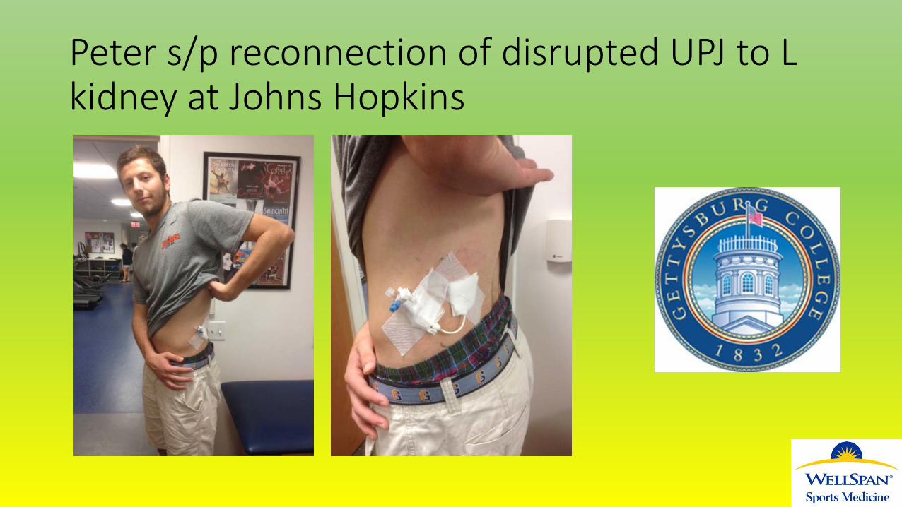
Peter s/p reconnection of disrupted UPJ to L kidney at Johns Hopkins

Objectives
• Understand the mechanism of injury of abdominal trauma in sports • Learn the evaluation and presentation of abdominal injury on the
sidelines • Understand the evaluation and management of sports-related
abdominal trauma in the inpatient setting • Be able to distinguish between intra-abdominal injury vs injury to the
abdominal wall musculature • Learn return to play guidelines for managing abdominal injury in
athletes
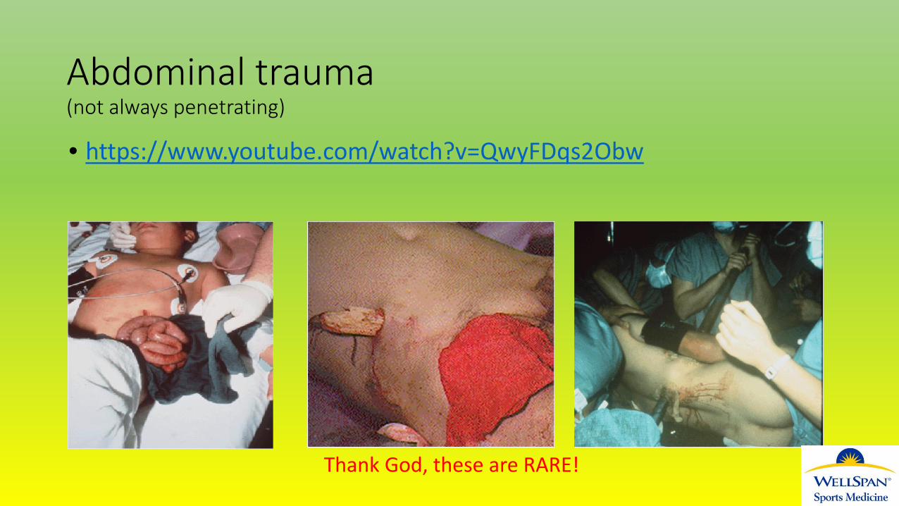
Abdominal trauma (not always penetrating)
• https://www.youtube.com/watch?v=QwyFDqs2Obw
Thank God, these are RARE!
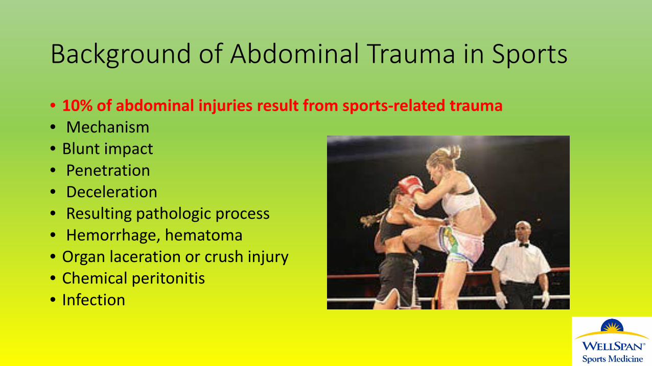
Background of Abdominal Trauma in Sports
• 10% of abdominal injuries result from sports-related trauma • Mechanism • Blunt impact • Penetration • Deceleration • Resulting pathologic process • Hemorrhage, hematoma • Organ laceration or crush injury • Chemical peritonitis • Infection
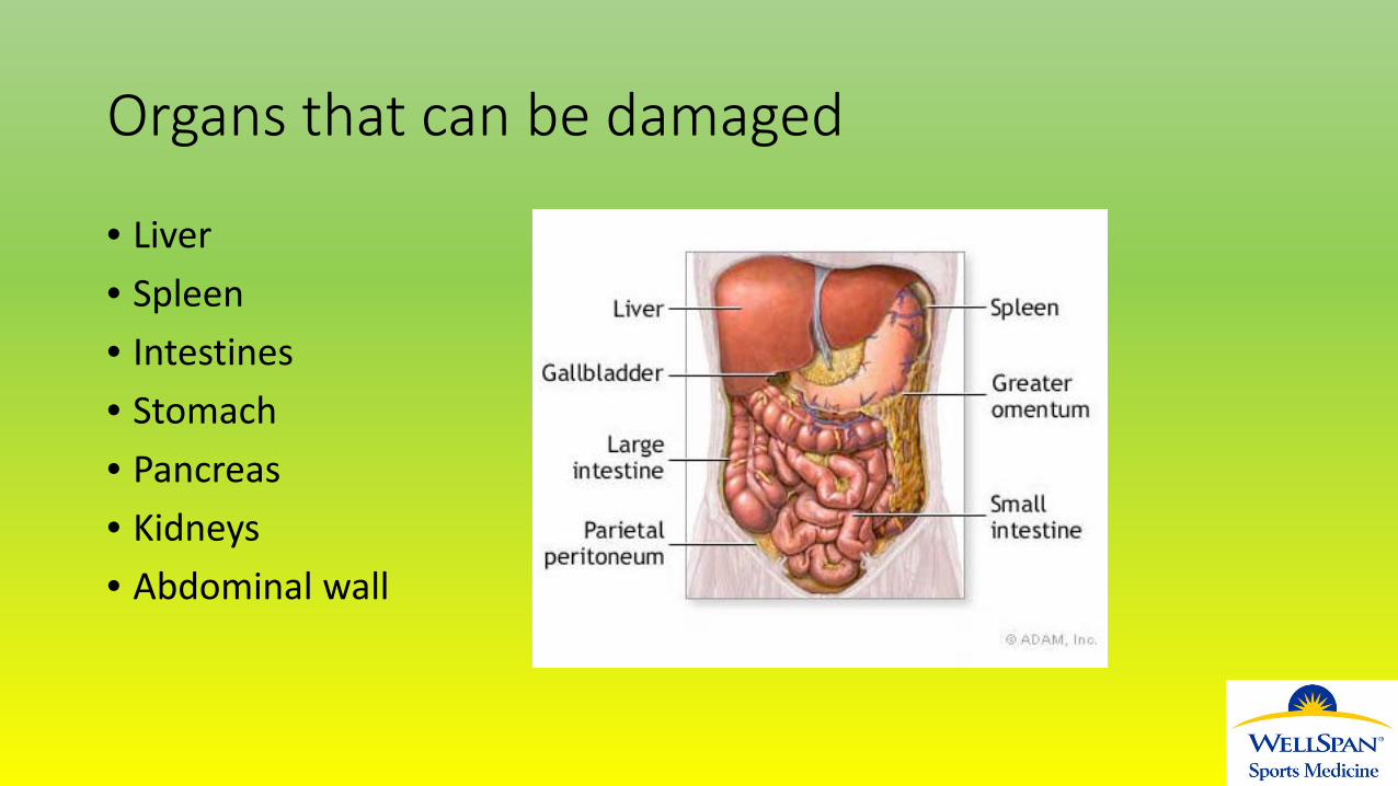
Organs that can be damaged
• Liver • Spleen • Intestines • Stomach • Pancreas • Kidneys • Abdominal wall
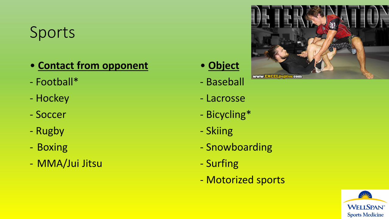
Sports
• Contact from opponent - Football* - Hockey - Soccer - Rugby - Boxing - MMA/Jui Jitsu
• Object - Baseball - Lacrosse - Bicycling* - Skiing - Snowboarding - Surfing - Motorized sports

Difficult Decision facing the Team Doctor
Return to the game? • Take out, observe on sideline? • Send to ER? • When can resume sport after injury/hospitalization recovery? • If suspect any intra-abdominal
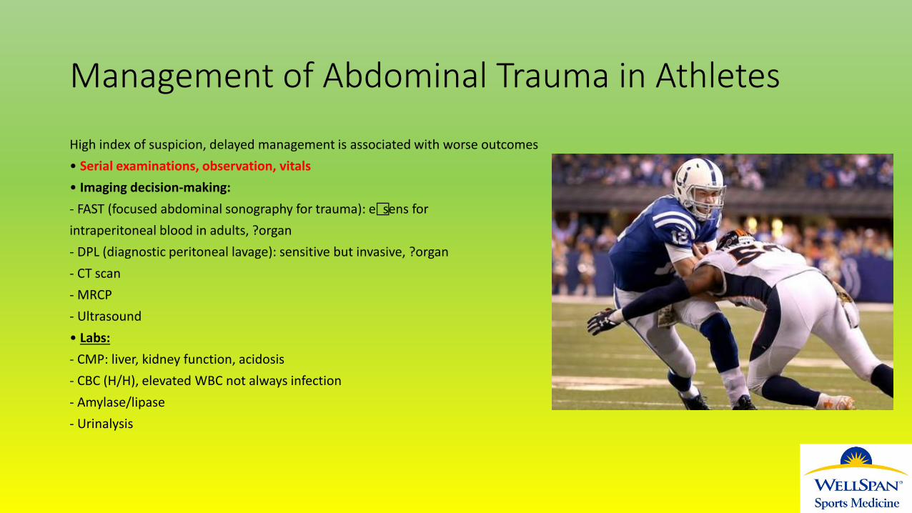
Management of Abdominal Trauma in Athletes
High index of suspicion, delayed management is associated with worse outcomes • Serial examinations, observation, vitals • Imaging decision-making: - FAST (focused abdominal sonography for trauma): esens for intraperitoneal blood in adults, ?organ - DPL (diagnostic peritoneal lavage): sensitive but invasive, ?organ - CT scan - MRCP - Ultrasound • Labs: - CMP: liver, kidney function, acidosis - CBC (H/H), elevated WBC not always infection - Amylase/lipase - Urinalysis
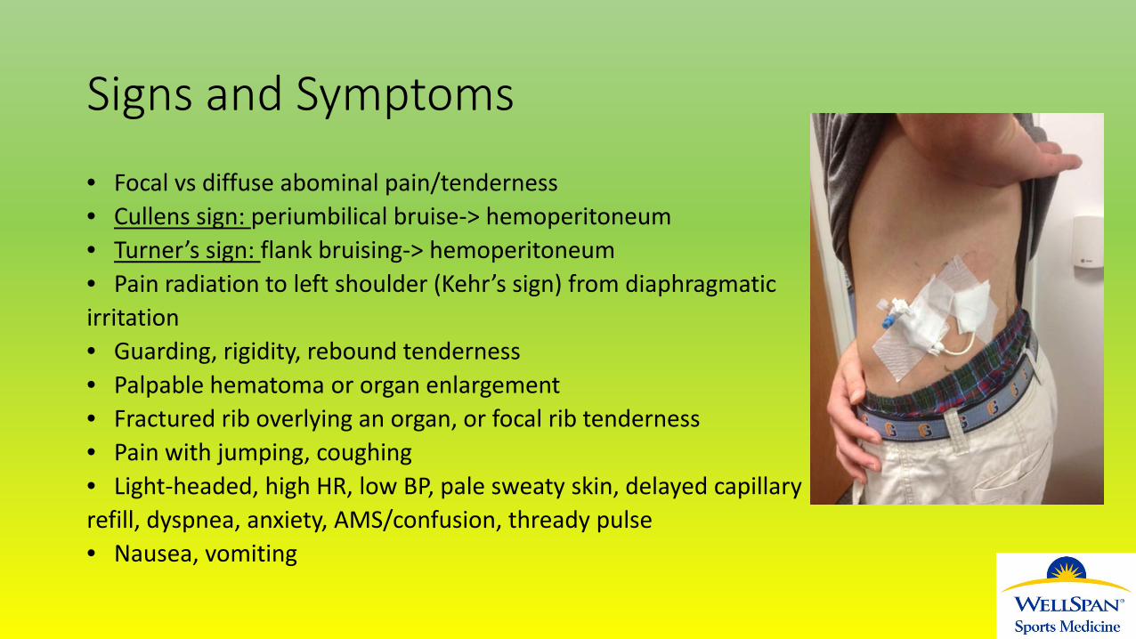
Signs and Symptoms
• Focal vs diffuse abominal pain/tenderness • Cullens sign: periumbilical bruise-> hemoperitoneum • Turner’s sign: flank bruising-> hemoperitoneum • Pain radiation to left shoulder (Kehr’s sign) from diaphragmatic irritation • Guarding, rigidity, rebound tenderness • Palpable hematoma or organ enlargement • Fractured rib overlying an organ, or focal rib tenderness • Pain with jumping, coughing • Light-headed, high HR, low BP, pale sweaty skin, delayed capillary refill, dyspnea, anxiety, AMS/confusion, thready pulse • Nausea, vomiting
Transient Diaphragmatic Spasm
• “Wind knocked out” • Blow to the upper abdomen in epigastric region causes transient paralysis of the diaphragm muscle • Athlete presents w/ difficulty breathing, dyspnea, that resolves spontaneously • If doesn’t quickly resolve consider other diagnoses
Liver Injury
• The most commonly injured abdominal organ overall • 2 possible mechanisms: deceleration vs direct blow • Deceleration -> laceration of liver capsule and underlying attached parenchyma as it continues to move • Direct blow -> crush injury to the liver… hematoma (subcapsular or intraparenchymal), while contusions are rare
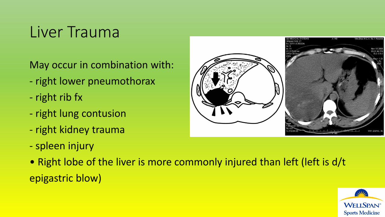
Liver Trauma
May occur in combination with: - right lower pneumothorax - right rib fx - right lung contusion - right kidney trauma - spleen injury • Right lobe of the liver is more commonly injured than left (left is d/t epigastric blow)
Liver Trauma
• RUQ pain/tenderness • Pain may radiate to shoulder (Kehr’s sign) • Overlying ribs may be tender • Nausea, vomiting • +/- Abdominal guarding, peritoneal signs • +/- Hemodynamic instability
Liver Trauma
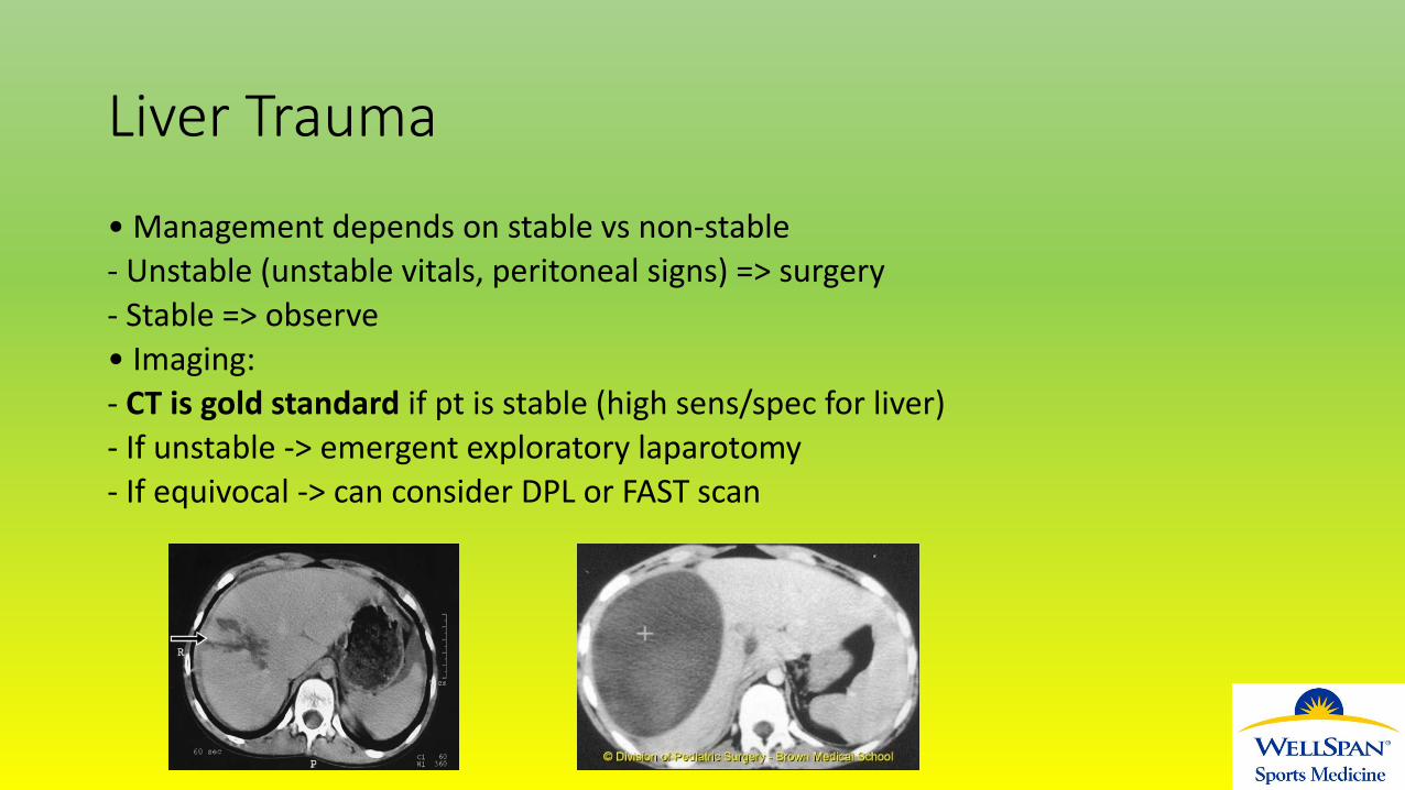
• Management depends on stable vs non-stable - Unstable (unstable vitals, peritoneal signs) => surgery - Stable => observe • Imaging: - CT is gold standard if pt is stable (high sens/spec for liver) - If unstable -> emergent exploratory laparotomy - If equivocal -> can consider DPL or FAST scan
Liver Trauma
50-80% of all liver injuries stop bleeding spontaneously, thus non-op management can be successful up to 94% of liver injuries - Bowel rest, close observation/serial examinations, IVF - Transcatheter embolization considered if slow persistent bleeding - Surgery is to stop uncontrolled bleeding and/or repair/resect liver • Hematomas can grow before they regress • Contusions may resolve in 5-7 days • Lacerations can take weeks
Liver Trauma
Return to play when: - Anatomic and functional healing - Liver enzymes, H/H normalized - Vitals stabilized - Surgical scar healed
Splenic Injury
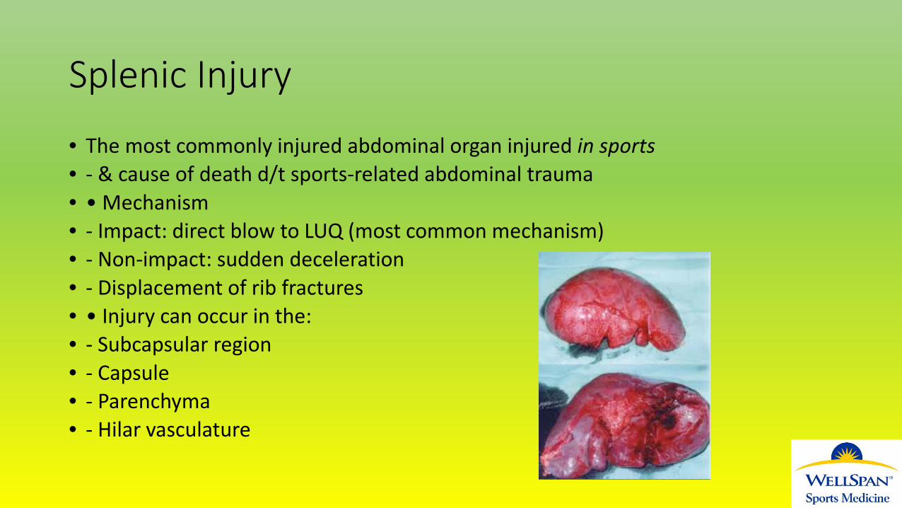
• The most commonly injured abdominal organ injured in sports • - & cause of death d/t sports-related abdominal trauma • • Mechanism • - Impact: direct blow to LUQ (most common mechanism) • - Non-impact: sudden deceleration • - Displacement of rib fractures • • Injury can occur in the: • - Subcapsular region • - Capsule • - Parenchyma • - Hilar vasculature
Splenic Injury
• Very vascular structure (receives 5-6% of the C.O., filters 10-15% of total blood volume/min) • B and T cells in parenchyma, produces IgM, involved in hematopoiesis & phagocytosis • In adults spleen is protected under the ribs… unless enlarged from mononucleosis, infection, pregnancy, portal HTN (increased risk) • In kids, rib cage does not fully cover the spleen and is more compliant/ transmits more energy from trauma. However, pediatric spleen bleeds less b/c of thicker capsule, more elastic parenchyma. - In other words… kids are more likely to injury the spleen than adults, but also have better chance of healing w/o surgery
Splenic Injury
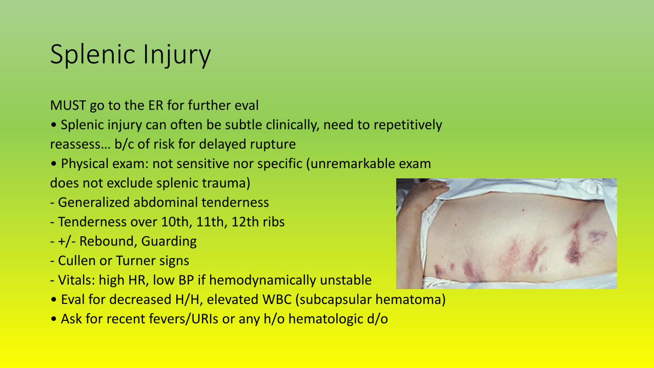
MUST go to the ER for further eval • Splenic injury can often be subtle clinically, need to repetitively reassess… b/c of risk for delayed rupture • Physical exam: not sensitive nor specific (unremarkable exam does not exclude splenic trauma) - Generalized abdominal tenderness - Tenderness over 10th, 11th, 12th ribs - +/- Rebound, Guarding - Cullen or Turner signs - Vitals: high HR, low BP if hemodynamically unstable • Eval for decreased H/H, elevated WBC (subcapsular hematoma) • Ask for recent fevers/URIs or any h/o hematologic d/o
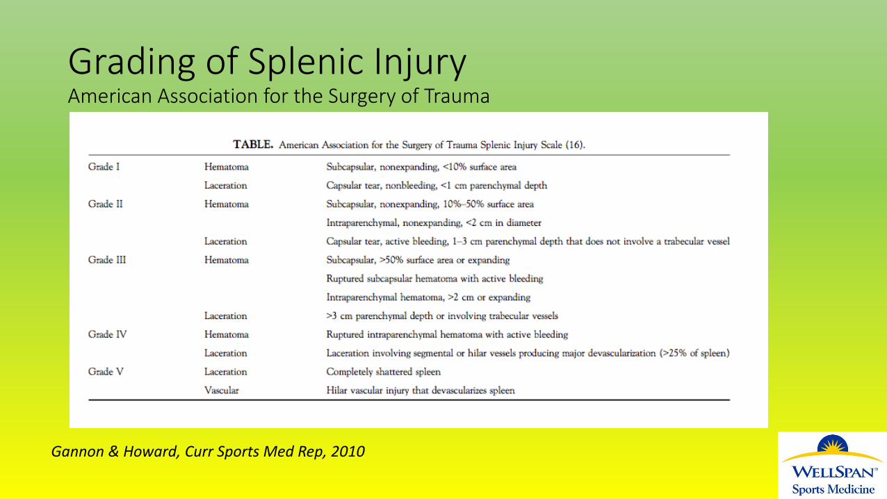
Grading of Splenic Injury American Association for the Surgery of Trauma
Gannon & Howard, Curr Sports Med Rep, 2010
Splenic Injury Evaluation
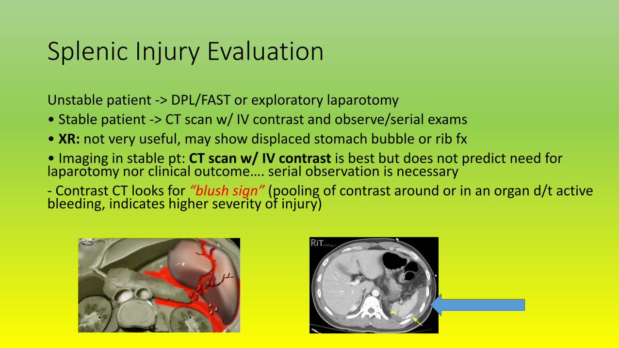
Unstable patient -> DPL/FAST or exploratory laparotomy • Stable patient -> CT scan w/ IV contrast and observe/serial exams • XR: not very useful, may show displaced stomach bubble or rib fx • Imaging in stable pt: CT scan w/ IV contrast is best but does not predict need for laparotomy nor clinical outcome…. serial observation is necessary - Contrast CT looks for “blush sign” (pooling of contrast around or in an organ d/t active bleeding, indicates higher severity of injury)
Splenic Injury
• Non-op tx may work in stable pts with grade I-III injury, not in higher grades (IV-VI) • Suggested duration of observation is [ASSI grade] + 1 - 5 days captures 95% of pts who will require intervention • 75-93% of children are successfully managed with non-op tx (regardless of injury grade as long as hemodynamically stable) vs 35-65% of adults • Increased morbidity in adults is d/t risk for delayed splenic rupture, which occurs in 1-8% of adults vs 0-7.5% in kids - Often results from delayed hemorrhage or a slowly growing hematoma that develops into a pseudoaneurysm that can rupture
Spleen Injury: Return to Play
Faster return to play in post-splenectomy pts, but future risk for infection (will need to vaccinate against Haemphilus influenza B, Neisseria meningitides, Pneumococcus; higher risk for malaria and babesiosis) • Return to play: 3 weeks to 3 months (the shorter timeframe seen in some post-splenectomy pts) • - According to one study, 84% of splenic injury pts had a fully healed spleen on imaging at 2-2.5 months • Guidelines do not recommend repeating CT scan, as radiographic healing can lag behind functional healing, and risk of radiation exposure… unless there is a change in status (sudden worsening) • No sports until symptoms, vitals, and exam have normalized; consider starting light activity starting at 3 months, then gradual return to play

Remember in Mononucleosis
Mononucleosis causes transient splenomegaly • 0.1-0.2% incidence of splenic rupture in mono pts, often atraumatic/spontaneous but need to avoid sports during this time when the spleen is vulnerable to rupture • Recommendation is to wait at least 3-4 weeks symptom onset in before returning to sport (assuming they are asymptomatic, afebrile, no e/o splenomegaly)
Injury to Bowel or Stomach
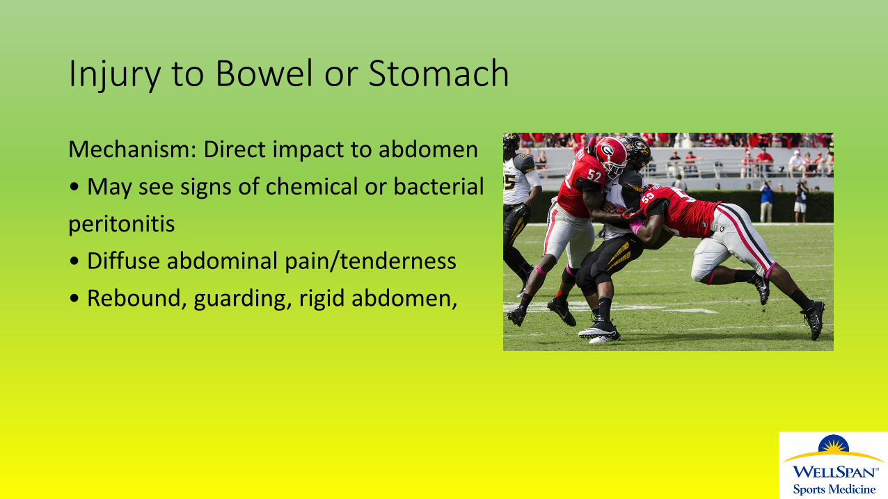
Mechanism: Direct impact to abdomen • May see signs of chemical or bacterial peritonitis • Diffuse abdominal pain/tenderness • Rebound, guarding, rigid abdomen,
Injury to Bowels or Stomach
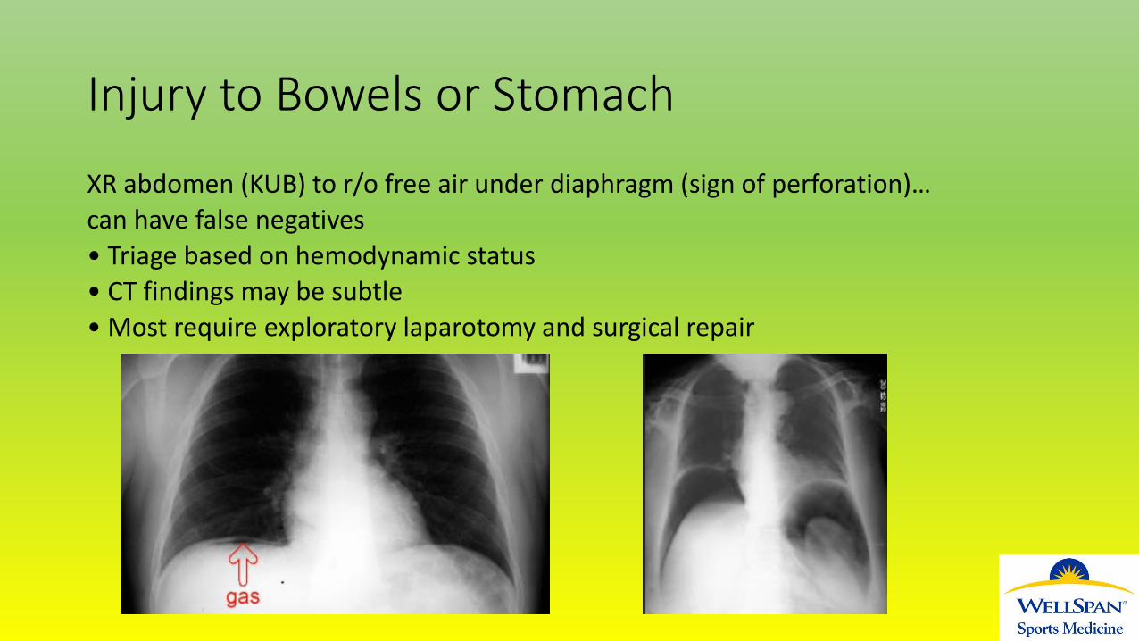
XR abdomen (KUB) to r/o free air under diaphragm (sign of perforation)… can have false negatives • Triage based on hemodynamic status • CT findings may be subtle • Most require exploratory laparotomy and surgical repair
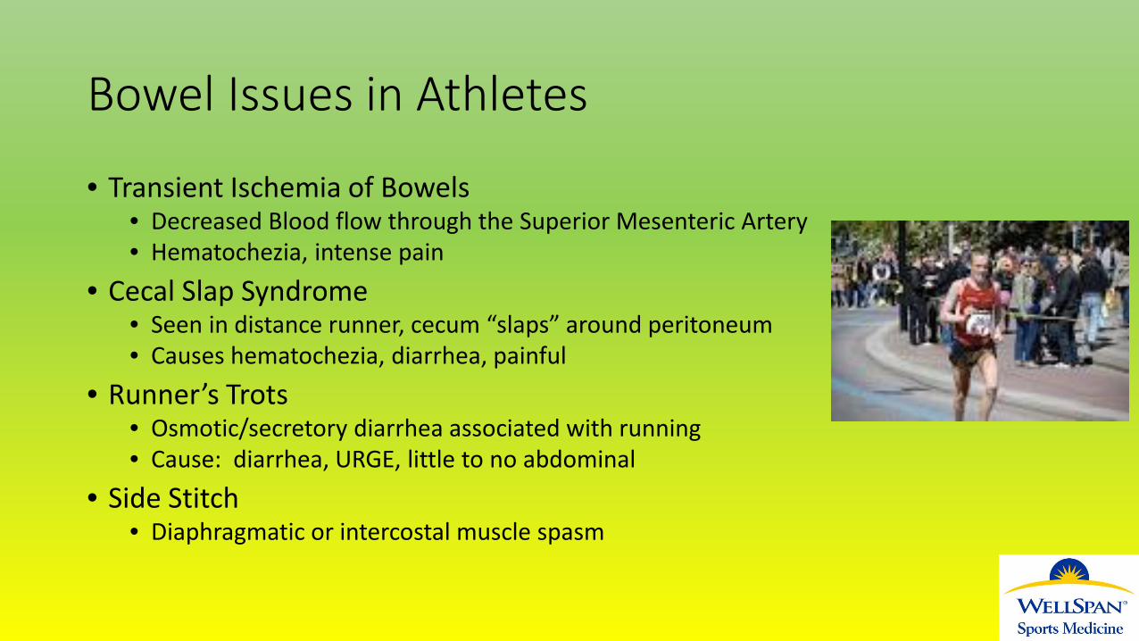
Bowel Issues in Athletes
• Transient Ischemia of Bowels • Decreased Blood flow through the Superior Mesenteric Artery • Hematochezia, intense pain
• Cecal Slap Syndrome • Seen in distance runner, cecum “slaps” around peritoneum • Causes hematochezia, diarrhea, painful
• Runner’s Trots • Osmotic/secretory diarrhea associated with running • Cause: diarrhea, URGE, little to no abdominal
• Side Stitch • Diaphragmatic or intercostal muscle spasm
Pancreatic Injury
Very rare… pancreas is protected in retroperitoneal space and relatively immobile • Pancreas injury is present in 3-12% of adult blunt abdominal trauma, and 2-10% of pediatric abd trauma • MOI: pancreas gets compressed b/t spine and external force - Majority are d/t MVA or bicycle injury/handlebar • Can get laceration of the pancreas, most commonly at head-neck junction • Abdominal pain/tenderness ein 1st 2hrs, then e again over 6-8 hrs - Pain/TTP can be diffuse or focal (epigastric), rebound is rare (9%), +abdominal wall ecchymosis in 1/3 cases
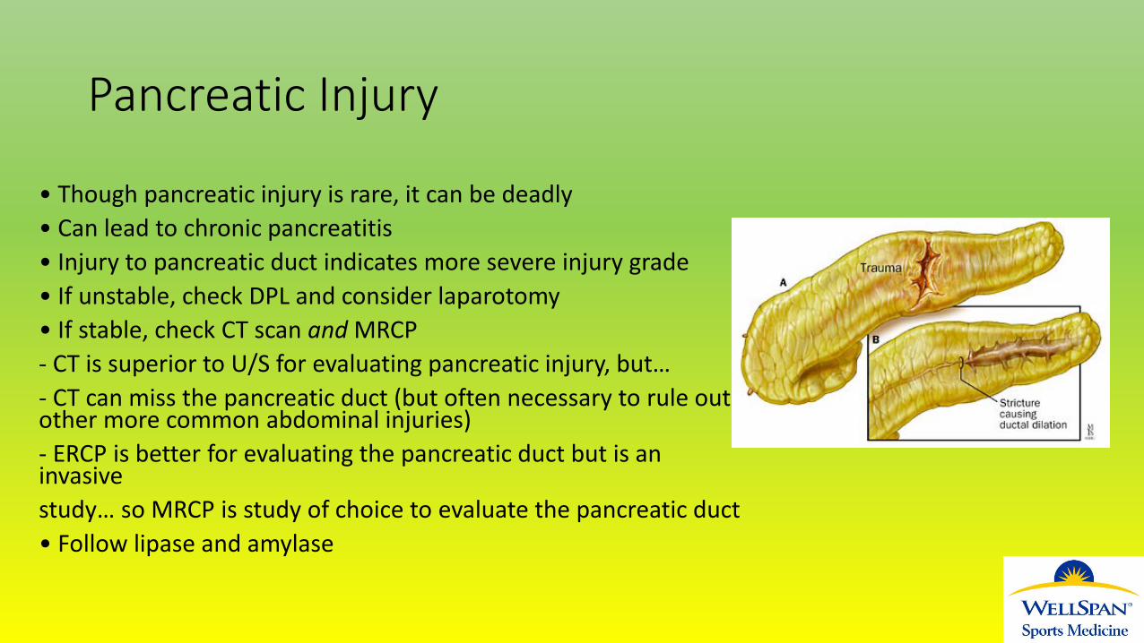
Pancreatic Injury
• Though pancreatic injury is rare, it can be deadly • Can lead to chronic pancreatitis • Injury to pancreatic duct indicates more severe injury grade • If unstable, check DPL and consider laparotomy • If stable, check CT scan and MRCP - CT is superior to U/S for evaluating pancreatic injury, but… - CT can miss the pancreatic duct (but often necessary to rule out other more common abdominal injuries) - ERCP is better for evaluating the pancreatic duct but is an invasive study… so MRCP is study of choice to evaluate the pancreatic duct • Follow lipase and amylase
Renal Trauma and Hematuria
• Stay awake a little longer cause that will be in the next lecture!!
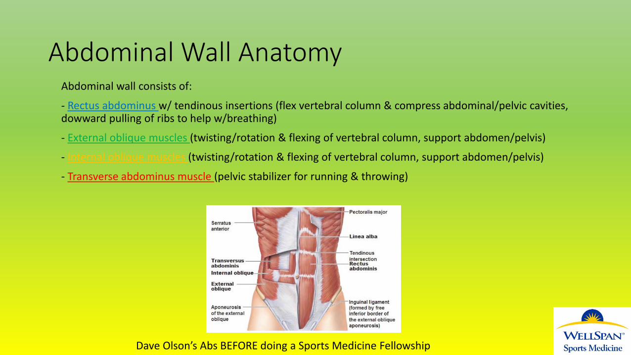
Abdominal Wall Anatomy Abdominal wall consists of:
- Rectus abdominus w/ tendinous insertions (flex vertebral column & compress abdominal/pelvic cavities, dowward pulling of ribs to help w/breathing)
- External oblique muscles (twisting/rotation & flexing of vertebral column, support abdomen/pelvis)
- Internal oblique muscles (twisting/rotation & flexing of vertebral column, support abdomen/pelvis)
- Transverse abdominus muscle (pelvic stabilizer for running & throwing)
Dave Olson’s Abs BEFORE doing a Sports Medicine Fellowship
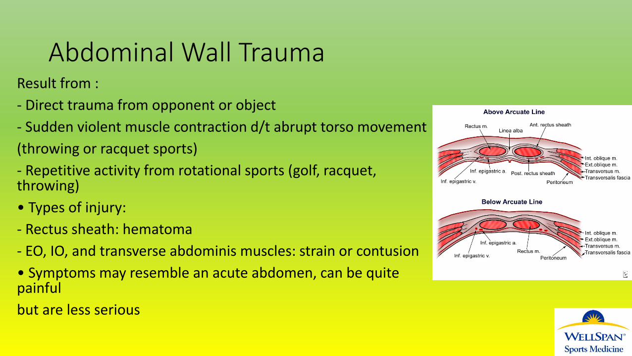
Abdominal Wall Trauma Result from : - Direct trauma from opponent or object - Sudden violent muscle contraction d/t abrupt torso movement (throwing or racquet sports) - Repetitive activity from rotational sports (golf, racquet, throwing) • Types of injury: - Rectus sheath: hematoma - EO, IO, and transverse abdominis muscles: strain or contusion • Symptoms may resemble an acute abdomen, can be quite painful but are less serious
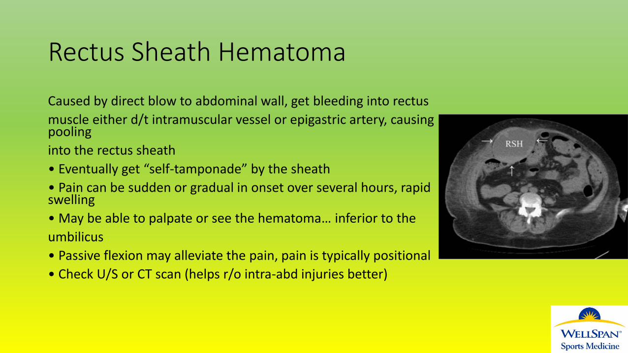
Rectus Sheath Hematoma
Caused by direct blow to abdominal wall, get bleeding into rectus muscle either d/t intramuscular vessel or epigastric artery, causing pooling into the rectus sheath • Eventually get “self-tamponade” by the sheath • Pain can be sudden or gradual in onset over several hours, rapid swelling • May be able to palpate or see the hematoma… inferior to the umbilicus • Passive flexion may alleviate the pain, pain is typically positional • Check U/S or CT scan (helps r/o intra-abd injuries better)
Rectus Sheath Hematoma
According to one study, 60% are on right side, and greater than 80% occur in the lower quadrants (inferior to umbilicus) • Majority will resolve with time, rest, close observation • Eval for hemodynamic compromise • Type I: unilateral focal, managed as outpatient, resolves in 1month • Type II: blood in muscle and fascia, hospitalized for 2-3d, resolves 2-4months, usually doesn’t need transfusion • Type III: blood in fascia, peritoneum, & prevesical space; typically needtransfusion, 4-5d for hemodynamic stabilization, hospitalized 1wk,resolves 3+ months, rarely may need surgical evacuation or surgical ligation of epigastric artery

Other type of Abdominal Wall Trauma
EO, IO, and transverse abdominus muscle strains/contusions will have no obvious hematoma • Rectus abdominis can be injured without a hematoma • Abdominal wall pain will be positional • Pain is typically focal, +TTP • Sx’s vary & may or may not include: N/V, +/- rebound/guarding/ rigidity • +Carnett’s sign: when abd pain is same or increased while abd muscles are being tensed -> indicates abd wall injury accurately if there is also no N/V/rebound/guarding/rigidity • Treatment: rest, ice, analgesics… rehab, gradual return to play when can resume sports activities effectively & w/o pain
Summary of Abdominal Trauma
• Exam doesn’t always correlate with severity of abdominal injury • Presentation of abdominal injury often evolves, need to monitor/ observe serial examination and re-check vitals • Have high index of suspicion: if there is any concern for abdominal injury send to ER for workup, can decompensate suddenly • Workup includes: CT abdomen if stable, ex lap or FAST scan if unstable, labwork. • Return to play depends on symptom resolution, vitals and labs normalized, asymptomatic, scar healed, gradual transition to play

Thank You!!!