abdominal surgery: advances in the use of ultrasound-guided
TRANSCRIPT

Chapter 3
© 2012 Børglum and Jensen, licensee InTech. This is an open access chapter distributed under the terms of the Creative Commons Attribution License (http://creativecommons.org/licenses/by/3.0), which permits unrestricted use, distribution, and reproduction in any medium, provided the original work is properly cited.
Abdominal Surgery: Advances in the Use of Ultrasound-Guided Truncal Blocks for Perioperative Pain Management
Jens Børglum and Kenneth Jensen
Additional information is available at the end of the chapter
http://dx.doi.org/10.5772/48255
1. Introduction
1.1. Anatomical considerations
In the order to get an overview of the mechanisms for the conduction of pain stimulus from the abdominal area it is clearly important to refresh the memory concerning some of the basic anatomical considerations. This knowledge constitutes an important factor with the on-going quest of providing efficient and safe post- and perioperative pain management to patients undergoing abdominal surgery.
1.2. Visceral innervation
The viscera are innervated by the vagal nerve (parasympathetic innervation) and by the splanchnic nerves (sympathetic innervation). The splanchnic nerves carry both visceral efferent and afferent nerve fibers. The sensory (or afferent) part of the splanchnic nerves reach the spinal column at certain spinal segments. Table 1 attempts to give a brief overview of the visceral innervation by the sympathetic autonomic system.
If post- or perioperative pain sensations are predominantly transmitted via the autonomic nervous system, then the choice of analgesic management would today primarily rely on continuous intrathecal and especially epidural infusions of local anaesthetic. It is also possible to block central visceral pain conduction with thoracic paravertebral blockade or maybe even with the novel quadratus lumborum block (Carney 2011). We will deal with these blocks later in this chapter. Finally, opioids administered either orally or intravenously will also reduce visceral pain significantly.

Abdominal Surgery 70
Greater splanchnic nerve Th5-Th9(10) Celiac ganglia Lesser splanchnic nerve Th10-Th11 Superior mesenteric & Aorticorenal
ganglia Least splanchnic nerve Th10 Renal plexusLumbar splanchnic nerves L1-L2 Inferior mesenteric ganglia & ganglia of
intermesenteric and hypogastric plexuses Sacral splanchnic nervers Sacral part of
sympathetic trunkInferior hypogastric plexus and ganglia to the pelvic vicera
Table 1. Visceral innervation by the sympathetic autonomic system
1.3. Innervation of the anterolateral abdominal wall by the thoracolumbar spinal nerves
The innervation of the anterolateral abdominal wall by the somatic nervous system arises from the anterior rami of the thoracolumbar spinal nerves (Th6-L1) (Børglum 2011). Branches from the anterior rami include the intercostal nerves (Th6-T11), the subcostal nerve (Th12), and the iliohypogastric and ilioinguinal nerves (L1). Furthermore, Th6-Th12 nerves provide motor innervation to the pyramidalis and rectus muscles in the anterior abdomen, and Th6-L1 nerves innervate the intercostal muscles, the external and internal oblique muscles, the transversus abdominis muscles and also provide sensory innervation to the parietal peritoneum (Børglum 2011). However, many previous descriptions of the thoracolumbar spinal nerves innervating the abdominal wall have been inconsistent leading to misunderstanding and faulty attempts to provide sufficient anaesthesia (Rozen 2008). Conducting a thorough cadaveric study including comprehensive tracing of nerves and their branches Rozen et al. were able to describe the pattern and course of all thoracolumbar nerves innervating the anterior abdominal wall. The thoracolumbar nerves were found to travel as multiple mixed segmental nerves (running with their accompanying blood vessels), which branch and communicate widely within the neurovascular plane called the transversus abdominis plane (TAP) (Rozen 2008). Such large branch communications were found antero-laterally (the intercostal plexus – Th6-Th9), and in plexuses that run with the deep circumflex iliac artery (DCIA) (the classical TAP plexus – Th10-L1) and the deep inferior epigastric artery (DIEA) (rectus sheath plexus – Th6-L1) (Børglum 2012, Rozen 2008). Segmental nerves Th6 to Th9 emerged from the costal margin to enter the TAP between the midline and the anterior axillary line. Th6 entered the TAP just lateral to the linea alba, while Th7-Th9 emerged from the costal margin at increasingly lateral positions. It was also found that Th9 emerged from the costal margin either medial (predominantly) or lateral to the anterior axillary line (Rozen 2008).
2. Truncal blocks
2.1. History
Dr. Louis Gaston Labat was a pioneer in the world of regional anaesthesia. In the early 20th century, he brought to the United States knowledge he had acquired from his mentor, the

Abdominal Surgery: Advances in the Use of Ultrasound-Guided Truncal Blocks for Perioperative Pain Management 71
French surgery professor Victor Pauchet. Thus, the spread of regional anaesthesia in the United States was greatly facilitated by the work of Dr. Gaston Labat. Recruited to work at the Mayo Clinic, Dr. Labat there published his original textbook, Regional Anesthesia, in which he laid out his techniques to the next generation of physician specialists (Labat 1922, Bacon 2002). Regional anaesthesia in the United States was popularized by Dr. Labat’s book, and many physician anaesthesiologists in the 1920s and 1930s learned regional techniques this way. Most interestingly concerning the current chapter, in relation to abdominal operations, regional anaesthesia had already then been found to provide superior muscle relaxation with fewer complications than deep ether anaesthesia. Dr. Labat has also been given credit for the posterior approach to splanchnic nerve block,use of intercostal block instead of paravertebral block for breast surgery, the use of abdominal field blocks, level of dural puncture and many other regional anaesthetic techniques (Côté 2003). Further, in the early 1920s Dr. Labat wrote about the combined caudal, trans-sacral and paravertebral (lower lumbar) block for resection of the rectum. He also elaborated extensively on other regional techniques: “With the abdominal field block procedure, colostomy is performed painlessly, provided the patient is not too obese and the mesocolon is not too short. Exploration is possible in the majority of cases, if gentleness is used. The sacral block, consisting of the caudal or epidural and transsacral block, added to the paravertebral block of the last three lumbar nerves on both sides, constitutes the method of choice for the posterior resection of the carcinomatous rectum and rectosigmoid”. Thus, this very brief historical entry highlights the fact that the use of truncal blocks certainly is not a new phenomenon in anaesthesia practice. The old masters relied on landmark-based techniques as well as a thorough understanding of anatomy and anatomical variations.
2.2. Evidence-based medicine
In 2010 Abrahams et al. performed a systematic search of the medical literature in the quest to describe evidence-based medicine in relation to ultrasound (US) guidance for truncal block administration (Abrahams 2010). In this review article it is mentioned that anaesthesia and analgesia of the trunk can be achieved with perineural injections, which could have several advantages compared with neuraxial blockade; i.e. reduced sympathectomy, less severe consequences of infection or bleeding at the injection site, minimal interference with bladder and bowel function, and less incidence of lower extremity motor weakness (Abrahams 2010). It is also clearly stated that Thoracic Paravertebral blocks (TPVB) from Th6-L1, TAP blocks, Rectus Sheath (RS) blocks, and Ilioinguinal and Iliohypogastric nerve (IIN and IHN) blocks can provide anaesthesia and analgesia of the abdominal wall. Abraham et al. did not compare the efficacy of various truncal blocks against each other or against the golden standard of the continuous epidural blockade. In the following we will go through the most common truncal blocks suitable for the purpose of alleviating the patients from pain following abdominal surgery. Table 2 provides an overview of the recommended ultrasound-guided (USG) truncal nerve blocks specifically in relation to abdominal surgical procedures.

Abdominal Surgery 72
Truncal nerve blockade Indications
Bilateral Dual – Transversus Abdominal Plane (BD-TAP) block
Fig. 1: Intercostal TAP block (IC-TAP) block providing anaesthesia to the upper abdomen
(Th6-Th9) (PPM) Fig. 2: Classical TAP block (CL-TAP) providing anaesthesia to the lower abdomen (Th10-Th12)
(PPM)Ilioinguinal/iliohypogastric nerve
(IIN/IHN) block (L1) Fig. 3: Open and laparoscopic inguinal hernia
repair (PPM) Rectus sheath (RS) block (Th6-L1) Fig. 4: Midline incisions and trochar holes (PPM)
Intercostal nerve (ICN) block (single or multiple injection technique)
Fig.5: Cholecystectomy, trochar holes high in the epigastric area (PPM)
Thoracic paravertebral block (TPVB)
Fig. 6: Providing anaesthesia both to the upper (Th6-Th9) and lower (Th10-L1) abdomen
depending on the site of administration and the volume of local anaesthetic. Has the potential to block the visceral pain in addition to the somatic
sensory pain.
Quadratus lumborum (QL) block
Fig. 7: Seems to be able to provide anaesthesia from the Th5 to the L1. Is currently still rather
inadequately described. Seems to have the potential to block the visceral pain in addition to
the somatic sensory pain.
Table 2. Overview of recommended truncal nerve blocks specifically in relation to abdominal surgical procedures. Indications only include postoperative pain management (PPM).
2.3. Education in ultrasound-guided (USG) peripheral nerve blocks
In order to enhance the clinical implementation process, to support further education and to advance improvements in clinical practice, the American Society of Regional Anesthesia and Pain Medicine (ASRA) and the European Society of Regional Anaesthesia and Pain Therapy (ESRA) encouraged all institutions that conduct USG PNB to support a quality improvement process (Sites 2010). The joint committee of ASRA and ESRA advocated a focus on the following issues: (i) ten common tasks used when performing an ultrasound-guided nerve block, (ii) the core competencies and skills associated with UGS PNB, and (iii) a training practice pathway for postgraduate anaesthesiologists and a residency-based training pathway (Sites 2010). Table 3 lists the first proposal from the joint committee of ASRA and ESRA.
High block expertise requires both anatomical knowledge and extensive hands-on experience (Jensen 2011, Orebaugh 2009). In this pursuit it is probably wise to adhere to the principles or EFSUMB (www.efsumb.org) when dividing practitioners into various catagories (levels of expertice) when formulating a strategy for enhancing the continuous

Abdominal Surgery: Advances in the Use of Ultrasound-Guided Truncal Blocks for Perioperative Pain Management 73
education of physicians. As to the other proposals regarding core competencies and skills associated with USG peripheral nerve blocks and the proposed training practice pathway at any institution, this chapter refers to the original publication (Sites 2010). Our primary aim with this chapter is to provide the reader with an easy pathway to perform USG truncal nerve blocks in daily clinical practise. The USG truncal nerve blocks can in reality be performed by any trained physician qualified in the field of emergency medicine, acute pain management and trauma as well as anaesthesiologists providing for surgical anaesthesia and postoperative pain management. In the following we will provide recommendations on how to perform the various USG truncal blocks and show relevant clinical photographs together with ultrasound recordings to advice the reader accordingly. However, it is necessary to mention early on, that the special field of USG truncal blocks differs from the performance of USG peripheral nerve blocks on the upper and lower extremities; i.e. with USG truncal blocks you very rarely actually see the nerves ultrasonographically. Rather, the focus for the physician must be on the surrounding perineural structures (muscle layer, fascia, neurovascular plane, bone etc.) and a thorough knowledge of anatomy.
1 Visualize key landmark structures including blood vessels, muscles, fascia, and bone
2 Identify the nerves or plexus on short-axis imaging 3 Confirm normal anatomy and recognize anatomic variation(s) 4 Plan for a needle approach that avoids unnecessary tissue trauma 5 Maintain an aseptic technique with respect to the ultrasound equipment 6 Follow the needle under real-time visualization as it advances toward the target 7 Consider a secondary confirmation technique, such as nerve stimulation 8 When the needle tip is presumed to be in the correct position, inject a small volume
of a test solution. If solution is not visualized during this test injection, presume that the needle tip is intravascular or out of the imaging plane.
9 Make necessary needle adjustments if an undesired pattern of local anaesthetic spread is visualized. The visualization of local anaesthetic should occur through the
entirety of the injection to avoid an intravascular injection 10 Maintain traditional safety guidelines including the presence of resuscitation
equipment, frequent aspiration, intravascular test dosing, standard monitoring, patient response, and assessment of injection characteristics
Table 3. Ten tasks helpful in performing USG peripheral nerve blocks. (Sites 2012)
2.4. Transversus abdominis plane (TAP) block
The idea of the TAP block is to anaesthetize part of - or the entire - abdominal wall instead of using intrathecal or epidural techniques, that may or may not elicit more negative side effects by the application. By adhering to this principle one would block the nerves as peripheral as possible but only as centrally a necessary (to quote Professor Peter Marhofer, Austria). The technique builds on anaesthetizing the peripheral nerves to the abdomen

Abdominal Surgery 74
using a direct approach. Since the first description of the TAP block technique by Rafi (or something very similar to what it is conceived as today), this block has been increasingly used to provide somatic anaesthesia of the antero-lateral abdominal wall (Rafi 2001, Abrahams 2010, Petersen 2010, Børglum 2011, Koscielniak-Nielsen 2011, Børglum 2012). This landmark-based blind approach to deposit local anaesthetic at the neurovascular plane was since thoroughly described by McDonnell et al. and further documented using computerized tomography (McDonnell 2004, 2007). As of today, it would seem that four different TAP block approaches are in common use. First, let us begin with the landmark-based blind approach at the triangle of Petit. McDonnell et al. and Carney et al. have provided ample evidence with many scientific publications for the huge success when using their TAP block approach where ultrasound guidance is not used (McDonnell 2007, Carney 2009, 2011), but where extensive dermatomal anaesthesia is achived and postoperatively pain management is improved significantly for a great many surgical procedures. Second, we find the USG approach to the TAP very well described by El-Dawlatly et al. and Shibata et al. Both use two separate injections deposited at the lateral classical TAP plexus; i.e. one injection on each hemi-abdomen (above the iliac crest and below the thoracic cage) (El-Dawlatly 2009, Shibata 2007). This simple and very efficient technique is probably the method most commonly used today. Thirdly, we find the brilliant classification by Hebbard et al. where the USG continuous oblique subcostal transversus abdominis plane blockade technique is described (Hebbard 2010). Dr. Hebbard must be thankfully accredited to address the issue of providing consistent anaesthesia to both the lower (Th10-L1) and upper (Th6-Th9) abdominal wall on a continuous basis. In addition, Dr. Hebbard´s research has showed that it possible to use more peripheral approaches to continuous block the entire abdominal wall. Dr. Hebbard has however clearly expressed that his technique for providing safe and continuous anaesthesia to the entire abdomen requires considerable skills and serious anatomical knowledge (Hebbard 2010). Having said that, one must not rule out that McDonnell et al. have also found their landmarked-based and blind approach to be able to anaesthetize the entire abdominal wall for an extended period. Finally – and fourth – is our own approach called the bilateral dual TAP (BD-TAP) block based on four single shot injections with the aim to provide anaesthesia to the entire abdominal wall in a fast and safe sequence (Børglum 2011, Børglum 2012). Our method does not rely on relatively lengthy or sophisticated methods for the insertion of catheters; rather our technique relies on simple anatomical knowledge and structured ultrasonographic recognition (Børglum 2011, Børglum 2012). The BD-TAP block will normally take the well-trained anaesthesiologist approximately 5-6 minutes to perform. Probably even shorter time if the block is administered prior to surgery. The BD-TAP block will anaesthetize the dermatomes Th6-Th12, the antero-medial muscles of the abdominal wall and the underlying parietal peritoneum. It would also be fair to say that the BD-TAP block builds to some extent on a “mixture” of previously described techniques by El-Dawlatly et al. and Shibata et al. as well as Dr. Hebbards research (El-Dawlatly 2009, Shibata 2007, Hebbard 2010). In addition, the BD-TAP technique has been proved not to result in inhibition of the accessory

Abdominal Surgery: Advances in the Use of Ultrasound-Guided Truncal Blocks for Perioperative Pain Management 75
respiratory function attributed to the abdominal wall muscles – mainly forced expiration (Petersen 2011). The efficacy of the BD-TAP block technique has been ascertained by magnetic resonance imaging (MRI) (Børglum 2012), and it seems obvious that it is not possible to anaesthetize the entire abdominal wall (TH6-L1) with the so-called lateral classical TAP block technique alone, but that the intercostal TAP plexus in the upper abdomen (epigastric area) must also be anaesthetized by a direct approach in addition.
Generally speaking when considering outcome measures of the various techniques, TAP blocks have been described as an effective component of multimodal postoperative analgesic protocols for a wide variety of abdominal surgical procedures including laparotomy for colorectal surgery, open and laparoscopic appendectomy, caesarean section, abdominal hysterectomy, laparoscopic cholecystectomy, open prostatectomy and renal transplant surgery. In an uncontrolled study, patients undergoing lower abdominal gynaecological surgery received bilateral TAP block catheters, and the authors found an average pain at rest and on movement below 2 on a 10-point VAS scale for up to 48 hours postoperatively, with no occurrences of nausea or side effects (Fujita 2012). Compared to systemic opioids, patients receiving TAP blocks after major abdominal surgery had less pain up to 24 hours postoperatively than non-TAP block groups, but in that study no statistical differences were found with respect to nausea (Siddiqui 2011). The benefits of TAP blocks are so far measured in relation to reduced postoperative opioid requirements, lower pain scores or a reduction in opioid-related side effects (Shin 2011). As an example, a meta-analysis of 7 studies demonstrated an average reduction in 24-hour morphine consumption of 22 mg compared with systemic opioids, and TAP blocks were associated with reduced early postoperative pain VAS in 4 of the 7 studies (Petersen 2010). Postoperative sedation, as well as PONV, was marginally reduced in patients having TAP blocks administered. Newer studies confirm these findings, and also observe a higher patient satisfaction in the TAP block groups (Hivelin 2011). Despite the numerous descriptive studies on TAP blocks, however, results of comparative studies have been inconsistent. The current scientific evidence is lacking to definitively identify the surgical procedures, dosing, techniques, and timing that provide optimal analgesia following TAP block (Abdallah 2012).
2.4.1. Detailed description of the BD-TAP block
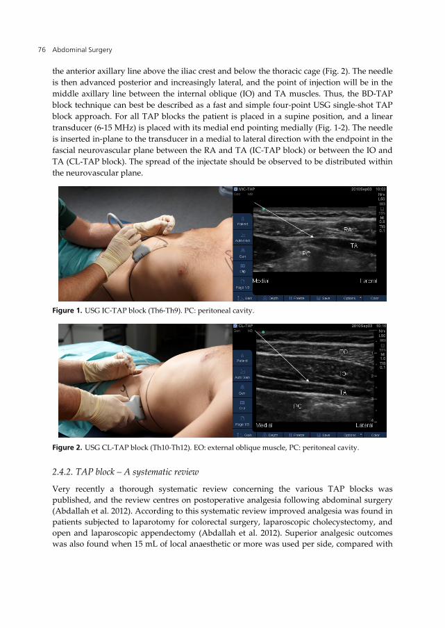
With the aim to render the entire abdominal wall pain-free after surgery (or during the surgical procedure) one must anaesthetize all the antero-lateral rami of the thoracoabdominal nerves (Th6-Th12). In doing so, one must anaesthetize both the intercostal TAP plexus (Th6-Th9) situated in the epigastric area just below the xiphoid process medially to the costal curvature (Fig. 1), and one must also target the lateral classical TAP plexus (Th10-Th12) situated in the lower abdomen. This must be done on both hemi-abdomens. When administering local anaesthetic to the uppermost branches of the intercostal TAP plexus the physician must use the USG intercostal TAP (IC-TAP) block, where the IC-TAP plexus lies in the fascial plane between the rectus abdominis (RA) muscle (or rather deep to the posterior rectus sheath) and the transversus abdominis (TA) muscle (Fig. 1). When blocking the lateral classical TAP (CL-TAP) plexus the point of skin penetration must be in
Abdominal Surgery 76
the anterior axillary line above the iliac crest and below the thoracic cage (Fig. 2). The needle is then advanced posterior and increasingly lateral, and the point of injection will be in the middle axillary line between the internal oblique (IO) and TA muscles. Thus, the BD-TAP block technique can best be described as a fast and simple four-point USG single-shot TAP block approach. For all TAP blocks the patient is placed in a supine position, and a linear transducer (6-15 MHz) is placed with its medial end pointing medially (Fig. 1-2). The needle is inserted in-plane to the transducer in a medial to lateral direction with the endpoint in the fascial neurovascular plane between the RA and TA (IC-TAP block) or between the IO and TA (CL-TAP block). The spread of the injectate should be observed to be distributed within the neurovascular plane.
Figure 1. USG IC-TAP block (Th6-Th9). PC: peritoneal cavity.
Figure 2. USG CL-TAP block (Th10-Th12). EO: external oblique muscle, PC: peritoneal cavity.
2.4.2. TAP block – A systematic review
Very recently a thorough systematic review concerning the various TAP blocks was published, and the review centres on postoperative analgesia following abdominal surgery (Abdallah et al. 2012). According to this systematic review improved analgesia was found in patients subjected to laparotomy for colorectal surgery, laparoscopic cholecystectomy, and open and laparoscopic appendectomy (Abdallah et al. 2012). Superior analgesic outcomes was also found when 15 mL of local anaesthetic or more was used per side, compared with

Abdominal Surgery: Advances in the Use of Ultrasound-Guided Truncal Blocks for Perioperative Pain Management 77
lesser volumes, and TAP blocks performed in the triangle of Petit and along the midaxillary line both demonstrated some analgesic advantages (Abdallah et al. 2012). Finally, this systematic review also found that although the majority of trials reviewed suggested superior early pain control, they were unable to definitively identify the surgical procedures, local anaesthetic doses, techniques, and timing (pre- or post-incisional) that would ensure optimal analgesia following the TAP blocks. Thus, there is still much work to be done.
2.5. USG ilioinguinal/iliohypogastric nerve (IIN/IHN) block
This is a selective block of the ventral ramus of the L1. The IIN provides sensation to the upper medial part of the thigh and the upper part of the genitalia. The IHN provides sensation to the buttock and abdominal wall above the pubis (Abrahams 2010). In our own previous studies, we have been unable to register effective dermatomal anaesthesia of the L1 branch with our BD-TAP block technique, but other studies have shown the L1 branches to be blocked by other versions of the TAP block technique (Børglum 2012, Carney 2011). For the selective USG IIN/IHN block the patient is placed in a supine position, and the anterior superior iliac spine (ASIS) is localized by palpation first and since ultrasonographically (Fig. 3). A linear transducer (6-15 MHz) is placed with its lateral end at or just superior to the ASIS. The needle is inserted in-plane to the transducer in a lateral to medial direction, and the neurovascular plane between the IO and the TA is located. The IIN and IHN can often be seen together with the deep circumflex iliac artery in the neurovascular plane. This is rather specific for this particular truncal block, because trunk nerves are not as easily discovered by US as are the peripheral nerves of the upper and lower extremities. The tip of the needle is placed in this plane, and the spread of the injectate should be observed to expand in the fascial neurovascular plane (3).
Figure 3. USG IIN/IHN block. EO: external oblique muscle.
Traditional techniques were landmark-based and relied on one or two facial “clicks”, but the old techniques are largely abandoned now, since US imaging subsequent to the so-called blind blocks has demonstrated incorrect placement of the local anaesthetic administered (Weintraud 2008). The US technique is very well established already (Willschke 2005, Willschke 2006, Eichenberger 2007). Most IIN/IHN blocks are placed for analgesia after inguinal hernia repairs in children, but have also been shown to provide similar analgesia to

Abdominal Surgery 78
caudal blocks during orchidopexy and hydrocele repair (Abrahams 2010). In his review article Dr. Abrahams gave the use of US guidance for the IIN/IHN block a Grade A recommendation (Abrahams 2010). Our own very recent study concerning primary open inguinal hernia repair (ad modum Lichtenstein) in adult males showed significant reduction in pain scores at mobilization and at rest in the group of patients having active bupivacaine USG IIN/IHN blocks administered prior to surgery (Bærentzen 2012). The pain scores were recorded when the patients arrived at the post anaesthesia care unit (PACU) and after 30 minutes stay. Pain at rest was similarly reduced in the active group at the time of discharge. Most importantly, patients with severe (NRS>5) and moderate (NRS>3) pain at mobilization and rest, respectively, were significantly reduced in the group of patients having the block (Bærentzen 2012). Thus, it would seem that the USG IIN/IHN block also has a place in the post- and perioperative pain management in adult patients.
2.6. USG rectus sheath (RS) block
The central portion of the anterior abdominal wall is innervated by the ventral branches of the thoracolumbar nerves (Th6-L1), and in the beginning of this chapter we have already mentioned the rectus sheath plexus and its anatomical position. The ventral branches lie deep to the RA muscle but ventral to the posterior rectus sheath. Since the tendinous inscriptions of the rectus muscle are not attached to the posterior RS the local anaesthetic administered into the spatial space can in theory spread both in the cranial and caudal direction. However, the RS block may have been over-shadowed by the various TAP block techniques, but the evidence base for its use is very good; i.e. a grade A recommendation for the use of US guidance for the RS block has been granted (Abrahams 2010). The RS block has been utilized to provide analgesia for midline incisions and laparoscopic procedures (Ferguson 1996, Abrahams 2010). RS blocks may also be effective in reducing postoperative pain in upper abdominal surgery as an alternative method to epidural analgesia in anti-coagulated patients (Osaka 2010).
However, we find that the RS block has a potential drawback, since the risk for inadvertent injections deep to the posterior RS (intra-peritoneal) seems to be higher that for the USG TAP blocks where the TA muscle lies deep to the point of injection (Dolan 2009). To our knowledge, no comparison between TAP blocks and RS blocks has yet been done. For the selective USG RS block, the patient is placed in a supine position, and a linear transducer (6-15 MHz) is placed with its medial end just above the linea alba (LA) (Fig. 4). The needle is inserted in-plane to the transducer in a medial to lateral direction, and the division between the belly of the rectus abdominis muscle and the posterior rectus sheath is visualized. The tip of the needle is placed in this space. The spread of the injectate should be observed to advance in a lateral direction.
2.7. USG intercostal nerve (ICN) block: Parasagittal plane
The thoracoabdominal nerves Th6-Th11 are all intercostal nerves per se, before they become abdominal nerves when they leave the thoracic cage and contribute to the formation of the

Abdominal Surgery: Advances in the Use of Ultrasound-Guided Truncal Blocks for Perioperative Pain Management 79
IC-TAP, CL-TAP and RS-TAP plexuses in the anterolateral abdominal wall (Rozen 2008). Thus the potential to provide efficient abdominal analgesia employing US guidance to block the intercostal nerves are obviously there if a multiple injection technique is used. In the past, the landmark-based (blind) technique has been employed to provide analgesia for various surgical procedures in the abdominal area; i.e. following renal transplantation, cholecystectomy and appendectomy (Knowles 1998, Vieira 2003, Bunting 1988). It would seem to be obvious that the USG ICN block (multiple injection technique) could very well be used postoperatively; i.e. either as an effective rescue block or because TAP blocks were not possible due to surgical incisions, tissue swelling etc. For the selective USG ICN block the patient is placed in the lateral decubitus position, and a linear transducer (6-15 MHz) is placed in a sagittal paravertebral plane (Fig. 5). The needle is inserted in-plane to the transducer in a cranial to caudal direction, and the three intercostal muscles (external, internal and innermost) are visualized between two costae. The tip of the needle is placed in the fascial plane between the internal and innermost intercostal muscles. The spread of the injectate should be observed to occur in this fascial plane. It is very important to visualize the tip of the needle at all times and its close proximity to the parietal pleura.
Figure 4. USG RS block. PC: peritoneal cavity, LA: linia alba.
Figure 5. USG ICN block: parasagittal plane. CO: costa, EX,IN,INM: external, internal, innermost intercostal muscles, PL: pleura.

Abdominal Surgery 80
2.8. USG Thoracic Paravertebral block (TPVB)
The conventional technique of a TPVB involves inserting the needle perpendicular to all planes, making contact with the transverse process, and then walking off the bone with the needle until the physician feels the loss of resistance when penetrating deep to the internal intercostal membrane and entering the thoracic paravertebral space (TPVS). The TPVB has been used to provide pain relief for many surgical procedures in the abdominal area (Naja 2002, Moussa 2008, Ho 2004, Berta 2008). Conducting an USG TPVB or using the traditional landmark-based methods is the technique of injecting local anaesthetic adjacent to the thoracic vertebra close to the actual site where spinal nerves emerge from the intervertebral foramina. This results in ipsi-lateral somatic and sympathetic nerve blockade in multiple contiguous thoracic dermatomes above and below the site of injection (Karmakar 2001). How much dermatomal anaesthesia in the abdominal area results from specific volumes of local anaesthetic is to our knowledge still not fully elucidated, at least when it concerns the USG TPVBs. There is bound to be considerable individual variations as well. From previous studies it would seem that the point of injection within the TPVS must influence the distribution pattern of a paravertebral blockade. Apparently, injections made in the more ventral part of the TPVS, supposedly anterior to the endothoracic fascia, will result in a multisegmental longitudinal spreading pattern (evaluated by radiographic spreading patterns), whereas injections dorsal to the endothoracic fascia will result in a cloud-like spreading pattern, with only limited distribution over adjacent segments (Naja 2004). Whether US guidance can make the administration of local anaesthetic more beneficial remains to be evaluated in future studies. Even when using the USG technique, the endothoracic fascia is very difficult to visualize if at all possible with the current ultrasound machines. The TPVB is effective in treating pain of resulting from surgery in the chest and abdomen (Karmakar 2001). The potential advantage over the various TAP blocks could be that the visceral pain is more reliably blocked with the TPVB. Further, the potential failure rates and complications using the traditional techniques have already been brilliantly described previously (Lönnqvist 1995). Finally, insertion of catheters using the USG technique in the TPVS is indeed possible with at high success rate, thus making this technique a potential replacement of the epidural continuous infusion catheters (Renes 2010).
Figure 6. USG TPVB block. IIM: Internal intercostal membrane (continuous with the superior costotransverse ligament medially), PL: Pleura, TP: Transverse process

Abdominal Surgery: Advances in the Use of Ultrasound-Guided Truncal Blocks for Perioperative Pain Management 81
For the selective USG TPVB block the patient is placed in the lateral decubitus position, and a linear transducer (6-15 MHz) is placed parallel to and in-between two costae in an axial transverse plane (Fig. 6). It is important to visualize the pleura very clearly at all times. The transducer is then gradually aligned in a medial direction until the acoustic shadow of the transverse process is clearly visualized in the medial part of the sonographic image. The needle is inserted in-plane to the transducer in a lateral to medial direction until the tip of the needle is seen to penetrate the internal intercostal membrane. We do not recommend that the needle tip should be advanced under the acoustic shadow of the transverse process. Rather, we recommend that the spread of the injectate should be observed to occur above the pleura in the triangular space (which is the TPVS) and thus depressing the pleura and filling up the TPVS. It is very important to visualize the tip of the needle at all times and its close proximity to the parietal pleura.
2.9. USG Quadratus Lumborum (QL) block – the “new kid on the block”
As we are approaching the end of the description of the various USG truncal blocks we would like to introduce the reader to the “new kid on the block”. The so-called Blanco block (as it is known by some anaesthetists in the United Kingdom) is an USG block administered to the quadratus lumborum space first described by Professor R. Blanco in May 2007 during his presentation at ESRA 2007 at the XXVI Annual ESRA Congress in Valencia, Spain. Professor Blanco describes a potential space posterior to the abdominal wall muscles and lateral to the quadratus lumborum muscle. Thus, this new block has also been called the Quadratus lumborum (QL) block. It has been used in abdominoplasties, caesarean sections and lower abdominal operations since 2006 providing complete pain relief in the distribution area from Th6 to L1 dermatomes. Apparently, in operations with peritoneal involvement the morphine consumption was significantly reduced to less than 30% of the control groups. It is hard however, to find any solid scientific evidence to support these findings in the literature, and much of the knowledge of the QL block relies regrettably to this day on personal communication, which is certainly not the best of documentation. Much research effort at many centres is currently directed towards the description and qualification of this new block, and we have found it highly relevant to include the block in this chapter, since the block holds some very positive potential benefits. If may well be seen as a lumbar approach to the TPVS. The block apparently produces distribution of the local anaesthetic extending proximally and over both sides of the surface of the QL muscle, in between the anterior and intermediate layers of the thoracolumbar fascias. It also pushes the fascia transversalis and the perinephric fat towards the peritoneum without the risk of intrabdominal puncture. The block does not rely on the feeling of any pops or fascial clicks because depending of the angle of the needle several pops can be felt without reaching the target zone, which is lateral to the quadratus lumborum muscle. Actually, the block has never been intended to be conducted without the use of US guidance, and the block is thus a purely USG block. In an absolutely brilliant paper by Carney et al. the block is compared to other TAP block techniques using volunteers rather than patients (Carney 2011). Dr. Carney found that there was a non-contiguous paravertebral, epidural and lymphatic contrast enhancement Th5-Th10 in one subject, and similarly contrast at Th6-Th10 in two other subjects (Carney 2011).

Abdominal Surgery 82
Carney et al. concluded that the posterior USG approach (as they have named the QL block in their recent publication) produced a more extensive, predictable and posterior spread of contrast, similar to that seen with their own landmark-based and blind approach at the triangle of Petit. The contrast extended postero-medially to the paravertebral region from the 5th thoracic vertebral level rostrally, to the first lumbar vertebral level caudally, indicating that this US guidance approach is the optimal site for injection to reproduce the analgesia of the blind landmark TAP block favoured by Dr. McDonnell and Dr. Carney. Fig. 7 depicts one method to administer the USG guided QL block as we have found it most easy to perform in our daily clinical practise (Jensen 2012). Again, the patient rests in a supine position. We have found it easier to conduct the block using a low frequency transducer (2-6 MHz) as compared to a linear transducer. The low frequency transducer is placed on the lateral abdomen above the iliac crest and below the thoracic cage. The transducer is then gradually aligned in a more posterior and lateral direction parallel to the inter-crista line. It is always possible to observe, that the TA muscle becomes aponeurotic, and this aponeurosis is followed until the QL muscle is clearly visualized. Thus, it is indeed possible to visualize the QL muscle lateral and posterior to the abdominal wall muscles. It is also clearly possible to visualize the thoracolumbar fascia at the lateral edge of the QL muscle. We have set this to be the point of injection of local anaesthetic. Following the injection we could observe the local anaesthetic spread along the ventral side of the QL muscle. Apparently this block results in a block that is longer lasting and more extensive than what we have previously observed with the BD-TAP block, but it remains to be further elucidated in RCT trials.
Figure 7. USG QL block. Arrows in upper left corner indicating needle shaft approaching in a medial to lateral-posterior direction towards the injection point (IP). PC: Peritoneal cavity, IP: Injection point, QL: Quadratus lumborum muscle, PNF: Perinephric fat.

Abdominal Surgery: Advances in the Use of Ultrasound-Guided Truncal Blocks for Perioperative Pain Management 83
3. Epidural analgesia
3.1. General physiology of the epidural
Epidural analgesia is an effective method of anaesthetizing the sensory nerves to the abdominal wall. The impulse conduction of sensory roots protruding from the spinal cord is considerably reduced in a dermatomal fashion, optimally extending from the 4th to the 12th thoracic dermatome. Not all spinothalamic nerve transmission is reduced however, and some sensory input is perceived by the brain (Lund 1991). An element of habituation (so-called tachyphylaxis) is also present, in which the nerve roots require increasing amounts of local anaesthetic to maintain a sufficient nerve block over days. This inconvenient effect may be reduced by the addition of opioids, administered epidurally or systemically. Common side effects include a subsidiary block of the sympathetic trunk in the dermatomes anaesthetized, causing arteriole relaxation, reduced peripheral vascular resistance with hypotension and reflex tachycardia; motor block of the lower extremities when the epidural extends below thoracic dermatomes; and central nervous system side effects such as drowsiness, nausea or pruritus when opioids or other drugs are added to the local anaesthetic. The placement of an epidural catheter requires some experience, and the handling of an epidural catheter is particularly resource heavy for infusion maintenance and monitoring of potential side effects.
3.2. The epidural catheter in a historical perspective
The benefits and risks of epidural analgesia have been extensively documented ever since its introduction almost 100 years ago (Thompson 1917). In the past ten years alone, more than two publications have appeared every single day on the subject, equally distributed between analgesic effects in major surgery, obstetric anaesthesia, side effects and technical aspects (Jensen 2012). Apart from subarachnoidal analgesia, which was first introduced in 1898 (Bier 1899), it is hard to imagine a method of analgesia having been similarly subjected to scientific study. In the case of spinal anaesthesia, the foundation for its use is still overwhelming by its significant reduction in perioperative morbidity and mortality compared to general anaesthesia (Rodgers 2000). Epidural analgesia is a well-established technique that is often regarded as the gold standard in postoperative pain management. However, newer and evidence-based outcome data show that its benefits are not as significant as previously believed, and that these benefits are probably limited to high-risk patients undergoing major abdominal surgery receiving epidural analgesia with local anaesthetic drugs only. There is increasing evidence that less invasive regional analgesic techniques are as effective as epidural analgesia, and while pain relief associated with epidurals can be outstanding, clinicians expect more from this invasive, high-cost, labour-intensive technique (Niraj 2009; Rawal 2012). A plethora of cardiovascular, neurologic and infectious side effects of epidural analgesia have been published, and given its modest success rate (at 70-80%) and the potential of motor block of the lower extremities, the scale is beginning to tip in the direction of USG truncal nerve blocks, in particular when the need for short-term analgesia of the abdominal wall is anticipated or when patients are not high-risk.

Abdominal Surgery 84
3.3. Physiological reactions to surgery
Invasive surgery induces a combination of local response to tissue injury and generalized activation of systemic metabolic and hormonal pathways via afferent nerve pathways and the central nervous system. The local inflammatory responses and the parallel neurohumoral pathways are linked through complex signalling networks. The magnitude of the response is related to the site of injury (greater in abdomen or thorax) and the extent of the trauma. The changes include alterations in metabolic, hormonal, inflammatory, and immune systems that collectively are termed the stress response. Integral to the stress response are the effects of nociceptive afferent stimuli on systemic and pulmonary vascular resistance, heart rate and blood pressure. Opioid doses required to provide analgesia are less than those required for haemodynamic stability in response to surgery, and these are in turn less than those required to suppress most aspects of the stress response. In contrast to this considerable dose dependency, neuraxial nerve blocks allow blockade of the afferent and efferent sympathetic pathways at relatively low doses resulting in profound suppression of hemodynamic and stress responses to surgery (Wolf 2012). Intraoperative stress may therefore suppress the adaptive immune system. Abolished pro-inflammatory lymphocyte function is associated with higher risk of infection and postoperative complications. During major abdominal surgery, plasma concentrations of epinephrine and cortisol are significantly lower in the epidural group compared to the non-epidural group. Lymphocyte numbers and T-helper cells are significantly higher in the epidural group on day one, whereas no significant differences may be detected among IL-2, HLA-DR, or the postoperative clinical course. Intraoperative use of a thoracic epidural catheter may therefore reduce stress response and prevent stress-induced perioperative impairment of pro-inflammatory lymphocyte function (Ahlers 2008). However, other studies find that epidural analgesia cannot suppress postoperative lymphocyte apoptosis, increases in cortisol, CRP or ESR compared with general anaesthesia, so the evidence is equivocal (Papadima 2009). Epidurals modify the electrical activity of the heart in addition to ventricular function and wall motion. Improvements in regional blood flow and a reduction of the major determinants of cardiac oxygen consumption lead to less severity of the ischemic injury. Although epidural analgesia negatively affects the performance of intercostal muscles, is spares diaphragmatic function and, when limited to the first five thoracic segments, affects pulmonary volumes to a lesser extent. Improved gastrointestinal blood flow and motility are clear in animals, and in clinical studies epidurals have been shown to improve recovery after major abdominal surgery. Liver perfusion increases with thoracic but not lumbar epidural analgesia after major abdominal surgery in most patients (Kortgen 2009). However, its use alone cannot prevent postoperative morbidity and mortality (Clemente 2008). Overall though, the evidence is strong that epidural analgesia is superior to systemic opioids after major abdominal surgery (www.postoppain.org). Postoperative pulmonary function, mobilization, food intake and general well-being are all increased (Catro-Alves 2011). Its benefit on postoperative analgesia is most evident in surgery involving high-risk surgery or high-risk patients (Siriussawakul 2010; van Lier 2011; Panaretou 2012).

Abdominal Surgery: Advances in the Use of Ultrasound-Guided Truncal Blocks for Perioperative Pain Management 85
3.4. Outcome studies with epidurals
Data generally indicate that the perioperative use of regional anaesthesia and analgesia may be associated with improvement in both major outcomes and rehabilitation. The majority of evidence favours an ability of epidural analgesia to reduce postoperative cardiovascular and pulmonary complications, and there is also consistent evidence that epidural analgesia with LA is associated with faster resolution of postoperative ileus after major abdominal surgery, compared to systemic opioids (Hanna 2009). But while there is evidence favouring epidural analgesia following major surgery in high-risk patients, controversy exists as to whether epidural analgesia also reduces the intensive care resources following major surgery. In a study where patients were followed after thoraco-abdominal oesophagectomy, higher calculated costs of epidural versus systemic pain treatment were outweighed by lower postoperative costs of intensive care, and the overall costs of postoperative care were in fact the same in the two groups (Bartha 2008). In a prospective but non-randomized study on pancreato-duodenectomy, patients receiving epidurals had, surprisingly, significantly higher rates of major complications (pancreatic fistulae, postoperative ileus), and more often required discharge to rehabilitation facilities. Also, 31% of epidural infusions were aborted before anticipated because of haemodynamic compromise or inadequate analgesia (Pratt 2008). Similar adverse events were observed in a study on liver resections; 20% epidurals failed, and patients with epidurals required more intravenous colloid than patients on systemic opioids (Revie 2011). Few individual clinical trials have had sufficient subject numbers to definitively determine the effects of postoperative analgesia on major outcomes. In two comprehensive systematic reviews, the majority of evidence favours an ability of epidural analgesia to reduce postoperative cardiovascular and pulmonary complications only after major vascular surgery or in high-risk patients. However, this finding may become irrelevant because of rapid conversion of major surgery to minimally invasive techniques that carry less risk of complications. There is also consistent evidence that epidural analgesia with local anaesthetics is associated with faster resolution of postoperative ileus after major abdominal surgery, but this finding may also become irrelevant with increasing use of laparoscopic and multimodal fast-track protocols (Liu 2007). No differences were found in mortality, length of stay in hospital, or other morbidity variables (Seller 2008). In yet another exhaustive meta-analysis comparing epidural versus systemic analgesia, the authors found that epidurals carried a reduced risk of pneumonia, independent of site of surgery, catheter insertion, duration of analgesia, or regimen. Epidural analgesia reduced the need for prolonged ventilation or re-intubation, improved lung function and blood oxygenation, but also increased the risk of hypotension, urinary retention, and pruritus. In addition, the beneficial effect on pulmonary function has in fact lessened considerably over the last 35 years because of an overall decrease in the baseline risk (Pöpping 2008). As for the risk of bladder paresis, urinary retention requiring catheterization carries the risk of infection and is generally a problem after abdominal surgery. In a recent meta-analysis, the authors found that the duration of detrusor dysfunction following neuraxial anaesthesia was correlated with LA dose and potency, and the incidence of urinary retention was increased by the presence of neuraxial opioids (Choi 2012).

Abdominal Surgery 86
3.5. Analgesic efficacy of truncal blocks compared to epidural analgesia
There can be no doubt that the administration of continuous epidural analgesia following abdominal surgery has remained - to this day - the golden standard for the provision of post- and perioperative pain management following major abdominal surgery. Only few studies have compared the efficacy of TAP blocks to epidural analgesia. In a matched-control study comparing continuous TAP block catheters to thoracic epidurals, no differences in pain scores were seen over a 3-day follow-up period. Therapeutic failure rate was higher in the epidural group, and the incidence of hypotension was also greater (Kadam 2011). Niraj et al. are also amongst the few having compared the new techniques versus the older and more established techniques (Niraj 2011). Dr. Niraj compared the analgesic efficacy of the subcostal TAP block catheter technique (very much resembling the technique described by Dr. Hebbard) with the epidural analgesia for patients undergoing elective open hepatobiliary or renal surgery. The primary outcome measure was visual analogue pain scores during coughing at 8, 24, 48 and 72 hours after surgery, and they found no significant differences in median VAS during coughing. Tramadol consumption was, however, significantly greater in the TAP group. Very recently, one of the major pioneers of modern regional anaesthesiological practices has published a rather controversial special article (Rawal 2012). The conclusion of the paper is very direct: “It is therefore no exaggeration to suggest that the diminishing role of epidural analgesia can be expected to diminish further. Epidural analgesia remains the gold standard for pain relief in labour because there are currently no good alternatives.This can no longer be said of the use of the epidural analgesia after surgery, and it can therefore no longer be described as the gold standard in postoperative analgesia. The continued use of epidural techniques in your institution should be based on a careful evaluation of its risks and benefits drawn from local audit data, rather than on a tradition that is increasingly being viewed as outdated”.
Continuing along this venue of argumentation, a recent study on continuous wound installation after laparotomy found that this method was in fact the most cost-effective compared to epidural or systemic therapy (Tilleul 2012). A pre-peritoneal catheter reduced the demand for epidural analgesia after colonic surgery (Ozturk 2011). Continuous paravertebral nerve blocks provided excellent analgesia after major abdominal or retroperitoneal procedures (Burns 2008). Finally, continuous paravertebral nerve blocks provided better pain at rest and during coughing, less opioid consumption, superior pulmonary function, and were associated with less nausea and hypotension than epidural analgesia in patients undergoing thoracotomy (Davis 2006). Similar benefits of the paravertebral nerve blocks were observed in abdominal, pelvic and urological surgery (Bigler 1989, Burns 2008; Ben-Ari 2009). Although still tentative, these studies suggest that a diverse group of truncal blocks may at the very least be as effective as the epidural block. Some of the physiological effects and potential analgesic and side effects of these techniques are outlined in Table 4. Future research will no doubt be able to enhance our knowledge concerning direct comparison between the various techniques.

Abdominal Surgery: Advances in the Use of Ultrasound-Guided Truncal Blocks for Perioperative Pain Management 87
Parameter Epidural block Truncal block
Physiology
Lung volumes Reduced No change
Postoperative pulmonary dysfunction
Reduced ?
Postoperative pneumonia Probably reduced ?
Heart frequency Increased No change
Blood pressure Reduced No change
Coronary blood flow Increased ?
Myocardial infarction risk Slightly reduced ?
Splanchnic venous pooling Increased Probably no change
Gastrointestinal circulation Probably increased ?
Postoperative bowel function Increased if LA alone ?
Urinary bladder paralysis Yes No
Perioperative immune suppression
Probably reduced ?
Postoperative rehabilitation
Postoperative pain Reduced Reduced
Continuous analgesia Yes Yes, if intermittent boluses or catheter
Prevention of chronic pain No ?
Opioid demands Reduced Reduced
Out-of-bed mobilization Increased Probably increased
Side effects and logistics
Block failure rate 20-30% 10-20%
Lower extremity motor weakness
5-15% 0%
Risk of systemic LA toxicity Yes Yes
Risk of neurological damage Yes Probably not
Usable during anticoagulation
No Yes
Pruritus Yes, if opioids No
References to Table 4: Liu 1995, Jørgensen 2000, Groeben 2006, Liu 2007, McDonnell 2007, Burns 2008, Clemente 2008, Nakayoshi 2008, Pratt 2008, Jensen 2009, Königsrainer 2009, Børglum 2011, Kadam 2011, Niraj 2011, Petersen 2011.
Table 4. Comparison of the epidural and truncal blocks

Abdominal Surgery 88
Author details
Jens Børglum* and Kenneth Jensen Copenhagen University Hospital: Bispebjerg, Department of Anaesthesia and Intensive Care Medicine, Bispebjerg, Denmark
4. References
Abdallah FW, Chan VW, Brull R. Transversus abdominis plane block. A systematic review. Reg Anesth Pain Med 2012; 37: 193-209.
Abrahams MS, Horn J-L, Noles M, Aziz MF. Evidenc-based medicine. Ultrasound guidance for truncal blocks. Reg Anesth Pain Med 2010; 35; S36-S42
Ahlers O, Nachtigall I, Lenze J, Goldmann A, Schulte E, Höhne C, Fritz G, Keh D. Intraoperative thoracic epidural anaesthesia attenuates stress-induced immunosuppression in patients undergoing major abdominal surgery. Br J Anaesth 2008; 101: 781-787
Bacon, DR. Gaston Labat, John Lundy, Emery Rovenstine, and the Mayo Clinic: the spread of regional anesthesia in America between the world wars. Journal of Clinical Anesthesia 2002; 14: 315-320.
Bartha E, Rudin A, Flisberg P, Lundberg CJ, Carlsson P, Kalman S. Could benefits of epidural analgesia following oesophagectomy be measured by perceived perioperative patient workload? Acta Anaesth Scand 2008; 52: 1313-1318
Ben-Ari AY, Moreno GM, Chelly JE, Bigeleisen PE. Ultrasound guided approach for a continuous intercostal approach to the paravertebral space. Anesth Analg 2009; 109: 1691-1694
Berta E, Spanhel J, Smakal O, Smolka V, Gabrhelik T, Lönnqvist PA. Single injection paravertebral block for renal surgery in children. Paediatr Anaesth 2008; 18: 593-597
Bier A. Versuche über cocainisierung des rückenmarkes. Deutsch Zeitschr Chir 1899; 51: 361-368
Bigler D, Dirkes W, Hansen R, Rosenberg J, Kehlet H. Effects of thoracic paravertebral block with bupivacaine vs. combined thoracic epidural block with bupivacaine and morphine on pain and pulmonary function after cholecystectomy. Acta Anaesthesiol Scand 1989; 33: 561-564
Blackford D, Hewitt P, Pande G, Nguyen H, Chilvers C, Robertson I. Future of analgesia for abdominal laparotomy. ANZ J Surg 2008; 78: 527-529
Bunting P, McGeachie JF. Intercostal nerve blockade producing analgesia after appendectomy. Br J Anaesth 1988; 61: 169-172
Burns DA, Ben-David B, Chelly JE, Greensmith JE. Intercostally placed paravertebral catheterization: an alternative approach to continuous paravertebral blockade. Anesth Analg 2008; 107: 339-341
* Corresponding Author

Abdominal Surgery: Advances in the Use of Ultrasound-Guided Truncal Blocks for Perioperative Pain Management 89
Bærentzen F, Maschmann C, Jensen K, Belhage B, Hensler M, Børglum J. Ultrasound-guided nerve block for inguinal hernia repair: A randomized, controlled double-blinded study. Reg Anesth Pain Med 2012 (Accepted for publication April 2012).
Børglum J, Maschmann C, Belhage B, Jensen K. Ultrasound-guided bilateral dual transversus abdominis plane block: a new four-point approach. Acta Anaesthesiol Scand 2011; 55: 658-663
Børglum J, Jensen K, Christensen AF, Hoegberg LCG, Johansen SS, Lönnqvist P-A, Jansen T. Distribution patterns, dermatomal anesthesia, and ropivacaine serum concentrations after bilateral dual transversus abdominis plane block. Reg Anesth Pain Med 2012; 37: 294-301
Carney J, McDonnell JG, Ochana A, Bhinder R, Laffey JG. The transversus abdominis place block provides effective postoperative analgesia in patients undergoing total abdominal hysterectomy. Anesth Analg 2008; 107: 2056-2060
Carney J, Finnerty O, Rauf J, Curley G, McDonnell JG, Laffey JG. Ipsilateral transversus abdominis plane block provides effective analgesia after appendectomy in children: a randomized controlled trial. Anesth Analg 2010; 111: 998-1003
Carney J, Finnerty O, Rauf J, Bergin D, Laffey JG, McDonnel JG. Studies on the spread of local anaesthetic solution in transversus abdominis plane blocks. Anaesthesia 2011; 66: 1023-1030.
Catro-Alves LJ, De Azevedo VL, De Freitas Braga TF, Goncalves AC, De Oliveira GS. The effect of neuraxial versus general anesthesia techniques on postoperative quality of recovery and analgesia after abdominal hysterectomy: a prospective, randomized, controlled trial. Anesth Analg 2011; 113: 1480-1486
Choi S, Mahon P, Awad IT. Neuraxial anesthesia and bladder dysfunction in the perioperative period: a systematic review. Can J Anaesth 2012 [Epub ahead of print]
Clemente A, Carli F. The physiological effects of thoracic epidural anesthesia and analgesia on the cardiovascular, respiratory and gastrointestinal systems. Minerva Anestesiol 2008; 74: 549-563
Côté AV, Vachon CA, Horlocker TT, Bacon DR.From Victor Pauchet to Gaston Labat: The Transformation of Regional Anesthesia from a Surgeon’s Practice to the Physician Anesthesiologist. Anesth Analg 2003; 96: 1193-1200
Davies RG, Myles PS, Graham JM. A comparison of the analgesic efficacy and side-effects of paravertebral vs. epidural blockade for thoracotomy: a systematic review and meta-analysis of randomized trials. Br J Anesth 2006; 96: 418-426
Dolan J, Lucie P, Geary T, Smith M, Kenny GN. The rectus sheath block: accuracy of local anesthetic placement by trainee anesthesiologists using loss of resistance or ultrasound guidance. Reg Anesth Pain Med 2009; 34: 247-250
El-Dawlatly AA, Trkistani A, Kettner SC, Machata A.-M., Delvi MB, Thallaj A, Kapral S, Marhofer P. Ultrasound-guided transversus abdominis plane block: description of a

Abdominal Surgery 90
new technique and comparison with conventional systemic analgesia during laparoscopic cholecystectomy. Br J Anaesth 2009; 109: 763-767
Eichenberger U, Greher M, Kirchmair L, Curatolo M, Moriggl B. Ultrasound-guided blocks of the ilioinguinal and iliohypogastric nerve: accuracy of a selective new technique confirmed by anatomical dissection. Br J Anaesth 2006;97:238-243
Ferguson S, Thomas V, Lewis I. The rectus sheath block in paediatric anaesthesia: new indications for an old technique? Paediatr Anaesth 1996; 6: 463-466
Fujita Y, Horiguchi Y, Nakamura K, Ikeda D, Kaneko M, Tomioka K, Tokunaga C, Iwakura T. Effect of postoperative analgesia by repeated transversus abdominis plane blocks via a placed catheter in patients undergoing ovarian cystectomy. Masui 2012; 61: 155-158
Groeben H. Epidural anesthesia and pulmonary function. J Anesth 2006; 20: 290-299 Hanna MN, Murphy JD, Kumar K, Wu CL. Regional techniques and outcome: what is the
evidence? Curr Opin Anaesthesiol 2009; 22: 672-677 Hebbard PD, Barrington MJ, Vasey C. Ultrasound-guided continuous oblique subcostal
transversus abdominis plane blockade: description of anatomy and clinical technique. Reg Anesth Pain Med 2010; 35: 436-441
Hivelin M, Wyniecki A, Plaud B, Marty J, Lantieri L. Ultrasound-guided bilateral transversus abdominis plane block for postoperative analgesia after breast reconstruction DIEP flap. Plast Reconstr Surg 2011; 128: 44-55
Ho AM, Karmakar MK, Cheung M, Lam GC. Right thoracic paravertebral analgesia for hepatectomy. Br J Anaesth 2004; 93:458-461
Jensen K, Børglum J. Predictors of failure of interscalene nerve blocks for shoulder surgery. A 4-year cohort study. Reg Anesth Pain Med 2011; 36: 508-520
Jensen K, Børglum J. The fate of the epidurals. European Society of Regional Anaesthesia, 31st Congress, Bordeaux 2012 (Submitted)
Jensen K, Børglum J. Ultrasound-guided quadratus lumborum block with a curved probe for a morbidly obese patient. European Society of Regional Anaesthesia, 2012 Congress, Bordeaux (Submitted)
Jensen K, Kehlet H, Lund CM. Postoperative recovery profile after elective abdominal hysterectomy: a prospective, observational study of a multimodal anaesthetic regime. Eur J Anaesth 2009; 26: 382-388
Jørgensen H, Wetterslev J, Møiniche S, Dahl JB. Epidural local anaesthetics versus opioid-based analgesic regimens on postoperative gastrointestinal paralysis, PONV and pain after abdominal surgery. Cochrane Database Syst Rev 2000: CD 001893
Kadam VR, Moran JL. Epidural infusions versus transversus abdominis plane (TAP) block infusions: retrospective study. J Anesth 2011; 25: 786-787
Kamarkar MK. Thoracic paravertebral block. Anesthesiology 2001; 95: 771-780 Knowles P, Hancox D, Letheren M, Eddleston J. An evaluation of intercostal nerve blockade
for analgesia following renal transplantation. Eur J Anaesthesiol 1998; 15: 457-461

Abdominal Surgery: Advances in the Use of Ultrasound-Guided Truncal Blocks for Perioperative Pain Management 91
Koscielniak-Nielsen ZJ. Transversus abdominis plane blocks for rescue analgesia after major abdominal surgery. Acta Anaesthesiol Scand 2011; 55: 635-637
Kortgen AM, Silomon M, Pape-Becker C, Buchinger H, Grundmann U, Bauer M.l Thoracic but not lumbar epidural anaesthesia increases liver blood flow after major abdominal surgery. Eur J Anaesth 2009; 26: 111-116
Königsrainer I, Bredanger S, Drewel-Frohnmeyer R, Vonthein R, Krueger WA, Königsrainer A, Unerti KE, Schroeder TH. Audit of motor weakness and premature catheter dislodgement after epidural analgesia in major abdominal surgery. Anaesthesia 2009; 64: 27-31
Labat G. Regional anaesthesia. Its technique and clinical application. WB Saunders, New York 1922
Liu SS, Carpenter RL, Neal JM. Epidural anesthesia and analgesia: their role in postoperative outcome. Anesthesiology 1995; 82: 1474-1506
Liu SS, Wu CL. Effect of postoperative analgesia on major postoperative complications: a systematic update of the evidence. Anesth Analg 2007; 104: 689-702
Lund C, Hansen OB, Kehlet H, Mogensen T, Qvitzau S. Effects of etidocaine administered epidurally on changes in somatosensory evoked potentials after dermatomal stimulation. Reg Anesth 1991; 16: 38-42
McDonnell J, O´Donnell B, Tuite D, Farrell T, Power C. The regional abdominal field infiltration (R.A.F.I.) technique: computerized tomographic and anatomical identification of a novel approach to the transversus abdominis neuro-vascular fascial plane. Anesthesiology 2004; 101: A899
McDonnell J, O´Donnell B, Farrell T, Gough N, Tuite D, Power C, Laffey J. Transversus abdominis plane block: a cadaveric and radiological evaluation. Reg Anesth Pain Med 2007; 32: 399-404
Moussa AA. Opioid saving strategy: bilateral single-site thoracic paravertebral block in right lobe donor hepatectomy. Middle East J Anesthesiol 2008; 19: 789-801
Nakayoshi T, Kawasaki N, Suzuki Y, Urashima M, Hanyu N, Yanaga K. Epidural analgesia and gastrointestinal motility after open abdominal surgery – a review. J Smooth Muscle Res 2008; 44: 57-64
Naja MZ, Ziade MF, Lönnqvist PA. Bilateral paravertebral somatic nerve block for ventral hernia repair. Eur J Anaesthesiol 2002, 19: 197-202.
Naja MZ, Ziade MF, El Rajab M, El Tayara K, Lönnqvist PA. Varying anatomical injection points within the thoracic paravertebral space: effect on spread of solution and nerve blockade. Anaesthesia 2004; 59: 459–463
Narinder R. Epidural Technique for Postoperative Pain. Gold Standard No More? Reg Anesth Pain Med 2012; 37: 310-317
Niraj G, Kelkar A, Jeyapalan I, Graff-Baker P, w-illiamas O, Darbar A, Maheshwaran A, Powell R. Comparison of analgesic efficacy of subcostal transversus abdominus plane blocks with epidural analgesia following upper abdominal surgeryAnaesthesia 2011; 66: 465-471

Abdominal Surgery 92
Niraj G, Kelkar A, Fox AJ. Oblique sub-costal transversus abdominis plane (TAP) catheters: an alternative to epidural analgesia after upper abdominal surgery. Anaesthesia 2009; 64: 1137-1140
Orebaugh SL et al. Interscalene block using ultrasound guidance: impact of experience on resident performance. Acta Anaesth Scand 2009; 53: 1268-1274
Osaka Y, Kashiwagi M, Nagatsuka Y, Oosaku M, Hirose C. Ultrasound-guided rectus sheath block for upper abdominal surgery. Masui 2010; 59: 1039-1041
Ozturk E, Yilmazlar A, Coskun F, Isik O, Yilmazlar T. The beneficial effects of preperitoneal catheter analgesia following colon and rectal resections: a prospective, randomized, double-blind, placebo-controlled study. Tech Coloproctol 2011; 15: 331-336
Panaretou V, Toufektzian L, Siafaka I, Kouroukli I, Sigala F, Vlachopoulos C, Katsaragakis S, Zografos G, Filis K. Postoperative pulmonary function after open abdominal aortic aneurysm repair in patients with chronic obstructive pulmonary disease: epidural versus intravenous analgesia. Ann Vasc Surg 2012; 26: 149-155.
Papadima A, Boutsikou M, Lagoudianakis EE, Kataki A, Konstadoulakis M, Georgiou L, Katergiannakis V, Manouras A. Lymphocyte apoptosis after major abdominal surgery is not influenced by anesthetic technique: a comparative study of general anesthesia versus combined general and epidural analgesia. J Clin Anesth 2009; 21: 414-421
Petersen PL, Mathiesen O, Thorup H, Dahl JB. The transversus abdominis plane block: a valuable option for post-operative analgesia? A topical review. Acta Anaesthesiol Scand 2010; 54: 529-539
Petersen M, Elers J, Børglum J, Belhage B, Mortensen J, Maschmann C. Is Pulmonary Function Affected by Bilateral Dual Transversus Abdominis Plane Block? A Randomized,Placebo-Controlled, Double-Blind, Crossover Pilot Study in Healthy Male Volunteers. Reg Anesth Pain Med 2011; 36: 568-571
Pratt WB, Steinbrook RA, Maithel SK, Vanounou T, Callery MP, Vollmer CM. Epidural analgesia for pancreatoduodenectomy: a critical appraisal. J Gastrointest Surg 2008; 12: 1207-1220
Pöpping DM, Elia N, Marret E, Remy C, Tramer MR. Protective effects of epidural analgesia on pulmonary complications after abdominal and thoracic surgery: a meta-analysis. Arch Surg 2008; 143: 990-999
Rafi AN. Abdominal field block: a new approach via the lumbar triangle. Anaesthesia. 2001; 56: 1024-1026
Renes SH, Bruhn J, Gielen MJ, Scheffer GJ, Van Geffen GJ. In-Plane Ultrasound-Guided Thoracic Paravertebral Block. A Preliminary Report of 36 Cases With Radiologic Confirmation of Catheter Position. Reg Anesth Pain Med 2010; 35: 212-216
Revie EJ, Massie LJ, McNally SJ, McKeown DW, Garden OJ, Wigmore SJ. Effectiveness of epidural analgesia following open liver resection. HPB (Oxford); 13: 206-211
Rodgers A, Walker N, Schug S, McKee A, Kehlet H, van Zundert A, Sage D, Futter M, Saville G, Clark T, MacMahon S. Reduction of postoperative mortality and morbidity

Abdominal Surgery: Advances in the Use of Ultrasound-Guided Truncal Blocks for Perioperative Pain Management 93
with epidural or spinal anaesthesia: results from overview of randomised trials. BMJ 2000; 321: 1493-1505
Rozen WM, Tran TMN, Ashton MW, Barrington MJ, Ivanusic JJ, Taylor GI. Refining the Course of the thoracolumbar nerves: A new understanding of the innervation of the anterior abdominal wall. Clin Anat 2008, 21: 325-333
Seller Losada JM, Sifre Julio C, Ruiz Garcia V. Rev Esp Anestesiol Reanim 2008; 55: 360-366 Shin HJ, Kim ST, Yim KH, Lee HS, Sim JH, Shin YD. Preemptive analgesic efficacy of
ultrasound-guided transversus abdominis plane block in patients undergoing gynecologic surgery via a transverse lower abdominal skin incision. Korean J Anesth 2011; 61: 413-418
Shibata Y, Sato Y, Fujiwara Y, Komatsu T. Transversus abdominis plane block. Anesth Analg 2007; 105:883
Siddiqui MR, Sajid MS, Uncles DR, Cheek L, Baig MK. A meta-analysis on the clinical effectiveness of transversus abdominis plane block. J Clin Anesth 2011; 23: 7-14
Siriussawakul A, Mandee S, Thonsontia J, Vitayaburananont P, Areewatana S, Laonarinthawoot J. Obesity, epidural analgesia, and subcostal incision are risk factors for postoperative desaturation. Can J Anaesth 2010; 57: 415-422
Sites BD, Chan VW, Neal JM, Weller R, Grau T, Koscielniak-Nielsen ZJ, Ivani G. The American Society of Regional Anesthesia and Pain Medicine and the European Society of Regional Anaesthesia and Pain Therapy joint committee recommendations for education and training in ultrasound-guided regional anesthesia. Reg Anesth Pain Med. 2010, 35(2): 74-80
Thompson JE. An anatomical and experimental study of sacral anaesthesia. Ann Surg 1917; 66: 718-727
Tilleul P, Aissou M, Bocquet F, Thiriat N, le Grelle O, Burke MJ, Hutton J, Beaussier M. Cost-effectiveness analysis comparing epidural, patient-controlled intravenous morphine, and continuous wound infiltration for postoperative pain management after open abdominal surgery. Br J Anaesth 2012 (Epub ahead of print)
Van Lier F, van der Geest PJ, Hoeks SE, van Gestel YR, Hol JW, Sin DD, Stolker RJ, Poldermans D. Epidural analgesia is associated with improved health outcomes of surgical patients with chronic obstructive pulmonary disease. Anesthesiology 2011; 115: 315-321
Vieira AM, Schnaider TP, Brandao AC, Campos Neto JP. Comparative study of intercostal and interpleural block for post-cholecystectomy analgesia. Rev Bras Anestesiol 2003; 53: 346-350
Weintraud M, Marhofer P, Bosenberg A et al. Ilioinguinal/iliohypogastric blocks in children: where do we administer the local anesthetic without direct visualization? Anesth Analg 2008; 106:89-93
Willschke H, Bosenberg A, Marhofer P, Johnston S, Kettner S, Eichenberger U, Wanzel O, Kapral S. Ultrasonographic-guided ilioinguinal/iliohypogastric nerve block in pediatric anesthesia: what is the optimal volume? Anesth Analg 2006;102: 1680-1684

Abdominal Surgery 94
Willschke H, Marhofer P, Bosenberg A, Johnston S, Wanzel O, Cox S G, Sitzwohl C, Kapral S. Ultrasonography for ilioinguinal/Iliohypo-gastric nerve blocks in children. Br J Anaesth 2005;95:226-230
Wolf AR. Effects of regional analgesia on stress responses to pediatric surgery. Paediatr Anaesth 2012; 22: 19-24
Young MJ, Gorlin AW, Modest VE, Quarishi SA. Clinical implications of the transversus abdominis plane block in adults. Anesthesiol Res Pract 2012 (Epub ahead of print)