aavp displaying octreotide for ligand-directed …aavp displaying octreotide for ligand-directed...
TRANSCRIPT

AAVP displaying octreotide for ligand-directedtherapeutic transgene delivery in neuroendocrinetumors of the pancreasTracey L. Smitha,b, Ziqiang Yuanc, Marina Cardó-Vilaa,b, Carmen Sanchez Clarosc, Asha Ademc, Min-Hui Cuid,e,Craig A. Branchd,e, Juri G. Gelovanif, Steven K. Libuttic,g, Richard L. Sidmanh,1, Renata Pasqualinia,b,1,2,and Wadih Arapa,i,1,2
aUniversity of New Mexico Comprehensive Cancer Center, Albuquerque, NM 87131; bDivision of Molecular Medicine, Department of Internal Medicine,University of New Mexico School of Medicine, Albuquerque, NM 87131; cDepartment of Surgery, Albert Einstein College of Medicine, Bronx, NY 10461;dDepartment of Radiology, Albert Einstein College of Medicine, Bronx, NY 10461; eDepartment of Medicine, Albert Einstein College of Medicine, Bronx, NY10461; fDepartment of Biomedical Engineering, Wayne State University, Detroit, MI 48201; gDepartment of Genetics, Albert Einstein College of Medicine,Bronx, NY 10461; hDepartment of Neurology, Beth Israel Deaconess Medical Center, Harvard Medical School, Boston, MA 02215; and iDivision ofHematology/Oncology, Department of Internal Medicine, University of New Mexico School of Medicine, Albuquerque, NM 87131
Contributed by Richard L. Sidman, January 4, 2016 (sent for review December 7, 2015; reviewed by Herbert Chen, James Howe, and Raphael E. Pollock)
Patients with inoperable or unresectable pancreatic neuroendocrinetumors (NETs) have limited treatment options. These rare humantumors often express somatostatin receptors (SSTRs) and thus areclinically responsive to certain relatively stable somatostatin analogs,such as octreotide. Unfortunately, however, this tumor response isgenerally short-lived. Here we designed a hybrid adeno-associatedvirus and phage (AAVP) vector displaying biologically active octreo-tide on the viral surface for ligand-directed delivery, cell internaliza-tion, and transduction of an apoptosis-promoting tumor necrosisfactor (TNF) transgene specifically to NETs. These functional attributesof AAVP-TNF particles displaying the octreotide peptidemotif (termedOct-AAVP-TNF) were confirmed in vitro, in SSTR type 2-expressingNET cells, and in vivo using cohorts of pancreatic NET-bearing Men1tumor-suppressor gene KO mice, a transgenic model of function-ing (i.e., insulin-secreting) tumors that genetically and clinicallyrecapitulates the human disease. Finally, preclinical imaging andtherapeutic experiments with pancreatic NET-bearing mice dem-onstrated that Oct-AAVP-TNF lowered tumor metabolism and in-sulin secretion, reduced tumor size, and improved mouse survival.Taken together, these proof-of-concept results establish Oct-AAVP-TNF as a strong therapeutic candidate for patients with NETs of thepancreas. More broadly, the demonstration that a known, short,biologically active motif can direct tumor targeting and receptor-mediated internalization of AAVP particles may streamline the po-tential utility of myriad other short peptide motifs and provide ablueprint for therapeutic applications in a variety of cancers andperhaps many nonmalignant diseases as well.
AAVP | neuroendocrine tumor | pancreas | phage display | preclinical study
Along-standing goal of targeted anticancer therapy is to de-liver the drug solely to the tumor and eliminate toxicity to
nonmalignant tissues. To this end, bacteriophage (phage)-basedvectors are attractive, because ligand peptide motifs can be dis-played on the pIII coat protein to allow viral binding to andinternalization into target cells in vitro and in vivo (1), and thephage can be genetically modified to deliver gene products thatkill tumor cells (2–7). In previous work, we combined phage andadeno-associated virus (AAV) to form a hybrid AAV/phage(termed AAVP) vector that enables superior ligand-directeddelivery and cellular transduction of transgenes as a targetedplatform (2). Indeed, combining the functional attributes ofprokaryotic phage (i.e., robust nontoxic particles multipliable atlow cost) with those of eukaryotic AAV (i.e., strong mammaliancell transduction) allows incorporation of the beneficial aspectsof each into the new particle. The targeting capacities andengineered tropism for cells specifically expressing a receptor ofinterest permits gene expression with limited amounts of circulating
AAVP after systemic administration, increasing efficacy withminimal toxicity.The ligand-directed AAVP-based system has enabled a
theranostic approach wherein a single transgene functions as anoninvasive molecular imaging reporter and therapeutic gene inpreclinical models of prostate and breast cancer (2), soft-tissuesarcomas (3), and glioblastomas (4). These previous chimericAAVP vectors were each targeted on the specific tumor type byligand motifs identified by phage display from combinatorialpeptide libraries. To date, however, no studies have shown thatshort peptides with known biological activity would function as aligand displayed in viral systems used for genetic delivery, in-cluding phage, AAV, or AAVP. The use of known, biologicallyactive peptides eliminates the need to a priori screen, identify, andvalidate ligand–receptor pairs with substantial time and cost savings,
Significance
There are literally thousands of biologically active, clinically rel-evant peptide motifs in mammalian species. Surprisingly, how-ever, despite this abundance of potential peptide reagents forligand-directed delivery, applications for targeted gene therapyare generally lacking. Here we used a hybrid AAV/phage (AAVP)vector for octreotide ligand-directed therapeutic gene delivery topancreatic neuroendocrine tumors in a transgenic mouse modelthat faithfully recapitulates the cognate human disease. Thisplatform is readily available for a translational clinical trial. In abroader context, this proof-of-concept work establishes a uniquetargeting paradigm in which existing ligand/receptors may beexploited in nature while minimizing or eliminating several rate-limiting steps of conventional phage display library selection thatrequire cumbersome experimental discovery work.
Author contributions: T.L.S., M.C.-V., C.A.B., S.K.L., R.L.S., R.P., and W.A. designed research; T.L.S.,Z.Y., M.C.-V., C.S.C., A.A., and M.-H.C. performed research; T.L.S., Z.Y., A.A., M.-H.C., and J.G.G.contributed new reagents/analytic tools; T.L.S., Z.Y., M.C.-V., C.S.C., A.A., M.-H.C., J.G.G., S.K.L.,R.L.S., R.P., and W.A. analyzed data; and T.L.S., M.C.-V., S.K.L., R.L.S., R.P., and W.A. wrotethe paper.
Reviewers: H.C., University of Alabama; J.H., University of Iowa Carver College of Medi-cine; and R.E.P., Ohio State University Wexner Medical Center.
Conflict of interest statement: J.G.G., S.K.L., R.P., and W.A. are founders of and equityholders in AAVP BioSystems. R.P. andW.A. are inventors listed on patent applications relatedto this work and will be entitled to standard royalties if licensing and/or commercializationoccurs. The University of New Mexico Health Sciences Center currently manages these ar-rangements in accordance with its established institutional conflict of interest policy.
Freely available online through the PNAS open access option.1To whom correspondence may be addressed. Email: [email protected],[email protected], or [email protected].
2R.P. and W.A. contributed equally to this work.
This article contains supporting information online at www.pnas.org/lookup/suppl/doi:10.1073/pnas.1525709113/-/DCSupplemental.
2466–2471 | PNAS | March 1, 2016 | vol. 113 | no. 9 www.pnas.org/cgi/doi/10.1073/pnas.1525709113
and reveals a minimal risk avenue for tumor attack via knownligands or receptors overexpressed specifically in tumors.Brazeau’s landmark discovery of somatostatin two decades ago
(8), along with a large body of ensuing investigations (9, 10), haveidentified the avidity of malignant tumors overexpressing receptorsfor somatostatin or its analogs (SSTRs). Generally, tumors arisingfrom neuroendocrine tissues often express at least one SSTR familymember at the cell membrane level, most often SSTR type2 (SSTR2), and thereby provide a specific path for ligand-directed targeting and molecular imaging (11). Unfortunately,the half-life of native somatostatin in the circulation is only∼2–3 min (12), which essentially eliminates its medical utility andhas prompted the development of synthetic analogs (13). Onesuch synthetic cyclic somatostatin analog, termed octreotide(single-letter residue sequence, FCFWKTCT), mimics nativesomatostatin but with a much longer half-life (∼90 min), specificSSTR2 affinity, and potent inhibition of growth hormone andglucagon activity and insulin release (14, 15).Human pancreatic NETs are relatively rare. The sole potentially
curative intervention is surgical resection, but patients are oftenineligible (i.e., with unresectable, metastatic, or inoperable tumors),leaving limited therapeutic options (16). However, given that thesetumors frequently overexpress SSTR2, they often clinically respondto synthetic somatostatin analogs such as octreotide and lanreotide(16–19). Indeed, diagnostic imaging studies of SSTR targets arestandard for so-called “octreotide-avid” tumors (20), and peptide-based receptor radionuclide therapy also has been used to clinicaleffect (21). Thus, we reasoned that octreotide-targeted cellulartransduction would enable the expression of tumor necrosisfactor (TNF) within the SSTR2-expressing tumor cells aftersystemic administration through the vulnerable tumor-associatedblood vessels of pancreatic NETs without off-target vascular orparenchymal toxicity to normal organs.TNF is an inflammatory cytokine with variable physiological and
pathological functions, including antivascular and antitumor effects.Unfortunately, TNF has limited application clinically because ofpredictable but severe systemic toxicity, which calls for localizedadministration or targeted delivery (5, 6). Here we introduce anAAVP particle displaying the octreotide peptide motif to enableligand-directed genetic TNF delivery (termed Oct-AAVP-TNF) inthe preclinical context of pancreatic NETs. Our central hypothesis,evaluated in vitro, in cellulo, and in vivo, is that the motif display ofa clinically active peptide, such as octreotide, in the setting ofAAVP particles can functionally emulate the specific binding at-tributes of the native biological ligand–receptor system.
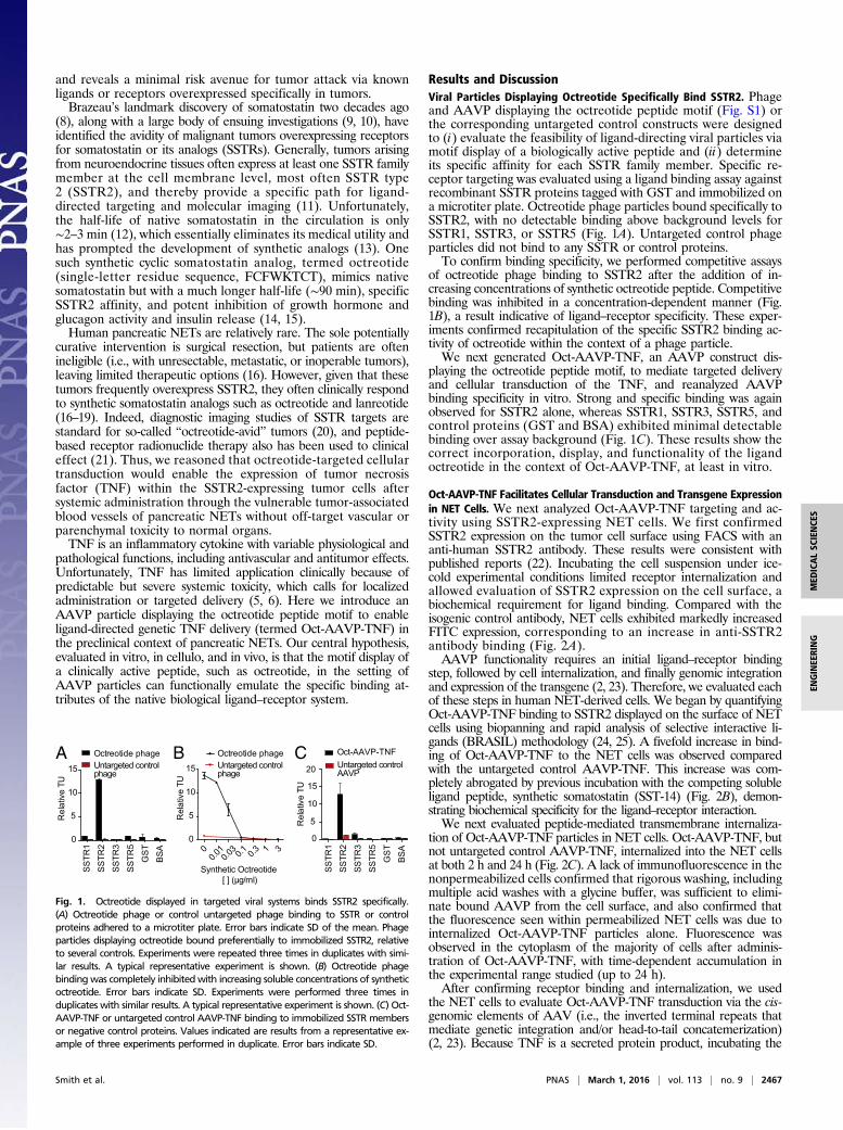
Results and DiscussionViral Particles Displaying Octreotide Specifically Bind SSTR2. Phageand AAVP displaying the octreotide peptide motif (Fig. S1) orthe corresponding untargeted control constructs were designedto (i) evaluate the feasibility of ligand-directing viral particles viamotif display of a biologically active peptide and (ii) determineits specific affinity for each SSTR family member. Specific re-ceptor targeting was evaluated using a ligand binding assay againstrecombinant SSTR proteins tagged with GST and immobilized ona microtiter plate. Octreotide phage particles bound specifically toSSTR2, with no detectable binding above background levels forSSTR1, SSTR3, or SSTR5 (Fig. 1A). Untargeted control phageparticles did not bind to any SSTR or control proteins.To confirm binding specificity, we performed competitive assays
of octreotide phage binding to SSTR2 after the addition of in-creasing concentrations of synthetic octreotide peptide. Competitivebinding was inhibited in a concentration-dependent manner (Fig.1B), a result indicative of ligand–receptor specificity. These exper-iments confirmed recapitulation of the specific SSTR2 binding ac-tivity of octreotide within the context of a phage particle.We next generated Oct-AAVP-TNF, an AAVP construct dis-
playing the octreotide peptide motif, to mediate targeted deliveryand cellular transduction of the TNF, and reanalyzed AAVPbinding specificity in vitro. Strong and specific binding was againobserved for SSTR2 alone, whereas SSTR1, SSTR3, SSTR5, andcontrol proteins (GST and BSA) exhibited minimal detectablebinding over assay background (Fig. 1C). These results show thecorrect incorporation, display, and functionality of the ligandoctreotide in the context of Oct-AAVP-TNF, at least in vitro.
Oct-AAVP-TNF Facilitates Cellular Transduction and Transgene Expressionin NET Cells. We next analyzed Oct-AAVP-TNF targeting and ac-tivity using SSTR2-expressing NET cells. We first confirmedSSTR2 expression on the tumor cell surface using FACS with ananti-human SSTR2 antibody. These results were consistent withpublished reports (22). Incubating the cell suspension under ice-cold experimental conditions limited receptor internalization andallowed evaluation of SSTR2 expression on the cell surface, abiochemical requirement for ligand binding. Compared with theisogenic control antibody, NET cells exhibited markedly increasedFITC expression, corresponding to an increase in anti-SSTR2antibody binding (Fig. 2A).AAVP functionality requires an initial ligand–receptor binding
step, followed by cell internalization, and finally genomic integrationand expression of the transgene (2, 23). Therefore, we evaluated eachof these steps in human NET-derived cells. We began by quantifyingOct-AAVP-TNF binding to SSTR2 displayed on the surface of NETcells using biopanning and rapid analysis of selective interactive li-gands (BRASIL) methodology (24, 25). A fivefold increase in bind-ing of Oct-AAVP-TNF to the NET cells was observed comparedwith the untargeted control AAVP-TNF. This increase was com-pletely abrogated by previous incubation with the competing solubleligand peptide, synthetic somatostatin (SST-14) (Fig. 2B), demon-strating biochemical specificity for the ligand–receptor interaction.We next evaluated peptide-mediated transmembrane internaliza-
tion of Oct-AAVP-TNF particles in NET cells. Oct-AAVP-TNF, butnot untargeted control AAVP-TNF, internalized into the NET cellsat both 2 h and 24 h (Fig. 2C). A lack of immunofluorescence in thenonpermeabilized cells confirmed that rigorous washing, includingmultiple acid washes with a glycine buffer, was sufficient to elimi-nate bound AAVP from the cell surface, and also confirmed thatthe fluorescence seen within permeabilized NET cells was due tointernalized Oct-AAVP-TNF particles alone. Fluorescence wasobserved in the cytoplasm of the majority of cells after adminis-tration of Oct-AAVP-TNF, with time-dependent accumulation inthe experimental range studied (up to 24 h).After confirming receptor binding and internalization, we used
the NET cells to evaluate Oct-AAVP-TNF transduction via the cis-genomic elements of AAV (i.e., the inverted terminal repeats thatmediate genetic integration and/or head-to-tail concatemerization)(2, 23). Because TNF is a secreted protein product, incubating the
A B C
Fig. 1. Octreotide displayed in targeted viral systems binds SSTR2 specifically.(A) Octreotide phage or control untargeted phage binding to SSTR or controlproteins adhered to a microtiter plate. Error bars indicate SD of the mean. Phageparticles displaying octreotide bound preferentially to immobilized SSTR2, relativeto several controls. Experiments were repeated three times in duplicates with simi-lar results. A typical representative experiment is shown. (B) Octreotide phagebindingwas completely inhibitedwith increasing soluble concentrations of syntheticoctreotide. Error bars indicate SD. Experiments were performed three times induplicates with similar results. A typical representative experiment is shown. (C) Oct-AAVP-TNF or untargeted control AAVP-TNF binding to immobilized SSTR membersor negative control proteins. Values indicated are results from a representative ex-ample of three experiments performed in duplicate. Error bars indicate SD.
Smith et al. PNAS | March 1, 2016 | vol. 113 | no. 9 | 2467
MED
ICALSC
IENCE
SEN
GINEE
RING
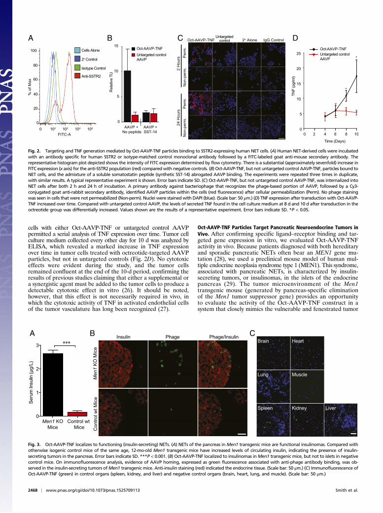
cells with either Oct-AAVP-TNF or untargeted control AAVPpermitted a serial analysis of TNF expression over time. Tumor cellculture medium collected every other day for 10 d was analyzed byELISA, which revealed a marked increase in TNF expressionover time in tumor cells treated with octreotide-targeted AAVPparticles, but not in untargeted controls (Fig. 2D). No cytotoxiceffects were evident during the study, and the tumor cellsremained confluent at the end of the 10-d period, confirming theresults of previous studies claiming that either a supplemental ora synergistic agent must be added to the tumor cells to produce adetectable cytotoxic effect in vitro (26). It should be noted,however, that this effect is not necessarily required in vivo, inwhich the cytotoxic activity of TNF in activated endothelial cellsof the tumor vasculature has long been recognized (27).
Oct-AAVP-TNF Particles Target Pancreatic Neuroendocrine Tumors inVivo. After confirming specific ligand–receptor binding and tar-geted gene expression in vitro, we evaluated Oct-AAVP-TNFactivity in vivo. Because patients diagnosed with both hereditaryand sporadic pancreatic NETs often bear an MEN1 gene mu-tation (28), we used a preclinical mouse model of human mul-tiple endocrine neoplasia syndrome type 1 (MEN1). This syndrome,associated with pancreatic NETs, is characterized by insulin-secreting tumors, or insulinomas, in the islets of the endocrinepancreas (29). The tumor microenvironment of the Men1transgenic mouse (generated by pancreas-specific eliminationof the Men1 tumor suppressor gene) provides an opportunityto evaluate the activity of the Oct-AAVP-TNF construct in asystem that closely mimics the vulnerable and fenestrated tumor
A B C
Fig. 3. Oct-AAVP-TNF localizes to functioning (insulin-secreting) NETs. (A) NETs of the pancreas inMen1 transgenic mice are functional insulinomas. Compared withotherwise isogenic control mice of the same age, 12-mo-old Men1 transgenic mice have increased levels of circulating insulin, indicating the presence of insulin-secreting tumors in the pancreas. Error bars indicate SD. ***P < 0.001. (B) Oct-AAVP-TNF localized to insulinomas inMen1 transgenic mice, but not to islets in negativecontrol mice. On immunofluorescence analysis, evidence of AAVP homing, expressed as green fluorescence associated with anti-phage antibody binding, was ob-served in the insulin-secreting tumors ofMen1 transgenic mice. Anti-insulin staining (red) indicated the endocrine tissue. (Scale bar: 50 μm.) (C) Immunofluorescence ofOct-AAVP-TNF (green) in control organs (spleen, kidney, and liver) and negative control organs (brain, heart, lung, and muscle). (Scale bar: 50 μm.)
A B C D
Fig. 2. Targeting and TNF generation mediated by Oct-AAVP-TNF particles binding to SSTR2-expressing human NET cells. (A) Human NET-derived cells were incubatedwith an antibody specific for human SSTR2 or isotype-matched control monoclonal antibody followed by a FITC-labeled goat anti-mouse secondary antibody. Therepresentative histogram plot depicted shows the intensity of FITC expression determined by flow cytometry. There is a substantial (approximately sevenfold) increase inFITC expression (x axis) for the anti-SSTR2 population (red) comparedwith negative controls. (B) Oct-AAVP-TNF, but not untargeted control AAVP-TNF, particles bound toNET cells, and the admixture of a soluble somatostatin peptide (synthetic SST-14) abrogated AAVP binding. The experiments were repeated three times in duplicate,with similar results. A typical representative experiment is shown. Error bars indicate SD. (C) Oct-AAVP-TNF, but not untargeted control AAVP-TNF, was internalized intoNET cells after both 2 h and 24 h of incubation. A primary antibody against bacteriophage that recognizes the phage-based portion of AAVP, followed by a Cy3-conjugated goat anti-rabbit secondary antibody, identified AAVP particles within the cells (red fluorescence) after cellular permeabilization (Perm). No phage stainingwas seen in cells that were not permeabilized (Non-perm). Nuclei were stained with DAPI (blue). (Scale bar: 50 μm.) (D) TNF expression after transduction with Oct-AAVP-TNF increased over time. Compared with untargeted control AAVP, the levels of secreted TNF found in the cell culture medium at 8 d and 10 d after transduction in theoctreotide group was differentially increased. Values shown are the results of a representative experiment. Error bars indicate SD. *P < 0.05.
2468 | www.pnas.org/cgi/doi/10.1073/pnas.1525709113 Smith et al.
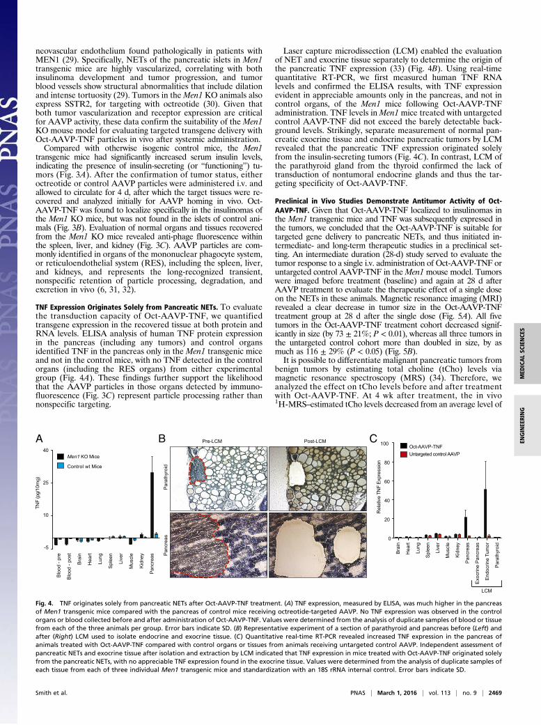
neovascular endothelium found pathologically in patients withMEN1 (29). Specifically, NETs of the pancreatic islets in Men1transgenic mice are highly vascularized, correlating with bothinsulinoma development and tumor progression, and tumorblood vessels show structural abnormalities that include dilationand intense tortuosity (29). Tumors in theMen1 KO animals alsoexpress SSTR2, for targeting with octreotide (30). Given thatboth tumor vascularization and receptor expression are criticalfor AAVP activity, these data confirm the suitability of the Men1KO mouse model for evaluating targeted transgene delivery withOct-AAVP-TNF particles in vivo after systemic administration.Compared with otherwise isogenic control mice, the Men1
transgenic mice had significantly increased serum insulin levels,indicating the presence of insulin-secreting (or “functioning”) tu-mors (Fig. 3A). After the confirmation of tumor status, eitheroctreotide or control AAVP particles were administered i.v. andallowed to circulate for 4 d, after which the target tissues were re-covered and analyzed initially for AAVP homing in vivo. Oct-AAVP-TNF was found to localize specifically in the insulinomas ofthe Men1 KO mice, but was not found in the islets of control ani-mals (Fig. 3B). Evaluation of normal organs and tissues recoveredfrom the Men1 KO mice revealed anti-phage fluorescence withinthe spleen, liver, and kidney (Fig. 3C). AAVP particles are com-monly identified in organs of the mononuclear phagocyte system,or reticuloendothelial system (RES), including the spleen, liver,and kidneys, and represents the long-recognized transient,nonspecific retention of particle processing, degradation, andexcretion in vivo (6, 31, 32).
TNF Expression Originates Solely from Pancreatic NETs. To evaluatethe transduction capacity of Oct-AAVP-TNF, we quantifiedtransgene expression in the recovered tissue at both protein andRNA levels. ELISA analysis of human TNF protein expressionin the pancreas (including any tumors) and control organsidentified TNF in the pancreas only in the Men1 transgenic miceand not in the control mice, with no TNF detected in the controlorgans (including the RES organs) from either experimentalgroup (Fig. 4A). These findings further support the likelihoodthat the AAVP particles in those organs detected by immuno-fluorescence (Fig. 3C) represent particle processing rather thannonspecific targeting.
Laser capture microdissection (LCM) enabled the evaluationof NET and exocrine tissue separately to determine the origin ofthe pancreatic TNF expression (33) (Fig. 4B). Using real-timequantitative RT-PCR, we first measured human TNF RNAlevels and confirmed the ELISA results, with TNF expressionevident in appreciable amounts only in the pancreas, and not incontrol organs, of the Men1 mice following Oct-AAVP-TNFadministration. TNF levels inMen1mice treated with untargetedcontrol AAVP-TNF did not exceed the barely detectable back-ground levels. Strikingly, separate measurement of normal pan-creatic exocrine tissue and endocrine pancreatic tumors by LCMrevealed that the pancreatic TNF expression originated solelyfrom the insulin-secreting tumors (Fig. 4C). In contrast, LCM ofthe parathyroid gland from the thyroid confirmed the lack oftransduction of nontumoral endocrine glands and thus the tar-geting specificity of Oct-AAVP-TNF.
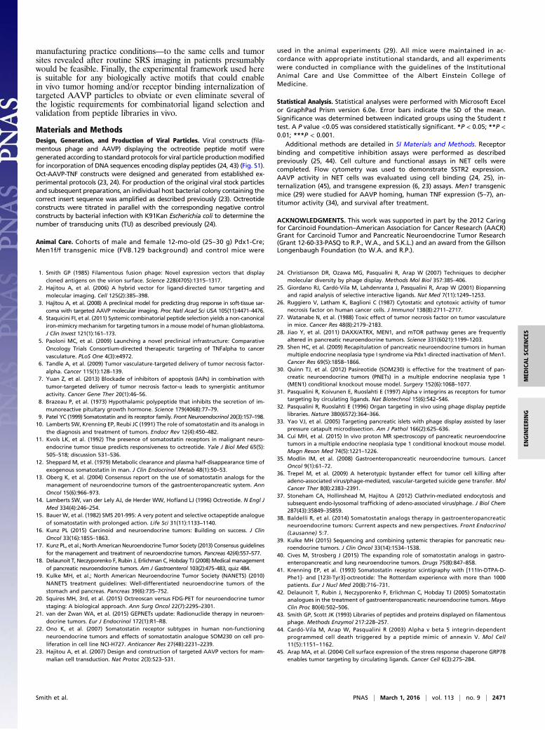
Preclinical in Vivo Studies Demonstrate Antitumor Activity of Oct-AAVP-TNF. Given that Oct-AAVP-TNF localized to insulinomas inthe Men1 transgenic mice and TNF was subsequently expressed inthe tumors, we concluded that the Oct-AAVP-TNF is suitable fortargeted gene delivery to pancreatic NETs, and thus initiated in-termediate- and long-term therapeutic studies in a preclinical set-ting. An intermediate duration (28-d) study served to evaluate thetumor response to a single i.v. administration of Oct-AAVP-TNF oruntargeted control AAVP-TNF in theMen1mouse model. Tumorswere imaged before treatment (baseline) and again at 28 d afterAAVP treatment to evaluate the therapeutic effect of a single doseon the NETs in these animals. Magnetic resonance imaging (MRI)revealed a clear decrease in tumor size in the Oct-AAVP-TNFtreatment group at 28 d after the single dose (Fig. 5A). All fivetumors in the Oct-AAVP-TNF treatment cohort decreased signif-icantly in size (by 73 ± 21%; P < 0.01), whereas all three tumors inthe untargeted control cohort more than doubled in size, by asmuch as 116 ± 29% (P < 0.05) (Fig. 5B).It is possible to differentiate malignant pancreatic tumors from
benign tumors by estimating total choline (tCho) levels viamagnetic resonance spectroscopy (MRS) (34). Therefore, weanalyzed the effect on tCho levels before and after treatmentwith Oct-AAVP-TNF. At 4 wk after treatment, the in vivo1H-MRS–estimated tCho levels decreased from an average level of
A B C
Fig. 4. TNF originates solely from pancreatic NETs after Oct-AAVP-TNF treatment. (A) TNF expression, measured by ELISA, was much higher in the pancreasof Men1 transgenic mice compared with the pancreas of control mice receiving octreotide-targeted AAVP. No TNF expression was observed in the controlorgans or blood collected before and after administration of Oct-AAVP-TNF. Values were determined from the analysis of duplicate samples of blood or tissuefrom each of the three animals per group. Error bars indicate SD. (B) Representative experiment of a section of parathyroid and pancreas before (Left) andafter (Right) LCM used to isolate endocrine and exocrine tissue. (C) Quantitative real-time RT-PCR revealed increased TNF expression in the pancreas ofanimals treated with Oct-AAVP-TNF compared with control organs or tissues from animals receiving untargeted control AAVP. Independent assessment ofpancreatic NETs and exocrine tissue after isolation and extraction by LCM indicated that TNF expression in mice treated with Oct-AAVP-TNF originated solelyfrom the pancreatic NETs, with no appreciable TNF expression found in the exocrine tissue. Values were determined from the analysis of duplicate samples ofeach tissue from each of three individual Men1 transgenic mice and standardization with an 18S rRNA internal control. Error bars indicate SD.
Smith et al. PNAS | March 1, 2016 | vol. 113 | no. 9 | 2469
MED
ICALSC
IENCE
SEN
GINEE
RING
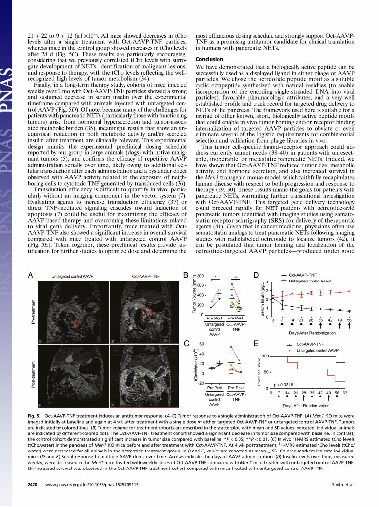
21 ± 22 to 9 ± 12 (all ×104). All mice showed decreases in tCholevels after a single treatment with Oct-AAVP-TNF particles,whereas mice in the control group showed increases in tCho levelsafter 28 d (Fig. 5C). These results are particularly encouraging,considering that we previously correlated tCho levels with surro-gate development of NETs, identification of malignant lesions,and response to therapy, with the tCho levels reflecting the well-recognized high levels of tumor metabolism (34).Finally, in a long-term therapy study, cohorts of mice injected
weekly over 2 mo with Oct-AAVP-TNF particles showed a strongand sustained decrease in serum insulin over the experimentaltimeframe compared with animals injected with untargeted con-trol AAVP (Fig. 5D). Of note, because many of the challenges forpatients with pancreatic NETs (particularly those with functioningtumors) arise from hormonal hypersecretion and tumor-associ-ated metabolic burden (35), meaningful results that show an un-equivocal reduction in both metabolic activity and/or secretedinsulin after treatment are clinically relevant. This experimentaldesign mimics the experimental preclinical dosing schedulereported by our group in large animals (dogs) with native malig-nant tumors (5), and confirms the efficacy of repetitive AAVPadministration serially over time, likely owing to additional cel-lular transduction after each administration and a bystander effectobserved with AAVP activity related to the exposure of neigh-boring cells to cytotoxic TNF generated by transduced cells (36).Transduction efficiency is difficult to quantify in vivo, partic-
ularly without an imaging component in the vector system (7).Evaluating agents to increase transduction efficiency (37) ordirect TNF-mediated signaling cascades toward induction ofapoptosis (7) could be useful for maximizing the efficacy ofAAVP-based therapy and overcoming these limitations relatedto viral gene delivery. Importantly, mice treated with Oct-AAVP-TNF also showed a significant increase in overall survivalcompared with mice treated with untargeted control AAVP(Fig. 5E). Taken together, these preclinical results provide jus-tification for further studies to optimize dose and determine the
most efficacious dosing schedule and strongly support Oct-AAVP-TNF as a promising antitumor candidate for clinical translationin humans with pancreatic NETs.
ConclusionWe have demonstrated that a biologically active peptide can besuccessfully used as a displayed ligand in either phage or AAVPparticles. We chose the octreotide peptide motif as a solublecyclic octapeptide synthesized with natural residues (to enableincorporation of the encoding single-stranded DNA into viralparticles), favorable pharmacologic attributes, and a very wellestablished profile and track record for targeted drug delivery toNETs of the pancreas. The framework used here is suitable for amyriad of other known, short, biologically active peptide motifsthat could enable in vivo tumor homing and/or receptor bindinginternalization of targeted AAVP particles to obviate or eveneliminate several of the logistic requirements for combinatorialselection and validation from phage libraries in vivo.This tumor cell-specific ligand–receptor approach could ad-
dress currently unmet needs (38–40) in patients with unresect-able, inoperable, or metastatic pancreatic NETs. Indeed, wehave shown that Oct-AAVP-TNF reduced tumor size, metabolicactivity, and hormone secretion, and also increased survival inthe Men1 transgenic mouse model, which faithfully recapitulateshuman disease with respect to both progression and response totherapy (29, 30). These results mimic the goals for patients withpancreatic NETs, warranting further translational investigationwith Oct-AAVP-TNF. This targeted gene delivery technologycould proceed rapidly for NET patients with octreotide-avidpancreatic tumors identified with imaging studies using somato-statin receptor scintigraphy (SRS) for delivery of therapeuticagents (41). Given that in cancer medicine, physicians often usesomatostatin analogs to treat pancreatic NETs following imagingstudies with radiolabeled octreotide to localize tumors (42), itcan be postulated that tumor homing and localization of theoctreotide-targeted AAVP particles—produced under good
Pre-
treat
men
tPo
st-tr
eatm
ent
A
EC
B
0 7 14 21 28 35 42 49 56 630
50
100
Days After Randomization
Perc
ent S
urvi
val
p = 0.0316
D
Pre Post Pre Post-20
0
20
40
60
**
Pre Post Pre Post0
200
400
600
800
Tum
or V
olum
e (m
m3 )
Days After Randomization
*Oct-AAVP-TNFUntargeted control AAVP
Oct-AAVP-TNF
UntargetedcontrolAAVP
Oct-AAVP-TNF
Untargetedcontrol AAVP
0 7 14 21 28 35 42 49 560
1
2
3
4
Seru
m In
sulin
(μg/
L)
Oct-AAVP-TNFUntargeted control AAVP
Oct-AAVP-TNFUntargeted control AAVP
tCho
/Wat
er (x
104 )
Fig. 5. Oct-AAVP-TNF treatment induces an antitumor response. (A–C) Tumor response to a single administration of Oct-AAVP-TNF. (A) Men1 KO mice wereimaged initially at baseline and again at 4 wk after treatment with a single dose of either targeted Oct-AAVP-TNF or untargeted control AAVP-TNF. Tumorsare indicated by colored lines. (B) Tumor volume for treatment cohorts are described in the scatterplot, with mean and SD values indicated. Individual animalsare indicated by different-colored dots. The Oct-AAVP-TNF treatment cohort showed a significant decrease in tumor size compared with baseline. In contrast,the control cohort demonstrated a significant increase in tumor size compared with baseline. *P < 0.05; **P < 0.01. (C) In vivo 1H-MRS estimated tCho levels(tCho/water) in the pancreas of Men1 KO mice before and after treatment with Oct-AAVP-TNF. At 4 wk posttreatment, 1H-MRS estimated tCho levels (tCho/water) were decreased for all animals in the octreotide treatment group. In B and C, values are reported as mean ± SD. Colored markers indicate individualmice. (D and E) Serial response to multiple AAVP doses over time. Arrows indicate the days of AAVP administration. (D) Insulin levels over time, measuredweekly, were decreased in theMen1mice treated with weekly doses of Oct-AAVP-TNF compared withMen1mice treated with untargeted control AAVP-TNF.(E) Increased survival was observed in the Oct-AAVP-TNF treatment cohort compared with mice treated with untargeted control AAVP-TNF.
2470 | www.pnas.org/cgi/doi/10.1073/pnas.1525709113 Smith et al.
manufacturing practice conditions—to the same cells and tumorsites revealed after routine SRS imaging in patients presumablywould be feasible. Finally, the experimental framework used hereis suitable for any biologically active motifs that could enablein vivo tumor homing and/or receptor binding internalization oftargeted AAVP particles to obviate or even eliminate several ofthe logistic requirements for combinatorial ligand selection andvalidation from peptide libraries in vivo.
Materials and MethodsDesign, Generation, and Production of Viral Particles. Viral constructs (fila-mentous phage and AAVP) displaying the octreotide peptide motif weregenerated according to standard protocols for viral particle productionmodifiedfor incorporation of DNA sequences encoding display peptides (24, 43) (Fig. S1).Oct-AAVP-TNF constructs were designed and generated from established ex-perimental protocols (23, 24). For production of the original viral stock particlesand subsequent preparations, an individual host bacterial colony containing thecorrect insert sequence was amplified as described previously (23). Octreotideconstructs were titrated in parallel with the corresponding negative controlconstructs by bacterial infection with K91Kan Escherichia coli to determine thenumber of transducing units (TU) as described previously (24).
Animal Care. Cohorts of male and female 12-mo-old (25–30 g) Pdx1-Cre;Men1f/f transgenic mice (FVB.129 background) and control mice were
used in the animal experiments (29). All mice were maintained in ac-cordance with appropriate institutional standards, and all experimentswere conducted in compliance with the guidelines of the InstitutionalAnimal Care and Use Committee of the Albert Einstein College ofMedicine.
Statistical Analysis. Statistical analyses were performed with Microsoft Excelor GraphPad Prism version 6.0e. Error bars indicate the SD of the mean.Significance was determined between indicated groups using the Student ttest. A P value <0.05 was considered statistically significant. *P < 0.05; **P <0.01; ***P < 0.001.
Additional methods are detailed in SI Materials and Methods. Receptorbinding and competitive inhibition assays were performed as describedpreviously (25, 44). Cell culture and functional assays in NET cells werecompleted. Flow cytometry was used to demonstrate SSTR2 expression.AAVP activity in NET cells was evaluated using cell binding (24, 25), in-ternalization (45), and transgene expression (6, 23) assays. Men1 transgenicmice (29) were studied for AAVP homing, human TNF expression (5–7), an-titumor activity (34), and survival after treatment.
ACKNOWLEDGMENTS. This work was supported in part by the 2012 Caringfor Carcinoid Foundation–American Association for Cancer Research (AACR)Grant for Carcinoid Tumor and Pancreatic Neuroendocrine Tumor Research(Grant 12-60-33-PASQ to R.P., W.A., and S.K.L.) and an award from the GillsonLongenbaugh Foundation (to W.A. and R.P.).
1. Smith GP (1985) Filamentous fusion phage: Novel expression vectors that displaycloned antigens on the virion surface. Science 228(4705):1315–1317.
2. Hajitou A, et al. (2006) A hybrid vector for ligand-directed tumor targeting andmolecular imaging. Cell 125(2):385–398.
3. Hajitou A, et al. (2008) A preclinical model for predicting drug response in soft-tissue sar-coma with targeted AAVP molecular imaging. Proc Natl Acad Sci USA 105(11):4471–4476.
4. Staquicini FI, et al. (2011) Systemic combinatorial peptide selection yields a non-canonicaliron-mimicry mechanism for targeting tumors in a mouse model of human glioblastoma.J Clin Invest 121(1):161–173.
5. Paoloni MC, et al. (2009) Launching a novel preclinical infrastructure: ComparativeOncology Trials Consortium-directed therapeutic targeting of TNFalpha to cancervasculature. PLoS One 4(3):e4972.
6. Tandle A, et al. (2009) Tumor vasculature-targeted delivery of tumor necrosis factor-alpha. Cancer 115(1):128–139.
7. Yuan Z, et al. (2013) Blockade of inhibitors of apoptosis (IAPs) in combination withtumor-targeted delivery of tumor necrosis factor-α leads to synergistic antitumoractivity. Cancer Gene Ther 20(1):46–56.
8. Brazeau P, et al. (1973) Hypothalamic polypeptide that inhibits the secretion of im-munoreactive pituitary growth hormone. Science 179(4068):77–79.
9. Patel YC (1999) Somatostatin and its receptor family. Front Neuroendocrinol 20(3):157–198.10. Lamberts SW, Krenning EP, Reubi JC (1991) The role of somatostatin and its analogs in
the diagnosis and treatment of tumors. Endocr Rev 12(4):450–482.11. Kvols LK, et al. (1992) The presence of somatostatin receptors in malignant neuro-
endocrine tumor tissue predicts responsiveness to octreotide. Yale J Biol Med 65(5):505–518; discussion 531–536.
12. Sheppard M, et al. (1979) Metabolic clearance and plasma half-disappearance time ofexogenous somatostatin in man. J Clin Endocrinol Metab 48(1):50–53.
13. Oberg K, et al. (2004) Consensus report on the use of somatostatin analogs for themanagement of neuroendocrine tumors of the gastroenteropancreatic system. AnnOncol 15(6):966–973.
14. Lamberts SW, van der Lely AJ, de Herder WW, Hofland LJ (1996) Octreotide. N Engl JMed 334(4):246–254.
15. Bauer W, et al. (1982) SMS 201-995: A very potent and selective octapeptide analogueof somatostatin with prolonged action. Life Sci 31(11):1133–1140.
16. Kunz PL (2015) Carcinoid and neuroendocrine tumors: Building on success. J ClinOncol 33(16):1855–1863.
17. Kunz PL, et al.; North American Neuroendocrine Tumor Society (2013) Consensus guidelinesfor the management and treatment of neuroendocrine tumors. Pancreas 42(4):557–577.
18. Delaunoit T, Neczyporenko F, Rubin J, Erlichman C, Hobday TJ (2008) Medical managementof pancreatic neuroendocrine tumors. Am J Gastroenterol 103(2):475–483, quiz 484.
19. Kulke MH, et al.; North American Neuroendocrine Tumor Society (NANETS) (2010)NANETS treatment guidelines: Well-differentiated neuroendocrine tumors of thestomach and pancreas. Pancreas 39(6):735–752.
20. Squires MH, 3rd, et al. (2015) Octreoscan versus FDG-PET for neuroendocrine tumorstaging: A biological approach. Ann Surg Oncol 22(7):2295–2301.
21. van der Zwan WA, et al. (2015) GEPNETs update: Radionuclide therapy in neuroen-docrine tumors. Eur J Endocrinol 172(1):R1–R8.
22. Ono K, et al. (2007) Somatostatin receptor subtypes in human non-functioningneuroendocrine tumors and effects of somatostatin analogue SOM230 on cell pro-liferation in cell line NCI-H727. Anticancer Res 27(4B):2231–2239.
23. Hajitou A, et al. (2007) Design and construction of targeted AAVP vectors for mam-malian cell transduction. Nat Protoc 2(3):523–531.
24. Christianson DR, Ozawa MG, Pasqualini R, Arap W (2007) Techniques to deciphermolecular diversity by phage display. Methods Mol Biol 357:385–406.
25. Giordano RJ, Cardó-Vila M, Lahdenranta J, Pasqualini R, Arap W (2001) Biopanningand rapid analysis of selective interactive ligands. Nat Med 7(11):1249–1253.
26. Ruggiero V, Latham K, Baglioni C (1987) Cytostatic and cytotoxic activity of tumornecrosis factor on human cancer cells. J Immunol 138(8):2711–2717.
27. Watanabe N, et al. (1988) Toxic effect of tumor necrosis factor on tumor vasculaturein mice. Cancer Res 48(8):2179–2183.
28. Jiao Y, et al. (2011) DAXX/ATRX, MEN1, and mTOR pathway genes are frequentlyaltered in pancreatic neuroendocrine tumors. Science 331(6021):1199–1203.
29. Shen HC, et al. (2009) Recapitulation of pancreatic neuroendocrine tumors in humanmultiple endocrine neoplasia type I syndrome via Pdx1-directed inactivation of Men1.Cancer Res 69(5):1858–1866.
30. Quinn TJ, et al. (2012) Pasireotide (SOM230) is effective for the treatment of pan-creatic neuroendocrine tumors (PNETs) in a multiple endocrine neoplasia type 1(MEN1) conditional knockout mouse model. Surgery 152(6):1068–1077.
31. Pasqualini R, Koivunen E, Ruoslahti E (1997) Alpha v integrins as receptors for tumortargeting by circulating ligands. Nat Biotechnol 15(6):542–546.
32. Pasqualini R, Ruoslahti E (1996) Organ targeting in vivo using phage display peptidelibraries. Nature 380(6572):364–366.
33. Yao VJ, et al. (2005) Targeting pancreatic islets with phage display assisted by laserpressure catapult microdissection. Am J Pathol 166(2):625–636.
34. Cui MH, et al. (2015) In vivo proton MR spectroscopy of pancreatic neuroendocrinetumors in a multiple endocrine neoplasia type 1 conditional knockout mouse model.Magn Reson Med 74(5):1221–1226.
35. Modlin IM, et al. (2008) Gastroenteropancreatic neuroendocrine tumours. LancetOncol 9(1):61–72.
36. Trepel M, et al. (2009) A heterotypic bystander effect for tumor cell killing afteradeno-associated virus/phage-mediated, vascular-targeted suicide gene transfer. MolCancer Ther 8(8):2383–2391.
37. Stoneham CA, Hollinshead M, Hajitou A (2012) Clathrin-mediated endocytosis andsubsequent endo-lysosomal trafficking of adeno-associated virus/phage. J Biol Chem287(43):35849–35859.
38. Baldelli R, et al. (2014) Somatostatin analogs therapy in gastroenteropancreaticneuroendocrine tumors: Current aspects and new perspectives. Front Endocrinol(Lausanne) 5:7.
39. Kulke MH (2015) Sequencing and combining systemic therapies for pancreatic neu-roendocrine tumors. J Clin Oncol 33(14):1534–1538.
40. Cives M, Strosberg J (2015) The expanding role of somatostatin analogs in gastro-enteropancreatic and lung neuroendocrine tumors. Drugs 75(8):847–858.
41. Krenning EP, et al. (1993) Somatostatin receptor scintigraphy with [111In-DTPA-D-Phe1]- and [123I-Tyr3]-octreotide: The Rotterdam experience with more than 1000patients. Eur J Nucl Med 20(8):716–731.
42. Delaunoit T, Rubin J, Neczyporenko F, Erlichman C, Hobday TJ (2005) Somatostatinanalogues in the treatment of gastroenteropancreatic neuroendocrine tumors. MayoClin Proc 80(4):502–506.
43. Smith GP, Scott JK (1993) Libraries of peptides and proteins displayed on filamentousphage. Methods Enzymol 217:228–257.
44. Cardó-Vila M, Arap W, Pasqualini R (2003) Alpha v beta 5 integrin-dependentprogrammed cell death triggered by a peptide mimic of annexin V. Mol Cell11(5):1151–1162.
45. Arap MA, et al. (2004) Cell surface expression of the stress response chaperone GRP78enables tumor targeting by circulating ligands. Cancer Cell 6(3):275–284.
Smith et al. PNAS | March 1, 2016 | vol. 113 | no. 9 | 2471
MED
ICALSC
IENCE
SEN
GINEE
RING