a188 t cell priming with deep™ il-15 improves preclinical
TRANSCRIPT
• Deep IL-15 Primed PMEL cells:– Preferentially expand CD8+ T cells in circulation
and intratumorally– Do not increase bystander circulating neutrophils
or lymphocytes, including NK cells, which are associated with the immunotoxicity for systemic IL-15
– Result in dramatically lower systemic exposure to IL15-Fc
– Do not induce significant systemic cytokine release
– Do not result in significant body weight loss– Result in histopathology findings of lower
severity compared to IL15-Fc across multiple organs
• Deep IL-15 Primed PMEL cells do show different biodistribution in naïve vs tumor – bearing mice:
– Reduced accumulation in spleen of tumor –bearing mice
– Enhanced accumulation in tumor-draining LN compared to contralateral LN
• Loading of PMEL cells with Deep IL-15 results in increased persistence of PMEL cells in circulation as well as in the periphery and at the tumor site.
• Deep IL-15 primed PMEL cells show improved in vivo expansion and anti-tumor activity compared to PMEL.
• Clinical trials with Deep IL-15 Primed multi-target T cells (TRQ15-01) are expected to start in 2018.
Results
A188Presented at the 4th CRI-CIMT-EATI-AACR International Cancer Immunotherapy ConferenceSeptember 30–October 3, 2018 New York, NY
T cell priming with Deep™ IL-15 improves preclinical safety compared to systemic IL-15, and increases in vivo persistence and activity Elena Geretti, Philip Bardwell, Xiaoyan Liang, Santina Caruso, De-Kuan Chang, Jesse Lyons, Austin Boesch, Aaron Handler, Carlos Tassa, Sanela Bilic, Janice Lansita, Becker Hewes, Jonathan Fitzgerald, Thomas Andresen. Torque Therapeutics, Cambridge, MA
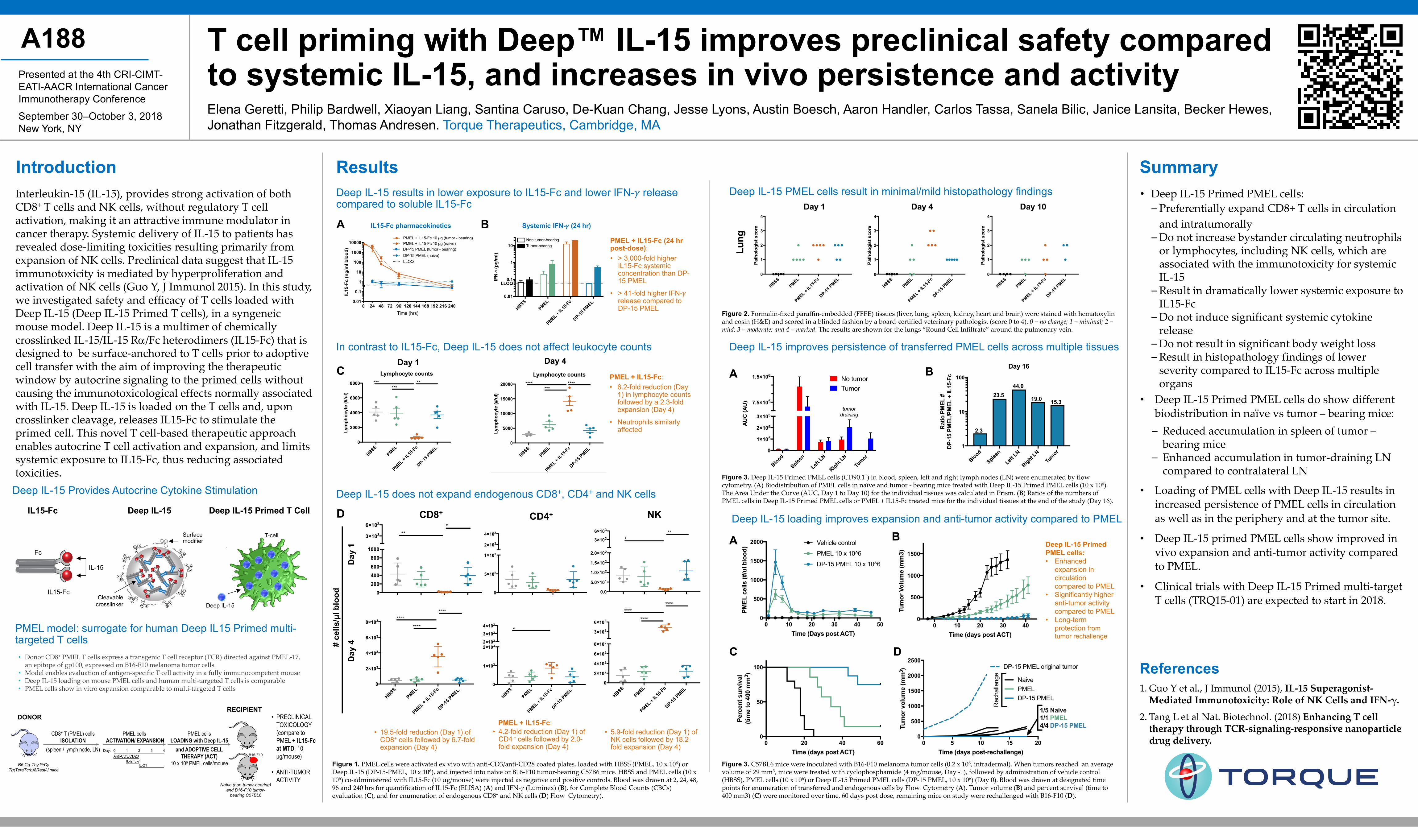
Figure 1. PMEL cells were activated ex vivo with anti-CD3/anti-CD28 coated plates, loaded with HBSS (PMEL, 10 x 106) or Deep IL-15 (DP-15-PMEL, 10 x 106), and injected into naïve or B16-F10 tumor-bearing C57B6 mice. HBSS and PMEL cells (10 x 106) co-administered with IL15-Fc (10 μg/mouse) were injected as negative and positive controls. Blood was drawn at 2, 24, 48, 96 and 240 hrs for quantification of IL15-Fc (ELISA) (A) and IFN-𝛾𝛾 (Luminex) (B), for Complete Blood Counts (CBCs) evaluation (C), and for enumeration of endogenous CD8+ and NK cells (D) Flow Cytometry).
References 1. Guo Y et al., J Immunol (2015), IL-15 Superagonist-
Mediated Immunotoxicity: Role of NK Cells and IFN-γ.
2. Tang L et al Nat. Biotechnol. (2018) Enhancing T cell therapy through TCR-signaling-responsive nanoparticle drug delivery.
DDeep IL-15 does not expand endogenous CD8+, CD4+ and NK cells
Deep IL-15 results in lower exposure to IL15-Fc and lower IFN-𝛾𝛾 release compared to soluble IL15-Fc
Interleukin-15 (IL-15), provides strong activation of both CD8+ T cells and NK cells, without regulatory T cell activation, making it an attractive immune modulator in cancer therapy. Systemic delivery of IL-15 to patients has revealed dose-limiting toxicities resulting primarily from expansion of NK cells. Preclinical data suggest that IL-15 immunotoxicity is mediated by hyperproliferation and activation of NK cells (Guo Y, J Immunol 2015). In this study, we investigated safety and efficacy of T cells loaded with Deep IL-15 (Deep IL-15 Primed T cells), in a syngeneic mouse model. Deep IL-15 is a multimer of chemically crosslinked IL-15/IL-15 Rα/Fc heterodimers (IL15-Fc) that is designed to be surface-anchored to T cells prior to adoptive cell transfer with the aim of improving the therapeutic window by autocrine signaling to the primed cells without causing the immunotoxicological effects normally associated with IL-15. Deep IL-15 is loaded on the T cells and, upon crosslinker cleavage, releases IL15-Fc to stimulate the primed cell. This novel T cell-based therapeutic approach enables autocrine T cell activation and expansion, and limits systemic exposure to IL15-Fc, thus reducing associated toxicities.
Deep IL-15 PMEL cells result in minimal/mild histopathology findings
In contrast to IL15-Fc, Deep IL-15 does not affect leukocyte counts
NKCD8+
IL15-Fc pharmacokinetics
Introduction
Day 1 Day 4
Systemic IFN-𝛾𝛾 (24 hr)A B
Day
1
Day
4
PMEL + IL15-Fc:
C
Deep IL-15 improves persistence of transferred PMEL cells across multiple tissues
Figure 3. Deep IL-15 Primed PMEL cells (CD90.1+) in blood, spleen, left and right lymph nodes (LN) were enumerated by flow cytometry. (A) Biodistribution of PMEL cells in naïve and tumor - bearing mice treated with Deep IL-15 Primed PMEL cells (10 x 106). The Area Under the Curve (AUC, Day 1 to Day 10) for the individual tissues was calculated in Prism. (B) Ratios of the numbers of PMEL cells in Deep IL-15 Primed PMEL cells or PMEL + IL15-Fc treated mice for the individual tissues at the end of the study (Day 16).
PMEL + IL15-Fc: • 6.2-fold reduction (Day
1) in lymphocyte counts followed by a 2.3-fold expansion (Day 4)
• Neutrophils similarly affected
Summary
A B
PMEL + IL15-Fc (24 hrpost-dose): • > 3,000-fold higher
IL15-Fc systemic concentration than DP-15 PMEL
• > 41-fold higher IFN-𝛾𝛾release compared to DP-15 PMEL
CD4+
# ce
lls/μlb
lood
• 4.2-fold reduction (Day 1) of CD4 + cells followed by 2.0-fold expansion (Day 4)
• 5.9-fold reduction (Day 1) of NK cells followed by 18.2-fold expansion (Day 4)
• 19.5-fold reduction (Day 1) of CD8+ cells followed by 6.7-fold expansion (Day 4)
Figure 2. Formalin-fixed paraffin-embedded (FFPE) tissues (liver, lung, spleen, kidney, heart and brain) were stained with hematoxylin and eosin (H&E) and scored in a blinded fashion by a board-certified veterinary pathologist (score 0 to 4). 0 = no change; 1 = minimal; 2 = mild; 3 = moderate; and 4 = marked. The results are shown for the lungs “Round Cell Infiltrate” around the pulmonary vein.
Deep IL-15 loading improves expansion and anti-tumor activity compared to PMEL
Figure 3. C57BL6 mice were inoculated with B16-F10 melanoma tumor cells (0.2 x 10⁶, intradermal). When tumors reached an average volume of 29 mm3, mice were treated with cyclophosphamide (4 mg/mouse, Day -1), followed by administration of vehicle control (HBSS), PMEL cells (10 x 106) or Deep IL-15 Primed PMEL cells (DP-15 PMEL, 10 x 106) (Day 0). Blood was drawn at designated time points for enumeration of transferred and endogenous cells by Flow Cytometry (A). Tumor volume (B) and percent survival (time to 400 mm3) (C) were monitored over time. 60 days post dose, remaining mice on study were rechallenged with B16-F10 (D).
Deep IL-15 Primed PMEL cells:• Enhanced
expansion in circulation compared to PMEL
• Significantly higher anti-tumor activity compared to PMEL
• Long-term protection from tumor rechallenge
A B
DC
Deep IL-15 Provides Autocrine Cytokine Stimulation
T-cellSurfacemodifier
Cleavable crosslinker
IL15-Fc
IL-15
Fc
Deep IL-15
• Donor CD8+ PMEL T cells express a transgenic T cell receptor (TCR) directed against PMEL-17, an epitope of gp100, expressed on B16-F10 melanoma tumor cells.
• Model enables evaluation of antigen-specific T cell activity in a fully immunocompetent mouse• Deep IL-15 loading on mouse PMEL cells and human multi-targeted T cells is comparable • PMEL cells show in vitro expansion comparable to multi-targeted T cells
PMEL model: surrogate for human Deep IL15 Primed multi-targeted T cells
B6.Cg-Thy1a/Cy Tg(TcraTcrb)8Rest/J mice
RECIPIENT
CD8+ T (PMEL) cells ISOLATION
(spleen / lymph node, LN)
PMEL cells ACTIVATION/ EXPANSION
• PRECLINICAL TOXICOLOGY (compare to PMEL + IL15-Fc at MTD, 10 μg/mouse)
• ANTI-TUMOR ACTIVITY
B16-F10
Naïve (non-tumor-bearing) and B16-F10 tumor-
bearing C57BL6
PMEL cells LOADING with Deep IL-15
and ADOPTIVE CELLTHERAPY (ACT)
10 x 106 PMEL cells/mouse
DONOR
IL15-Fc Deep IL-15 Deep IL-15 Primed T Cell