a tan to die for? dan magrill taz singh laura tincknell
TRANSCRIPT

A Tan to Die For?
Dan Magrill
Taz Singh
Laura Tincknell

Mr. RB History and Examination
Background - 53 yrs, male, unemployed PC - RIF pain HPC - RIF pain for 1/12, radiating to the back.
Loss of appetite, weight loss, tiredness, indigestion. Loose bowels 1/52, no blood or mucus. No N+V.
PMH - No THREAD. L Testicular lump 18/12 - under observation.

History and Examination cont...
FH - Paternal Grandfather - Bowel Ca. Father - Diverticulitis.
SH - Unmarried, living alone and unemployed. Smoking 20+/day. Social drinker.
SE: CVS - No chest pain, palpitations,
breathlessness, orthopnoea, collapse, nocturnal dyspnoea...
History and Examination cont...
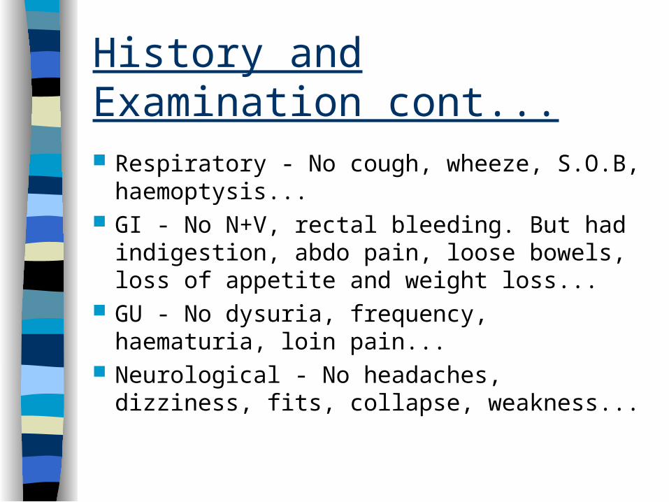
Respiratory - No cough, wheeze, S.O.B, haemoptysis...
GI - No N+V, rectal bleeding. But had indigestion, abdo pain, loose bowels, loss of appetite and weight loss...
GU - No dysuria, frequency, haematuria, loin pain...
Neurological - No headaches, dizziness, fits, collapse, weakness...

History and Examination cont
O/E - Anxious, thin. T-37.5C, Pulse-regular, 75 bpm, BP 120/60 mmHg. O2 Sats- 97%
CVS : Unremarkable Resp: Unremarkable Neuro: Unremarkable GI: Abdomen distended. Large firm mass in
whole of abdomen apart from LIF which was tender on palpation. Liver percussed out to 35cm. BS present. PR-NAD

Plan
USS: showing multiple liver metastases. Biopsy - melanoma.
Ix to find primary: colonoscopy, CXR, ENT and opthalmology.
Opthalmology report: R eye, smooth melanotic mass in anterior chamber.
Management: Palliative Chemotherapy.

Presentation of a disease
In Incidence A Age Surgeon’s Sex Gown Geography Physicians Predisposing factors Might Macro/Micro Pathology Make Management Progress Prognosis

Incidence
UK incidence of 10 / 100 000 (per year) Rising by 7% every Year
Least common of the “Big Three”, but highest mortality.
Over last 20 years, incidence risen by over 80%

Age
Superficial Spreading and Nodular Malignant Melanoma - 20-60 year olds
Lentigo Malignant Melanoma - >60y.

Sex
In the UK, women are affected twice as often as men
In Men, the commonest site is the back In Women it is the Lower Leg (50%)

Geography
The worldwide incidence is proportional to the Geographic Latitude
Caucasians living closest to equator at highest risk
This suggests an effect of UV radiation
People living outside their indigenous climate are at risk

Predisposing Factors
Fair Skin Red Hair Living close to Equator Freckles Exposure to the Sun Melanocytic Naevus (found in 30%) Genetics - 5% of Pt have Family History


Macro/Micro Pathology 1
Superficial Spreading Malignant Melanoma– 50% of UK cases, especially female– Commonest in Lower Leg– Macular Tumour with Variable
Pigmentation


Macro/Micro Pathology 2
Nodular Malignant Melanoma– Seen in 25% of UK cases, especially Male– Commonest site is the Trunk– Pigmented Nodule– Grows rapidly and can Ulcerate


Macro/Micro Pathology 3
Lentigo Malignant Melanoma– 15% of UK cases– Malignant melanoma growing in long
standing Lentigo Maligna• These arise form sun damaged skin• Often in elderly, especially who have worked
outside for many years


Macro/Micro Pathology 4
Acral Lentiginous Malignant Melanoma– 10% of UK cases– Commonest form in Mongoloids– Tumour affects Palms, Soles and Nail
Beds– Often diagnosed late - poor prognosis


Staging
Local Staging assessed using the BRESLOW method– Measured mm between granular cell layer
and deepest identifiable melanoma cell Metastasis are uncommon if confined to
epidermis


Diagnosis
The following changes in a Naevus or Pigmented lesion– Size, usually a recent increase– Shape, irregular in outline– Colour, variation - darker or lighter– Inflammation, especially at edge– Crusting, may ooze or bleed– Itch

Differential Diagnosis
Benign melanocytic naevus Seborrhoeic wart Haemangioma Dermatofibroma Pigmented Basal Cell Carcinoma Benign Lentigo

Management 1
Surgical Excision– If <1mm, use a 1cm clearance margin– If >1mm, need a 1-2cm clearance
• As this is quite a large area a skin graft may be indicated
Regular follow up to detect recurrence– Local– Lymphatic, regional or distant– Blood Bourne - to distant sites (eg Liver)

Management 2
Elective Lymph node dissection and Sentinel node biopsy not recommended as routine.
Radiotherapy of limited use– Interferon-alfa may increase survival if
tumour >1.5mm thick

Prognosis
Related to tumour depth 5 year survival:
– <1mm 95-100%– 1-2mm 80-96%– 2.1-4mm 60-75%– >4mm 50%

Prevention and Public Education
If caught early have good prognosis Public should be encouraged to visit
doctor early if changing pigmented lesion
Sun exposure should be discouraged– Especially if fair skinned or with multiple
melanocytic naevi