a systematic capsid evolution approach performed in vivo ... · a systematic capsid evolution...
TRANSCRIPT

A systematic capsid evolution approach performed invivo for the design of AAV vectors with tailoredproperties and tropismMarcus Davidssona,1, Gang Wanga,1,2, Patrick Aldrin-Kirka, Tiago Cardosob, Sara Nolbrantb, Morgan Hartnora,Janitha Mudannayakea, Malin Parmarb, and Tomas Björklunda,3
aMolecular Neuromodulation, Department of Experimental Medical Science, Lund University, 221 84 Lund, Sweden; and bDevelopmental and RegenerativeNeurobiology, Department of Experimental Medical Science, Lund Stem Cell Center, Lund University, 221 84 Lund, Sweden
Edited by Tomas Hökfelt, Karolinska Institute, Stockholm, Sweden, and approved November 11, 2019 (received for review June 12, 2019)
Adeno-associated virus (AAV) capsid modification enables thegeneration of recombinant vectors with tailored properties andtropism. Most approaches to date depend on random screening,enrichment, and serendipity. The approach explored here, calledBRAVE (barcoded rational AAV vector evolution), enables efficientselection of engineered capsid structures on a large scale usingonly a single screening round in vivo. The approach stands in con-trast to previous methods that require multiple generations ofenrichment. With the BRAVE approach, each virus particle displaysa peptide, derived from a protein, of known function on the AAVcapsid surface, and a unique molecular barcode in the packagedgenome. The sequencing of RNA-expressed barcodes from a single-generation in vivo screen allows the mapping of putative bindingsequences from hundreds of proteins simultaneously. Using the BRAVEapproach and hidden Markov model-based clustering, we present25 synthetic capsid variants with refined properties, such as retro-grade axonal transport in specific subtypes of neurons, as shownfor both rodent and human dopaminergic neurons.
vector evolution | gene therapy | barcoding | retrograde transport |dopamine
The recombinant adeno-associated virus (AAV) vector hasbecome the de facto standard for in vivo gene transfer (1). In
clinical gene therapy, AAV has been proven to be safe and resultin stable gene expression over many years (2, 3). However, withfew exceptions, specificity and efficacy are still issues hinderingsome critical applications and have delayed broad clinicaladoption in other areas (4). Engineering of the AAV capsid hasbeen conducted over the last 2 decades to address these short-comings and to generate capsids with altered tropism and func-tion (5–15). AAV capsid engineering can broadly be divided intodirected evolution and rational design. In directed evolution,random processes are utilized such as capsid gene shuffling ofavailable serotypes (9, 16, 17), random peptide insertion into aknown site of the AAV capsid (10, 14, 18–21), or phage display(22, 23). This approach, however, involves a selection processthat requires multiple generations of screening to identify func-tional capsids (6–8), which excludes the use of tools such assingle-cell or in situ sequencing. Due to the random nature of thisprocess, it is also inherently unreproducible, and the resultingcapsid variants provide little mechanistic insights into the molec-ular targets engaged. The alternative approach is rational design,where a priori knowledge is utilized to design fewer capsid variantsand through systematic evaluation refine the capsid structure toachieve the desired function. Examples of this include the dis-ruption of native cellular binding motifs and insertion of high-affinity ligands in the Cap gene, and through the use of structuraland evolutionary shared sequences infer putative ancestral var-iants (24–29). What unifies the rational design approaches is thatthe resulting tested variants are counted in the tens or hundredscompared with the thousands to millions of capsids assessed usinga directed evolution approach. While AAV capsid engineering has
been around for over 25 y, few have broadly replaced wild-typevariants in preclinical applications, and none has to date reachedlate-stage clinical trials despite distinct intellectual property in-centives. The reason is that the improved features and functionshave seldom transferred from the screening host species (mostoften mouse) to humans or the nonhuman primate (30).To improve on this, we have developed a method for capsid
engineering named barcoded rational AAV vector evolution(BRAVE), which encompasses all of the benefits of rationaldesign (18, 26, 31–35) while maintaining the broad screeningdiversity permitted by directed evolution. The key to this methodis a viral library production approach where each virus particledisplays a protein-derived peptide on the surface, which is linkedto a unique barcode in the packaged genome (36). Throughhidden Markov model-based clustering (37), we were able toidentify consensus motifs for neuronal cell type-specific retro-grade transport and expression in the brain. The BRAVE ap-proach enables the selection of functional capsid structures usingonly a single-generation screening. Furthermore, it can be usedto map, with high resolution, the putative binding sequences of
Significance
A challenge with the available synthetic viruses used for thetreatment of genetic disorders is that they originate from wild-type viruses. These viruses benefit from infecting as many cellsas possible in the body, while therapies should most oftentarget a particular cell type, for example, dopamine neurons inthe brain. In this paper, we present a technique for developingtargeted synthetic viruses suitable for clinical therapy. Weshow that such viruses can be designed to target human do-pamine neurons in vivo and to transport along the connectivepathways in the brain, allowing for unprecedented accuracy ofthe therapy.
Author contributions: M.D., G.W., P.A.-K., M.P., and T.B. designed research; M.D., G.W.,P.A.-K., T.C., S.N., M.H., and J.M. performed research; M.D., G.W., P.A.-K., and T.B. ana-lyzed data; and M.D., G.W., P.A.-K., M.P., and T.B. wrote the paper.
Competing interest statement: M.D. and T.B. are authors of a patent application coveringcomponents of the presented study.
This article is a PNAS Direct Submission.
This open access article is distributed under Creative Commons Attribution-NonCommercial-NoDerivatives License 4.0 (CC BY-NC-ND).
Data deposition: Datasets supporting the conclusions of this article have been deposited inthe National Center for Biotechnology Information (NCBI) Sequence Read Archive (SRA)(accession no. PRJNA473475). The R-based workflow is publicly available as a Git repositoryat https://bitbucket.org/MNM-LU/aav-library and as a Docker image: Bjorklund/aavlib:v0.2.1M.D. and G.W. contributed equally to this work.2Present address: National Institute for Viral Disease Control and Prevention, ChineseCenter for Disease Control and Prevention, 102206 Beijing, China.
3To whom correspondence may be addressed. Email: [email protected].
This article contains supporting information online at https://www.pnas.org/lookup/suppl/doi:10.1073/pnas.1910061116/-/DCSupplemental.
First published December 9, 2019.
www.pnas.org/cgi/doi/10.1073/pnas.1910061116 PNAS | December 26, 2019 | vol. 116 | no. 52 | 27053–27062
NEU
ROSC
IENCE
Dow
nloa
ded
by g
uest
on
May
16,
202
0

large protein libraries. Through the use of peptide sequences de-rived from proteins with a known mechanism in the human CNS,we believe that this approach provides significantly higher chancesof success in clinical translation. Here, we have explored and val-idated this approach in the rodent brain as well as in human em-bryonic stem cell-derived dopaminergic (DA) neurons in vitro andin the brain of rats with human-derived DA transplants in vivo.
ResultsIn this, the first application of BRAVE screening, we utilizedpeptides derived from proteins with documented synapse in-teraction. The aim was to develop AAV capsid variants that areefficiently taken up and transported retrogradely in neurons invivo. This combinatorial method enabled the generation andsimultaneous functional mapping of close to 4 million uniqueAAV virion variants in parallel, both in vitro and in vivo. In asingle round of screening, we selected 25 de novo capsid variants,all of which could be packaged into functional viruses with thecapacity to become retrogradely transported in vivo and effi-ciently transduce neurons in vitro. We further characterized acapsid variant expressing highly efficient retrograde transport inboth rat and human (stem cell-derived) DA neurons in vivo, anda pan-neuronal retrogradely transported AAV which we went onto use in an experiment aimed to elucidate the function of asubset of neurons in the basolateral nucleus of the amygdalaprojecting to the rostromedial striatum.
Design of Barcoded AAVs. To link capsid structure to an in vivoexpressed molecular barcode, we generated a refined AAV pro-duction plasmid. The barcode is inserted into the 3′ untranslatedregion (UTR) of GFP of a gutted self-complementary AAV ge-nome (38, 39) and the AAV2 Rep/Cap genes are expressed fromthe same plasmid, but outside the inverted terminal repeats (ITRs),creating a linkage between capsid structure and barcode. Here,peptides are displayed at N587 of the VP1 capsid protein (18) (SIAppendix, Fig. S1). The insertion mutates the wild-type (WT) AAV2heparan sulfate proteoglycan-binding motif (32). The AAV pro-duced from this plasmid without an inserted peptide is hereafterreferred to as MNMnull.With the aim of targeting neurons at their terminals, we se-
lected 131 proteins based on their documented affinity to syn-apses (Fig. 1A and SI Appendix, Fig. S2). We digested their aminoacid (aa) sequences computationally into overlapping 14- or 22-aa-long polypeptides, and 3 alternative linkers were added to the14-aa polypeptides (Fig. 1B, 1 and 2). The resulting 92,358 oligo-nucleotides were synthesized in parallel on a gene chip array (Fig.1B, 3). They were then assembled into the backbone plasmid toallow for packaging of replication-deficient AAV particles, wherethe peptide is displayed on the capsid surface and a 20-bp randommolecular barcode is included as part of the genome (Fig. 1B,4 and SI Appendix, Fig. S1). In parallel, the same plasmid librarywas utilized to generate a look-up table (LUT), linking the ran-dom barcodes to the respective peptide (36) (see Fig. 1B, 5a andSI Appendix, Fig. S3 andMethods for details). The resulting librarycontained 3,934,570 unique combinations of peptide and barcode.On average, 50 barcodes point to the same peptide, but only 1 pep-tide is represented per barcode (40). This oversampling is essentialfor noise filtration and mapping of oligonucleotide array-inducedmutations (see SI Appendix,Methods on LUT generation for details).To ensure that each virion is assembled using only 1 mutated
capsid protein variant and that the corresponding barcode ispackaged inside, the AAV library plasmid was supplied at a 100-or 1,000-fold dilution compared with standard production (10, 41),resulting in 30 or 3 copies per cell (SI Appendix, Fig. S3), re-spectively. Using the AAV vector library, we performed multipleparallel screening experiments in vitro and in vivo, followed by thesequencing of barcodes expressed in the mRNA. Efficacy could bemapped back to the original 131 proteins with the help of the LUT
and consensus motifs determined using the Hammock method (Fig.1B, 6 and 7). The Hammock method utilizes a hidden Markovmodel approach to find underlying patterns in the complete datasetand cluster together peptides that share sequence homology.
Validation of the BRAVE Screening Principle. As an initial proof ofconcept, we utilized the BRAVE technology to screen for thereintroduction of tropism for HEK293T cells in vitro (Fig. 2 A–B″′),which was lost when the heparin sulfate proteoglycan (HS)-bindingmotif was removed in the MNMnull capsid (Fig. 2 B and B′). In thescreening of the 4 million uniquely barcoded capsid variants, wefound several regions from the 131 included proteins that conferred asignificantly improved infectivity over the parent MNMnull capsidstructure (SI Appendix, Fig. S4 and Dataset S1). One peptide fromherpes simplex virus 2 (HSV-2) surface protein pUL44 was selected,and a first capsid variant was generated (MNM001) (Fig. 2A). Thiscapsid indeed displayed a recovered tropism to the HEK293T cells(Fig. 2B″). Through a second BRAVE screen in primary corticalneurons, we identified several peptides clustering over a C-terminalregion of the HSV-2 pUL1 protein (Fig. 2C). From these data, wegenerated an AAV capsid (MNM002) which improved the infectivityof primary neurons in culture dramatically compared with both theAAV2-WT and the MNMnull vector (Fig. 2D–D″′, SI Appendix, Fig.S4, and Dataset S1). Together, these data provide compelling evi-dence of the efficiency of the BRAVE screening approach and in-dications that the peptides can be utilized more broadly than theinitial protein inclusion criteria (synaptic association) would imply.
Identification of Capsid Variants for Retrograde Axonal Transport. Inan experiment aimed to identify individual AAV capsid variantswith efficient retrograde transport in neurons in vivo, we injectedthe AAV library (MNMlib) into the forebrain of adult rats.Compared with the standard AAV2-WT vector, the insertedpeptides confer a striking change in the transduction pattern withboth a broader spread of transduction and retrograde transportto the connecting afferent regions (Fig. 2E). Eight weeks afterinjection, total RNA was extracted from the injection site, and3 connected areas (Fig. 2F) and the transcribed barcodes weresequenced (Dataset S1). Analysis of the unique peptides (iden-tified using the barcodes and the LUT) throughout the BRAVEpipeline provides unique insights into the efficacy of the process(Fig. 2G). More than 98% of the designed oligonucleotides(array; black circle) were successfully cloned into the plasmidlibrary and barcoded (purple ring) and 72% of the peptidespermitted complete assembly of the AAV (as assessed by DNasetreatment; gray ring). Barcodes recovered at the dissected re-gions reveal that ∼13% of the inserted peptides promoted effi-cient transduction in the brain (green ring segment) and ∼4%promoted retrograde transport in neurons (red ring segment).Pooling all experiments, including in vitro experiments, revealedthat at least 19% of the peptides could promote infectivity in thecell types tested (dark blue ring segment). The large fraction,compared with what is observed in random peptide approaches,provides evidence that the rational design, even at this relativelyhigh throughput, provides a significant advantage in the gener-ation of functional capsids compared with a random approach.Even without any consideration of unsuitable peptide sequencesfor capsid insertion, only the introduction of a premature stopcodon would remove 54 to 68% of all capsids with a randominserted peptide at this length.
Validation of Capsid Variants. From this comprehensive in vivoBRAVE screening, we selected 23 peptides from 20 proteins thatall were represented by multiple barcodes and found in multipleanimals (Fig. 2H, SI Appendix, Fig. S5, and Dataset S1). Twenty-one of the 23 de novo AAV capsid structures found in vivo (andthe 2 from the in vitro screening) allowed for higher than or onpar with AAV2-WT packaging efficacy (SI Appendix, Fig. S1). Of
27054 | www.pnas.org/cgi/doi/10.1073/pnas.1910061116 Davidsson et al.
Dow
nloa
ded
by g
uest
on
May
16,
202
0

note is that this experiment suggests that the insertion of 16 aa(14-aa peptide + 2 alanine [A] linkers) is approaching the upperlimit of insertion into this site in the AAV capsid, as only 1 out ofthe 23 selected peptides was 24 aa in length (14 + 2 G4S/A5 linkers or 22 aa + 2 A linkers). Moreover, in the infectivelibrary, the 24-aa sequences were heavily underrepresented de-spite making up more than half of the total diversity in the peptidelibrary (see Dataset S1 for details). To confirm the predictivenature of the single-generation BRAVE screen, we injected the23 individually produced capsid variants into the rat striatum, oneby one, and assessed retrograde transport to afferent regions (SIAppendix, Fig. S5). All 21 capsids selected for this capacity showeddrastically improved transport compared with the parent AAV,while the 2 selected in vitro showed no improvement. Using ahidden Markov model (HMM)-based clustering (37), of all pep-tides with retrograde transportability, we could determine theputative consensus motif for each of the 23 capsids, providing afoundation for directed optimization (Dataset S1).In the BRAVE design, we can compare the function of pep-
tides across multiple regions of the brain and also between ani-mals (Dataset S1). This data aggregation allows us to mapdomains of proteins that promote specific transport or uptakeabilities. Exemplified by HSV-derived pUL22 protein, peptidesthroughout the protein can drive uptake at the injection site inthe striatum (Fig. 3A). However, only peptide sequences fromthe C-terminal region of the protein displayed reproducible transportto all afferent brain structures: the cortex, thalamus, and substantianigra (Fig. 3 A and B). HMM clustering of all peptides recovered atthese sites revealed 2 overlapping consensus motifs (Fig. 3 B–D).These were generated into the MNM004 (VMSVLLVDTDATQQ)and MNM023 (QQIAAGPTEGAPSV) (Fig. 3C) capsid structures,respectively, with both displaying similar transport patterns in vivo (SIAppendix, Fig. S5 E and X). The MNM004 capsid promoted ef-ficient retrograde transport to all afferent regions as far backas the medial entorhinal cortex (Fig. 3E and Movie S1). Theparent AAV2-WT capsid, by contrast, promoted efficient trans-duction at the site of injection but very little retrograde trans-port of the vector (Fig. 3E). While this manuscript was beingfinalized, another peptide, LADQDYTKTA (Retro peptide),was published promoting strong retrograde transport when dis-played in the same location on the AAV2 capsid surface (AAV2-Retro) (13). The paper also reports 2 additional point mutantsin the capsid but without any data showing their influence onthe transport. To allow the comparison between the peptidesthose were here omitted. When compared in vivo, the 2 vectorsdisplayed very similar retrograde transport properties, withMNM004 showing equal or higher transport efficacy (Fig. 3 F–Land SI Appendix, Fig. S6).
A Capsid Variant for the Retrograde Infectivity of Dopamine Neurons.In the final component of the BRAVE screening, we aimed todevelop a AAV capsid variant with efficient retrograde transportto dopamine neurons of the substantia nigra from injections intothe striatal target region. In this screening, we identified 2 do-mains of the canine adenovirus (CAV-2) capsid protein [anoften-used but immunogenic vector for targeting of DA neuronsfrom their terminals in vivo (42)]. Using a Cre-inducible AAVgenome (CMV-loxP-GFP) injected into the striatum of TH-Creknockin rats, we found that the CAV-2–based MNM008 improvedthe retrograde infectivity of nigral neurons dramatically comparedwith the parent AAV2, from only a few scattered neurons transduced
A
B
Fig. 1. Barcoded rational AAV vector evolution screening approach. (A) Piechart displaying the selected 131 proteins with documented affinity tosynapses. (B) Schematics of the BRAVE procedure. 1: NCBI reference aminoacid sequences were computationally digested into 14-aa (or 22-aa) -longpolypeptides with a 1-aa shifting sliding window. 2: Three alternative linkerswere added to the 14-aa polypeptides, for example, a rigid linker with5 alanine residues (14aaA5). 3: Codon-optimized sequences (92,358) weresynthesized in parallel on an oligonucleotide array. 4: The pool of oligonu-cleotides was assembled into an AAV production backbone with cis-actingAAV2 Rep/Cap and ITR-flanking CMV-GFP. A 20-bp random molecular bar-code (BC) was simultaneously inserted into the 3′ UTR of the GFP gene. 5a:Using a fraction of the plasmid preparation, exposed to Cre-recombinase invitro, a look-up table was generated linking the BC to the peptide. 5b: Usingthe same plasmid pool from step 4, an AAV batch was produced (MNMlib),and multiple parallel screening experiments were performed both in vitroand in vivo followed by BC sequencing from mRNA. 6: Through the combi-nation of the sequenced barcodes and the LUT, transduction efficacy can bemapped back to each of the original 131 proteins, here exemplified by the
aa sequence of HSV-1 pUL21. The height of each bar represents the rela-tive abundance of that amino acid, normalized to the total library andsummed from all peptides covering the region. 7: Consensus motifs can bedetermined using the Hammock, hidden Markov model-based clusteringapproach.
Davidsson et al. PNAS | December 26, 2019 | vol. 116 | no. 52 | 27055
NEU
ROSC
IENCE
Dow
nloa
ded
by g
uest
on
May
16,
202
0

by AAV2-WT to the vast majority of the substantia nigra parscompacta neurons targeted by MNM008 (Fig. 4 A and A′). Next,we utilized this capsid to revisit a long-standing question ofnigrostriatal innervation topography. The rodent striatum isfunctionally divided along each axis: the mediolateral, dorsoven-
tral, and rostrocaudal (43). This topography is maintained in thestriatal afferents from the forebrain neocortex, amygdala, andthalamic nuclei (43). Here we utilized 3 genes with available high-specificity antibodies, mCherry, TagBFP2, and alpha-synuclein(labeled red, blue, and green, respectively, in Fig. 4B). The latter
PFCStr
ThalSNpc
0
15000
30000
45000
60000
75000
90000
Trsp.3991
Injection site 12240
Infective AAV
17401
DNase resistant AAV
66531
9 0 6 3 5
Plasmid library
Array92343
AAV-2MNMlib
G
FE
Infective library
Plasmid library
HSV-1 | pUL22 HSV-1 | pUL22#023
AAV1 AAV1#020
BoNT | C-Hc#024
BoNT | C-Hc
AAV1 AAV1#014
BoNT | E-Hc#010
BoNT | E-Hc
BV | G#007
BV | G
#019CAV-2 | F-SH01
CAV-2 | F-SH01#008
CAV-2 | F-SH01
EV71 | VP3#005
EV71 | VP3
HRP#013
HRP
HSV-1 | pUL37#006
HSV-1 | pUL37
HSV-1 | pUS6#016
HSV-1 | pUS6
HSV-2 | pUS6#018
HSV-2 | pUS6
HSV-2 | pUL10#021
HSV-2 | pUL10
HSV-2 | pUL19#022
HSV-2 | pUL19
HSV-1 | pUL22#004
HSV-1 | pUL22
HSV-2 | pUL27#003
HSV-2 | pUL27
MV | H-Edmonston#012
MV | H-Edmonston
PRV | Becker-gE #015
PRV | Becker-gE
Tau#017
Tau
Tau#009
Tau
TBEV | E-4387-B7#011
TBEV | E-4387-B7
CAV-2 | F-SH01
Transported in vivo 3cpc
Transported in vivo 30cpc
SA SA#025
#024
H | Selected for improved retrograde transport in vivo
MNM001
293T
AAV2
293T
MNMnull
293T
MNM002
293TB B’ B’’ B’’’
MNM001
Neurons
AAV2
Neurons
MNMnull
Neurons
MNM002
NeuronsD D’ D’’ D’’’
A | MNM001 – recovered HEK293T tropism in vitro
C | MNM002 – Selected for improved neuronal tropism in vitro
Infective library
Plasmid library
HSV-2 | pUL44 HSV-2 | pUL44 HEK293T in vitro 30cpc
HSV-2 | pUL1 HSV-2 | pUL1Primary Neurons in vitro 30cpc
Primary Neurons in vitro 3cpc
Infective library
Plasmid library
Fig. 2. Assessment of improved in vitro infectivity and retrograde transport in the brain using BRAVE screening. (A) Infectivity mapping of HSV-2 pUL44peptides is displayed comparing infective peptides (Top Left) plotted against total plasmid library (Bottom Left) and compared with peptides infectingHEK293T cells in vitro (Right). (B–B″′) The MNMnull capsid disrupts this binding (B′), compared to AAV2-WT (B). The first capsid generated from single-generation BRAVE screening in HEK293T, MNM001, displayed a significantly recovered tropism (B″), which is not observed in a capsid screened for in primaryneurons (B″′). (C) Infectivity mapping of HSV-2 pUL1 peptides for introduced tropism to primary neurons. (D–D″′) In a second experiment, we used the BRAVEtechnology to improve the infectivity of primary cortical rat neurons in vitro. Both AAV2-WT and the MNMnull vector display very poor infectivity of primaryneurons (D and D′) and the MNM001 displays some improvement (D″). BRAVE screening in primary neurons identified several peptides clustering over aC-terminal region of the HSV-2 pUL1 protein, which improved the infectivity of primary neurons in culture dramatically (D″′). (E) In vivo expression pattern ofGFP after injection of a 30-cpc (copies per cell) library into the rat forebrain compared with an AAV2-WT vector at the same titer. (F) Connectivity diagramshowing the injection site (striatum; Str) and connected neuronal populations frontal cortex (PFC), thalamus (Thal), and substantia nigra (SNpc) utilized forBRAVE screening in vivo for retrograde transport; 2.5 × 10¹⁰ or 4.4 × 10⁸ vector genomes of the AAV library with capsid concentration of 30 or 3 cpc, re-spectively (5-μL total volume), were injected into the striatum (“3 cpc” and “30 cpc” refer to copies per cell of the capsid/genome plasmid utilized at the AAVproduction stage). (G) Polar plot showing the absolute quantities of the unique peptides recovered at each step of the BRAVE assay. (H) From the in vivoscreening for improved retrograde transport capacity, we selected 23 peptides from 20 proteins that all were represented by multiple barcodes and found inmultiple animals (Dataset S1). Twenty-three of the 25 de novo AAV capsid structures allowed for higher than or at par with AAV2-WT packaging efficacy (SIAppendix, Fig. S1), all with retrograde transportability (SI Appendix, Fig. S5).
27056 | www.pnas.org/cgi/doi/10.1073/pnas.1910061116 Davidsson et al.
Dow
nloa
ded
by g
uest
on
May
16,
202
0

was chosen due to the protein’s prominent localization to axonterminals and its excellent axonal transport properties in thenigrostriatal system. With this approach, we could confirm thatthe rostrocaudal topography of the projections to the striatum islinked to a mediolateral topography of the substantia nigra (Fig. 4B–C″). Also, this experiment showed abundant collaterals fromthe dopamine neurons projecting to the central striatum reachingthe entire ipsilateral neocortex, including as far back as theentorhinal cortex (Fig. 4B′).
A significant drawback of de novo capsid design using animalmodels has been the lack of predictability with regard to thetranslation to human cells. The BRAVE approach offers anexciting possibility to solve this problem through the use of nat-urally occurring peptides from viruses and proteins with knownfunction in the human brain. To establish that the properties ofMNM008 were not limited to rodent DA neurons, we performedan experiment on DA neurons derived from Cre-expressing hu-man embryonic stem cells (hESCs) both in vitro and in vivo aftertransplantation into immunocompromised rats (44) (Fig. 4 B–D).Six months after transplantation, when the grafted neurons havegrown to establish axonal projections with their usual forebraintarget areas, the MNM008 vector was injected into the frontalcortex of the animals. MNM008 was efficiently transported backto the transplanted neurons innervating this region (Fig. 4E), withthe majority of these cells being dopaminergic (Fig. 4E′).Interestingly, MNM004, which does not have this ability in the
rat brain, showed similar potency in the transplanted human neurons(Fig. 4 F and F′). Using the same in vitro hESC differentiation pro-tocol, we confirmed that the de novo capsid variants (MNM002, 008,and 010) which displayed high tropism on primary rodent neuronsalso showed much higher tropism than the wild-type AAV variants(Fig. 4D and SI Appendix, Fig. S8) in human neurons in vitro. Ofnote is that the MNM004 capsid variant, so efficient in vivo, wasnot at all suitable for in vitro transduction (Fig. 4D).
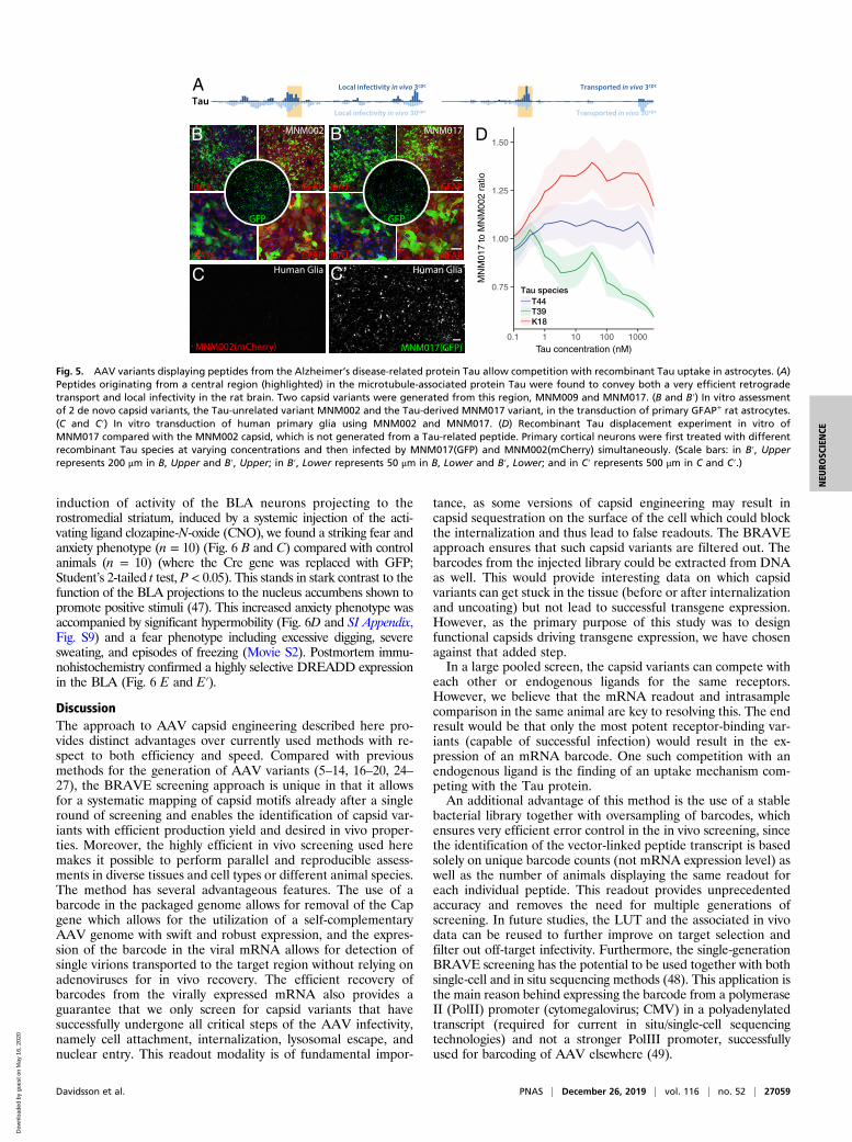
Mapping of Tau-Derived Peptides for Neuronal Uptake of AAV. TheBRAVE approach provides a unique possibility to map proteinfunction systematically. We therefore utilized this approach to displaypeptides from endogenous proteins involved in the pathogenesisof Alzheimer’s disease, amyloid precursor protein (APP) andmicrotubule-associated protein Tau, to provide insights into themechanism underlying the proposed cell-to-cell communication ofthese proteins in the diseased brain (45, 46). In the mapping of APP,we found 2 regions that conferred retrograde transport, 1 in the sol-uble APP N-terminal region and 1 in the amyloid beta region (SIAppendix, Fig. S7). The functional properties of peptides originatingfrom Tau were even more striking. In this protein, a central regionconveyed very efficient retrograde transport (Fig. 5A, SI Appendix, Fig.S7, and Dataset S1). Two capsid structures were generated from thisregion, MNM009 and MNM017. Both capsids promoted retrogradetransport in vivo (SI Appendix, Fig. S5), but MNM017 also displayedadditional noteworthy properties. MNM017 infected both primary ratneurons and glial fibrillary acidic protein (GFAP)-positive primary ratglial cells in vitro with very high efficacy (Fig. 5B), including humanprimary astrocytes (Fig. 5 C–C″). Using this property, we then per-formed a displacement experiment comparing the MNM017 with theneurotrophic MNM002 capsid, which is not generated from a Tau-derived peptide (Fig. 5D). Three groups of primary neuron pop-ulations were pretreated with different recombinant Tau variants(T44, T39, and K18). The T44 variant had no apparent effect on theMNM017-to-MNM002 ratio of infectivity, while K18 enhancedthe infectivity of MNM017 and the T39 variant efficiently blockedthe infectivity compared with MNM002 (Fig. 5D). This outcomesuggests that the Tau peptide is utilizing a receptor on the neuronsthat also has a binding activity of full-length human Tau protein.
Basolateral Amygdala Regulates Anxiety Behavior via the RostromedialStriatum. The BRAVE-generated capsid variants provide a pow-erful tool for brain connectivity studies. We utilized the MNM004capsid variant to answer an outstanding question regarding thefunctional contribution of the afferents from the basolateralamygdala (BLA) to the rostromedial striatum. The experiment wasconducted using a retrogradely induced chemogenetics (designerreceptor exclusively activated by designer drugs, DREADDs) ap-proach, using an injection of an MNM004-Cre vector in therostromedial striatum combined with an injection of a Cre-inducible,activating DREADD vector in the BLA (Fig. 6A). After selective
A
B
C
D
E
F
G H
I
J
K L
Fig. 3. Characterization of the MNM004 capsid for retrograde axonaltransport in the rat brain. (A) A C-terminal region of the HSV pUL22 protein(highlighted) displayed reproducible transport to all afferent brain areas(cortex, thalamus, and substantia nigra) while not showing the same bias atthe injection site (striatum). As this domain of the pUL22 protein is highlyconserved between HSV strains (HSV-1 and HSV-2), both are visualized forcomparison. (B–D) HMM clustering of all peptides displaying these proper-ties revealed 2 overlapping consensus motifs (C). Those were generated intothe MNM004 (B) and MNM023 (D) AAV capsid variants. (E) In vivo compar-ison between MNM004 and the parent AAV2 after unilateral striatal in-jection. Both vectors express GFP, and sections were developed using brownDAB (3, 3-diaminobenzidine)-peroxidase reaction (see Movie S1 for a 3Dvisualization). (F–L) Same animal comparison between MNM004 andAAV2 capsid with the previously published LADQDYTKTA peptide (AAV2-Retro). AAV2-Retro expressing mCherry was injected into the left striatum(G) while MNM004 expressing GFP was injected into right striatum (H) atmatched titers. Monitored afferent regions include the prefrontal cortex (F),anterodorsal (I) and intralaminar thalamic nuclei (J), and basolateral amygdala(K and L) (see SI Appendix, Fig. S6 for additional information). (Scale bars,200 μm.)
Davidsson et al. PNAS | December 26, 2019 | vol. 116 | no. 52 | 27057
NEU
ROSC
IENCE
Dow
nloa
ded
by g
uest
on
May
16,
202
0

DIO-GFP
MNM008
SN in TH-Cre ratDIO-GFP
AAV2-WT
SN in TH-Cre rat
A A’
GFP/TH/Map2
D
AAV2
MNM008MNM004 MNM010MNM002
AAV9AAV8 MNM001
F
hESC-TX DIO-GFP
MNM004
MNM004F’
GFP/THhESC-TX
E
hESC-TX DIO-GFP
MNM008
MNM008E’
GFP/THhESC-TX
MNM008 DIO vectors in Striatum
mCherry/aSyn/tagBFP2
Str
CC
Thal
Ctx
SN
SN
mCherry aSyn tagBFP2
B
C C’ C’’
B’
aSyn
EC
Fig. 4. Validation of AAV capsids infecting rat and human dopaminergic neurons in vivo and in vitro. (A and A′) In vivo comparison between the CAV-2–derived MNM008 and AAV2-WT assessing infectivity of dopamine neurons from their terminals in the striatum. Both vectors express Cre-inducible GFP(double-floxed inverted orientation, DIO-GFP) and were injected into the striatum of TH-Cre knockin rats. (B and C″) Mapping of nigral afferent topographyto the striatum using 3 Cre-inducible genes (mCherry, red; alpha-synuclein, green; tagBFP2, blue) packaged in separate MNM008 vectors and injected into thestriatum along the rostrocaudal axis (B), as illustrated in a horizontal section including both striatum (Str) and substantia nigra pars compacta (SN) imagedusing a laser-scanning confocal microscope. (C–C″) The nigro-striatal topology is visible in the transgene expression pattern in the SN. (B′) transduced DA-neurons send collaterals as far back as the entorhinal cortex (EC). (D) Assessment of retained neuronal tropism in human embryonic stem cell-derived DAneuroblasts in vitro. (E and F) Assessment of retrograde infectivity in humanized rats. These animals first received an hESC-derived DA-rich neuronal transplant(expressing Cre) into the striatum. Six months later, the MNM008 or MNM004 vectors (expressing DIO-GFP) were injected into the frontal cortex. hESC-derivedneurons in the graft were efficiently labeled by both vectors (E and F) and the vast majority expressed the DA neuron marker tyrosine hydroxylase (TH) determinedusing confocal microscopy (E′ and F′). (Scale bars: in A′ represents 100 μm in A and A′; in B represents 500 μm in B, C, and C″; in B′ represents 100 μm; in D represents100 μm; in E represents 50 μm in E and F; and in E′ represents 50 μm in E′ and F′.)
27058 | www.pnas.org/cgi/doi/10.1073/pnas.1910061116 Davidsson et al.
Dow
nloa
ded
by g
uest
on
May
16,
202
0
induction of activity of the BLA neurons projecting to therostromedial striatum, induced by a systemic injection of the acti-vating ligand clozapine-N-oxide (CNO), we found a striking fear andanxiety phenotype (n = 10) (Fig. 6 B and C) compared with controlanimals (n = 10) (where the Cre gene was replaced with GFP;Student’s 2-tailed t test, P < 0.05). This stands in stark contrast to thefunction of the BLA projections to the nucleus accumbens shown topromote positive stimuli (47). This increased anxiety phenotype wasaccompanied by significant hypermobility (Fig. 6D and SI Appendix,Fig. S9) and a fear phenotype including excessive digging, severesweating, and episodes of freezing (Movie S2). Postmortem immu-nohistochemistry confirmed a highly selective DREADD expressionin the BLA (Fig. 6 E and E′).
DiscussionThe approach to AAV capsid engineering described here pro-vides distinct advantages over currently used methods with re-spect to both efficiency and speed. Compared with previousmethods for the generation of AAV variants (5–14, 16–20, 24–27), the BRAVE screening approach is unique in that it allowsfor a systematic mapping of capsid motifs already after a singleround of screening and enables the identification of capsid var-iants with efficient production yield and desired in vivo proper-ties. Moreover, the highly efficient in vivo screening used heremakes it possible to perform parallel and reproducible assess-ments in diverse tissues and cell types or different animal species.The method has several advantageous features. The use of abarcode in the packaged genome allows for removal of the Capgene which allows for the utilization of a self-complementaryAAV genome with swift and robust expression, and the expres-sion of the barcode in the viral mRNA allows for detection ofsingle virions transported to the target region without relying onadenoviruses for in vivo recovery. The efficient recovery ofbarcodes from the virally expressed mRNA also provides aguarantee that we only screen for capsid variants that havesuccessfully undergone all critical steps of the AAV infectivity,namely cell attachment, internalization, lysosomal escape, andnuclear entry. This readout modality is of fundamental impor-
tance, as some versions of capsid engineering may result incapsid sequestration on the surface of the cell which could blockthe internalization and thus lead to false readouts. The BRAVEapproach ensures that such capsid variants are filtered out. Thebarcodes from the injected library could be extracted from DNAas well. This would provide interesting data on which capsidvariants can get stuck in the tissue (before or after internalizationand uncoating) but not lead to successful transgene expression.However, as the primary purpose of this study was to designfunctional capsids driving transgene expression, we have chosenagainst that added step.In a large pooled screen, the capsid variants can compete with
each other or endogenous ligands for the same receptors.However, we believe that the mRNA readout and intrasamplecomparison in the same animal are key to resolving this. The endresult would be that only the most potent receptor-binding var-iants (capable of successful infection) would result in the ex-pression of an mRNA barcode. One such competition with anendogenous ligand is the finding of an uptake mechanism com-peting with the Tau protein.An additional advantage of this method is the use of a stable
bacterial library together with oversampling of barcodes, whichensures very efficient error control in the in vivo screening, sincethe identification of the vector-linked peptide transcript is basedsolely on unique barcode counts (not mRNA expression level) aswell as the number of animals displaying the same readout foreach individual peptide. This readout provides unprecedentedaccuracy and removes the need for multiple generations ofscreening. In future studies, the LUT and the associated in vivodata can be reused to further improve on target selection andfilter out off-target infectivity. Furthermore, the single-generationBRAVE screening has the potential to be used together with bothsingle-cell and in situ sequencing methods (48). This application isthe main reason behind expressing the barcode from a polymeraseII (PolII) promoter (cytomegalovirus; CMV) in a polyadenylatedtranscript (required for current in situ/single-cell sequencingtechnologies) and not a stronger PolIII promoter, successfullyused for barcoding of AAV elsewhere (49).
TauTransported in vivo 30cpc
Transported in vivo 3cpc
Local infectivity in vivo 30cpc
Local infectivity in vivo 3cpc
D
A
0.75
1.00
1.25
1.50
0.1 1 10 100 1000
Tau concentration (nM)
MN
M01
7 to
MN
M00
2 ra
tio
T39T44
K18
Tau species
IBA1
IBA1
GFP
GFAP
GFAP
MNM002
IBA1
IBA1
GFP
GFAP
GFAP
MNM017B’
Human Glia
MNM002(mCherry) MNM017(GFP)
C’ Human Glia
B
C
Fig. 5. AAV variants displaying peptides from the Alzheimer’s disease-related protein Tau allow competition with recombinant Tau uptake in astrocytes. (A)Peptides originating from a central region (highlighted) in the microtubule-associated protein Tau were found to convey both a very efficient retrogradetransport and local infectivity in the rat brain. Two capsid variants were generated from this region, MNM009 and MNM017. (B and B′) In vitro assessmentof 2 de novo capsid variants, the Tau-unrelated variant MNM002 and the Tau-derived MNM017 variant, in the transduction of primary GFAP+ rat astrocytes.(C and C′) In vitro transduction of human primary glia using MNM002 and MNM017. (D) Recombinant Tau displacement experiment in vitro ofMNM017 compared with the MNM002 capsid, which is not generated from a Tau-related peptide. Primary cortical neurons were first treated with differentrecombinant Tau species at varying concentrations and then infected by MNM017(GFP) and MNM002(mCherry) simultaneously. (Scale bars: in B′, Upperrepresents 200 μm in B, Upper and B′, Upper; in B′, Lower represents 50 μm in B, Lower and B′, Lower; and in C′ represents 500 μm in C and C′.)
Davidsson et al. PNAS | December 26, 2019 | vol. 116 | no. 52 | 27059
NEU
ROSC
IENCE
Dow
nloa
ded
by g
uest
on
May
16,
202
0

The BRAVE approach depends on 2 fundamental advances:the tailored massively parallel array synthesis of DNA sequencesand the generation of a look-up table linking a genetic barcodeto the capsid structure. These techniques provide significantadvantages to the screening approach such as the single-generation selection, potential mapping of changes over theentire capsid structure, and scanning of sequences derived fromproteins with known function, but they also confer some lim-
itations. The principal limitation is the comparatively small sizeand diversity of the libraries compared with libraries generatedthrough processes such as capsid shuffling, degenerate primers,or error-prone PCR. However, this limitation is outweighed bythe power of the rational design, which ensures the inclusion ofonly potentially functional sequences in the library withoutsequence repetitions or bias. While this approach cannot ac-commodate the full theoretical diversity of an entirely random
A B B’ C
D E E’
F G H I
Fig. 6. Selective activation of basolateral amygdala projections to the rostromedial striatum induced using the MNM004 capsid drives fear and anxietyphenotypes. (A) Experimental injection paradigm for functional assessment of the afferents from the basolateral amygdala to the rostromedial striatum(CPu). We injected the Cre-expressing MNM004 vector bilaterally into the CPu and a Cre-inducible (DIO) chemogenetic (DREADD) vector into the BLA bi-laterally into wild-type rats (n = 20). Eight weeks later the animals received an s.c. injection of the DREADD-activating ligand clozapine-N-oxide. (B and C)Assessment of the fear and anxiety phenotype using the elevated plus maze, where the animals spent significantly less time in the open arms (n = 10) (B)compared with control animals (n = 10) (B′ and C), where the Cre gene was replaced with GFP. (D) The increased anxiety phenotype was accompanied bysignificant hypermobility in the open-field arena and a fear phenotype including excessive digging, severe sweating, and episodes of freezing (Movie S2).(E and E′) Visualization of selective DREADD expression in the BLA using immunohistochemistry together with brown DAB-peroxidase precipitation reaction.(F–I) After selective induction of the activity of the BLA neurons projecting to the dorsal striatum using the DREADD ligand CNO, we found a striking fear andanxiety phenotype in the animals with excessive defecation, sweating, digging, and freezing (Movie S2). This behavior was accompanied by an increase inboth ipsilateral and contralateral rotation (F), high-speed rushes (G), and significantly elevated mobility (H). However, this did not result in any conditioning,as seen in the preference test performed the next day (I). Both animals in the active group and the control group spent equal time in both chambers with nosigns of conditioned place aversion. (Scale bar, 50 μm in E and E′.) The asterisk in C, F, and H represents P < 0.05 in Student’s 2-tailed t test, corrected formultiple comparisons in F and H using Bonferroni correction. Error bars in C and F–I display the standard error of the mean (SEM).
27060 | www.pnas.org/cgi/doi/10.1073/pnas.1910061116 Davidsson et al.
Dow
nloa
ded
by g
uest
on
May
16,
202
0

approach, the continuous increase in synthesis size (now in the 106
range) together with a decrease in cost will further broaden thepotential applications.It is important to realize that the principle behind the BRAVE
approach is not limited to the screening of capsid variants car-rying modifications restricted to a single, small domain, such asthe HS-binding motif used in this study (36, 50). It is possiblethat the BRAVE screening approach can be used where rationalchanges are introduced at any site of the 3 capsid proteins. In thiscase, where the long-read sequencing would be required to mapthe changes throughout the Cap gene, the implementation isgreatly facilitated by the efficiency of the barcoding paradigm,allowing for this costly sequencing to be conducted only at thegeneration of the look-up table, not when assessing the functionin vivo. However, the feasibility of such an approach remains tobe shown.In the presented studies, microdissection was used to separate
afferent brain regions and allowed for the identification oftransported variants, for example, to the DA neurons of the SN.However, the current library can potentially, without modifica-tion, be used together with FACS, single-cell/single-nucleusRNA-seq (sequencing) paradigms (e.g., from 10× genomics),or spatial sequencing [e.g., Slide-seq (51)]. This screen couldallow for direct discrimination of capsids with specific, cell type-specific infectivity from those with high ubiquitous tropism. Suchmethods could further reduce the off-target infectivity, improvepotency, and make possible the development of vector capsidswith high selectivity and tropism of cellular subtypes.In this first validation of the technique, we focused on se-
quences derived from proteins with known synaptic interactions.This approach was remarkably efficient, allowing us in a singleround of screening to generate 25 AAV vector variants with theproperty of being efficiently transported retrogradely in diverseneuronal populations in the brain. Five of these were exploredfurther in vitro and in vivo for their usefulness as research toolsfor the study of neuronal connectivity and function in the rodentbrain. The HSV-derived peptide sequence used in MNM004 andthe CAV-2–derived peptide sequence used in MNM008 pro-vided these vectors with radically improved retrograde transportproperties relative to the parent AAV2 vector, on par with orsuperior to previously published AAV variants. The propertiesof these AAV variants make them highly attractive as toolsfor studies of functional connectivity in the brain, as illustratedby the experiment performed on the neurons connecting thebasolateral amygdala and the rostromedial striatum, where we
used the MNM004 vector as a retrogradely induced chemogenetictool.Moreover, the remarkable properties ofMNM009 andMNM017,which express Tau-derived peptide sequences, indicate that theinfectivity of these vectors depends on their ability to bind to areceptor that has binding activity for the human Tau protein, andthus possibly involved in the cell-to-cell spread of this disease-causing protein.In summary, the BRAVE screening approach described here
provides a tool for the design of AAV vector variants with tai-lored in vivo properties and tropism based on a highly efficientmethod for screening and selection of engineered capsid struc-tures. This strategy, which can be applied in vivo in both rodentsand primates, as well as in human cells in vitro, opens up thedesign and development of synthetic AAV vectors expressingcapsid structures with unique properties and broad potential forclinical applications and brain connectivity studies. The resultswill provide possibilities to broaden the AAV toolbox for theexploration of vectors for use in future clinical gene therapies.
Data and Materials Availability. The datasets supporting the con-clusions of this article are available in the National Center forBiotechnology Information (NCBI) Sequence Read Archive(SRA) with accession no. PRJNA473475. The R-based workflowis publicly available as a Git repository at https://bitbucket.org/MNM-LU/aav-library and as a Docker image: Bjorklund/aav-lib:v0.2 (https://hub.docker.com/r/bjorklund/aavlib). The com-plete bioinformatics output and annotated code can be found inDataset S1.
ACKNOWLEDGMENTS. We thank the staff at the National GenomicsInfrastructure of SciLifeLab, Sweden, and UCLA Clinical Microarray Core,United States, for expert assistance in the sequencing performed using theIllumina NextSeq technology. We also thank Anna Hammarberg forassistance and much appreciated help with in vitro fluorescence imaging.pscAAV-GFP was a gift from John T. Gray (Addgene plasmid 32396), pHGTI-adeno was a gift from Julie Tordo, and recombinant Tau was a gift fromVirginia Lee and Alexander Crowe. This work was supported by Parkinson’sDisease Foundation International Research Grant PDF-IRG-1303, the SwedishResearch Council (K2014-79X-22510-01-1 and ÄR-MH-2016-01997 startinggrant), Swedish Parkinson Foundation, Swedish Alzheimer Foundation, CrafoordFoundation, The Bagadilico Linnaeus Consortium, Multipark, Schyberg Foun-dation, Thuring Foundation, Kocks Foundation, Åke Wiberg Foundation,Åhlén Foundation, Magnus Bergvall Foundation, Tore Nilsson Foundation,The Swedish Neuro Foundation, O. E. and Edla Johanssons Foundation, andLars Hierta Foundation. M.P. is a New York Stem Cell Foundation RobertsonInvestigator. T.B. is supported by an associate senior lectureship from theBente Rexed Foundation.
1. M. Hocquemiller, L. Giersch, M. Audrain, S. Parker, N. Cartier, Adeno-associated virus-
based gene therapy for CNS diseases. Hum. Gene Ther. 27, 478–496 (2016).2. B. E. Deverman, B. M. Ravina, K. S. Bankiewicz, S. M. Paul, D. W. Y. Sah, Gene therapy for
neurological disorders: Progress and prospects. Nat. Rev. Drug Discov. 17, 641–659 (2018).3. S. Russell et al., Efficacy and safety of voretigene neparvovec (AAV2-hRPE65v2) in
patients with RPE65-mediated inherited retinal dystrophy: A randomised, controlled,
open-label, phase 3 trial. Lancet 390, 849–860 (2017).4. P. Colella, G. Ronzitti, F. Mingozzi, Emerging issues in AAV-mediated in vivo gene
therapy. Mol. Ther. Methods Clin. Dev. 8, 87–104 (2017).5. S. J. Gray et al., Directed evolution of a novel adeno-associated virus (AAV) vector that
crosses the seizure-compromised blood-brain barrier (BBB). Mol. Ther. 18, 570–578
(2010). Correction in: Mol. Ther. 18, 1054 (2010).6. K. Y. Chan et al., Engineered AAVs for efficient noninvasive gene delivery to the
central and peripheral nervous systems. Nat. Neurosci. 20, 1172–1179 (2017).7. B. E. Deverman et al., Cre-dependent selection yields AAV variants for widespread
gene transfer to the adult brain. Nat. Biotechnol. 34, 204–209 (2016).8. D. S. Ojala et al., In vivo selection of a computationally designed SCHEMA AAV library
yields a novel variant for infection of adult neural stem cells in the SVZ. Mol. Ther. 26,
304–319 (2018).9. D. Grimm et al., In vitro and in vivo gene therapy vector evolution via multispecies in-
terbreeding and retargeting of adeno-associated viruses. J. Virol. 82, 5887–5911 (2008).10. N. Maheshri, J. T. Koerber, B. K. Kaspar, D. V. Schaffer, Directed evolution of adeno-
associated virus yields enhanced gene delivery vectors. Nat. Biotechnol. 24, 198–204 (2006).11. O. J. Müller et al., Random peptide libraries displayed on adeno-associated virus to
select for targeted gene therapy vectors. Nat. Biotechnol. 21, 1040–1046 (2003).
12. L. Yang et al., A myocardium tropic adeno-associated virus (AAV) evolved by DNAshuffling and in vivo selection. Proc. Natl. Acad. Sci. U.S.A. 106, 3946–3951 (2009).
13. D. G. Tervo et al., A designer AAV variant permits efficient retrograde access toprojection neurons. Neuron 92, 372–382 (2016).
14. J. Körbelin et al., Pulmonary targeting of adeno-associated viral vectors by next-generation sequencing-guided screening of random capsid displayed peptide librar-ies. Mol. Ther. 24, 1050–1061 (2016).
15. D. Marsic, H. R. Méndez-Gómez, S. Zolotukhin, High-accuracy biodistribution analysisof adeno-associated virus variants by double barcode sequencing.Mol. Ther. MethodsClin. Dev. 2, 15041 (2015).
16. W. Li et al., Engineering and selection of shuffled AAV genomes: A new strategy forproducing targeted biological nanoparticles. Mol. Ther. 16, 1252–1260 (2008).
17. P. Asuri et al., Directed evolution of adeno-associated virus for enhanced gene deliveryand gene targeting in human pluripotent stem cells. Mol. Ther. 20, 329–338 (2012).
18. A. Girod et al., Genetic capsid modifications allow efficient re-targeting of adeno-associated virus type 2. Nat. Med. 5, 1052–1056 (1999).
19. P.Wu et al., Mutational analysis of the adeno-associated virus type 2 (AAV2) capsid geneand construction of AAV2 vectors with altered tropism. J. Virol. 74, 8635–8647 (2000).
20. D. Dalkara et al., In vivo-directed evolution of a new adeno-associated virus for thera-peutic outer retinal gene delivery from the vitreous. Sci. Transl. Med. 5, 189ra76 (2013).
21. J. E. Rabinowitz, W. Xiao, R. J. Samulski, Insertional mutagenesis of AAV2 capsid andthe production of recombinant virus. Virology 265, 274–285 (1999).
22. Y. H. Chen, M. Chang, B. L. Davidson, Molecular signatures of disease brain endotheliaprovide new sites for CNS-directed enzyme therapy. Nat. Med. 15, 1215–1218 (2009).
23. S. A. Nicklin et al., Efficient and selective AAV2-mediated gene transfer directed tohuman vascular endothelial cells. Mol. Ther. 4, 174–181 (2001).
Davidsson et al. PNAS | December 26, 2019 | vol. 116 | no. 52 | 27061
NEU
ROSC
IENCE
Dow
nloa
ded
by g
uest
on
May
16,
202
0

24. J. Tordo et al., A novel adeno-associated virus capsid with enhanced neurotropism
corrects a lysosomal transmembrane enzyme deficiency. Brain 141, 2014–2031 (2018).25. E. Zinn et al., In silico reconstruction of the viral evolutionary lineage yields a potent
gene therapy vector. Cell Rep. 12, 1056–1068 (2015).26. R. C. Münch et al., Displaying high-affinity ligands on adeno-associated viral vectors
enables tumor cell-specific and safe gene transfer. Mol. Ther. 21, 109–118 (2013).27. N. M. Kanaan et al., Rationally engineered AAV capsids improve transduction and
volumetric spread in the CNS. Mol. Ther. Nucleic Acids 8, 184–197 (2017).28. M. Grifman et al., Incorporation of tumor-targeting peptides into recombinant
adeno-associated virus capsids. Mol. Ther. 3, 964–975 (2001).29. Q. Yang et al., Development of novel cell surface CD34-targeted recombinant
adenoassociated virus vectors for gene therapy. Hum. Gene Ther. 9, 1929–1937 (1998).30. J. Hordeaux et al., The neurotropic properties of AAV-PHP.B are limited to C57BL/6J
mice. Mol. Ther. 26, 664–668 (2018).31. K. Adachi, T. Enoki, Y. Kawano, M. Veraz, H. Nakai, Drawing a high-resolution functional
map of adeno-associated virus capsid by massively parallel sequencing. Nat. Commun. 5,
3075 (2014).32. S. R. Opie, K. H. Warrington, Jr, M. Agbandje-McKenna, S. Zolotukhin, N. Muzyczka, Iden-
tification of amino acid residues in the capsid proteins of adeno-associated virus type 2 that
contribute to heparan sulfate proteoglycan binding. J. Virol. 77, 6995–7006 (2003).33. L. Perabo et al., Heparan sulfate proteoglycan binding properties of adeno-associated
virus retargeting mutants and consequences for their in vivo tropism. J. Virol. 80,
7265–7269 (2006).34. M. U. Ried, A. Girod, K. Leike, H. Büning, M. Hallek, Adeno-associated virus capsids
displaying immunoglobulin-binding domains permit antibody-mediated vector re-
targeting to specific cell surface receptors. J. Virol. 76, 4559–4566 (2002).35. B. H. Albright et al., Mapping the structural determinants required for AAVrh.10
transport across the blood-brain barrier. Mol. Ther. 26, 510–523 (2018).36. M. Davidsson et al., A novel process of viral vector barcoding and library preparation
enables high-diversity library generation and recombination-free paired-end se-
quencing. Sci. Rep. 6, 37563 (2016).37. A. Krejci, T. R. Hupp, M. Lexa, B. Vojtesek, P. Muller, Hammock: A hidden Markov
model-based peptide clustering algorithm to identify protein-interaction consensus
motifs in large datasets. Bioinformatics 32, 9–16 (2016).
38. D. M. McCarty et al., Adeno-associated virus terminal repeat (TR) mutant generatesself-complementary vectors to overcome the rate-limiting step to transduction invivo. Gene Ther. 10, 2112–2118 (2003).
39. D. M. McCarty, P. E. Monahan, R. J. Samulski, Self-complementary recombinantadeno-associated virus (scAAV) vectors promote efficient transduction independentlyof DNA synthesis. Gene Ther. 8, 1248–1254 (2001).
40. M. Davidsson et al., BRAVE AAV library for in vivo screening of retrograde transportin the CNS. National Center for Biotechnology Information (NCBI) Sequence ReadArchive (SRA). https://www.ncbi.nlm.nih.gov/bioproject/PRJNA473475. Deposited 29May 2018.
41. M. Jordan, A. Schallhorn, F. M. Wurm, Transfecting mammalian cells: Optimization ofcritical parameters affecting calcium-phosphate precipitate formation. Nucleic AcidsRes. 24, 596–601 (1996).
42. T. S. Hnasko et al., Cre recombinase-mediated restoration of nigrostriatal dopaminein dopamine-deficient mice reverses hypophagia and bradykinesia. Proc. Natl. Acad.Sci. U.S.A. 103, 8858–8863 (2006).
43. P. Voorn, L. J. Vanderschuren, H. J. Groenewegen, T. W. Robbins, C. M. Pennartz, Puttinga spin on the dorsal-ventral divide of the striatum. Trends Neurosci. 27, 468–474 (2004).
44. S. Nolbrant, A. Heuer, M. Parmar, A. Kirkeby, Generation of high-purity humanventral midbrain dopaminergic progenitors for in vitro maturation and intracerebraltransplantation. Nat. Protoc. 12, 1962–1979 (2017).
45. J. Brettschneider, K. Del Tredici, V. M. Lee, J. Q. Trojanowski, Spreading of pathologyin neurodegenerative diseases: A focus on human studies. Nat. Rev. Neurosci. 16, 109–120 (2015).
46. S. Dujardin et al., Neuron-to-neuron wild-type Tau protein transfer through a trans-synapticmechanism: Relevance to sporadic tauopathies. Acta Neuropathol. Commun. 2, 14 (2014).
47. S. S. Correia, A. G. McGrath, A. Lee, A. M. Graybiel, K. A. Goosens, Amygdala-ventralstriatum circuit activation decreases long-term fear. eLife 5, e12669 (2016).
48. P. L. Ståhl et al., Visualization and analysis of gene expression in tissue sections byspatial transcriptomics. Science 353, 78–82 (2016).
49. M. Xu et al., High-throughput quantification of in vivo adeno-associated virustransduction with barcoded non-coding RNAs. Hum. Gene Ther. 30, 946–956 (2019).
50. M. Davidsson et al., Molecular barcoding of viral vectors enables mapping and op-timization of mRNA trans-splicing. RNA 24, 673–687 (2018).
51. S. G. Rodriques et al., Slide-seq: A scalable technology for measuring genome-wideexpression at high spatial resolution. Science 363, 1463–1467 (2019).
27062 | www.pnas.org/cgi/doi/10.1073/pnas.1910061116 Davidsson et al.
Dow
nloa
ded
by g
uest
on
May
16,
202
0