a system for the surgical staging of musculoskeletal … system for the surgical staging of...
TRANSCRIPT

A System for the Surgical Staging of Musculoskeletal Sarcoma
WILLIAM F. ENNEKING, M.D.,* SUZANNE S. SPANIER, M.D.,** A N D
MARK A. GOODMAN, M.D.t
Historically, an adequate surgical pro- cedure has been the most effective means of treating the majority of primary musculo- skeletal sarcomas, and amputation has figured prominently in the surgical arma- mentari~rn.~~~~".'"~'~~~~~~ The recent evidence that certain chemotherapeutic agents may have significant anti-sarcoma a c t i ~ i t y * ~ ~ ~ * ~ ~ * ~ * and coincident technical advances in ir- radiation therapy, radiographic localiza- tion, and reconstructive surgery have fos- tered enthusiastic interest in extremity- saving treatments. Almost all such treatments emphasize limb salvage as an alternative to amputation and are usually performed under a protective cloak of adjunctive chemotherapy, irradiation or immunoac- tive agents .20,23*24,30,37,39 Since neither che- motherapy nor irradiation therapy alone has been shown to assure long-term local
From the Department of Orthopaedic Surgery, Uni-
Supported in part by NIH Grant CA 16559 and
* Eugene L. Jewett Professor, Chairman, Depart- ment of Orthopaedics, University of Florida Health Center, Gainesville, Florida.
** Assistant Professor of Orthopaedics and Pathology, Department of Orthopaedics, University of Florida Health Center, Gainesville, Florida.
t Tumor Fellow, Department of Orthopaedics, Uni- versity of Florida Health Center, Gainesville, Florida.
Reprint requests to William F. Enneking, M.D., Department of Orthopaedics, Box 5-246, JHMHC, University of Florida, Gainesville, FL 32610.
versity of Florida, Gainesville, Florida.
5T32AM07 14 1-03,
control of bulk disease, surgical inter- vention remains an essential step in the overall management of musculoskeletal sar- c o m a ~ . ~ , ~ ~ ~ ~ ~ ~ ~ ~ ~ ~ Questions concerning the magnitude and timing of the surgical pro- cedure are as unanswered as those relating to the most appropriate use of the adjuncts themselves. Increasingly, the surgeon and his patient are confronted with a bewildering array of therapeutic options, the long-term outcomes of which are unknown.
These relatively rare sarcomas increas- ingly are distributed among a variety of treatment protocols in which multiple parameters differ. This trend necessitates interinstitutional cooperation if sufficient numbers of patients are to be available for the timely evaluation of treatments in clini- cal use.
Such cooperation and even effective interinstitutional communication are seri- ously hampered by the lack of uniform language, so that meaningful comparison of treatments is currently impossible. Prime factors include the lack of a consistent definition of the surgery performed and a serviceable surgical staging system encom- passing bone and soft tissue. Standard terminology will assure that like and unlike treatments are appropriately compared. Al- though an effective staging system should serve all members of the multidisciplinary team, the biologic behavior of musculo-
0009-921X/80/1100/106 $01.25 0 J . B . Lippincott Co.
106
Number 153 November-December, 1980 Surgical Staging of Sarcomas 107
skeletal sarcomas suggests that the most use- ful staging system will articulate with the surgical procedure.
SURGICAL STAGING
A surgical staging system for sarcoma should:
1. Incorporate the most significant prog- nostic factors into a system which de- scribes progressive degrees of risk to which a patient is subject.
2 . Delineate progressive stages of disease that have specific implications for surgi- cal management.
3. Provide guidelines to the use of adjunc- tive therapies.
Since its organization in 1959, the Ameri- can Joint Committee for Cancer Staging and End Results Reporting (AJC) has under- taken responsibility for developing clini- cally useful staging systems for many kinds of cancer. The intent of staging is to desig- nate “the state of a cancer at various points in time and is related to the natural course of this particular type of cancer.” The pur- pose is to: “provide a way by which this information can be readily communicated to others: to assist in decisions regarding treat- ment; and to be a factor in judgement as to prognosis. Ultimately, it provides a mecha- nism for comparing like or unlike groups of cases, particularly in regard to the results of different therapeutic procedures.” The AJC philosophy expresses the idea that “for most types of cancer, the extent to which the disease has spread is probably the most important factor determining prog- nosis and must be given prime consideration in evaluating and comparing different therapeutic regimens.” To this end, the TNM system, where T designates the local extent of disease (often translated into size) of the primary tumor, N designates nodal extent, and M, metastatic extent, has been consistently In addition to ana- tomic extent, the histopathologic analysis
and grade of the tumor are other recognized
The single attempt to develop a staging system for sarcomas of bone by the Task Force on Malignant Bone Tumors of the AJC failed to yield a satisfactory system. They recommended that institutions with access to large numbers of patients, consis- tency in management, and long-term follow- up undertake this task.” The staging system for soft tissue sarcomas proposed by the AJC in 197731 and the recent modification suggested by Hajdu16 have, in our experi- ence, been of limited value in the surgical management of soft-tissue lesion^.'^^^^^^'
A surgical staging system for musculo- skeletal sarcomas is most logically ac- complished by assessment of the surgical grade (G), the local extent (T), and the pres- ence or absence of regional or distant metastases (M).
The sarcomas for which this system was designed are those arising from the mesen- chymal connective tissue of the musculo- skeletal system. Lesions derived from the marrow, reticuloendothelial tissue housed within bone and mesenchymal soft tissue, and the skull are not included in this system because their natural history, surgical man- agement, and response to treatment are quite different. Thus, leukemias, plasmacy- toma, lymphomas, Ewing’s sarcoma, un- differentiated round-cell lesions, and metastatic carcinomas are excluded.
prime determinantS~6,13,14.16,19,22,26,27,28,31-33
SURGICAL GRADE (G)
From the standpoint of surgical planning, neoplasms of any histogenesis are divided into two grades: low (GI) and high (G2). The majority of low-grade lesions may be man- aged with relatively conservative pro- cedures while the high-grade lesions require more aggressive procedures to achieve the primary goal of a definitive oncologic surgi- cal procedure-local control. 12~13328329 In general, low-grade lesions correspond to Broder’s I or I1 and have a low risk for
108 Enneking, et al. Clinical Orthopaedics and Related Research
metastases (<25%). Histologically, they are well-differentiated, have few mitoses, and moderate cytologic atypia. Their clinical course is marked by indolence. When they occur in bone, there is a tendency toward circumscription by reactive new bone.
High-grade lesions (Broder’s I11 and IV) have a significantly higher incidence of metastases. They are characterized by poor differentiation, a high celVmatrix ratio, a high mitotic rate, necrosis, and microvascu- lar invasion. Their clinical course is corre- spondingly marked by activity. Radiograph- ically, the bone primaries are poorly mar- ginated and have a permeated pattern. Angiographically, a reactive neovascula- ture usually rims the lesion.
The surgical grade may differ slightly from the purely histologic grade by consid- eration of clinical and radiographic features.
Thus, the surgical grade may be weighted by the radiographic characteristics in chondrosarcoma, by the histologic appear- ance in fibrosarcoma, or by the clinical course in giant-cell tumor of bone. Usually there is good correspondence among the clinical, radiologic, and histologic findings.
The surgical grades (G) of a number of musculoskeletal sarcomas are given in Table 1. Each lesion ultimately is assessed on its own clinicopathologic features; not all parosteal osteosarcomas are low-grade,’ nor are all intraosseous osteosarcomas high- grade.40 In the absence of metastases, this method of separating lesions determines the stage: Stage I = GI; Stage I1 = G,. The stage is linked to surgical planning through providing information about what kind of surgical margin is required for definitive local control.
TABLE 1. Surgical Grade (G)
Low (GJ High (Gd
Parosteal osteosarcoma Endosteal osteosarcoma
Secondary chondrosarcoma
Fibrosarcoma, Kaposi’s sarcoma Atypical malignant fibrous histiocytoma
Giant-cell tumor, bone
Hemangioendothelioma Hemangioperic ytoma
Myxoid liposarcoma
Classic osteosarcoma Radiation sarcoma Paget’s sarcoma
Primary chondrosarcoma
Fibrosarcoma Malignant fibrous histiocytoma Undifferentiated primary sarcoma
Giant-cell sarcoma, bone
Angiosarcoma Hemangiopericytoma
Pleomorphic liposarcoma Neurofibrosarcoma Rhabdom yosarcoma Synovial sarcoma
Clear-cell sarcoma, tendon sheath Epithelioid sarcoma
Chordoma
Adamantinoma
Alveolar cell sarcoma
Other and undifferentiated
Alveolar cell sarcoma
Other and undifferentiated
Number 153 November-December, 1980 Surgical Staging of Sarcomas 109
SURGICAL SITE (T)
Just as the surgical grade is a measure of the overall biologic aggressivity of a lesion and indicates what kind of surgical margin is a p p r ~ p r i a t e , ~ ~ , ' ~ , ~ ~ , ~ ~ the anatomic extent or setting (T) indicates how the surgical pro- cedure is most likely to be a ~ h i e v e d ~ * " ~ . ' ~ , ~ ~ . -
or even whether the desired margin can be achieved at all. The prime factor in determininghow a surgical margin is accom- plished is whether the lesion is within a well- delineated anatomic compartment or is dif- fusely infiltrating poorly demarcated ad- ventitial planes and spaces. Therefore, the two stages are subdivided by whether the lesion is intracompartmental (A) or extra- compartmental (B). Anatomic compart- ments have natural barriers to occult tu- mor extension: in bone, the barriers are cortical bone and articular cartilage: in joints, articular cartilage and joint capsule; and in soft tissues, the major fascial septae and the tendinous origins and insertions of muscles. In contrast, the ill-defined interfas- cia1 spaces and planes are limited only by loose areolar tissues that favor occult micro- extension. Because major neurovascular bundles lie in these interfascial extracom- partmental tissues, a lesion involving these structures is by definition extracompart- mental.
Both lesion size and its physical distance from vital structures are related to compart- mentalization, but they are not determinants in surgical planning.33 Although the larger lesions are more likely to become extracom- partmental, neither large intracompart- mental nor small extracompartmental lesions are unusual. Similarly, a lesion may be separated by only a few millimeters from a major nerve or vessel and yet be con- tained by a fascial septum that provides an adequate plane of dissection without sacri- fice of the adjacent structures. Because satellite micronodules are routinely found in the pseudocapsular and reactive zones
25,26,33.41
about all sarcomas, these zones must be considered an integral part of the lesion. Whether or not the lesion and its reaction is contained within a well-defined anatomic compartment more accurately indicates the feasibility of a local procedure than does the size or proximity to vital structures.'"
The various surgical compartmental sites (T) are listed in Table 2 . The left hand column lists the defined anatomic compart- ments: intraosseous, intra-articular. sub- cutaneous, paraosseous, and intrafascial. The skin and subcutaneous tissues are designated as an anatomic compartment be- cause the deep fascia is a barrier to direct extension. In the same sense, the potential paraosseous space is a compartment: a lesion that has neither invaded the underly- ing bone nor penetrated the overlying muscle is intracompartmental. If a paraos- seous lesion invades either the underlying bone or overlying muscle, it is extracom- part mental.
Extracompartmental anatomic sites are listed in the right hand column of Table 2 . A lesion is extracompartmental if it arises in these tissues or if it secondarily extends into them from an original intracompartmental site. Thus, a synovial sarcoma arising in the popliteal space is extracompartmental: an osteosarcoma of the femur extending into the quadriceps muscle is extracompart- mental; and a fibrosarcoma of the quadri- ceps invading bone is extracompartmental. A superficial lesion which penetrates the deep fascia is extracompartmental, as is a deep lesion when it penetrates the fascia and becomes superficial. An intraosseous lesion that lifts periosteum from cortical bone or an intra-articular lesion that pene- trates a joint capsule is extracompartmental. Surgical manipulation of a lesion without complete removal of the lesion places any tissue planes exposed to the lesion or post- surgical hematoma at risk for subsequent re- currence. Thus, most intracompartmental lesions are converted to extracompart-
110 Enneking, et al. Clinical Orthopaedics and Related Research
~~~~
TABLE 2. Surgical Sites (T)
Intracompartmental ( T J Extracompartmental (T.J
Intraosseous Intra-articular Superficial to deep fascia
Paraos s eou s
Intrafascial compartments Ray of hand or foot Posterior calf Anterolateral leg Anterior thigh Medial thigh Posterior thigh Buttocks Volar forearm Dorsal forearm Anterior arm Posterior arm Periscapular
Soft-tissue extension Soft-tissue extension Deep fascia1 extension Intraosseous or extrafascial Extrafascial planes or spaces
Mid and hind foot Popliteal space Groin-femoral triangle Intrapelvic Mid-hand Antecubital fossae Axilla Periclavicular Paraspinal Head and neck
mental lesions by any surgical manipulation which does not completely remove the lesion.
Detailed pathologic examimtion of speci- mens and surgical observations have docu- mented that a reliable preoperative distinc- tion between intra- and extracompartmental involvement may be made by the appro- priate combinations of history, physical findings, roentgenograms, tomograms, angiograms, computed assisted tomography (CAT) scans, isotope scans, and other specialized studies.
REGIONAL OR DISTANT EXTENT (M)
The presence or absence of metastases is the third major factor related to both prog- nosis and surgical planning. In sarcomas the common route of hematogenous metastasis to the lung and the less common regional metastasis to lymph nodes have the same ominous prognostic significance. They indi- cate the failure of local control, and the presence of either indicates little chance for prolonged s ~ r v i v a l . ” ~ ~ ~ ~ ’
SUMMARY OF STAGING
Based on these considerations, a Surgical Staging System (SSS) that stratifies both bone and soft-tissue lesions by grade (G, or G2), anatomic setting (T, or TJ, and metastases (M, or M,) has been con- structed. The stages are based upon con- siderations of grade and metastasis. The stages are subdivided into A and B based upon the compartmentalization of the lesion. The stages and their subdivisions are summarized in Table 3. Stage I comprises those low-grade lesions shown in Table 1 (GI); Stage 11, the high-grade lesions in Table 1 (G2); and Stage I11 lesions, those with either regional or distant metastases (G, or G 2 , MI). Stages I (G,, M,) and I1 (G2, M,) are further subdivided by the intra- (TI) and extracompartmental (T2) settings shown in Table 2. Thus, Stage IA is a low- grade, intracompartmental lesion with no regional or distant metastases (G,, T,, Mu); Stage IB is a low-grade, extracompart- mental lesion without metastases (G,, T2. MJ; Stage IIA is a high-grade, intracom-
Number 153 November-December, 1980 Surgical Staging of Sarcomas 111
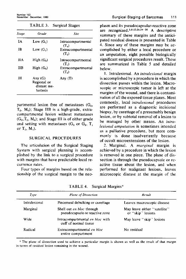
TABLE 3. Surgical Stages
Stage Grade Site
IA LOW (GI) Intracompartmental
IB LOW (GI) Extracompartmental
IIA High (G,) Intracompartmental
IIB High (G,) Extracom part mental
(TI)
(T,)
(TI)
(T2) I11 Any (GI Any (TI
Regional or distant me- tastasis
partmental lesion free of metastases (G2, TZ, MJ; Stage IIB is a high-grade, extra- compartmental lesion without metastases (G2,T2, MU); and Stage I11 is of either grade and setting with metastases (GI or G2, T, or Tz, MA.
SURGICAL PROCEDURES
The articulation of the Surgical Staging System with surgical planning is accom- plished by the link to a surgical procedure with margins that have predictable local re- currence rates.
Four types of margins based on the rela- tionship of the surgical margin to the neo-
plasm and its pseudocapsular-reactive zone are r e c ~ g n i ~ e d . ~ * ~ * ~ ~ * ~ ~ ~ ~ ~ - ~ ~ A descriptive summary of these margins and the antici- pated residual disease is presented in Table 4. Since any of these margins may be ac- complished by either a local procedure or an amputation, eight possible biologically significant surgical procedures result. These are summarized in Table 5 and detailed below.
1. Intralesional. An intralesional margin is accomplished by a procedure in which the dissection passes within the lesion. Macro- scopic or microscopic tumor is left at the margins of the wound, and there is contami- nation of all the exposed tissue planes. Most commonly, local intralesional procedures are performed as a diagnostic incisional biopsy, by curettage of a presumably benign lesion, or by subtotal removal of a lesion to be managed by other means. An intra- lesional amputation is sometimes intended as a palliative procedure, but more com- monly is done inadvertently because of occult microextensions of the lesion.
2 . Marginal. A marginal margin is achieved by a procedure in which the lesion is removed in one piece. The plane of dis- section is through the pseudocapsule or re- active tissue about the lesion, and when performed for malignant lesions, leaves microscopic disease at the margin of the
TABLE 4. Surgical Margins*
Type Plane of Disseciion Result
Intralesional Piecemeal debulking or curettage Leaves macroscopic disease
Marginal Shell out en bloc through May leave either “satellite” pseudocapsule or reactive zone or “skip” lesions
Wide Intracompartmental en bloc with May leave “skip” lesions cuff of normal tissue
Radical Extracompartmental en bloc No residual entire compartment
* The plane of dissection used to achieve a particular margin is shown as well as the result of that margin in terms of residual lesion remaining in the wound.

112 Enneking, et al. Clinical Orthopaedics and Related Research
TABLE 5. Surgical Procedures*
Margin Local Amputation
Intralesional Curettage or Debulking debulking amputation
Marginal Marginal Marginal excision amputation
Wide Wide local Wide through- excision bone ampu-
tation
Radical Radical local Radical dis- resection articulation
* Classified by the type of margin they achieve and whether it is obtained by a local or ablative procedure.
wound in a high percentage of the case^.^*^*- 9*33 As a local procedure, marginal excision is usually described as excisional biopsy or “shell ’em out” of a presumed benign lesion. Marginal amputation is usually done as either a palliative procedure, an at- tempted definitive procedure constrained by anatomic inaccessibility, or as an adjunc- tive procedure.
3. Wide. A wide margin is accomplished by a procedure in which the lesion, its pseu- docapsule and/or reactive zone, and a sur- rounding cuff of normal tissue are taken as a single block. The plane of dissection is entirely through normal tissue but within the involved compartment. No effort is made to remove the entire length of involved muscle from origin to insertion or bone from joint to joint. The local wide procedure probably corresponds to what is referred to as “wide local excision,” “en bloc excision,” and “radical en bloc excision.” A wide margin is definitive surgical management for Stage I lesions and can usually be accom- plished by a local procedure for IA le- s i o n ~ . ’ ~ . ’ ~ Because Stage IB lesions usually invclve some combination of bone, soft parts, and neurovascular structures, ampu- tation is more likely to be required.
4. Radical. A radical margin is achieved by a procedure in which the lesion, pseudo-
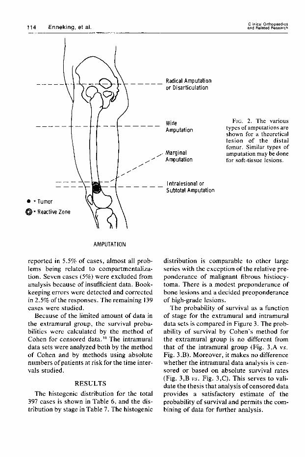
capsule, reactive zone, and the entire mus- cles or bone involved are removed as one block. Longitudinally, the plane of dissec- tion goes through or beyond the joint proxi- mally and distally to the bone involved and through the tendinous origin and insertion of involved muscles. Transversely, the dis- section passes beyond the major fascia1 septa of the involved soft tissue compart- ments or beyond the periosteum of intra- osseous lesions. A radical margin does not necessarily imply a greater distance from the lesion to the margin of the wound than a wide margin. A margin on the other side of the intermuscular septum of a lesion in the vastus lateralis will constitute a radical margin but may be considerably closer than a wide margin achieved by amputation. A radical margin is definitive for Stage I1 lesions. A radical local resection can often be done for a Stage IIA lesion. If a lesion involves more than one compartment, or extends into or arises in the extracompart- mental planes or spaces, compartmental containment is lost, and a radical margin is usually not attainable with a local pro- cedure. Thus, radical amputation is usually carried out to achieve a radical margin in Stage IIB lesions, and it often requires a disarticulation or amputation proximal to the joint in question. These various pro- cedures are illustrated diagramatically in Figures 1 and 2.
A total myectomy for a lesion within a single muscle may be either a wide local ex- cision or a radical local resection, depend- ing upon the muscle involved. If the muscle also constitutes a compartment, i .e., the deltoid, then myectomy accomplishes a radical local resection. If the muscle is one of several muscles separated by loose areolar tissue within a large fascially con- tained compartment such as the rectus femoris, then myectomy is radical in the longitudinal sense but only wide in the trans- verse sense and is, by definition, a wide local excision.

Number 153 November-December, 1980 Surgical Staging of Sarcomas 113
APPLICATION I
I I \ \
The utility and general applicability of this surgical staging system has been evaluated in two quite different situations: (1) intra- murally by the University of Florida mus- culoskeletal oncology service, and (2) extramurally by an interinstitutional pilot study conducted by the Musculoskeletal Tumor Society.
The intramural evaluation involved pa- tients treated on the musculoskeletal on- cology service at the University of Florida since 1959. The service has well-established patient referral patterns, effective mecha- nisms for patient follow-up, and a consis- tent, well-defined surgical philosophy. A great deal of prospective primary observa- tional data have been collected since 1959 and is stored in computers. Histogenic classi- fication and grading have been done prospec- tively since 1974. Cases treated prior to 1974 have been retrospectively reviewed and graded, allowing reclassification along the lines of new histogenic concepts. The sur- gical grade, site, and stage were estimated preoperatively for surgical planning. The final stage was assigned after pathologic re- view of the surgical specimen. Two-hun- dred-fifty-eight cases form the basis of the in- tramural evaluation of the staging system.
The extramural evaluation of the system was done among 13 institutions (M. D. Anderson Hospi ta l , S U N Y -Buffalo, UCLA, UCSF, Case Western Reserve, University of Chicago, University of Iowa, Massachusetts General Hospital, Mayo Clinic, Memorial Hospital for Cancer- New York, University of Miami, Univer- sity of Minnesota, and Rizzoli Institute of Orthopaedics, Bologna). Many of the par- ticipants are established investigators with extensive experience in the management of musculoskeletal neoplasms. The spectrum of their surgical philosophies ranges from conservative to highly innovative. These 13 institutions each have their own patholo-
RADICAL \
RESECTION \ \
WIDE EXCISION ,
MARGINAL
,
EXCISION - -
INTERLESIONALOR /’
SUBTOTAL EXCIS ION
0 = T u m o r
F c i \ @ = Reactive Zone
LOCAL PROCEDURE
FIG. 1. The various local procedures are shown. The dotted lines indicate the plane of dis- section and the amount of tissue removed to achieve the various procedures for a theoretical lesion within the anterior compartment of the thigh. Similar types of procedures may be done for bone lesions.
gists, referral patterns, methods of follow- up and mechanisms for handling data, and therefore represent a reasonable sample of the spectrum of musculoskeletal surgical oncology practice as it exists today.
Each participant was mailed a question- naire along with a precis of the staging sys- tem (Project Manager, Michael Simon, Uni- versity of Chicago), and asked to retrospec- tively stage and submit ten consecutive cases of musculoskeletal sarcoma per- sonally treated since 1970. The only restrictions were that the cases have a mini- mum two-year follow-up period. Patients could be entered without regard for treat- ment. This diverse group contributed 146 cases. Difficulty in utilizing the system was
114 Enneking, et al. Clinical Orthopaedics and Related Research
Radical Amputation or Disarticulation
AMPUTATION
reported in 5.5% of cases, almost all prob- lems being related to compartmentaliza- tion. Seven cases (5%) were excluded from analysis because of insufficient data. Book- keeping errors were detected and corrected in 2.5% of the responses. The remaining 139 cases were studied.
Because of the limited amount of data in the extramural group, the survival proba- bilities were calculated by the method of Cohen for censored data.lo The intramural data sets were analyzed both by the method of Cohen and by methods using absolute numbers of patients at risk for the time inter- vals studied.
RESULTS The histogenic distribution for the total
397 cases is shown in Table 6, and the dis- tribution by stage in Table 7. The histogenic
Wide Amputation
Marginal Amputation
FIG. 2 . The various types of amputations are shown for a theoretical lesion of the distal femur. Similar types of amputation may be done for soft-tissue lesions.
I ntralesional or Subtotal Amputation
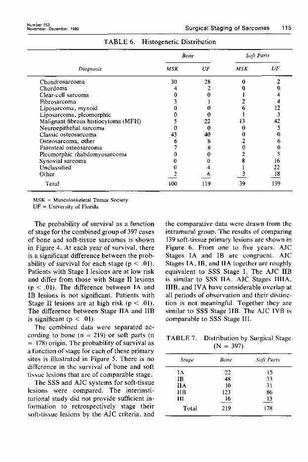
distribution is comparable to other large series with the exception of the relative pre- ponderance of malignant fibrous histiocy- toma. There is a modest preponderance of bone lesions and a decided preoponderance of high-grade lesions.
The probability of survival as a function of stage for the extramural and intramural data sets is compared in Figure 3. The prob- ability of survival by Cohen's method for the extramural group is no different from that of the intramural group (Fig. 3,A vs. Fig. 3,B). Moreover, it makes no difference whether the intramural data analysis is cen- sored or based on absolute survival rates (Fig. 3,B vs. Fig. 3,C). This serves to vali- date the thesis that analysis of censored data provides a satisfactory estimate of the probability of survival and permits the com- bining of data for further analysis.
Number 153 Novem ber-Decem ber. 1980 Surgical Staging of Sarcomas 115
TABLE 6. Histogenetic Distribution
Diagnosis
Bone Soft Parts
MSK UF MSK UF ~~
Chondrosarcoma Chordoma Clear-cell sarcoma Fibrosarcoma Liposarcoma, myxoid Liposarcoma, pleomorphic Malignant fibrous histiocytoma (MFH) Neuroepithelial sarcoma Classic osteosarcoma Osteosarcoma, other Parosteal osteosarcoma Pleomorphic rhabdomyosarcoma Synovial sarcoma Unclassified Other
Total
30 4 0 3 0 0 5 0
43 6 7 0 0 0 2
28 2 0 1 0 0
22 0
40 8 8 0 0 4 6
100 119
0 0 1 2 6 1
13 0 0 2 0 2 8 1 3
39 -
2 0 4 4
12 3
42 5 0 6 0 5
16 22 18
139 -
MSK = Musculoskeletal Tumor Society UF = University of Florida
The probability of survival as a function of stage for the combined group of 397 cases of bone and soft-tissue sarcomas is shown in Figure 4. At each year of survival, there is a significant difference between the prob- ability of survival for each stage (p < .01). Patients with Stage I lesions are at low risk and differ from those with Stage I1 lesions (p < .01). The difference between IA and IB lesions is not significant. Patients with Stage I1 lesions are at high risk (p < .01). The difference between Stage IIA and IIB is significant (p < .01).
The combined data were separated ac- cording to bone (n = 219) or soft parts (n = 178) origin. The probability of survival as a function of stage for each of these primary sites is illustrated in Figure 5 . There is no difference in the survival of bone and soft tissue lesions that are of comparable stage.
The SSS and AJC systems for soft-tissue lesions were compared. The interinsti- tutional study did not provide sufficient in- formation to retrospectively stage their soft-tissue lesions by the AJC criteria, and
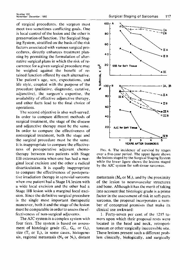
the comparative data were drawn from the intramural group. The results of comparing 139 soft-tissue primary lesions are shown in Figure 6. From one to five years, AJC Stages IA and IB are congruent. AJC Stages IA, IB, and IIA together are roughly equivalent to SSS Stage I . The AJC IIB is similar to SSS IIA. AJC Stages IIIIA, IIIB, and IVA have considerable overlap at all periods of observation and their distinc- tion is not meaningful. Together they are similar to SSS Stage IIB. The AJC IVB is comparable to SSS Stage 111.
TABLE 7 . Distribution by Surgical Stage (N = 397)
Stage Bone Soft Parts
IA 22 15 IB 48 33 IIA 10 31 IIB 123 86 111 16 13
Total 219 178 - -
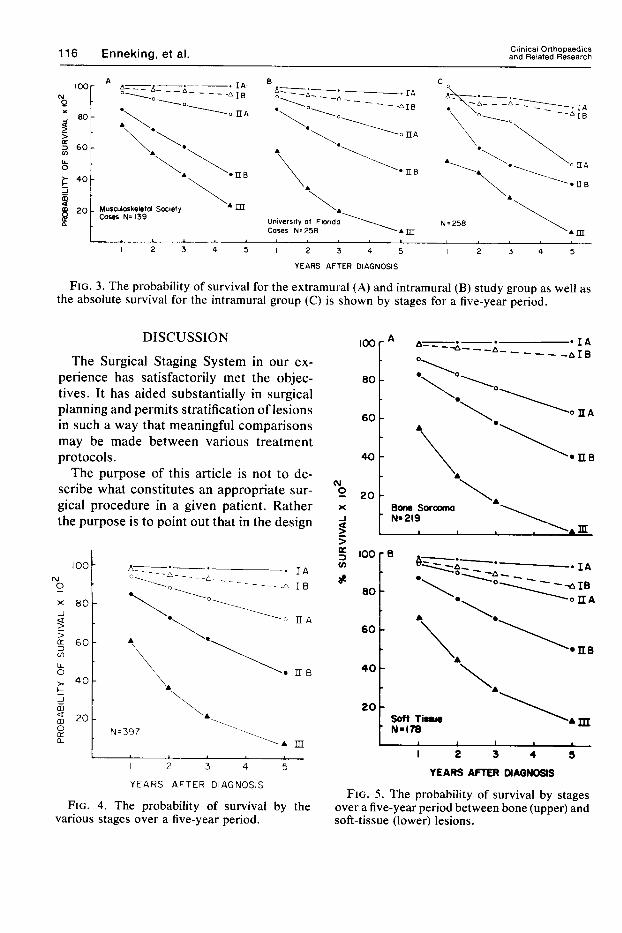
Clinical Orthopaedics 116 Enneking, et al. and Related Research
Musculoskeletal Society Cases N= 139 University of Florido
Cases N=258 I
1 2 3 4 5 1 2 3 4 5 1 2 3 4 5
YEARS AFTER DIAGNOSIS
FIG. 3. The probability of survival for the extramural (A) and intramural (B) study group as well as the absolute survival for the intramural group (C) is shown by stages for a five-year period.
DISCUSSION
The Surgical Staging System in our ex- perience has satisfactorily met the objec- tives. It has aided substantially in surgical planning and permits stratification of lesions in such a way that meaningful comparisons may be made between various treatment protocols.
The purpose of this article is not to de- scribe what constitutes an appropriate sur- gical procedure in a given patient. Rather the purpose is to point out that in the design
m L
I 2 3 4 5
Y E A R S AFTER DIAGNOSIS
FIG. 4. The probability of survival by the various stages over a five-year period.
(u
0 X
J s 5
L 'A.
I I I
I 2 3 4 5
YEARS AFfER MAGNOSIS
FIG. 5. The probability of survival by stages over a five-year period between bone (upper) and soft-tissue (lower) lesions.
Number 153 Novem ber-Decem ber, 1980 Surgical Staging of Sarcomas 117
of surgical procedures, the surgeon must meet two sometimes conflicting goals. One is local control of the lesion and the other is preservation of function. The Surgical Stag- ing System, stratified on the basis of the risk factors associated with various surgical pro- cedures, directly enhances treatment plan- ning by permitting the formulation of alter- native surgical plans in which the risk of re- currence for a given surgical procedure may be weighed against the benefit of re- tained function offered by each alternative. The patient’s age, sex, expectations, and life style, coupled with the purpose of the procedure (palliative, diagnostic, curative, adjunctive), the surgeon’s expertise, the availability of effective adjunctive therapy, and other facts lead to the final choice of operations.
The second objective is also well-served. In order to compare different methods of surgical treatment, the stage of the disease and adjunctive therapy must be the same. In order to compare the effectiveness of nonsurgical treatment, both the stage and the surgical procedure must be the same. It is inappropriate to compare the effective- ness of postoperative adjuvant chemo- therapy between two patients with Stage IIB osteosarcoma when one has had a mar- ginal local excision and the other a radical disarticulation. It is equally inappropriate to compare the effectiveness of postopera- tive irradiation therapy in synovial sarcoma when one patient had a Stage IA lesion with a wide local excision and the other had a Stage IIB lesion with a marginal local exci- sion. Since the definitive surgical procedure is the single most important therapeutic maneuver, both it and the stage of the lesion must be comparable in order to assess the ef- fectiveness of non-surgical adjuvants.
The AJC system is a complex system with four tiers. The system is based on assess- ment of histologic grade (Gl, G2, or G3) , size (TI or T2), in some cases, histogene- sis, regional metastasis (No or Nl), distant
lOOr A ‘0,
6 100-8 A--.-. -IA, m -1
f
‘-AIIA
60 - 40 om8
I 2 3 4 5 YEARS AFTER DIAGNOSIS
FIG. 6. The incidence of survival by stages over a five-year period. The upper figure shows the lesions staged by the Surgical Staging System while the lower figure shows the lesions staged by the AJC system for soft-tissue sarcomas.
metastasis (M, or Ml), and by the proximity of the lesion to neurovascular structures and bone. Although it has the merit of taking into account that histologic grade is a prime factor in the assessment of risk in soft-parts sarcoma, the proposal incorporates a num- ber of conceptual premises that make its clinical use awkward:
1. Forty-seven per cent of the 1215 tu- mors upon which their proposal rests were located in the head and neck, retroperi- toneum or other surgically inaccessible site. These lesions present such a different prob- lem clinically, biologically, and surgically
118 Ennek ing , e t al. Clinical Orthopaedics and Related Research
that they should not be grouped with lesions of the extremities for analysis.
2. The division of sarcomas into three histologic grades is a histologic nicety. Al- though it is likely to have great appeal to the pathologist, it has little to offer the surgeon in terms of surgical guidance because there is no “middle” surgical procedure.
3. The T designation (local extent) is represented by lesion size. We believe that lesion size has prognostic significance that is a complex composite of anatomic setting, growth rate, and time to physician interven- tion. Since neither growth rate nor time to diagnosis can be quantitated, this variable in the AJC system would have more rele- vance if it reflected the extent defined by anatomic setting, i . r . , compartmentaliza- tion (or compartmental escape). The latter designation is more consistent with the natural biologic behavior of the sarcomas, and has meaning for the surgeon.
4. Appended to Stage I11 as IIIc are le- sions with regional lymph node metastases. Lymph node involvement is so uncommon in the natural history of these lesions at the time of diagnosis as to not be worth a sepa- rate fa~tor.~.~.~,’~.’~,~~,~’ When this relatively rare phenomenon does occur, the prognosis is poor. If nodal metastases are given equal weight with other metastases, the surgeon knows that a contemplated procedure is likely to be palliative or must be supple- mented with other treatment modalities to be curative.
5. “Gross involvement of a major nerve, artery, or bone” (TJ is poorly defined, and the methods by which these judgments are to be made are not defined. Lesions with such involvement are assigned to a higher stage without regard for grade. Analysis of our soft-tissue sarcoma data by this method results in these Stage IVA lesions having a prognosis similar to AJC Stages IIIA and B lesions. Such involvement is a proper function of the anatomic setting (extent of the primary), and as such, does not require a separate category.
6. Lesions of certain histogenesis are assigned to at least Stage 111 because of their usual poor prognosis. This is a function of grade and should be treated as such. Occa- sional lower grades of these lesions do occur, and they should be staged accord- ingly.
The AJC system proposed for primary bone lesions is so complex that we have not retrospectively compared it with the Surgi- cal Staging System. However, it is different from the AJC soft tissue system and if gen- erally adopted would require the use of two complex systems that would not permit ready comparison between bone and soft- tissue sarcomas of the same histogenesis. Because definitive surgery is the primary treatment for sarcomas of both bone and soft tissue, and because the principles de- scribing their biologic behavior and surgical procedures are the same for both groups, a common staging system for both groups would be more useful than two separate and different systems.
It is ironic that the essentials of the staging system proposed here were recognized by Quick and Cutler’8 over 50 years ago. They divided 75 tumors which they believed to be of neurogenic origin into three progressive histologic grades and correlated their micro- scopic observations with the clinical course and treatment. Their clinical rate of metas- tases reinforces our view that a simple divi- sion into high and low grade is practical and sufficient to define the risk of distant spread. Lesion size was not an important determin- ing factor in survival but anatomic location and inadequacy of treatment were. They recognized the relationship between histo- logic grade and an adequate surgical pro- cedure to patient survival. Their statement that “whereas wide local excision of the acellular fibrous tumors may result in a cure, this procedure is frequently followed by local recurrence and pulmonary metastasis in the highly cellular and malignant tumors (their Grades I1 and III),” is a precise state- ment of the principles of tumor surgery re-
Number 153 November-December, 1980 Surgical Staging of Sarcomas 119
capitulated at our i n s t i t ~ t i o n . ’ ~ , ~ ~ , ~ ~ They ap- preciated the occasional need for adjunc- tive therapies and attempted to elucidate factors in their appropriate use by compar- ing treatment results. The dilemma in treat- ment choices: “Tumors of the extremity in which amputation offers a chance of com- pletely eradicating the disease present an important problem in treatment. The de- cision between amputation on the one hand and excision and radiation on the other is at times most difficult,” is as unresolved now as it was then.
The Surgical Staging System for sar- comas of bone and soft tissues presented here is simple, clear cut, and has a high degree of compliance and accuracy. It is relevant to both surgical planning and end- result studies. It is quite clear that in com- paring nonsurgical treatment protocols both the prognostic stage and the extent of the surgical procedure must be clearly de- fined and standardized before meaningful end-result studies can be made. The ab- sence of a generally accepted staging system articulated with clearly defined surgical pro- cedures has hampered the understanding of the proper role of various nonsurgical methods in managing musculoskeletal sar- comas. The surgical staging system and sur- gical definitions presented here form the basis for the ongoing interinstitutional stud- ies currently being conducted by the Mus- culoskeletal Tumor Society.
SUMMARY
A surgical staging system for musculo- skeletal sarcomas stratifies bone and soft- tissue lesions of any histogenesis by the grade of biologic aggressiveness, by the anatomic setting, and by the presence of metastasis. The three stages: I-low grade; 11-high grade; and 111-presence of metastases, are subdivided by (a) whether the lesion is anatomically confined within well-delineated surgical Compartments, or (b) beyond such compartments in ill-defined
fascia1 planes and spaces. Operative mar- gins are defined as intralesional, marginal, wide, and radical, and relate the surgical margin to the lesions, its reactive zone, and anatomic compartment. The system defines prognostically significant progressive stages of risk which also have surgical implica- tions. When the system is linked to clearly defined surgical procedures, it permits ap- propriate evaluation and comparison of the new treatment protocols designed to replace standard surgical treatment.
ACKNOWLEDGMENT
The authors are indebted to Mrs. Linda Hensdell for aid in manuscript preparation. Mr. Samy Suissa and John Shuster, Ph.D., performed the statistical analysis.
1.
2.
3.
4.
5 .
6.
7.
8.
9.
10.
11.
REFERENCES
Ahuja, S. C., Villacin, A. B., Smith, J . , Bullough, P. G., Huvos, A. G., and Marcove, R. C.: Juxtacortical (parosteal) osteogenic sarcoma. J. Bone Joint Surg. 59A:632, 1977. Beattie, E. J., Martini, N., and Rosen G.: The management of pulmonary metastases in children with osteogenic sarcoma with surgical resection combined with chemotherapy. Cancer 35:618, 1975. Beck, J. C., Wara, W. M., Bovell, E. G., and Phillips, T. L.: The role of radiation therapy in the treatment of osteosarcoma. Radiology 120: 163, 1976. Bowden, L., and Booher, R. J.: Surgical considera- tions in the treatment of sarcoma of the buttock. Cancer 6:89, 1953. Bowden, L., and Booher, R. J.: The principles and technique of resection of soft parts for sarcoma. Surgery 44:963, 1958. Broders, A. C., Hargrave, R., and Meyerding, H. W.: Pathological features of soft tissue fibrosar- coma. Surg. Gynecol. Obstet. 69:267, 1939. Cantin, J., McNeer, G. P., Chu, F. C., and Booher, R. J.: The problem of local recurrence after treatment of soft tissue sarcoma. Ann. Surg. 168:47, 1968. Castro, E. B., Hajdu, S. I. , and Fortner, J. G.: Surgical therapy of fibrosarcoma of extremities. Arch. Surg. 107:284, 1973. Clark, R. L., Martin, R. G., White, E. C., and Old, J. W.: Clinical aspects of soft-tissue tumors. AMA Arch. Surg. 74:859, 1957. Cohen, A. C.: Progressively censored sample in life testing. Technometrics 5:327, 1963. Copeland, M. M., Robbins, G. F., and Myers, M. H.: Development of aclinical staging system for primary malignant tumors of bone: a progress report. In Management of Primary Bone and Soft

120 Enneking, e t al. Clinical Orthopaedics and Related Research
Tissue Tumors, Chicago, Year Book Medical Publishers, Inc., 1977, p. 35.
12. Enneking, W. F., and Spanier, S.: Surgical staging of musculoskeletal tumors. Presented at the 47th Annual Meeting of the American Academy of Orthopaedic Surgeons, Atlanta, February, 1980.
13. Enneking, W. F., Spanier, S. S., and Malawer, M. M.:Theeffectoftheanatomicsettingon the results of surgical procedures for soft parts sarcoma of the thigh. Cancer (in press).
14. Evans, H. L., Ayala, A. G., and Romsdahl, M. M.: Prognostic factors in chondrosarcoma of bone: a clinico-pathologic analysis with emphasis on his- tologic grading. Cancer 40318, 1977.
15. Gottlieb, J. A., Baker, L. H., Quagliana, J. M., Luce, J. K., Witecare, Jr., J. P., Sinkovics, J. G., Rivkin, S. E., Brownlee, R., and Frei, E. 111: Chemotherapy of sarcomas with a combination of Adriamycin and dimethyl triazeno imidazole car- boxamides. Cancer 30: 1632, 1972.
16. Hajdu, S. I.: Pathology of Soft Tissue Tumors. Philadelphia, Lea & Febiger, 1979, p. 43.
17. Jaffe, N., Frei, E., Traggis, D., and Bishop, Y.: Adjuvant methotrexate citrovorum-factor treat- ment of osteogenic sarcoma. N. Engl. J. Med. 291:994, 1974.
18. Jenkin, R., and Derek, T.: The treatment of osteosarcoma with radiation: current indications. In Management of Primary Bone and Soft Tissue Tumors. Chicago, Year Book Medical Publishers, Inc., 1977, p. 151.
19. Lieberman, Z., and Ackerman, L. V.: Principles in management of soft tissue sarcomas. Surgery 35:350, 1954.
20. Lindberg, R. D., Martin, R. G., Romsdahl, M. M., and McMurtrey, M. J.: Conservative surgery and radiation therapy for soft tissue sarcomas. In Management of Primary Bone and Soft Tissue Tumors. Chicago, Year Book Medical Publishers, Inc., 1977, p. 289.
21. Lunseth, P. A., and Nelson, C. L.: Longitudinal amputation for the treatment of soft tissue fibrosar- coma. Clin. Orthop. 109:147, 1975.
22. Manual for Staging of Cancer, 1977, Chicago, American Joint Committee, p. 1.
23. Martin, R. G., Lindberg, R. D., and Russell, W. 0.: Preoperative radiotherapy and surgery in the management of soft tissue sarcoma. In Manage- ment of Primary Bone and Soft Tissue Tumors. Chicago, Year Book Medical Publishers, Inc.,1977, p. 299.
24. Morton, D. L., Eilber, F. R., Townsend, C. M., Grant, T: T., Mirra, J . , and Weisenburger, T. H.: Limb salvage from a multidisciplinary treatment approach for skeletal and soft tissue sarcomas of the extremity. Ann. Surg. 184:268, 1976.
25. Pack, G. T., and Ariel, I. M.: Principles of treatment of tumors of the soft somatic tissues. In Pack, G. T., and Ariel, I. M., (eds.): Treatment of Cancer and Allied Diseases, Tumors of Soft Somatic Tissues and Bone, 2nd edition. New York,
Hoeber Medical Division, Harper and Row, 1964, VIII, p. 8.
26. Pack, G. T., and Ariel, I. M.: Fibrosarcoma of the soft somatic tissues. Recent Adv. Surgery 31:443, 1952.
27. Pritchard, D. J. , Lunke, R. J., Taylor, W. F., Dahlin, D. C., and Medley, B. E.: Chondrosar- coma: a clinicopathologic and statistical analysis. Cancer 45: 149, 1980.
28. Quick, D., and Cutler, M.: Neurogenic sarcoma, a clinical and pathological study. Ann. Surg. 86:810, 1927.
29. Romsdahl, M. M., and Ayala, A. G.: Surgical management of osteosarcoma. In Management of Primary Bone and Soft Tissue Tumors. Chicago, Year Book Medical Publishers, Inc., 1977, p. 137.
30. Rosen, G., Murphy, M. L., Huvos, A. G., G u t i e r r e z , M . , a n d M a r c o v e , R . C . : Chemotherapy, en bloc resection and prosthetic bone replacement in the treatment of osteogenic sarcoma. Cancer 37: 1, 1976.
31. Russell, W. O., Cohen, J., Enzinger, F., Hajdu, S. I., Heise, H., Martin, R. G., Meissner, W., Miller, W. T., Schmitz, R. L., and Suit, H. D.: A clinical and pathological staging system for soft tissue sarcomas. Cancer 40: 1562, 1977.
32. Sanerkin, N. G.: The diagnosis and grading of chondrosarcoma of bone. Cancer 45582, 1980.
33. Shiu, M. H., Castro, E. B., Hajdu, S. I., and Fortner, J. G.: Surgical treatment of 297 soft tissue sarcomas of the lower extremity. Ann. Surg. 182:597, 1975.
34. Simon, M. A., and Enneking, W. F.: The management of soft tissue tumors of the ex- tremities. J. Bone Joint Surg. 58A:317, 1976.
35. Simon, M. A., Spanier, S., and Enneking, W. F.: Soft tissue sarcomas of the extremities. Surg. Annu. 11:363, (ed.) Nyhus, L., 1979.
36. Spanier, S., and Enneking, W. F.: Chapter 8: Musculoskeletal tissues. In Pierson, K. K. (ed.): Principles of Prosection. New York, John Wiley and Sons, 1980, p. 61.
37. Storm, F. K., Eilber, F. R., Mirra, J . , and Morton, D. L.: Neurofibrosarcoma. Cancer 45: 126, 1980.
38. Suit, H. D., Russel, W. O., and Martin, R. G.: Management of patients with sarcoma of soft tissue in an extremity. Cancer 31:1247, 1973.
39. Townsend, C. M., Jr., Eilber, F. R., and Morton, D. L.: Skeletal and soft tissue sarcomas: results of surgical adjuvant chemotherapy. AACR Proceed- ings 17:C- 116, 1976.
40. Unni, K. K., Dahlin, D. C., McLeod, R. A,, and Pritchard, D. J.: Intraosseous well-differentiated osteosarcoma. Cancer 40: 1337, 1977.
41. Wanebo, H. J., Shah, J., Knapper, W., Hajdu, S. I., and Booher, R.: Reappraisal of surgical management of sarcoma of the buttock. Cancer 31:97, 1973.
42. Weingrad, D. N., and Rosenberg, S. A.: Early lymphatic spread of osteogenic and soft-tissue sarcomas. Surgery 84:231, 1978.