a study of gonococcus by the method of “fixation of complement.”
TRANSCRIPT

T H E
Journal of Medical Research.(NEW SERIES, VOLUME XII.)
VOL. XVII. DECEMBER, I 907 No. 3.
A STUDY OF GONOCOCCUS BY THE METHOD OF " FIXA-TION OF COMPLEMENT." *
OSCAR TEAGUE AND JOHN C. TORREY.
(From the Department of Experimental Pathiology, Loomis Laboratory, CornellUniversity Medical College, Newu York.)
The method of fixation of complement, founded on thestudies of Bordet and Gengou (190o),' and further developedby Wassermann and Briick, has received extended applica-tion during the past year, and it would seem its possibilitieshave been by no means exhausted. Complete historicalsketches and literature references to the use of this methodin forensic and diagnostic fields may be found in most of therecent articles on the subject. Accordingly, to avoid need-less repetition, we shall confine ourselves to a discussion ofarticles in which this method has been employed in thestudy of the gonococcus and the diseases caused by thismicroorganism.
MUller and Oppenheim2 were the first to make use of thisprocess in the detection of antibodies in the blood of apatient infected with gonococcus. They showed that theserum of this patient, suffering with gonorrheal arthritis,when mixed with an emulsion of gonococci in normal saltsolution, possessed the property of absorbing complement,and thereby preventing hemolysis when " sensitized " erythro-cytes were added. On the other hand, the serum frompatients with certain other diseases absorbed no complementunder like conditions. From this fact they concluded that
* Received for publication Oct. iI, 1907.

TEAGUE AND TORREY.
the serum of the first patient contained specific " antibodies"for gonococcus. Subsequently Bruck,3 Meakins,4 Vannod,5and Wollstein 6 have published articles on this subject. Thework of these investigators will be discussed in detail in alater portion of this paper.
In German articles, the term " Komplementablenkung " isfrequently applied to this method. We believe that theterm, " deviation of complement," should be confined tothe " Neisser-Wechsberg " phenomenon. In this instancethe complement is supposed to be* drawn aside from itsnormal union with the antigen and immune body and to unitewith an excess of immune bodies. On the other hand in themethod under discussion the complement may be supposedto enter into its normal function in every instance, whetherit be "bacteria plus (bacteriolytic) antibody plus comple-ment" (no hemolysis) or "erythrocytes plus hemolyticantibody plus complement" (hemolysis). Accordinglyunder these conditions there is always a normal " fixationof complement" (Komplementverankerung). This latterterm has been employed by Wassermann from the first.
In carrying out these experiments it was hoped that lightmight be shed on several questions. Recently one of us 7
has shown by a study of the agglutinins in various anti-gonococcic serums that the cultures which were employedmight be divided into groups. The major or specific agglu-tinins raised by these various groups were not removed byinter-absorptions. This seems to indicate that the gonococ-cus family is heterogeneous rather than homogeneous. Wehave attempted, accordingly, to determine whether theresults of " fixation of complement" lead one to the sameconclusion. It is generally supposed that a positive resultby this method indicates that the serum of immunizedanimals contains specific bacteriolytic antibodies. Grantingfor the time being that this is true, an investigation of thischaracter would show whether the differences in the agglu-tinins of the cultures of gonococci employed are paralleledby similar diversities in bacteriolytic antibodies. Finally itwas considered possible that the experiments might indicate
224

A STUDY OF GONOCOCCUS.
the degree of reliance which this technic offers in the diag-nosis of gonorrheal infections.
Technic.- In conducting experiments of this character,five different agents are required:
i. Extract of the microorganism, the " Antigen."2. Inactivated serum immune to the antigen.3. Fresh normal serum, the complement.4. Inactivated hemolytic serum.5. Homologous erythrocytes.
i. Antigen: In our earlier experiments distilled waterextracts of several different strains of gonococcus were used.In preparing these extracts the technic of Wassermann andhis colleagues was followed. Fresh stock cultures weretransplanted to large tubes of rabbit-serum agar prepared inthe following way: Agar was prepared according to themethod of Thalmann. This has been described in detail ina previous article by one of us.7 After tubing and thor-oughly sterilizing the medium, one-half cubic centimeter ofnormal inactivated rabbit serum was added to each tube. Onmedium of this character we obtained a very luxuriantgroWth with all the strains of gonococcus used in the experi-ments. Rabbit serum was employed in this medium insteadof ascitic fluid to avoid all extraneous precipitation. It hasbeen shown 7 that if ascitic fluid is used both in the prepara-tion of the antigen and in the production of the immuneserum, precipitation occurs, due to the union of albumens ofthe ascitic fluid with their precipitins in the immune serum.This would fix the complement to a certain extent by itself.The cultures, accordingly, were incubated for twenty-fourhours on large slants of rabbit-serum medium, and then thegrowth from each tube was emulsified in two cubic centi-meters of sterile distilled water. This emulsion was nextplaced in a shaking apparatus for three days and centrifu-galized. As a result of this treatment the fluid was notcompletely cleared. To each tube of extract, one-half ofone per cent of carbolic acid was finally added. The results
225

TEAGUE AND TORREY.
with extracts of this character were quite satisfactory, buttheir preparation required much time.
In the following much shorter technic, the extracts aremore easily obtained in large amounts. The cultures wereproduced as before and then the growth from two to threetubes was emulsified in one cubic centimeter of sterile seven-teen per cent NaCl solution and shaken at intervals for onehour. At the expiration of this period, nineteen cubic centi-meters of sterile distilled water was added, thus bringing thesolution to the strength of normal. The extracts were finallycentrifugalized and preserved as described above. Ourtables are founded on results with these salt solution extracts.It may be said, however, that the results of the experimentswith distilled water extracts, as far as they were conducted,agreed with those carried out with salt solution extracts.Seven selected strains of gonococcus, which had beenemployed in the agglutination experiments, were investigated.For controls, extracts were made in the same way of M.catarrhalis, Meningococcus and B. typhosus. The antigenswhich we employed were never more than two weeks old.
2. Rabbits were highly immunized to the various strainsof gonococcus and to the control cultures. The methoddescribed in a previous communication I was followed, usingascitic agar cultures. The serum was inactivated by heatingfor one-half hour at 54.50 C. on the day after the blood wasdrawn.
3. Serum from normal rabbits served as " complement."The serum was always used about eighteen hours after beingdrawn. To prevent loss in potency, the flask containing thediluted serum was packed in ice until all the tubes had beenprovided with complement. Rabbit serum was employed ascomplement in preference to the guinea-pig serum recom-mended by Wassermann for two reasons: ( i) because by theuse of homologous serums the conditions of the experimentsare less complex; (2) the large amount of complementrequired in our tests would have necessitated the sacrifice of
226

A STUDY OF GONOCOCCUS.
a large number of guinea-pigs. As has been the experienceof others, we found that our rabbits varied greatly in theamount of complement which their serum contained. Thisseemed to be due to an individual difference rather than tovariation in the condition of the animal. The uncertainty inregard to the potency of the serum of a new animal was thesource of the principal difficulty encountered during theprogress of this research. A great deal of time may, accord-ingly, be saved by setting apart a number of normal animalsand determining, by preliminary experiments, the strengthof their complement.
4. The hemolytic serum was obtained by immunizingrabbits to washed sheep erythrocytes. Fresh blood cellswere employed for each inoculation. They were washedfour times in about twenty times as much salt solution eachtime. Five cubic centimeters of washed cells were injectedintraperitoneally at intervals of five to seven days. Afterfive inoculations the antiserum was found to be of sufficientstrength for the experiments.
5. Erythrocytes of the sheep were washed four times, asdescribed above, just before the experiments were started.The salt solution employed in washing and suspending thecells was prepared by dissolving eighteen grains of commer-cial sodium chloride in two liters of tap water, filtering andsterilizing. The cells do not begin to hemolyze in this solu-tion until after standing five or six days. They were neverused, however, after the third day. A five per cent suspen-sion of the erythrocytes was employed.The various substances were added in the order indicated
in the table. We used a constant dilution of the extractsone-fifth. Experiments showed that none of the bacterialextracts of themselves hemolyzed the red cells nor absorbedcomplement to any extent at this dilution. Seven differentdilutions of each of the bacterial-immune serums were made.The bacterial extract, the immune serum dilution, and thecomplement, one-half cubic centimeter of each, wvere
227

TEAGUE AND TORREY.
thoroughly mixed and -incubated for an hour at 370 C. Thenthe hemolytic serum and the sheep cells, one-half cubic centi-meter of each, were added, shaking each tube, and the wholeincubated for two hours longer. Instead of using doublethe lytic dose of hemolytic serum, as recommended byWassermann and others, we employed the minimum lyticdose, which produced complete hemolysis after the comple-ment had been subjected to similar physical conditions as inthe fixation tests. By avoiding thus the presence of anexcess of hemolytic amboceptors, the method is, we believe,rendered more delicate. After the second incubation period,the tubes were placed on ice over night.
Readings were taken at varying intervals, depending onthe activity of the complement. The observation at the endof eighteen to twenty-four hours is much more satisfactorythan the two-hour reading, as the cells have settled to thebottom of the tubes, and one can readily recognize even atrace of hemoglobin in the clear fluid above. In the follow-ing table, with the exception of serum " G," the results arefrom readings taken at the end of approximately twenty-four hours. The result with serum "G" is based on atwo-hour reading, as an excess of complement masked some-what the specific differences in the later observation.
If, at the final observation, the fluid in the tube was per-fectly colorless, the result is indicated in the table by aminus (-) sign. If, however, it was faintly tinged byhemoglobin, it is called " trace." If the fluid was distinctlycolored red, but there was a quantity of undissolved bloodcells at the bottom of the tube, the symbol "+" is used.Finally, if there was a very marked hemolysis and all or thegreater part of the cells were destroyed, the result isindicated by the sign "+ + ."
In the table it is noted that the complement is dilutedto . i or . i i. It was necessary to vary thus the strength ofthe dilution because of the differences in the potency of thenormal rabbit serums. In each complete test, however, witha single antigonococcic serum, the same dilution of a givencomplement was of course used throughout.
228

TABLE SHOWING FiXATION OF COMPLEMENT WITH VARIOUs ANTIGONOCOCCIC SERUMS.
Normal ResulitBacterial Extracts. Immune Rabbit Hemolytic Result Result Result Result Result Result Result with
Serum Serutn Serum Sheep with with with with with with with SerumDilution =.2 Dilutions. (Comple- (Rabbit v. Cells. Seruin A. Seruin B. Serum C. Serum F. Serum G. Serum H. Serum I. Meningo.-
ment). Sheep). COCCUS.
...... Gonococcus A... I i or.ii .02 5% + + + trace trace
2...... do ...... .02 .......... .......... .......... + trace +3...... ...... .005 .......... .......... .......... + + + + + +4...... .002 .......... .......... .......... trace + + + + + + +5...... ....... .001 .......... .......... .......... + + trace + + + ++++ +6...... ...... .0005 .......... .......... .......... + + + + ++ + + + + + +
7...... . . . . . . .0002 .......... .......... ... .. ... + + + + + + + + + + + + + +
8...... Gonococcus B ...... .1 .......... .......... trace trace + + trace trace +9...... 6 9 , 4. . . . . . .02 .......... .......... .......... - + + trace + +10 44 .005 .......... .......... .......... + + + + +11 ...... 44 do ...... .002 .......... .......... .......... - + + + + + + + + +12...... 64 do ...... .001 .......... .......... .......... trace + + + + + + + + + +13 ...... ...... .0005 .......... .......... .......... + + + + + + + + + + + +14 ...... ...... .0002 .......... .......... .......... + + + + + + + + + + + + + + +
iS...... Gonococcus C ...... I .......... .......... .......... + + + + +i6 ...... . . . . . . .02 .......... .......... .......... + + +17 ...... . . . . . . .005 . . . . . . . . . . . . . . . . . . . . . . . . . . . . . + + + +
i8 ...... . . . . . . .002 .......... .......... .......... + trace + + + + + + +ig ...... .001 .......... .......... . ........ + trace + + + + + + + + +w . . . . . . . . . . . .0005 .......... .. ....... .......... + + + + + + + + + + + + +21 ...... . . . . . . .0002 .......... .......... .......... + + + + + + + + + + + + + + +
22...... Gonococcus F ...... .1 .......... ......... .......... + + + + + + + +23...... 46 44
...... .02 .......... .......... .......... + + + + + + + + +24...... id 64
...... .005 .......... .......... .......... + + + + + + + + + + + +25 ...... it 46 . . . . . . .002 .......... .......... .......... + + + + + + + + + + + + + +26...... OOI .......... .......... .......... + + + + + + + + + + + + + +27...... . . . . . . .0005 . . . . . . . . . . . . . . . . . . . . . . . . . . . . . . +++ + + + + + + + + + + +
28...... . ..... .0002 .......... ......... .......... + + + + + + + + + + + + + + +
29...... Gonococcus G ...... .1 .......... .......... .......... + + + + + + + + + + +30 ...... 44 4....... .02 .......... .......... .......... + + + + + + +
31 ......do go ...... .005 .......... .......... .......... + + + + + + + + + +
32 ...... .002 .......... ......... .......... + + + + + + + + + + +
33------ 94 do ...... .001 .......... .......... .......... + + + + + + + + + + + +
34...... .0005 .......... .......... .......... + + + + + + + + trace + + + + +35 ...... . . . . . . .0002 .......... .......... .......... + + + + + + + + + + + + + +
36 ..... Gonococcus H ...... .1 .......... .......... .......... + + trace + +37 ...... 46 4. . . . . . . .02 .......... .......... ........ . + + + +
38 ...... .005 .......... .......... .......... + trace + + + +39...... ...... .002 .......... .......... .......... + + + + + trace + +40...... do ...... OOI .......... .......... .......... + + + + + + + + trace trace + +41 ...... 44...... .0005 .......... .......... .......... + + + + + + + + + + + + +
42 ...... ........ .0002 .......... .......... .......... + + + + + + + + trace + + + + +
43...... Gonococcus I ...... .1 .......... .......... .......... + + + + + trace +414- - - - -
di do ...... .02 .... .... .......... . ........ + + + +45 ...... 44 ...... .005 .......... .......... .......... trace + + + +46....... 4....... .002 ..........
............ .......... trace + + + trace + +
47.- - - - ....... .001 .......... .......... .......... + + + ++ + + + +48 ...... " ...... .0005 .......... ..... .... .......... + + + + + + + trace + + +49...... ....... .0002 .......... .......... .......... + + + + + + + trace + + + + +
So...... Meningococcus ..... .1 .......... .......... .......... + + + + ++ + trace
Si ...... fo..... .02 .......... .......... .......... + + + + + + + + + + +
tsS2...... ..... .005 .......... .......... .......... + + + + + + + + + + + +4 4S3...... .002 .......... ..... .... .......... + + + + + + + + + + + +idS4...... ..... .001 .......... .......... .......... + + + + + .......... + + + + + +
Ss ...... ..... .0005 . ........ .......... .......... + + + + + .......... + + + + + + trace
S6...... 0002 .......... ......... .......... + + + + + + .......... + + + + + + + +
S7...... Catarrhalis ......... .1 .......... .......... .......... + + + + + + trace + + + + +S8 ...... cc
......... .02 .......... .......... .......... + + + + + + + + + + + + +
S9...... di......... .005 .......... .......... .......... + + + + + + + + + + ++ +
(O .....44
......... .002 .......... .......... .......... + + + + + + .......... + ++ + +
6i ......Ss
......... .001 .......... .......... .......... + + + + + + .......... + + + + +62 ...... 46
......... .0005 .......... .......... .......... + + + + + + .......... + + + + +
63...... ......... .0002 ..... .... .......... .......... + + + + + + .......... + + + + +
64 ...... Typhoid ............ .1 ........... .......... .......... + + + + + + + +++ + + + + +65 ...... 4 4
........... .02 ......... .......... ..... .... + + + + + + + + + + + + + + + +66...... is
............ .005 .......... ... ...... .......... + + + + + + + + + + + + + + + +67 ...... 44
............ .002 .......... .......... .......... + + + + + + .......... + + ++ + + + +6S...... ............ .001 .......... .......... .......... + + + + + + .......... + + + + + + + +69...... ............ .0005 .......... .......... .......... + + + + + + .......... + + + + + + + +70 ...... ............ .0002 .......... .......... ... ..... + + + + + + .......... + + + + + + + +
A STUDY OF GONOCOCCUS.
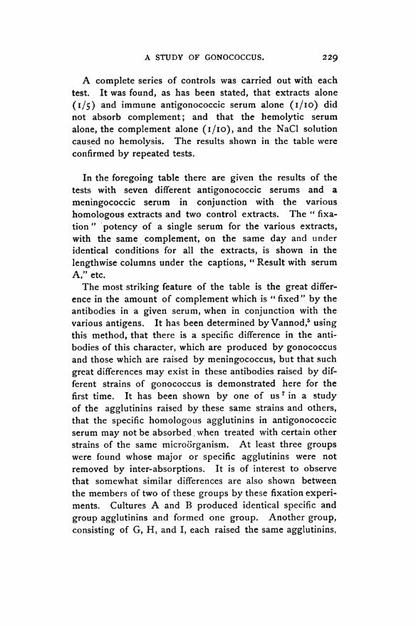
A complete series of controls was carried out with eachtest. It was found, as has been stated, that extracts alone(1/5) and immune antigonococcic serum alone (i/io) didnot absorb complement; and that the bemolytic serumalone, the complement alone (I/IO), and the NaCl solutioncaused no hemolysis. The results shown in the table wereconfirmed by repeated tests.
In the foregoing table there are given the results of thetests with seven different antigonococcic serums and ameningococcic serum in conjunction with the varioushomologous extracts and two control extracts. The " fixa-tion" 'potency of a single serum for the various extracts,with the same complement, on the same day and underidentical conditions for all the extracts, is shown in thelengthwise columns under the captions, " Result with serumA," etc.The most striking feature of the table is the great differ-
ence in the amount of complement which is " fixed" by theantibodies in a given serum, when in conjunction with thevarious antigens. It has been determined byVannod,5 usingthis method, that there is a specific difference in the anti-bodies of this character, which are produced by gonococcusand those which are raised by meningococcus, but that suchgreat differences may exist in these antibodies raised by dif-ferent strains of gonococcus is demonstrated here for thefirst time. It has been shown by one of us 7in a studyof the agglutinins raised by these same strains and others,that the specific homologous agglutinins in antigonococcicserum may not be absorbed. when treated with certain otherstrains of the same micro'organism. At least three groupswere found whose major or specific agglutinins were notremoved by inter-absorptions. It is of interest to observethat somewhat similar differences are also shown betweenthe members of two of these groups by these fixation experi-ments. Cultures A and B produced identical specific andgroup agglutinins and formed one group. Another group,consisting of G, H, and I, each raised the same agglutinins,
229

TEAGUE AND TORREY.
but their major agglutinins were not absorbed by the groupA, B. Turning now to these fixation experiments, we findthat serums A and B are practically identical in their anti-bodies. With their homologous extracts they fix comple-ment in dilutions as high as I/500 to I/1,000. With theextracts G, H, and I, on the other hand, the limit of completefixation is much lower: I/50 with G; 1/50 with H, and1/200 with I. If we examine now the results with the serumsraised by the other group we find conditions more or lessdirectly reversed. The extracts A and B fix complement,either not at all as with G serum or with more or lesscompleteness up to I/200 with H and I serums. WithG, H, and I antigens, however, there is an almost com-plete fixation in G serum up to I/2,000 to i/5,ooo. Thesame is true with H and I antigens with their respectiveserums, but extract G with H and I serum forms an excep-tion, as there is practically no fixation. The possibility issuggested by these variations that we are dealing withseveral sets of antibodies analogous to the specific and groupagglutinins which one encounters in agglutination work.The "fixing" antibodies of serum C are very similar tothose of group A, B, and have nothing in common withgroup G, H, I. The major agglutinins of C, however, werefound to be quite different from those of A and B and alsoof G, H, and I. Serum F is the most generalized and fixescomplement with all the gonococcic extracts in dilutions ofI/50 to I/200.The conclusion from this work, as with the agglutination
experiments, which stands out clearly, is that the familyGonococcus is heterogeneous rather than homogeneous.Whether there are groups in the gonococcus family whichare radically different, as is true with the dysentery family,or whether we are dealing here with slight variations fromthe family type, due to modification of receptors, can onlybe definitely determined by an extension of these studies toa large number of strains of this microorganism.With some of these serums there was a fixation of com-
plement at very high dilutions. The actual dilutio'ns of the
230
A STUDY OF GONOCOCCUS.
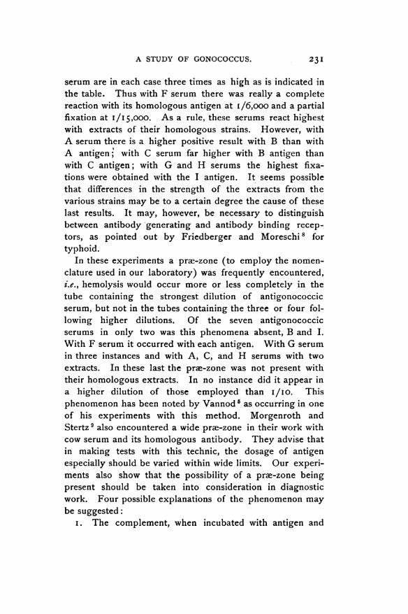
serum are in each case three times as high as is indicated inthe table. Thus with F serum there was really a completereaction with its homologous antigen at 1/6,ooo and a partialfixation at 1/15,000. As a rule, these serums react highestwith extracts of their homologous strains. However, withA serum there is a higher positive result with B than withA antigen; with C serum far higher with B antigen thanwith C antigen; with G and H serums the highest fixa-tions were obtained with the I antigen. It seems possiblethat differences in the strength of the extracts from thevarious strains may be to a certain degree the cause of theselast results. It may, however, be necessary to distinguishbetween antibody generating and antibody binding recep-tors, as pointed out by Friedberger and Moreschi 8 fortyphoid.
In these experiments a prae-zone (to employ the nomen-clature used in our laboratory) was frequently encountered,i.e., hemolysis would occur more or less completely in thetube containing the strongest dilution of antigonococcicserum, but not in the tubes containing the three or four fol-lowing higher dilutions. Of the seven antigonococcicserums in only two was this phenomena absent, B and I.With F serum it occurred with each antigen. With G serumin three instances and with A, C, and H serums with twoextracts. In these last the pre-zone was not present withtheir homologous extracts. In no instance did it appear ina higher dilution of those employed than I/IO. Thisphenomenon has been noted by Vannod 5 as occurring in oneof his experiments with this method. Morgenroth andStertz9 also encountered a wide proe-zone in their work withcow serum and its homologous antibody. They advise thatin making tests with this technic, the dosage of antigenespecially should be varied within wide limits. Our experi-ments also show that the possibility of a pre-zone beingpresent should be taken into consideration in diagnosticwork. Four possible explanations of the phenomenon maybe suggested:
i. The complement, when incubated with antigen and
23I

TEAGUE AND TORREY.
low (I/IO) dilution of immune serum, may, according toGay's 10 view, be absorbed through the union of precipitinand precipitinogen, whereas, when higher dilutions of theserum are employed, the complement combines with antigenplus amboceptor. The pre-zone in our experiments wouldbe due, then, to the fact that sensitized cells can utilize com-plement which is absorbed by a precipitate, but not thatwhich is fixed through union with amboceptor plus antigen.
2. Neisser and Sachs1 have shown that normal rabbitserum contains some hemolytic amboceptors for sheep cells.The pre-zone is then due to the fact that the increasedamount of these amboceptors in the low dilutions of anti-gonococcic serum allows even a trace of free complementto act on the sheep cells.
3. Again, it may be supposed that when complement isadded to a large amount of antigonococcic serum (I/10)and antigen, " deviation " takes place in the Neisser-Wechs-berg sense, i.e., the free amboceptors present combine withthe complement, so that the latter is no longer able to enterinto the antigen-antibody combination. On the addition ofred cells and hemolytic amboceptors, that portion of thecomplement which had before been held by free antigon-ococcic amboceptors is torn away and enters into thehemolytic system. This explanation assumes that the com-plement had in the first instance a greater affinity for freebacteriolytic amboceptors than for antigen plus these ambo-ceptors; in the second instance, however, this same com-plement has a greater affinity for antigen plus hemolyticamboceptors than for the free amboceptors with which it hadcombined. This seems at least unlikely.
4. The pre-zone phenomenon here, as well as in agglu-tination and bacteriolytic tests, may be explained by what wewill designate as the "optimum interaction zone of colloids."One of us,U2 in collaboration with Buxton, has shown that inflocculation tests colloids must be present in definite pro-portions in order to obtain the maximum effect. Increasingor diminishing either colloid causes a diminution in theamount of flocculation or its disappearance altogether. The
232

A STUDY OF GONOCOCCUS.
substances concerned in all immunity reactions are colloids,hence to obtain the maximum interaction they must bepresent in definite proportions. When one substance ispresent in great excess (in this instance gonococcic anti-bodies) no interaction occurs, - hence the pra-zonephenomenon. We regard this last as the most plausibleexplanation of the phenomenon under discussion.
In our discussion thus far we have assumed that a positiveresult with this method of " fixation of complement " indi-cates the presence of bacteriolytic amboceptors in theimmune serum tested, and we are here in accord with themajority of investigators who have employed this technic.Recently, however, Neufeld and Hiune 13 have cast somedoubt on this hypothesis. They found that the serum of twotyphoid patients, which had high bacteriolytic power asshown by plating out experiments, did not prevent hemoly-sis with Bordet's method. Hence they conclude that thecomplement deviating substance does not depend on theamount of bacteriolytic amboceptors present. It might beconsidered possible that the fixation of complement is dueto the interaction of the precipitin of the immune serumand the precipitinogen of the antigen (Gay"1). One greatobjection to such an explanation in this instance is thatfixation takes place strongly far above any possible precipi-tation zone. Wassermann and Bruck,14 too, have shown thatold extracts, which no longer give a precipitation withimmune serum, cause undiminished fixation of complement.This complement fixing substance in immune serum, in fact,may not be identical with any of the antibodies known atpresent. However this may prove to be, the evidence fur-nished by our experiments that the gonococcus family isheterogeneous, is in nowise affected.The technic under discussion has been employed thus far
by investigators with two objects in view: (a) For the differ-entiation of species of bacteria; (b) For diagnosis.
(a.) Ballner and Reibmayr ','studied the group of capsulebacteria (Friedlander's) but failed to obtain definite results,possibly because of faulty extracts. Gengou,'6 too, was
233

TEAGUE AND TORREY.
unable to differentiate between the acid-fast bacteria by thismethod. Using this technic in a study of the true choleraand " cholera-similar" micro'organisms, Schiitze 17 noteddistinct differences in the complement absorption, but con-cludes that " complement-fixation " does not offer reliablemeans of differentiations between these vibrios. Eysbroek 18tested a polyvalent antistreptococcic serum against variousstrains of streptococcus, pneumococcus, and meningococcus.As all these microorganisms caused fixation he concludesthat the method cannot be used to identify bacteria.Leuchs,"9 however, has shown that his typhoid, paratyphoid,and colon serums bind complement only with the correspond-ing extracts. He considers that the method may -prove tobe a better means of differentiation than agglutination andbacteriolytic tests. Vannod,5 also, in a comparative study ofmeningococcus and gonococcus concludes that the method isreliable since neither gonococcus serum plus meningococcusextract, nor meningococcus serum plus gonococcus extractabsorbed complement, whereas gonococcus extract plusgonococcus serum fixed complement in high dilutions of theserum. Finally, Wollstein,6 in a very recent paper, describesresults directly contradictory to those of Vannod, in that nodifferentiation between meningococcus and gonococcus couldbe obtained by this technic. The diametrically opposedresults obtained by Wollstein and Vannod, and also by Woll-stein and ourselves can only be due to differences in thetechnic employed. We have, accordingly, described ourmethods in great detail, although by doing so we were forcedto repeat much that has already been described by otherworkers. Nevertheless, in this way alone can differences inagents and procedure be detected.
If we had employed a single strain of gonococcus, weshould have been led to the same conclusion as Vannod,since the single meningococcic antigen does not absorb com-plement with any of our gonococcic serums, nor does men-ingococcic serum fix complement in conjunction with any ofthe gonococcic antigens, although with its homologous ex-tract there is a positive result with the meningococcic serum
234

A STUDY OF GONOCOCCUS.
in as high dilution as one to two thousand. Where onewould be likely to err, however, is in failing to recognize, bythe use of a single serum, cultures which are true membersof the gonococcus family. Thus, if serum C alone had beenemployed, cultures F, G, H, and I would not have beenrecognized; and if serum G had been used the same fatewould have befallen cultures A, B, C, and F. This shows,conclusively, that the method, as employed by Vannod, isnot reliable in differentiating between gonococcus and otherdiplococci. By the use of several serums or a polyvalentserum the results might, possibly, be rendered more trust-worthy.
(b.) Already several attempts have been made to applythis technic to the diagnosis of gonorrheal infections andmore especially to cases of gonorrheal arthritis. Muller andOppenheim,2 as has already been stated, were the first todetect by this method specific antibodies in the blood of apatient suffering with gonorrheal arthritis. Soon afterBriick3 obtained positive results in two out of six cases ofgonorrheal salpingitis and in one of two cases of chronicgonorrhea. Meakins4 has also arrived at like results in twoout of three cases of gonorrheal anthritis and in two cases ofprostatitis and chronic urethritis. All of these investigators,however, seem to have employed only one strain of gono-coccus in the preparation of the antigen. Such being thecase, it seems significant that Briick failed in five out of eightcases and Meakins in one out of five. The reason for thesenegative results has been explained, we think, by theseexperiments. Supposing the patient were infected withculture C, the antigens prepared with cultures F, G, H or Iwould have given negative results, or if the infection werewith culture G, then extracts from A, B, C or F would havebeen of no value. Our experiments, then, show clearly thatin order to apply this method of diagnosis to gonorrhealinfections, the serum of the suspected case should be testedagainst several different selected strains of gonococcus, beforea negative result is pronounced.
235
236 TEAGUE AND TORREY.
CONCLUSIONS.
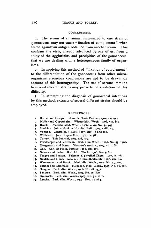
i. The serum of an animal immunized to one strain ofgonococcus may not cause " fixation of complement" whentested against an antigen obtained from another strain. Thisconfirms the view, already advanced by one of us, from astudy of the agglutinins and precipitins of the gonococcus,that we are dealing with a heterogeneous family of organ-isms.
2. In applying this method of " fixation of complement"to the differentiation of the gonococcus from other micro-organisms erroneous conclusions are apt to be drawn, onaccount of this heterogeneity. The use of serums immuneto several selected strains may prove to be a solution of thisdifficulty.
3. In attempting the diagnosis of gonorrheal infectionsby this method, extracts of several different strains should beemployed.
REFERENCES.
i. Bordet and Gengou. Ann. de l'Inst. Pasteur, I9OI, XV, 290.2. MUller and Oppenheim. Wiener klin. Woch., I9o6, Xix, 894.3. Bruck. Deutsche Med. Woc'h., I906, xxxii, No. 34, 945.4. Meakins. Johns Hopkins Hospital Bull., 1907, XViii, 225.5. Vannod. Centralbl. f. Bakt., 1907, xliv, io and I1o.6. Wollstein. Jour. Exper. Med., 1907, ix, 588.7. Torrey. This Journal, 1907, XVi, 329.8. Friedberger and Moreschi. Berl. klin. Woch., 1905, No. 45, I409.9. Morgenroth and Stertz. Virchow's Archiv., I907, viii, I66.
Io. Gay. Ann. de l'Inst. Pasteur, I905, xix, 593.ii. Neisser and Sachs. Berl. klin. Woch., I906, No. 3, 67.12. Teague and Buxton. Zeitschr. f. physikal Chem., I907, lx, 469.I3. Neufeld and HUne. Arb. a. d. Gesundheitsamte, 1907, xxv, I6.14. Wassermann and Bruck. Med. klin. Woch., i9oS, No. 55, I409.15. Ballner and Reibmayr. Munchen. Med. Woch., 1907, No. 13, 6oI.i6. Gengou. Berl. klin. Woch., I906, No. 48, 1531.17. SchUtze. Berl. klin. Woch., 1907, No. 26, 8oo.i8. Eysbroek. Berl. klin. Woch., I907, No. 32, ioi6.I9. Leuchs. Berl. klin. Woch., I907, Nos. 3 and 4.