a small conserved domain in the yeast spa2p is … as the shmoo tip and the incipient bud. the dy-...
TRANSCRIPT

The Rockefeller University Press, 0021-9525/97/07/17/20 $2.00The Journal of Cell Biology, Volume 138, Number 1, July 14, 1997 17–36 17
A Small Conserved Domain in the Yeast Spa2p Is Necessaryand Sufficient for Its Polarized Localization
Robert A. Arkowitz and Nick Lowe
Division of Cell Biology, Medical Research Council Laboratory of Molecular Biology, Cambridge, CB2 2QH, United Kingdom
Abstract.
SPA2
encodes a yeast protein that is one of the first proteins to localize to sites of polarized growth, such as the shmoo tip and the incipient bud. The dy-namics and requirements for Spa2p localization in liv-ing cells are examined using Spa2p green fluorescent protein fusions. Spa2p localizes to one edge of unbud-ded cells and subsequently is observable in the bud tip. Finally, during cytokinesis Spa2p is present as a ring at the mother–daughter bud neck. The bud emergence mutants
bem1
and
bem2
and mutants defective in the septins do not affect Spa2p localization to the bud tip. Strikingly, a small domain of Spa2p comprised of 150 amino acids is necessary and sufficient for localization to sites of polarized growth. This localization domain and the amino terminus of Spa2p are essential for its function in mating. Searching the yeast genome data-base revealed a previously uncharacterized protein
which we name, Sph1p (Spa2p homolog), with signifi-cant homology to the localization domain and amino terminus of Spa2p. This protein also localizes to sites of polarized growth in budding and mating cells.
SPH1
, which is similar to
SPA2
, is required for bipolar bud-ding and plays a role in shmoo formation. Overexpres-sion of either Spa2p or Sph1p can block the localization of either protein fused to green fluorescent protein, suggesting that both Spa2p and Sph1p bind to and are localized by the same component. The identification of a 150–amino acid domain necessary and sufficient for localization of Spa2p to sites of polarized growth and the existence of this domain in another yeast protein Sph1p suggest that the early localization of these pro-teins may be mediated by a receptor that recognizes this small domain.
P
olarized
cell growth and division are essential cel-lular processes that play a crucial role in the devel-opment of eukaryotic organisms. Cell fate can be de-
termined by cell asymmetry during cell division (Horvitzand Herskowitz, 1992; Cohen and Hyman, 1994; Rhyu andKnoblich, 1995). Consequently, the molecules involved inthe generation and maintenance of cell asymmetry are im-portant in the process of cell fate determination. Polarizedgrowth can occur in response to external signals such asgrowth towards a nutrient (Rodriguez-Boulan and Nelson,1989; Eaton and Simons, 1995) or hormone (Jackson andHartwell, 1990
a
,
b
; Segall, 1993; Keynes and Cook, 1995)and in response to internal signals as in
Caenorhabditis ele-gans
(Goldstein et al., 1993; Kimble, 1994; Priess, 1994) and
Drosophila melanogaster
(St Johnston and Nusslein-Vol-hard, 1992; Anderson, 1995) early development.
Saccharomyces cerevisiae
undergo polarized growth to-wards an external cue during mating and to an internal cueduring budding. Polarization towards a mating partner
(shmoo formation) and towards a new bud site requires anumber of proteins (Chenevert, 1994; Chant, 1996; Drubinand Nelson, 1996). Many of these proteins are necessaryfor both processes and are localized to sites of polarizedgrowth, identified by the insertion of new cell wall mate-rial (Tkacz and Lampen, 1972; Farkas et al., 1974; Lew andReed, 1993) to the shmoo tip, bud tip, and mother–daugh-ter bud neck. In yeast, proteins localized to growth sites in-clude cytoskeletal proteins (Adams and Pringle, 1984; Kil-martin and Adams, 1984; Ford, S.K., and J.R. Pringle.1986.
Yeast
. 2:S114; Drubin et al., 1988; Snyder, 1989; Sny-der et al., 1991; Amatruda and Cooper, 1992; Lew andReed, 1993; Waddle et al., 1996), neck filament components(septins) (Byers and Goetsch, 1976; Kim et al., 1991; Fordand Pringle, 1991; Haarer and Pringle, 1987; Longtine etal., 1996), motor proteins (Lillie and Brown, 1994), G-pro-teins (Ziman, 1993; Yamochi et al., 1994; Qadota et al.,1996), and two membrane proteins (Halme et al., 1996; Roe-mer et al., 1996; Qadota et al., 1996). Septins, actin, and ac-tin-associated proteins localize early in the cell cycle, be-fore a bud or shmoo tip is recognizable. How this group ofproteins is localized to and maintained at sites of cellgrowth remains unclear.
Spa2p is one of the first proteins involved in bud forma-tion to localize to the incipient bud site before a bud is rec-
Please address all correspondence to Robert A. Arkowitz, Division ofCell Biology, MRC Laboratory of Molecular Biology, Hills Road, Cam-bridge, CD2 2QH, United Kingdom. Tel.: (44) 1223-402229. Fax: (44)1223-412142. e-mail: [email protected]
The Journal of Cell Biology, Volume 138, 1997 18
ognizable (Snyder, 1989; Snyder et al., 1991; Chant, 1996).Spa2p has been localized to where a new bud will form atapproximately the same time as actin patches concentrateat this region (Snyder et al., 1991). An understanding ofhow Spa2p localizes to incipient bud sites will shed light onthe very early stages of cell polarization. Later in the cellcycle, Spa2p is also found at the mother–daughter budneck in cells undergoing cytokinesis. Spa2p, a nonessentialprotein, has been shown to be involved in bud site selec-tion (Snyder, 1989; Zahner et al., 1996), shmoo formation(Gehrung and Snyder, 1990), and mating (Gehrung andSnyder, 1990; Chenevert et al., 1994; Yorihuzi and Oh-sumi, 1994; Dorer et al., 1995). Genetic studies also sug-gest that Spa2p has a role in cytokinesis (Flescher et al.,1993), yet little is known about how this protein is local-ized to sites of polarized growth.
We have used Spa2p green fluorescent protein (GFP)
1
fusions to investigate the early localization of Spa2p to sitesof polarized growth in living cells. Our results demonstratethat a small domain of
z
150 amino acids of this large1,466-residue protein is sufficient for targeting to sites ofpolarized growth and is necessary for Spa2p function. Fur-thermore, we have identified and characterized a novelyeast protein, Sph1p, which has homology to both theSpa2p amino terminus and the Spa2p localization domain.Sph1p localizes to similar regions of polarized growth and
sph1
mutants have similar phenotypes as
spa2
mutants.
Materials and Methods
Strains, Media, and Microbiological Techniques
Yeast strains used in this study are listed in Table I. Strains were con-structed by standard genetic techniques and grown at 30
8
C (except tem-perature-sensitive [ts] strains) in rich media-yeast extract/peptone/dex-trose) or synthetic complete media lacking appropriate supplements forselection (Rose et al., 1991). RAY532 (
cdc3-1
), RAY520 (
cdc10-1
), andRAY525 (
cdc11-1
) strains were constructed by crossing mutants withSEY6210 and selecting for appropriate markers. Calcofluor was fromSigma Chemical Co. (St. Louis, MO) and
a
-factor was from Calbiochem-Novabiochem Corp. (La Jolla, CA).
Plasmid Construction and Mutagenesis
A 6.7-kb SalI–HindIII fragment of
SPA2
from p203 (Gehrung and Sny-der, 1990) was cloned into pRS406 (p406S2), pRS416 (p416S2), andpRS426 (p426S2).
GFP
was fused to the carboxyl terminus of
SPA2
usingPCR-amplified
GFP
coding region with an EcoRI site present at the 5
9
end of the coding region and HindIII–XbaI sites at the 3
9
end.
GFP
wasfused to the
SPA2
open reading frame (ORF) using the EcoRI site at theend of the
SPA2
ORF, resulting in the last three amino acids of
SPA2
be-ing replaced by Asn Ile followed by
GFP
with an additional Leu-Val at itscarboxyl terminus. We tested different versions of GFP to optimize sensi-tivity and minimize photobleaching.
GFPS65T
(Heim et al., 1995), madeby oligonucleotide-directed mutagenesis (Kunkel, 1985), and
mGFP5
(Sie-mering et al., 1996) were fused to
SPA2
similarly. Both
GFP
mutants weresignificantly more sensitive than wild-type
GFP
and enabled observationat the fluorescein excitation wavelength. For all experiments
SPA2GFPS65T
was used and cloned into pRS405 (p405S2G), pRS406 (p406S2G), pRS416(p416S2G), and pRS426 (p426S2G). Deletions were constructed usingp406S2G in which the EcoRI site within the
SPA2
ORF was removed byoligonucleotide-directed mutagenesis (Kunkel, 1985), and subsequentlythe EcoRI site upstream of the coding region was removed by partial di-gestion and subsequent filling in, leaving a single EcoRI site between
SPA2
and
GFP.
In addition, oligonucleotide-directed mutagenesis (Kunkel,1985) was used to replace the six nucleotides immediately 5
9
of the
SPA2
initiation codon with a BamHI restriction site, resulting in p406S2G1. Allcarboxyl-terminal deletions were generated by insertion of an EcoRI siteby oligonucleotide-directed mutagenesis (Kunkel, 1985), which resulted inAsn-Ser after amino acid 1,074 (
D
Z), 655 (
D
Y), 549 (
D
X), 511 (
D
W), andan Arg-Asn-Ser after amino acid 396 (
D
V). Amino-terminal deletionswere constructed by inserting a BamHI site by oligonucleotide-directedmutagenesis (Kunkel, 1985) 5
9
of amino acid 88 (
D
A), 288 (
D
B), 397 (
D
C),511 (
D
D), and 625 (
D
E), which resulted in Gly-Ser immediately precedinga methionine residue (in
D
C a methionine was also inserted after Gly-Ser). Deletions were made by restriction digestion and followed by religa-tion. All deletions were confirmed by dye terminator cycle sequencing(DNA Sequencing Kit; Perkin-Elmer Corp., Norwalk, CT) using anABI377 automated sequencer. Integration vectors containing
SPA2GFP
and derivatives were linearized with either StuI for pRS406 or XcmI forpRS405. Typically, integration yielded two to four copies per cell bySouthern analyses. For each integration, four independent transformantswere examined by fluorescence microscope for GFP expression and con-focal microscopy for localization.
A myc epitope tag was fused to the carboxyl terminus of
SPA2
by PCRusing p406S2G1 as a template, resulting in p406S2myc, which has the mycepitope (MEQKLISEEDL) followed by Lys-Leu-Val. Spa2myc fromp406S2myc was liberated by BamHI–SacII digestion and cloned intoBamHI–SacII-digested pRS425TPI, which is a derivative of pRS425 withthe triose phosphate isomerase (TPI) promoter cloned into XhoI–HindIIIsites, resulting in pRS425TPIS2myc.
Fluorescence and Confocal Microscopy
Transformants were initially screened using an Axiophot microscope(Zeiss, Oberkochen, Germany). Living cells were imaged at 22
8
C using aBiorad-MRC-600 confocal microscope (Bio Rad Laboratories, Hercules,CA) with a NA 1.4,
3
60 objective and FITC (488-nm) excitation filter.For analyses of localization of Spa2GFP deletions, temperature-sensitiveand mating mutants, 1–2
m
l of an exponentially growing culture (grown inthe appropriate synthetic deficient media) was spotted on a glass slide,and the coverslip was applied with gentle pressure. This resulted in cellssticking sufficiently to the slide to allow confocal image acquisition. Pho-tobleaching was minimized by decreasing illumination intensity with neu-tral density filters.
For time course analyses, slides were prepared by cutting a squarepiece of 0.5-mm-thick silicon rubber with a hole cut in the center using acork borer. This piece of silicon rubber was pressed onto a glass slideheated to 65
8
C, a drop of molten synthetic complete media lacking uracilwith 2% agarose was spotted in the hole of the silicon rubber, and a No. 1glass coverslip was placed on top to flatten the pad. The slide was cooledto room temperature for
z
10 min, and then the coverslip was slid off,leaving a flattened
2
ura agarose pad. 1–2
m
l of an exponentially growingculture (grown in the appropriate synthetic deficient media) was spottedon this pad, allowed to partially dry for about 5 min, and covered with acoverslip, applying slight pressure to make a seal. Vaseline was coatedalong the edges of the coverslip to prevent the pad from drying out. Cellson this slide were viable and formed microcolonies upon extended incuba-tion. For long confocal experiments, photobleaching and excessive irradi-ation were minimized by single scans with maximal neutral density filters atthree different focal planes. Cell growth appeared to slow during long con-focal experiments, yet cells continued to divide and Spa2GFP localizationwas identical to that in long experiments with infrequent scans. For exam-ination of mating cells, a and
a
cells were both spotted on the same aga-rose pad and mating was followed by light microscopy and confocal mi-croscopy.
Disruption of BEM1 and BEM2
BEM1
was cloned by PCR using Taq Polymerase (Perkin-Elmer Corp.)from yeast genomic DNA. A 2,136-bp fragment including 426 nucleotides5
9
of the initiation codon was cloned into the pGem-T vector (Promega,Madison, WI), resulting in pGBEM1. A
BEM1
disruption plasmid wasconstructed by replacing SmaI–XcmI from pGBEM1 with
S. pombe HIS5
(a homologue of
S. cerevisiae HIS3
) from pFA6a-HIS3MX6 (A. Wach,University of Basel, Switzerland), resulting in removal of all of
BEM1
ORF except for the last 81 codons.
BEM2
was disrupted using PCR-basedgene disruption (Baudin et al., 1993). Oligonucleotides were used eachwith homology to 60 nucleotides 5
9
and 3
9
of the
BEM2
ORF and 18 nu-cleotides to pFA6a-HIS3MX6, respectively, resulting in replacement of
1.
Abbreviations used in this paper
: cs, cold sensitive; GFP, green fluores-cent protein; ORF, open reading frame; TPI, triose phosphate isomerase;ts, temperature sensitive; WT, wild type.

Arkowitz and Lowe
Spa2p Polarized Localization
19
Table I. Yeast Strains Used in This Study
Strain Genotype Source
104 Mata ade1 ade2 ura1 his7 lys2 tyr1 gal1
cdc3-1
R. Mortimer (University of California, Berkeley)332 Mata ade1 ade2 ura1 his7 lys2 tyr1 gal1
cdc11-1
R. Mortimer (University of California, Berkeley)17012 Mata ade1 ade2 ura1 his7 lys2 tyr1 gal1
cdc10-1
R. Mortimer (University of California, Berkeley)JC-F5 Mata ura3-52 ade2-101 met1 bar1-1 bem1-s2 Chenevert et al., 1994JC-Gll Mata ura3-52 ade2-101 met1 bar1-1 bem1-s1 Chenevert et al., 1994JC-J9 Mata ura3-52 ade2-101 met1 bar1-1
pea1
(allelic to
SPA2
) Chenevert et al., 1994JY426 Mata his4-34 leu2-3112 ura3-52 fus1-
D
1 fus2-
D
3 Cold Spring Harbor Yeast Genetics CourseJY429 Mat
a
trp1
D
1 ura3-52 cyh2 fus1-
D
1 fus2-
D
3 Cold Spring Harbor Yeast Genetics CourseRAY416 same SEY6210 plus URA3::SPA2GFP This studyRAY520 Mat
a
ura3-52
‡
his3-
D
200
cdc10-1
This studyRAY525 Mat
a
leu2-3,112 ura3-52
‡
his3-
D
200 trp1-
D
901
cdc11-1
This studyRAY532 Mat
a
leu2-3,112 ura3-52
‡
his3-D200
cdc3-1
This studyRAY563 same as SEY6210 plus sph1-
D
1::HIS3 This studyRAY567 same as SEY6211 plus sph1-
D
1::HIS3 This studyRAY574 same as SEY6210 plus spa2-
D
1::TRP This studyRAY578 same as SEY6211 plus spa2-
D
1::TRP This studyRAY586 same as SEY6210 plus spa2-
D
1::TRP sph1-
D
1::HIS3 This studyRAY590 same as SEY6211 plus spa2-
D
1::TRP sph1-
D
1::HIS3 This studyRAY616 same as SEY6210/11 plus spa2-
D
1::TRP This studyRAY618 same as SEY6210/11 plus sph1-
D
1::HIS3 This studyRAY620 same as SEY6210/11 plus spa2-
D
1::TRP sph1-
D
1::HIS3 This studyRAY651 same as RAY574 plus URA3::SPA1GFP This studyRAY674 same as SEY6211 plus URA3::SPA2GFP This studyRAY685 same as RAY520 plus URA3::SPA2GFP This studyRAY691 same as RAY525 plus URA3::SPA2GFP This studyRAY696 same as RAY532 plus URA3::SPA2GFP This studyRAY697 same as SEY6210 plus URA3 This studyRAY698 same as RAY574 plus URA3 This studyRAY699 same as SEY6210 plus URA3::S2pSPH1GFP This studyRAY703 same as RAY574 plus URA3::S2pSPH1GFP This studyRAY705 same as JC-F5 plus URA3::SPA2GFP This studyRAY706 same as JC-G11 plus URA3::SPA2GFP This studyRAY709 same as RAY563 plus URA3* This studyRAY711 same as RAY586 plus URA3 This studyRAY712 same as RAY719 plus URA3::SPA2GFP This studyRAY719 same as SEY6210 plus bem1-
D
1::HIS5 This studyRAY765 same as RAY578 plus URA3::SPA2GFP This studyRAY773 same as RAY578 plus URA3 This studyRAY774 same as RAY578 plus URA3::S2pSH1GFP This studyRAY776 same as RAY616 plus URA3 This studyRAY778 same as RAY618 plus URA3 This studyRAY780 same as RAY620 plus URA3 This studyRAY775 same as RAY616 plus URA3::SPA2GFP This studyRAY777 same as RAY618 plus URA3::SPA2GFP This studyRAY779 same as RAY620 plus URA3::SPA2GFP This studyRAY786 same as RAY574 plus URA3::SPA2 This studyRAY787 same as RAY578 plus URA3::SPA2 This studyRAY811 same as RAY590 plus URA3::SPA2 This studyRAY836 same as SEY6210 plus bem2-
D
1::HIS5 This studyRAY854 same as RAY618 plus URA3::S2pPH1GFP This studyRAY867 same as RAY836 plus URA3::SPA2GFP This studyRAY871 same as RAY616 plus URA3::S2pPH1GFP This studyRAY872 same as RAY620 plus URA3::S2pPH1GFP This studyRAY873 same as RAY567 plus URA3 This studyRAY874 same as RAY567 plus URA3::SPA2GFP This studyRAY875 same as RAY567 plus URA3::S2pSPH1GFP This studyRAY876 same as SEY6211 plus URA3 This studyRAY877 same as SEY6211 plus URA3::S2pSPH1GFP This studySEY6210 Mat
a
leu2-3,112 ura3-52 his3-
D
200 trp1-
D
901 lys2-801 suc2-
D
9 S. Emr (University of California, San Diego)SEY6211 Mata leu2-3,112 ura3-52 his3-
D
200 trp1-
D
901 ade2 suc2-
D
9 S. Emr (University of California, San Diego)SEY6210/11 Mata/
a
leu2-3,112/leu2-3,112 ura3-52/ura3-52 his3-
D
200/his3-
D
200 S. Emr (University of California, San Diego)trp1-
D
901/trp1-
D
901 LYS/lys2-801 ADE2/ade2 suc2-
D
9/suc2-
D9
*Strains were transformed with a Bg1II URA3 fragment from pFL34 to make URA31.‡Progeny of crosses were transformed with pRS416 to select for ura3-52 mutants.

The Journal of Cell Biology, Volume 138, 1997 20
the entire BEM2 ORF with S. pombe HIS5 (a homologue of S. cerevisiaeHIS3) (Wach et al., 1994). Linearized disruption cassettes or the PCR dis-ruption cassette were used to create disruption strains using the one-stepknockout procedure of Rothstein (1983). Disruption strains were con-firmed by PCR analyses and both were temperature sensitive as previ-ously reported (Bender and Pringle, 1991).
Quantitative Mating and Shmoo FormationFor mating function analyses of SPA2GFP deletion mutants, strain JC-J9was transformed with StuI linearized p406S2G1 deletion mutants andtransformants were used that had observable fluorescence signals (by flu-orescence microscopy). Quantitative mating assays were carried out as de-scribed in Chenevert et al. (1994) using 1 3 106 cells from exponentiallygrowing cultures of the enfeebled mating partners JY429 or JY426. Mat-ing efficiency was calculated as the ratio of diploids to total cells.
Shmoo formation was determined by the addition of a-factor (final con-centration 12.9 mM) to 1 3 106 log growing cells. After 2 h of growth, cellswere fixed with 3.7% formaldehyde. Cells were sonicated and the numberof shmoos was quantitated using a phase microscope. This concentrationof a-factor and incubation time typically resulted in z80% shmooed cellsin wild-type strains with very few cells having two shmoos. Both peanut-and pear-shaped shmoos were counted as shmoos. For observation ofGFP fusions in shmooed cells, log cultures in selective media were grownfor 3 h in rich media before a-factor addition.
Budding Pattern AssaysCalcofluor staining of bud scars was carried out as described in Pringle(1991). Budding patterns were quantitated and cells were photographedusing a Zeiss Axiophot epifluorescence microscope with a NA 1.3, Plan-Neofluor 3100 objective and a UV-H 365 excitation filter.
Cloning and Sequencing of SPH1SPH1 was cloned by PCR using Taq Polymerase (Perkin-Elmer Corp.)from yeast genomic DNA. Initially a 2-kb fragment including 346 nucleo-tides 59 of the SPH1 initiation codon was cloned into the Promega pGem-T vector (pGSH1), and two independent clones were sequenced. Subse-quently, an additional 2-kb fragment starting at 1,018 nucleotides into theSPH1 ORF was isolated by PCR from genomic DNA and cloned intopGem-T (pGSH2). Three independent clones of this PCR product weresequenced, and the full-length SPH1 including 1,068 nucleotides 39 of theend of the coding region was constructed by replacement of a XhoI–NcoIrestriction fragment from pGSH1 with an XhoI–NcoI fragment frompGSH2, yielding pGSH3. CEN and 2-mm vectors carrying SPH1 wereconstructed by subcloning a SalI–SacII fragment from pGSH3 into SalI–SacII pRS415 (p415SH3) and pRS425 (p425SH3) and XhoI–SacII pRS406(p406SH3). The sequence data for SPH1 are available from EMBL/Gen-Bank/DDBJ under accession number AF008236.
Disruption of SPA2 and SPH1A SPA2 disruption plasmid was constructed by replacing the GFP se-quence in p406S2G1 with the 39 SPA2 noncoding sequence from p416S2(resulting in p406S2.1), and subsequently all of the SPA2 ORF was re-placed by TRP1 using the uniquely engineered BamHI site and the EcoRIsite. SPH1 disruption plasmid was constructed by replacing StuI–XhoIfrom pGSH1 with HIS3. Linearized disruption cassettes were used to cre-ate disruption strains using the one-step knockout procedure of Rothstein(1983). Disruption strains were confirmed by PCR analyses.
Localization of Sph1pAn EcoRI site at nucleotide 1,157 in the SPH1 coding region (p406SH3)was removed by oligonucleotide-directed mutagenesis (Kunkel, 1985). Inaddition, oligonucleotide-directed mutagenesis (Kunkel, 1985) was usedto replace the six nucleotides immediately preceding the initiation codonwith a BamHI site, resulting in p406SH4. An EcoRI site was placed at theend of the SPH1 coding region by PCR with Pfu polymerase (Stratagene,La Jolla, CA) using p406SH4 as a template, yielding p406SH5. This re-sulted in the addition of the amino acids Ala-Asn-Ser to the carboxyl ter-minus of Sph1p. The sequence of p406SH5 was confirmed by dye termina-tor cycle sequencing using an ABI377 automated sequencer. The codingregion of SPH1 from p406SH5 was liberated by BamHI–EcoRI digestion andcloned into BamHI–EcoRI-digested p406S2G1, resulting in p406SH5G in
which SPH1GFP is driven by the SPA2 promoter. p406SH5G was linear-ized with either BsmI or XcmI and used to transform various yeast strains.Two p406SH5G transformants were examined by confocal microscopy.
A myc epitope tag was fused to the carboxyl terminus of Sph1p by PCRusing p406SH4 as a template, resulting in p406SHmyc, which had two ad-ditional amino acids, Leu-Val, at the carboxyl terminus of Sph followed bythe myc epitope (MEQKLISEEDLV). Sph1myc from p406SHmyc was lib-erated by BamHI–SacII digestion and cloned into BamHI–SacII-digestedpRS425TPI, which is a derivative of pRS425 with the TPI promotercloned into XhoI–HindIII sites, resulting in pRS425TPISHmyc.
Results
Spa2GFP Is Functional and Localizes Correctly
To investigate the function and dynamics of Spa2p local-ization, we fused GFP to its carboxyl terminus. For thesestudies, it was necessary that the expression level of the fu-sion protein reflect the normal level and be essentiallyconstant throughout a population of cells. Hence we choseto use the endogenous SPA2 promoter and to integrate allconstructs at the URA3 locus. We first examined the func-tion of this fusion protein by testing its ability to comple-ment the various phenotypes of a Dspa2 strain, such as adefect in shmoo formation (Gehrung and Snyder, 1990;Chenevert et al., 1994; Yorihuzi and Ohsumi, 1994; Valtzand Herskowitz, 1996), a mating defect (Gehrung and Sny-der, 1990; Chenevert et al., 1994; Yorihuzi and Ohsumi,1994; Dorer et al., 1995), and a random bud site selectionpattern in a homozygous Dspa2 diploid (Snyder, 1989; Zah-ner et al., 1996). Fig. 1 A shows the results of a quantitativemating assay in which a Dspa2 strain carrying SPA2,SPA2GFP, or an empty plasmid is mated with an enfee-bled mating partner and diploids are subsequently se-lected. Deletion of SPA2 results in z90-fold decrease in mat-ing efficiency, which is fully complemented by SPA2GFP.Deletion of SPA2 results in a defect in shmoo formation(Gehrung and Snyder, 1990), which has subsequently beenshown to be dependent on pheromone concentration andincubation time (Valtz and Herskowitz, 1996). A Dspa2strain in the presence of saturating pheromone concentra-tions showed a lower percentage of shmooed cells thanwild-type cells. Consistent with previous observations (Valtzand Herskowitz, 1996), this strain exhibited primarily pea-nut-shaped shmoos in contrast with the pear-shapedshmoos of the wild-type strain (data not shown). SPA2GFPfully complements the defect in shmoo formation observedat high pheromone concentration in a Dspa2 strain (Table
Table II. SPA2GFP Complements Shmoo Formation Defect of Dspa2 Mutant
SPA2 construct Percentage of shmoos*
%
SPA2 772 58SPA2GFP 83
Cells were treated with a-factor and quantitated as described in Materials and Meth-ods. SPA2 denotes WT cells with pRS406 integrated at URA3 (RAY876), 2 areDspa2 cells with pRS406 integrated at URA3 (RAY578), and SPA2GFP are Dspa2cells with pRS406SPA2GFP integrated at URA3 (RAY765). For each strain 250 cellswere counted. SPA2GFP had no effect on shmoo formation or morphology of wild-type cells (see Table V).*Peanut- and pear-shaped shmoos were designated as shmoos.

Arkowitz and Lowe Spa2p Polarized Localization 21
II) and shmoo morphology (data not shown). Deletion ofSPA2 has been shown to have no effect on bud site selec-tion in haploids, yet homozygous diploid deletion mutantsare defective in bipolar budding (Snyder, 1989; Zahner etal., 1996; Valtz and Herskowitz, 1996). Haploids (a or a cells)generally bud in an axial pattern, adjacent to the previousdivision site; however, diploids (a/a cells) bud in a bipolarpattern, distal or proximal to the division site (Freifelder,1960; Hicks et al., 1977; Sloat et al., 1981). Fig. 1 B demon-strates that SPA2GFP is able to completely complement
the bud site selection defect in homozygous Dspa2 diploids.Furthermore, deletion of SPA2 results in round cells andSPA2GFP also complements this morphological defect(data not shown). Together, these assays demonstrate thatthe Spa2GFP fusion protein is fully functional.
Cells containing the Spa2GFP were examined by fluo-rescence confocal microscopy. All experiments used GFP-(S65T) (Heim et al., 1995) mutant, which optimized sensi-tivity and minimized photobleaching. Fig. 1 C shows thelocalization of Spa2GFP by confocal microscopy. Consis-
Figure 1. (A) Complementa-tion of spa2 mating defect bySPA2GFP. Dspa2 mutantswith SPA2 (RAY786),SPA2GFP (RAY651), or anempty plasmid (RAY698) in-tegrated at the URA3 locuswere mated with the enfee-bled mating tester strain(JY426). Diploids were se-lected on 2ura, 2lys plates.Mating efficiency with SPA2(30.9%) was set to 100% effi-ciency. (B) Complementa-tion of spa2 bipolar buddingdefect by SPA2GFP. Wild-type diploids (SEY6210/11)and homozygous Dspa2 dip-loids with either SPA2GFP(RAY775) or an empty plas-mid (RAY776) integrated atthe URA3 locus growing ex-ponentially were stained withCalcofluor as described inMaterials and Methods, andbudding pattern was ana-lyzed. For each strain, theposition of the bud relativeto the birth scar (see inset)was scored for z100 cellswith two or more bud scars.(C) Localization of Spa2GFPat sites of polarized growth.Confocal microscopy of liv-ing cells with SPA2GFP inte-grated at URA3 (RAY416)at different stages in cell cy-cle. Note unbudded cells withpatch of Spa2GFP, small-and medium-sized buds withSpa2GFP fluorescence onthe periphery of the tip, anda cell undergoing cytokinesiswith a “bar” of Spa2GFP atthe mother–daughter budneck. Bar, 5 mm.

The Journal of Cell Biology, Volume 138, 1997 22
Arkowitz and Lowe Spa2p Polarized Localization 23
tent with previous indirect immunofluorescence studies,Spa2p localizes to a crescent in unbudded cells, whichmarks the new bud site, the bud tip of small buds, and theneck between the mother and daughter cell just beforecytokinesis (Snyder, 1989; Snyder et al., 1991). SimilarSpa2GFP localization was observed in a Dspa2 strain (datanot shown), indicating that SPA2 is not required for the lo-calization of the fusion protein. These results confirm pre-vious immunolocalizations and show that they reflect thedistribution of this protein in vivo. We also examined theeffect of expression level on Spa2GFP localization to de-termine if localization was saturable. Expression level wasvaried by increasing the copy number of SPA2GFP from asingle copy (replacement), SPA2GFP integrated at URA3,a centromere plasmid carrying SPA2GFP, and a multi-copy 2-mm plasmid carrying SPA2GFP. The localizationof Spa2p to the bud tip and mother–daughter bud neckwas identical under all expression conditions; however,with expression from a multicopy 2-mm plasmid, cytoplasmicSpa2GFP was observed (data not shown). Furthermore,we did not observe a substantial increase in the intensity ofthe localized Spa2GFP fluorescence during overexpres-sion, suggesting that Spa2p localization is saturable.
Spa2GFP Dynamics: Localization to Two Spatially and Temporally Distinct Structures
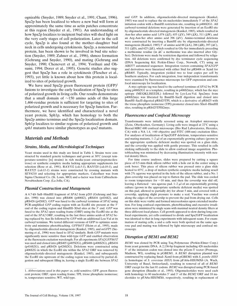
To examine the relationship between the two localizationsof Spa2GFP (bud tip and mother–daughter bud neck), weinvestigated the dynamics of Spa2GFP in haploid cells(Fig. 2 A). Initially we examined Spa2GFP in living cellsusing confocal microscopy, because we observed substan-tial phototoxicity with conventional fluorescence micros-copy, which was incompatible with such time course ex-periments. Fig. 2 A shows a time course of Spa2GFPdynamics at 228C in which Spa2GFP within two cells wasobserved approximately every 5 min at minimally threedifferent focal planes. This experiment involved a total ofz100 confocal scans over z3 h, and illustrated in Fig. 2 Aare the focal planes through the center of the cells. Itshould be noted that, although two cells are shown in thisfigure, Spa2GFP is found in a very specific location in thecell, making it difficult to observe such a structure in morethan one cell at a time. The upper cell is in focus through-out this time course. During the first hour, Spa2GFP canbe seen on the periphery of the bud and appears to spreadout during this time. After .1 h, weak fluorescence be-
Figure 2. (A) Dynamics of Spa2GFP localization in living cells. Spa2GFP distribution in two cells (RAY416) at 6-min intervals. TheSpa2GFP fluorescence in two cells is in different focal planes and the upper cell is in focus throughout the time course. Note the appear-ance at 76 min of a ring at the mother bud neck. Cells divided during and after confocal time course. (B) Localization of Spa2GFP inmating cells. Confocal microscopy of five representative mating pairs (RAY416 and RAY674) at different stages of mating/zygote for-mation. The diffuse background is due to the high gain setting to allow visualization of the entire cell. (C) Movement of Spa2GFP in bud-ding zygotes. Time course of zygotes from RAY416 mated to RAY674. In the center of each panel is a dumbbell-shaped zygote with aseparate round cell to its right and left. Note the ring of Spa2GFP fluorescence at the a/a shmoo neck at 0 min. Bars: (A and B) 1 mm;(C) 5 mm.
The Journal of Cell Biology, Volume 138, 1997 24
comes apparent at the neck between the mother anddaughter cell. At 66 and 71 min, Spa2GFP can be seen atthe bud periphery and mother–daughter bud neck, sug-gesting that these two structures can exist simultaneously.In panels (71–83 min), a ring structure is evident which ap-pears as two dots (the cross-section of a ring) at the nar-rowest point in the neck between the mother and daughtercells. Movement of one focal plane (1 mm) in either direc-
tion in the z-axis was consistent with such a ring structure,revealing a solid bar perpendicular to the mother–daugh-ter cell axis. At z1 h and 30 min, this Spa2p ring appearsto have closed, now appearing as a “bar” in all focalplanes. Subsequently, the Spa2p “bar” appears to get nar-rower in the axis perpendicular to the mother–daughtercell and thicker in the mother–daughter cell axis, indica-tive of cytokinesis. By z100 min, two Spa2p “bars” are ap-
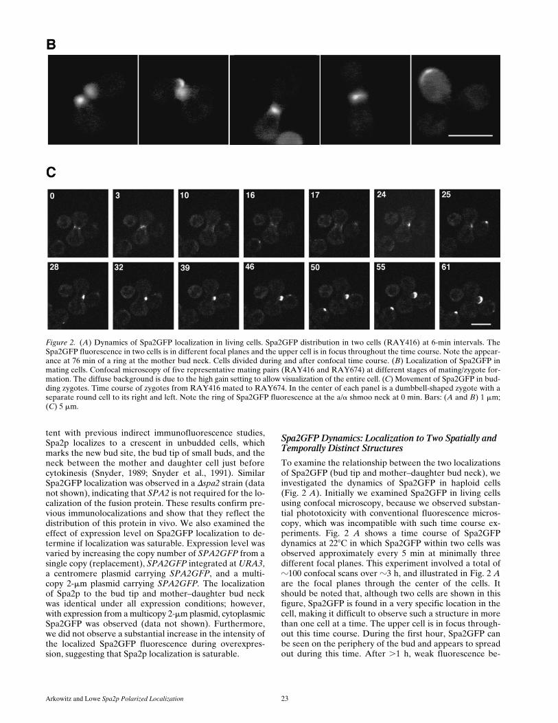
Figure 3. Spa2GFP local-ization in bem and septinmutants. Dbem1 (RAY712),Dbem2 (RAY836), cdc3-1(RAY696), cdc10-1 (RAY685),and cdc11-1 (RAY691) strainswith SPA2GFP integrated atthe URA3 locus were grownat 258C. Exponentially grow-ing cultures of the septin mu-tants were shifted to 378C for3 h, and cells were examinedby confocal microscopy.Note large cells in the bemmutants and the Spa2GFPfluorescence at both the budtip and mother–daughter budneck. Bar, 5 mm.

Arkowitz and Lowe Spa2p Polarized Localization 25
parent, one in the daughter and one in the mother cell. Inthe next 40 min (103–145-min panels), Spa2GFP fluores-cence becomes concentrated in an intense spot axial to thefirst bud/birth site marking the next bud site. During thistime and up to 160 min, the cells lose their round shapeand initiate the next round of polarized growth. Fromthese time courses Spa2p is observed to alternate betweenlocalized discrete structures (G1 phase unbudded cells, Sphase bud emergence, and M phase during cytokinesis)and more diffuse structures as the daughter cells increasein size. In addition, Spa2p structures appear to changefrom cup-like crescents to donut-like rings during the cellcycle. Despite the slow apparent movement of these struc-tures, these studies highlight the dynamic nature of Spa2pin living cells.
We also examined the localization and dynamics ofSpa2GFP in mating cells. Previous immunofluorescencestudies have revealed that Spa2p localizes to shmoo tips(Gehrung and Snyder, 1990) and Spa2GFP localization inmating cells showed similar localization (Fig. 2 B). Fur-thermore, Spa2GFP localized to the tips of mating projec-tions (shmoos) after mating pheromone addition and thislocalization was also observed in the absence of wild-typeSPA2 (data not shown). Upon zygote formation, Spa2GFP-labeled structures from each haploid appear to fuse intoone structure at the site of cell fusion, which subsequentlylocalizes to growing bud tip of the zygote. Fig. 2 C shows atime course of Spa2GFP localization in haploid cells thathave just fused to form a zygote. The ring of Spa2GFP flu-orescence at the neck of the a/a zygote persists for z20min and then relocalizes to one side of the neck where thenew bud will form. Subsequently, Spa2GFP can be seen onthe tip of the growing bud. These experiments demon-strate that within a time frame of 10 min the Spa2GFP re-localizes from a ring structure at the site of cell fusion tothe site of growth on the new bud.
Spa2GFP Localization in Mutants with Defects in Bud Emergence and Septation
We examined the effect of several cell cycle and bud emer-gence mutants on Spa2GFP localization to determine if itwas possible to separate the two observed Spa2p struc-tures during the cell cycle. Indirect immunofluorescencestudies have revealed that Spa2p localizes very early in thecell cycle, marking the site of the incipient bud. Further-more, neither actin mutants nor tubulin mutants, both ofwhich result in a delocalized cytoskeleton, mislocalize Spa2p(Snyder et al., 1991). These results suggest that Spa2p lo-calization is independent of the actin and microtubule cy-toskeleton. Recently, additional cytoskeletal componentstermed septins have been shown to be involved in cytoki-nesis (Haarer and Pringle, 1987; Kim et al., 1991; Ford andPringle, 1991; Cooper and Kiehart, 1996; Field et al., 1996;Longtine et al., 1996). The septins are encoded by CDC3,CDC10, CDC11, and CDC12, and these proteins arethought to form 10-nm neck filaments that have been ob-served by EM at the mother–daughter bud neck (Byers andGoetsch, 1976). Previous studies have shown that ts mu-tants in any of the septins result in a loss of localization ofall the septins (Kim et al., 1991; Ford and Pringle, 1991;Haarer and Pringle, 1987) and 10-nm neck filaments (By-
ers and Goetsch, 1976). The role of the septins, Cdc3p,Cdc10p, and Cdc11p, in Spa2p localization was investi-gated. Flescher et al. (1993) have previously demonstrateda genetic interaction between CDC10 and SPA2, by show-ing that spa2 and an ochre truncation mutant of CDC10,cdc10-10, are synthetically lethal. These experiments im-plicated a connection between SPA2 and septin formation,although it is unclear whether such an interaction isCDC10 specific. SPA2GFP was integrated at the URA3locus in the temperature-sensitive septin mutants cdc3-1,cdc10-1, and cdc11-1, and several transformants of eachwere examined by confocal microscopy after growth in liq-uid culture for 3 h at 258C, 3 h at 378C, and 6 h at 378C(Fig. 3). At the permissive temperature, despite a smallpercentage of cells displaying the characteristic septin mu-tant phenotype, misshapen long cell, Spa2GFP localiza-tion appeared normal, with bud tip and mother–daughterbud neck fluorescence observable. At the restrictive tem-perature, the septin mutant phenotype was readily observ-able in most cells, resulting in the inability of cells to sep-tate and thus an abundance of misshapen long cells withseveral long extended buds. In cdc3-1, cdc10-1, and cdc11-1cells at 378C, Spa2GFP was localized primarily to bud tips.Spa2GFP was not observed at the bud neck betweenmother and daughter cells (Fig. 3), consistent with Spa2plocalization in a cdc10-10 mutant (Flescher et al., 1993).These results suggest that the septins are either requiredfor Spa2p localization at the mother–daughter neck or,more likely, that these cdc mutants act at a point in the cellcycle before the Spa2p localization at the mother–daugh-
Figure 4. Diagram of Spa2GFP deletion constructs. DeletionsDA, DB, DC, DD, and DE contain Spa2p residues 88, 288, 397,511, and 625, respectively, to residue 1,463. Deletions DZ, DY,DX, DW, and DV contain Spa2p residue 1 to residue 1,074, 655,549, 511, and 396, respectively. Constructs DBX, DBV, and DCXcontain Spa2p residues 288–549, 288–396, and 397–549, respec-tively. All constructs contain Spa2p fused to GFP and were inte-grated at the URA3 locus in either SEY6210 for localization studiesor JC-J9 for mating function analysis. See Fig. 5 A for localizationdata and Fig. 5 B for results from mating assays.

The Journal of Cell Biology, Volume 138, 1997 26
ter neck. Perhaps, the mother–daughter neck needs to besufficiently constricted in order for the Spa2p localizationto occur and therefore is not possible in such septin mu-tants. In contrast, the persistent localization of Spa2GFP tobud tips in the septin mutants at the nonpermissive tem-perature indicates that septins are not required to main-tain Spa2p at this location.
In addition, we examined the localization of Spa2GFP in
bem1 and bem2 mutants that result in bud emergence de-fects (Bender and Pringle, 1991). BEM1 encodes a proteincontaining two SH3 domains (Chenevert et al., 1992) andBEM2 encodes a rho-GAP for Rho1p (Peterson et al.,1994; Kim et al., 1994). Bem1p has been shown to interactwith proteins required for bud formation (Peterson et al.,1994; Zheng et al., 1995), bud site selection (Chant et al.,1991), and components of the pheromone-responsive mi-
Figure 5. (A) Localization ofSpa2GFP deletion mutants. Ex-ponentially growing cultures ofSEY6210 with deletion con-structs integrated at the URA3locus were examined by confo-cal microscopy. In DA, DB, DC,DZ, DY, DX, and DCX, noteboth bud tip and mother–daugh-ter bud neck localization ofSpa2GFP deletions. Panels DD,DE, DC, DW, DV, and DCXshow general Spa2GFP fluores-cence. (B) Mating function ofSpa2GFP deletion mutants. Thespa2 mutant (JC-J9) withSPA2GFP deletion constructsintegrated at the URA3 locuswas mated with an enfeebledmating partner (JY429), anddiploids were selected on 2met,2ura plates. In each experiment,mating of JC-J9 carrying full-length Spa2GFP was taken as100% efficiency (absolute effi-ciency of 11.2% [left] and 7.9%[right]). Bar, 5 mm.

Arkowitz and Lowe Spa2p Polarized Localization 27
togen-activated protein kinase cascade (Leeuw et al.,1995). In addition, BEM1 has been shown to be involvedin cell mating and polarized growth during shmoo forma-tion (Chenevert et al., 1992, 1994; Yorihuzi and Ohsumi,1994). Bem2 mutants show genetic interactions with cy-toskeletal components (Wang and Bretscher, 1995). BEM1or BEM2 was deleted from haploid cells by one-step genereplacement (Rothstein, 1983); SPA2GFP was integratedat the URA3 locus; and several transformants of each wereexamined by confocal microscopy after growth in liquidculture (Fig. 3). Both bem1 and bem2 mutants show thebud emergence defects and are temperature sensitive aspreviously described (Bender and Pringle, 1991); however,Spa2GFP localization appears normal, with bud tip andmother–daughter bud neck fluorescence observable. Theseresults suggest that BEM1 and BEM2 are not requiredfor Spa2p localization. Furthermore, specific bem1 alleles,bem1-s1 and bem1-s2, affect cell mating (Chenevert et al.,1994) and both resultant truncated Bem1ps fail to interactwith Ste20p (Leeuw et al., 1995). Ste20 transduces the sig-nal from a membrane receptor, when it binds pheromone,to the mitogen-activated protein kinase cascade (Chene-vert, 1994). Despite the inability of bem1-s cells to shmoo,a distinct patch of Spa2GFP was observed on the side ofthe cells adjacent to their mating partner (data not shown),suggesting that shmoo formation is not necessary forSpa2p localization and BEM1 acts after Spa2p localizationin mating cells.
Delineation and Characterization of a SmallConserved Domain in Spa2p Both Necessary and Sufficient for its Localization and Function
To understand the early localization of Spa2p, we at-tempted to identify a region that was responsible for its
striking localization in vivo. Fig. 4 shows a schematic draw-ing of Spa2GFP with recognizable secondary structuralfeatures indicated. As previously mentioned by Gehrungand Snyder (1990), the amino terminus of Spa2p containsa region (amino acid residues 286–378) with a very highprobability, 0.99, of forming a coiled-coil structure usingthe Lupas program (Lupas et al., 1991). In addition, thecarboxyl-terminal half of Spa2p contains 25 imperfectly re-peated nonameric amino acid repeats between residues 816and 1,087 (Gehrung and Snyder, 1990). Analysis of theyeast genome sequence using the BLAST homology searchprogram (Altschul et al., 1990) revealed a significant ho-mology between the amino-terminal third of Spa2p (thefirst 536 residues) and an ORF, YSCL8543.8. This homol-ogy existed primarily in two regions, the first 120 aminoacid residues (32% identity) and amino acid residues 421–536 of Spa2p (42% identity), with the last 63 residues ofthis second region displaying even greater identity, 54%.Consequently, we have denoted this second region the“Spa2 box.” Further BLAST searches (Altschul et al., 1990)using the Spa2 box from Spa2p or YSCL8543.8 did not re-veal any other non-yeast proteins with significant homology.
We generated the series of truncation and deletion mu-tants shown in Fig. 4 to assess the contributions of each ofthe defined structural domains in Spa2p to its in vivo local-ization and function. These mutants were made by insert-ing unique restriction sites that further allowed us to con-struct double mutants either lacking specific regions (notshown) or consisting only of specific regions fused directlyto GFP, such as DBX, DBV, and DCX (Fig. 4). Each constructwas integrated at the URA3 locus and at least four trans-formants of each mutant were examined microscopically.Fig. 4 illustrates the summary of the results of mutant local-ization and function, and Fig. 5 shows representative confocalmicrographs of localization and mating function, respectively.
The Journal of Cell Biology, Volume 138, 1997 28
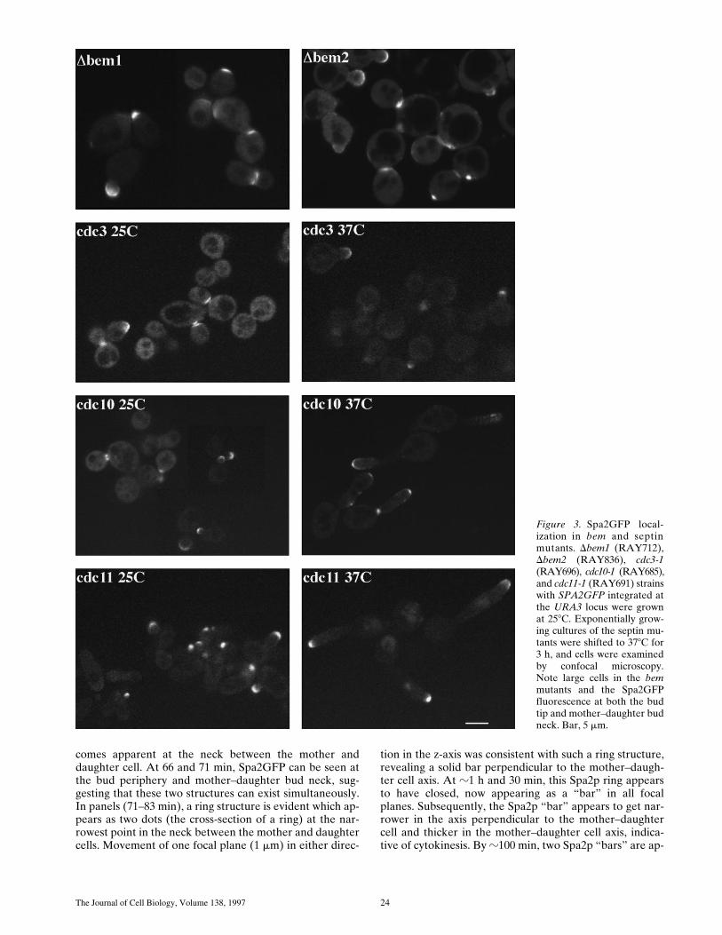
Amino-terminal truncations (DA–DC) and carboxyl-ter-minal truncations (DZ–DX) demonstrate that the aminoterminus, the coiled-coil region (residues 286–378), andthe nonameric amino acid repeat region (residues 816–1,087) are not required for Spa2GFP localization in vivo,indicating that the Spa2 box is necessary for correct local-ization. Strikingly, removal of the last 38 amino acids fromDX, resulting in DW which deletes the carboxyl-terminal 25amino acids of the Spa2 box, results in a fusion that doesnot localize (Fig. 5 A), defining the carboxyl-terminalboundary of the Spa2p localization domain to amino acidresidues 511–549. Furthermore, Spa2GFP lacking the B–Eregion (containing the coiled-coil and Spa2 box) or lackingthe C–E region (largely the Spa2 box) did not localize(data not shown). To address whether the 152-residueSpa2 box, defined maximally by residues 397–549, wassufficient for localization, we made three constructs con-sisting of the coiled-coil region and Spa2 box (DBX), thecoiled-coil region alone (DBV), and the Spa2 box alone(DCX) fused to GFP. Both constructs containing the Spa2box, BX and CX, localized correctly, whereas the coiled-coil region alone (BV) did not localize to regions ofgrowth. We were unable to detect expression of anySpa2GFP fusions in yeast by immunoblot analyses (usinganti-GFP); however, all of the constructs that did not lo-calize correctly were nonetheless expressed as seen by thesubstantial cytoplasmic fluorescence (Fig. 5 A). Alto-gether, these results show that the Spa2 box alone is neces-sary and sufficient for Spa2p bud tip and mother–daughterneck localization. Similar patterns of localization were ob-served in the spa2 mutant (pea1, strain JC-J9) isolatedby Chenevert et al. (1994) as well as a Dspa2 strain(RAY574), which is deleted for the entire SPA2 coding re-gion, demonstrating that the Spa2 box does not localize tosites of polarized growth via its interactions with Spa2p,i.e., oligomerization. Furthermore, we have not detectedany interactions between Spa2p and itself using these vari-ous constructs by two-hybrid assays (Orger, M., and R.Arkowitz, unpublished data).
We also analyzed this collection of Spa2GFP truncationand deletion mutants for their function in cell mating. Weexamined strains carrying these Spa2GFP fusions to deter-mine localization in cells exposed to high pheromone con-centration, their ability to complement shmoo formationand morphology defects of a Dspa2 mutant, and their abil-ity to complement the mating defect of a Dspa2 mutant.Spa2GFP localization in pheromone-treated cells and theability of various constructs to complement shmoo forma-tion defects were both carried out in a Mata Dspa2 strain(RAY578), and SPA2 mating function was assesed in Mataspa2 strains (JC-J9) by quantitative mating assays (Chen-evert et al., 1994). All experiments were carried out withintegrated copies of truncation or deletion constructs.
Localization and shmoo formation assays were carriedout by treating cells with high pheromone concentration(12.9 mM) for 2 h followed by fixation. These pheromoneconcentrations have been shown by Dorer et al. (1995) tosaturate the pheromone response pathway and shmoo for-mation. It was further shown that these saturating levels ofpheromone resulted in the execution of a default matingpathway. We chose such high concentrations of phero-mone both to increase shmoo formation and because SPA2
has been shown to be required for this default matingpathway (Dorer et al., 1995). Spa2GFP localization was as-sessed in Dspa2 cells by fluorescence and confocal micros-copy. Table III shows that all constructs that localized tosites of polarized growth in budding cells localize to theshmoo tip. While none of the Spa2GFP truncations exam-ined fully complemented the shmoo formation defect, DC,DD, and DY partially complemented this defect. These re-sults suggest that a region substantially larger than theSpa2 box is required for Spa2p-mediated shmoo formationduring the default mating pathway. Furthermore, the DXtruncation localizes to the shmoo tip, yet it does not com-plement the shmoo formation defect, consistent with dif-ferent requirements for these two processes.
The mating assays involved mixing equal amounts of anenfeebled tester strain with spa2 mutant JC-J9 carryingSPA2GFP constructs, allowing them to mate for 4 h at308C, and then determining mating efficiency by plating onnonselective and selective media. This functional assaywas not affected by SPA2 copy number (data not shown).The results of mating assays are presented in Fig. 5 B andsummarized in Fig. 4. The carboxyl-terminal two-thirds ofSpa2p, including the nonameric amino acid repeat do-main, is not necessary for mating function. Deletion DX,which removes amino acids 655–549, results in a fivefolddecrease in mating efficiency, and further truncationsdrastically reduce mating efficiency to background levels,implicating this region in SPA2 mating function. Theamino terminus of Spa2p has a greater role in mating func-tion with a truncation of as little as 88 amino acid residues(DA), resulting in a reduction in mating efficiency to back-ground levels, despite DA being expressed and localizingcorrectly (DA; Fig. 5 A). Together these results indicatethat the Spa2 box also plays a role in mating function be-ing necessary but not sufficient for mating. In addition, thefirst 90 amino acid residues are essential for mating func-tion. The Spa2p requirements for shmoo formation duringthe default mating pathway appear to overlap but are notidentical to the requirements for cell mating. For example,DC is partially functional in the shmoo formation assay,yet it is not functional for cell mating. These differencesmost likely reflect the roles of SPA2 in the different mat-ing processes: normal mating along a pheromone gradient,and the default mating pathway in high pheromone con-
Table III. Shmoo Formation and Localization of Spa2GFP Deletion Mutants
SPA2 constructPercentage of
shmoos*Shmoo tip
localization
%
SPA2GFP 83 1
DC 63 1
DD 66 2
DY 66 1
DX 54 1
DW 52 2
2 58 2
Dspa2 cells (RAY578) with the respective SPA2GFP deletion constructs integrated atURA3 were treated with a-factor and quantitated as described in Materials and Meth-ods. For the percentage of shmoos, 250 cells from each strain were counted. Shmootip localization was determined on fixed cells using confocal microscopy.*Peanut- and pear-shaped shmoos were designated as shmoos.
Arkowitz and Lowe Spa2p Polarized Localization 29
centration and the absence of a pheromone gradient. Ourresults suggest that Spa2p must be correctly localized (viathe Spa2 box) for mating function and, in addition, theSpa2p amino terminus is necessary for mating function.Consistent with this interpretation, constructs consisting ofthe coiled-coil region and Spa2 box (DBX) alone or theSpa2 box alone (DCX), both of which localize correctly,were not functional in the mating assay. Conversely,Spa2GFP lacking either the B–D region (containing thecoiled-coil and Spa2 box) or the C–D region (largely theSpa2 box), neither of which localized correctly, also werenonfunctional in mating (data not shown). The amino ter-minus of Spa2p, which is essential for mating, and the Spa2box, which is required for localization, are both conservedin ORF YSCL8543.8 (32% and 42% identity, respec-tively), suggesting common functions and localizations forthese two proteins.
SPH1, a Yeast Gene Containing the Spa2p Localization and Function Domains
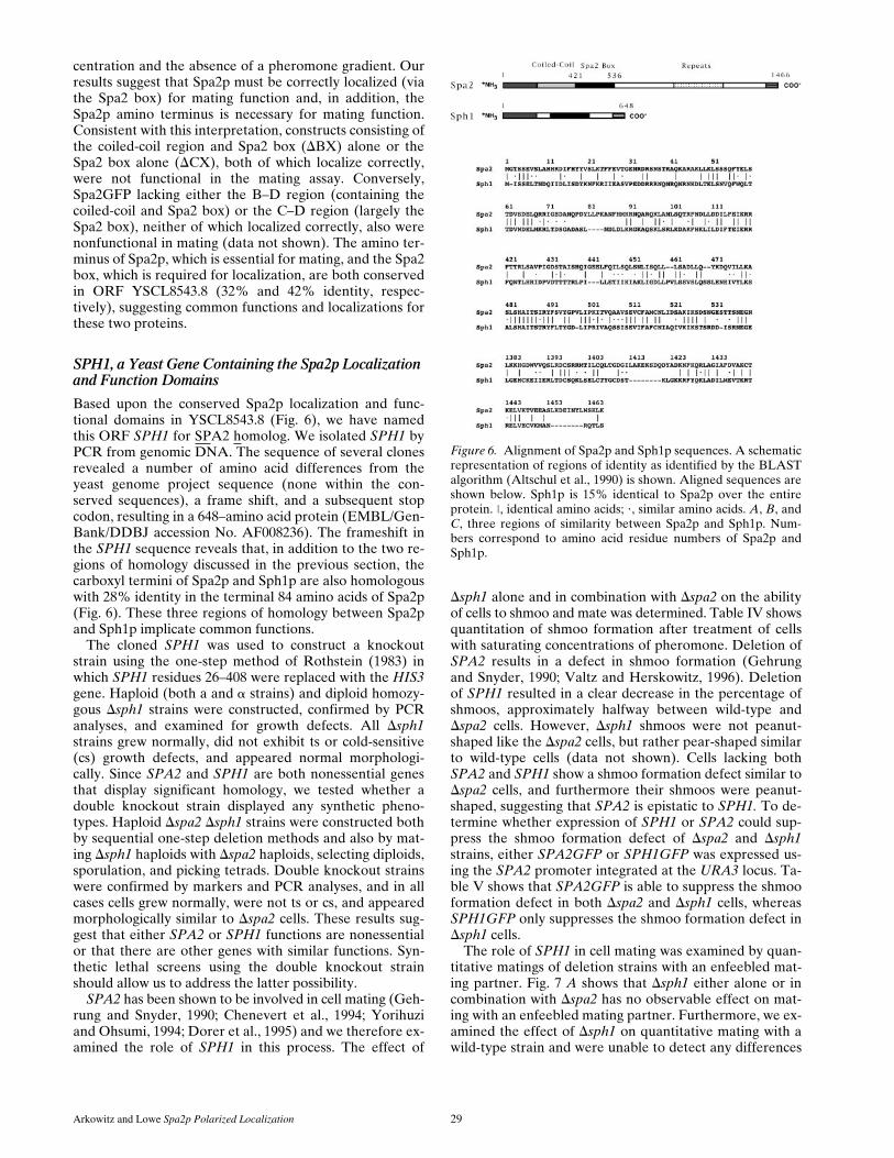
Based upon the conserved Spa2p localization and func-tional domains in YSCL8543.8 (Fig. 6), we have namedthis ORF SPH1 for SPA2 homolog. We isolated SPH1 byPCR from genomic DNA. The sequence of several clonesrevealed a number of amino acid differences from theyeast genome project sequence (none within the con-served sequences), a frame shift, and a subsequent stopcodon, resulting in a 648–amino acid protein (EMBL/Gen-Bank/DDBJ accession No. AF008236). The frameshift inthe SPH1 sequence reveals that, in addition to the two re-gions of homology discussed in the previous section, thecarboxyl termini of Spa2p and Sph1p are also homologouswith 28% identity in the terminal 84 amino acids of Spa2p(Fig. 6). These three regions of homology between Spa2pand Sph1p implicate common functions.
The cloned SPH1 was used to construct a knockoutstrain using the one-step method of Rothstein (1983) inwhich SPH1 residues 26–408 were replaced with the HIS3gene. Haploid (both a and a strains) and diploid homozy-gous Dsph1 strains were constructed, confirmed by PCRanalyses, and examined for growth defects. All Dsph1strains grew normally, did not exhibit ts or cold-sensitive(cs) growth defects, and appeared normal morphologi-cally. Since SPA2 and SPH1 are both nonessential genesthat display significant homology, we tested whether adouble knockout strain displayed any synthetic pheno-types. Haploid Dspa2 Dsph1 strains were constructed bothby sequential one-step deletion methods and also by mat-ing Dsph1 haploids with Dspa2 haploids, selecting diploids,sporulation, and picking tetrads. Double knockout strainswere confirmed by markers and PCR analyses, and in allcases cells grew normally, were not ts or cs, and appearedmorphologically similar to Dspa2 cells. These results sug-gest that either SPA2 or SPH1 functions are nonessentialor that there are other genes with similar functions. Syn-thetic lethal screens using the double knockout strainshould allow us to address the latter possibility.
SPA2 has been shown to be involved in cell mating (Geh-rung and Snyder, 1990; Chenevert et al., 1994; Yorihuziand Ohsumi, 1994; Dorer et al., 1995) and we therefore ex-amined the role of SPH1 in this process. The effect of
Dsph1 alone and in combination with Dspa2 on the abilityof cells to shmoo and mate was determined. Table IV showsquantitation of shmoo formation after treatment of cellswith saturating concentrations of pheromone. Deletion ofSPA2 results in a defect in shmoo formation (Gehrungand Snyder, 1990; Valtz and Herskowitz, 1996). Deletionof SPH1 resulted in a clear decrease in the percentage ofshmoos, approximately halfway between wild-type andDspa2 cells. However, Dsph1 shmoos were not peanut-shaped like the Dspa2 cells, but rather pear-shaped similarto wild-type cells (data not shown). Cells lacking bothSPA2 and SPH1 show a shmoo formation defect similar toDspa2 cells, and furthermore their shmoos were peanut-shaped, suggesting that SPA2 is epistatic to SPH1. To de-termine whether expression of SPH1 or SPA2 could sup-press the shmoo formation defect of Dspa2 and Dsph1strains, either SPA2GFP or SPH1GFP was expressed us-ing the SPA2 promoter integrated at the URA3 locus. Ta-ble V shows that SPA2GFP is able to suppress the shmooformation defect in both Dspa2 and Dsph1 cells, whereasSPH1GFP only suppresses the shmoo formation defect inDsph1 cells.
The role of SPH1 in cell mating was examined by quan-titative matings of deletion strains with an enfeebled mat-ing partner. Fig. 7 A shows that Dsph1 either alone or incombination with Dspa2 has no observable effect on mat-ing with an enfeebled mating partner. Furthermore, we ex-amined the effect of Dsph1 on quantitative mating with awild-type strain and were unable to detect any differences
Figure 6. Alignment of Spa2p and Sph1p sequences. A schematicrepresentation of regions of identity as identified by the BLASTalgorithm (Altschul et al., 1990) is shown. Aligned sequences areshown below. Sph1p is 15% identical to Spa2p over the entireprotein. |, identical amino acids; ?, similar amino acids. A, B, andC, three regions of similarity between Spa2p and Sph1p. Num-bers correspond to amino acid residue numbers of Spa2p andSph1p.
The Journal of Cell Biology, Volume 138, 1997 30
(data not shown). Because of the sequence conservationbetween Spa2p and Sph1p in two separate regions neces-sary for Spa2p mating function, we determined if SPH1could suppress the mating defect in Dspa2 mutants. Fig. 7 Bshows that expression of SPH1GFP using the SPA2 pro-moter integrated at the URA3 locus in a Dspa2 strain re-sulted in a substantial increase in mating efficiency with anenfeebled mating partner. In addition, overexpression ofSPH1 from a multicopy (2 mm) plasmid using the SPH1promoter also resulted in an increase in mating efficiencywith an enfeebled mating partner (data not shown). Theseresults suggest that Sph1p is involved in mating because itresults in a defect in shmoo formation and can partiallysubstitute for Spa2p mating function, implying that the se-quence conservation between these two proteins is func-tionally significant.
The effect of SPH1 deletion on bud site selection was in-vestigated. Deletion of SPA2 has been shown to have noeffect on bud site selection in haploids; however, homozy-gous diploid deletion mutants are defective in bipolar bud-
ding (Snyder, 1989; Zahner et al., 1996; Valtz and Her-skowitz, 1996). Specifically, spa2 mutants are defective inbud site selection after the correct positioning of the firstbud. Dsph1 haploid cells budded in an axial pattern identi-cal to that of wild-type cells (data not shown). The bud-ding pattern of homozygous diploids was determined bycounting cells with two or more bud scars and determiningthe position of the bud relative to the birth scar (Fig. 7, in-set). Fig. 7 C shows that SPH1 is required for bipolar budsite selection in diploids similar to SPA2. HomozygousDsph1 diploids were able to correctly position the firstbud, similar to spa2 mutants. The double homozygous dip-loid mutant Dsph1 Dspa2 showed a similar random budsite selection defect. Fig. 7 D shows representative pic-tures of Calcofluor staining of bud scars of the individualand double homozygous diploid deletions. It appears thatthe Dsph1 bud site selection defect is less random than thatof Dspa2; however, further analyses are required. Similarto the ability of SPA2GFP to suppress the Dsph1 shmooformation defect, SPA2GFP suppresses the bud site selec-tion defect of homozygous Dsph1 diploids. Together theseresults demonstrate that SPH1 is required for bipolar bud-ding after the positioning of the first bud and, togetherwith shmoo formation and mating experiments, show thatSPH1 and SPA2 have overlapping functions.
The conservation of the Spa2 box that is necessary andsufficient for localization to sites of polarized growth inSph1p and the similar functions of SPH1 and SPA2prompted us to examine the localization of Sph1p. To de-termine the localization of Sph1p, we used an SPH1GFPfusion that was driven by the SPA2 promoter, and thisconstruct was integrated at the URA3 locus. Fig. 8 A showsthe in vivo localization of Sph1GFP, which is strikinglysimilar to Spa2p localization. These experiments demon-strate that Sph1p also localizes to sites of polarizedgrowth: at the site where the bud will form, the bud tip,and the mother–daughter bud neck. Similar localization wasobserved in haploids of the opposite mating type (Mata)and diploids, indicating that Sph1p localization is not celltype specific. Furthermore, Sph1GFP localized to sites ofpolarized growth in Dsph1, Dspa2, and Dsph1 Dspa2 cells,demonstrating that neither SPH1 nor SPA2 is requiredfor Sph1GFP localization (data not shown). In addition,Sph1GFP localized to shmoo tips in Mata Dsph1 cells thathad been treated with saturating concentrations of matingpheromone (Fig. 8 B). Together these results suggest thatthe Spa2 box in Sph1p is sufficient for localization to po-larized growth during both budding and mating.
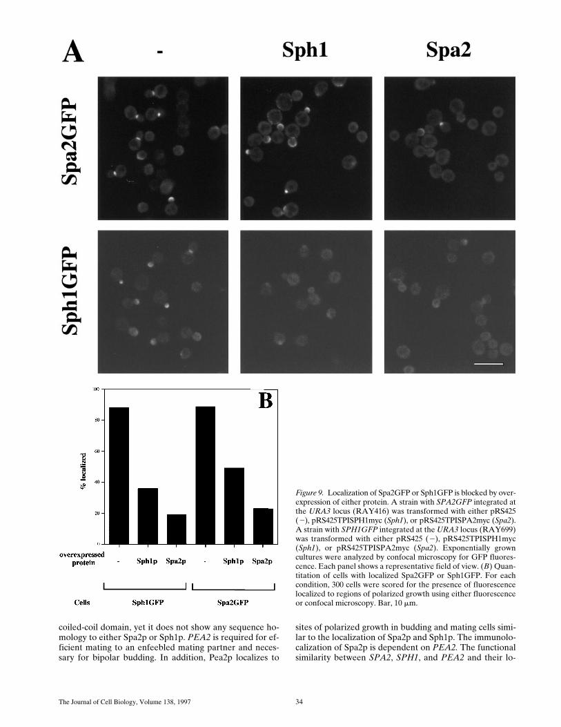
We also examined whether the localization of Spa2GFPor Sph1GFP could be blocked or competed out by over-expression of either protein. Cross competition of oneprotein for the other would suggest that these two proteinsbind to the same site in vivo. Epitope-tagged Spa2p andSph1p were overexpressed using a TPI promoter on a 2-mmmulticopy plasmid in strains with either integrated SPA2GFPor SPH1GFP (Fig. 9 A). Overexpression of either Spa2por Sph1p blocks localization of Spa2GFP. Conversely,overexpression of either Sph1p or Spa2p blocks localiza-tion of Sph1GFP. Immunoblot analyses demonstrated thatboth Spa2myc and Sph1myc were expressed (data notshown). Quantitation of the number of cells with localizedSpa2GFP or Sph1GFP revealed that overexpressed Spa2myc
Table IV. Shmoo Formation of Dspa2 and Dsph1 Mutants
Strain Percentage of shmoos*
%
WT 84WT 80
Dspa2 58Dspa2 58
Dsph1 74Dsph1 70
Dspa2 Dsph1 64Dspa2 Dsph1 63
Cells (SEY6211, RAY578, RAY567, and RAY590) were treated with a-factor andquantitated as described in Materials and Methods. Two independent isolates of eachstrain were used and for each isolate 250 cells were counted.*Peanut- and pear-shaped shmoos were designated as shmoos.
Table V. SPA2GFP Complements Shmoo Formation Defect and Bud Site Selection Defect of Dsph1 Mutant
Strain Integrated genePercentage of
shmoos*Buddingpattern
%
WT 2 77 BipolarWT SPA2GFP 78 NDWT SPA1GFP 76 ND
Dspa2 – 58 RandomDspa2 SPA2GFP 83 BipolarDspa2 SPH1GFP 55 Random
Dsph1 2 66 RandomDsph1 SPA2GFP 75 BipolarDsph1 SPH1GFP 79 Bipolar
Cells were treated with a-factor and quantitated as described in Materials and Meth-ods. Integrants were selected that showed localized fluorescence. For percentage ofshmoos, 250 Mata haploid cells from each strain were counted. Bud site selection wasdetermined using Calcofluor staining on homozygous diploid cells as described inMaterials and Methods.*Peanut- and pear-shaped shmoos were designated as shmoos.
Arkowitz and Lowe Spa2p Polarized Localization 31
blocked Spa2GFP or Sph1GFP localization more effec-tively than overexpressed Sph1myc (Fig. 9 B). Most cells(89%) showed localized Spa2GFP, whereas the number ofcells with localized Spa2GFP was reduced twofold in thepresence of overexpressed Sph1myc and reduced fourfoldin the presence of overexpressed Spa2myc. Conversely,88% of the cells had localized Sph1GFP that was reducedthreefold in the presence of overexpressed Sph1myc andreduced fourfold in the presence of overexpressed Spa2myc.These results are consistent with ability of SPA2GFP tofunctionally replace Dsph1 in shmoo formation and bipo-lar bud site selection, whereas SPH1GFP is unable to re-place Dspa2 function in shmoo formation and bipolar budsite selection. This cross competition of localization is con-
sistent with the notion that these two proteins may be lo-calized by the same cellular component and this interac-tion can be competed out by either protein.
DiscussionOur studies with Spa2GFP demonstrate that Spa2p local-izes to the bud tip, forming a cup-like crescent, and thebud neck between mother and daughter cell, forming a do-nut-like ring. These two localizations are distinct eventsthat follow one another during the cell cycle. The changesin shape and localization of these structures occur on theminute time scale. In septin mutants (cdc3, cdc10, and cdc11)at the nonpermissive temperature, Spa2GFP is localized
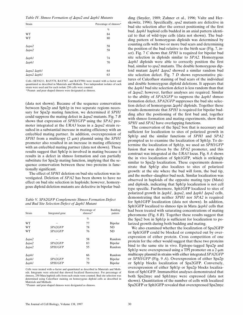
Figure 7. (A) Deletion of sph1 has no effect on mating efficiency.Exponential cultures of WT (RAY697), Dspa2 (RAY698), Dsph1(RAY709), and Dspa2 Dsph1 (RAY711) were mated with an en-feebled mating tester strain (JY426) as described in Materials andMethods, and diploids were selected on 2lys, 2ura plates. Wild-type (WT) mating efficiency (4.4%) was set to 100% efficiency. (B)SPH1 can partially complement for mating deficiency of Dspa2(RAY574). WT (RAY697), Dspa2 with SPH1GFP (RAY703),and Dspa2 (RAY698) were mated with an enfeebled matingtester strain (JY426) as described in Materials and Methods, anddiploids were selected on 2lys, 2ura plates. WT mating effi-ciency (6.5%) was normalized to 100% efficiency. (C) SPH1 is re-quired for bipolar bud site selection. Wild-type (SEY6210/11),homozygous Dspa2 (RAY616), homozygous Dsph1 (RAY618),and homozygous Dspa2 Dsph1 (RAY620) exponentially growingdiploids were stained with Calcofluor as described in Materialsand Methods, and budding pattern was analyzed. For each strain,the position of the bud relative to the birth scar (see inset) wasscored for z150 cells with two or more bud scars. (D) Buddingpattern of homozygous diploids sph1 and spa2 mutants. Repre-sentative fluorescence microscopy pictures of cells quantitated in C.Bar, 2.5 mm.
The Journal of Cell Biology, Volume 138, 1997 32

Arkowitz and Lowe Spa2p Polarized Localization 33
only to the bud tip, while the bud emergence mutantsbem1 and bem2 have no effect on Spa2GFP localization.We have identified a small domain in Spa2p that is bothnecessary and sufficient for localization to sites of polar-ized growth in vivo. This 150–amino acid residue domaincan target a heterologous protein to the sites of polarizedcell growth in yeast. In addition to the amino-terminal 100amino acids, the Spa2 box is also essential for Spa2p mat-ing function, suggesting that Spa2p must be correctly local-ized to be functional. We have identified a novel protein,Sph1p, in which both the Spa2p localization domain andamino terminus are conserved. Deletion of SPH1 resultsin a defect in shmoo formation and a random bud site se-lection pattern in diploids. In addition, SPH1 can partiallysuppress the mating defect of Dspa2 cells and localizes tothe same regions of polarized growth as Spa2p. Togetherthese two proteins with similar functions constitute a novelprotein family involved in polarized cell growth and ap-pear to localize to regions of polarized growth via an inter-action with the same cellular component.
Spa2p Dynamics and Localization
The biological and spectral characteristics of GFP haveenabled the analyses of Spa2p localization in vivo duringnormal cell growth and specifically have shed light on Spa2pdynamics. In contrast with actin cortical patch movements(Doyle and Botstein, 1996; Waddle et al., 1996), Spa2p-containing structures change shape and localization on amuch slower time scale. These movements could be due tosubcellular reorganizations or association/dissociation re-actions. While the ring of Spa2p at the mother–daughterbud neck is reminiscent of the septin ring (Kim et al., 1991;Ford and Pringle, 1991; Haarer and Pringle, 1987), it dif-fers in that it does not appear as two rings, like the septinrings, until the cells have divided. The septins appear astwo separate rings flanking the mother–daughter constric-tion in cells with large buds that are dividing, whereasSpa2GFP appears as a single ring until cell division andthereafter stays at the site of division in both cells.
What component localizes Spa2p to such specific re-gions and maintains this localization? Spa2p is one of thefirst proteins to localize to the site of the new bud (Snyder,199l; Snyder et al., 1991) at approximately the same timeas actin patches concentrate at the region of the incipientbud (Snyder et al., 1991). However, in budding cells whenactin patches are delocalized, Spa2p localization is normal(Snyder et al., 1991), indicating that Spa2p localization isnot dependent on a polarized cytoskeleton. Experimentsusing septin mutants, in which the neck filaments do notform at the nonpermissive temperature (Byers and Goetsch,1976; Kim et al., 1991; Ford and Pringle, 1991; Haarer andPringle, 1987), show that Spa2p is still localized to the budtip. These results suggest that the septins are not requiredfor maintaining this polarized localization. Our demon-
stration that a small domain of Spa2p is necessary and suf-ficient for this localization raises the attractive possibilitythat this region is binding to a receptor that marks the newbud. Consistent with the notion of a receptor is the resultthat Spa2p localization appears to be saturable. Further-more, Spa2GFP or Sph1GFP localization can be competedout by either Spa2p or Sph1p overexpression. While anumber of proteins localize to very similar regions asSpa2p and Sph1p, possible candidates for such a receptorinclude the polytopic membrane protein Fks1p, which en-codes a b(1→3)glucan synthase (Qadota et al., 1996), andAxl2p/Bud10p (Roemer et al., 1996; Halme et al., 1996),which encodes a type I single transmembrane protein,both of which localize to similar regions as Spa2p. Wehave begun to examine proteins involved in localizing Spa2pby screening for mislocalization mutants. Preliminary re-sults with ethylmethane sulfonate mutagenized cells indi-cate that it should be possible to identify and analyze suchmutants using Spa2GFP and a visual screen (Lowe, N.,and R. Arkowitz, unpublished data). Identification of pro-teins interacting with Spa2p and involved in its early local-ization will be greatly facilitated by the definition of a min-imal Spa2p domain that localizes correctly.
SPA2 and SPH1 Function
By sequence comparisons we have identified a homologueof SPA2 that contains both a region required for matingfunction and a localization domain necessary and suffi-cient for polarized growth localization. Furthermore, atpositions 2234 to 2239 in the promoter of SPA2 is thematch to a MluI cell cycle box (MCB), which also includesan imperfect MluI site typically found adjacent to the MluIsite. The position of this cell cycle box is in the range 2100to 2250 upstream of the start ATG where MCBs are typi-cally found (Johnston et al., 1991; McIntosh et al., 1991).SPH1 has an identical MCB (including adjacent imperfectMluI site) 2155 to 2160 in its potential promoter (Whiteet al., 1986; Kilmartin et al., 1993; Yamamoto et al., 1996).SPA2 and SPH1 have similar functions, with deletion ofeither gene resulting in a defect in shmoo formation at sat-urating pheromone concentration and random bud site se-lection in diploids. Furthermore, overexpression of SPH1can partially complement the mating defect of a Dspa2 strain,and overexpression of SPA2 can complement the shmooformation and bipolar bud site selection defect of Dsph1cells. Deletion of SPA2 results in a stronger defect in shmooformation and morphology at saturating pheromone con-centration than in Dsph1, and the double mutant appearsphenotypically similar to the Dspa2 mutant, suggestingthat SPA2 is epistatic to SPH1.
Recently, Valtz and Herskowitz (1996) cloned and char-acterized PEA2, which was originally identified as a mu-tant that formed a peanut-shaped shmoo in the presenceof mating pheromone. This protein contains a predicted
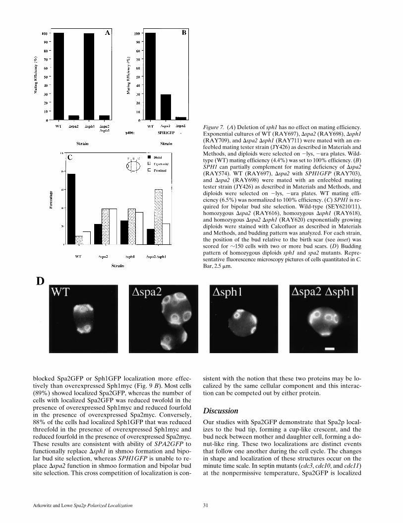
Figure 8. (A) Sph1p localizes to sites of polarized growth in budding cells. Exponentially grown cells with SPH1GFP integrated atURA3 (RAY699) were analyzed by confocal microscopy. Immunoblot analyses of RAY699 revealed that a GFP fusion protein of thecorrect molecular mass (z100 kD) was expressed. (B) Sph1p localizes to sites of polarized growth in shmoos. Exponentially grown MataDsph1 cells with SPH1GFP integrated at URA3 (RAY875) were treated with a-factor, as described in Materials and Methods, fixed, andviewed by confocal microscopy. Note that background fluorescence in cells is due to Ade fluorophore from ade2 mutation. Bar, 5 mm.
The Journal of Cell Biology, Volume 138, 1997 34
Figure 9. Localization of Spa2GFP or Sph1GFP is blocked by over-expression of either protein. A strain with SPA2GFP integrated atthe URA3 locus (RAY416) was transformed with either pRS425(2), pRS425TPISPH1myc (Sph1), or pRS425TPISPA2myc (Spa2).A strain with SPH1GFP integrated at the URA3 locus (RAY699)was transformed with either pRS425 (2), pRS425TPISPH1myc(Sph1), or pRS425TPISPA2myc (Spa2). Exponentially growncultures were analyzed by confocal microscopy for GFP fluores-cence. Each panel shows a representative field of view. (B) Quan-titation of cells with localized Spa2GFP or Sph1GFP. For eachcondition, 300 cells were scored for the presence of fluorescencelocalized to regions of polarized growth using either fluorescenceor confocal microscopy. Bar, 10 mm.
coiled-coil domain, yet it does not show any sequence ho-mology to either Spa2p or Sph1p. PEA2 is required for ef-ficient mating to an enfeebled mating partner and neces-sary for bipolar budding. In addition, Pea2p localizes to
sites of polarized growth in budding and mating cells simi-lar to the localization of Spa2p and Sph1p. The immunolo-calization of Spa2p is dependent on PEA2. The functionalsimilarity between SPA2, SPH1, and PEA2 and their lo-
Arkowitz and Lowe Spa2p Polarized Localization 35
calization to similar sites of polarized growth suggest thatthese proteins constitute a protein family. Interestingly, allthree of these genes function in bipolar bud site selection,shmoo formation, and cell mating. Both Pea2p and Spa2phave potential coiled-coil domains, whereas both Sph1pand Spa2p have the Spa2 box necessary for polarized lo-calization. An additional member of this family may beBUD6 (Zahner et al., 1996), which results in a similar bi-polar bud site selection defect. Analysis of Bud6p se-quence using the Lupas program (Lupas et al., 1991) alsoreveals a region with high probability of forming a coiled-coil domain. The localization as well as shmooing and mat-ing defects of bud6 mutants should help determine whetherit is a member of this family.
What is the function of this protein family? We proposethese proteins may be involved in initiating and maintain-ing the organization of the large number of proteinspresent at sites of polarized growth sites (in a sense, chap-erones for polarized growth), and that there must be anumber of additional components with such functions. It ispossible that CDC10 links such scaffolds to the septin ringinitially (Flescher et al., 1993); however, additional pro-teins are required to maintain the localization of theseproteins at the bud site. Further genetic and biochemicalexperiments will certainly shed light on the functions ofSpa2p and Sph1p. We have identified a saturable site thatlocalizes both Spa2p and Sph1p very early in the processof bud formation and a minimal domain of Spa2p that isable to localize to these sites of polarized growth. Thisminimal domain will be crucial for the identification of ad-ditional components involved in the early stages of cell po-larization.
We thank I. Herskowitz, M. Snyder, R. Mortimer, S. Emr, J. Haseloff, K.Siemering, and A. Wach for yeast strains and plasmids; H. Pelham, S. Munro,J. Kilmartin, and M. Bassilana for helpful discussions and critical readingof the manuscript; and A. Nern for cloning BEM1 and assistance withABI sequencing.
Received for publication 26 August 1996 and in revised form 1 May 1997.
Note Added in Proof. Bud6p has recently been shown to localize to sitesof polarized growth (Evangelista, M., K. Blundell, M.S. Longtine, C.J.Chow, N. Adames, J.R. Pringle, M. Peter, and C. Boone. 1997. Bni1p, ayeast formin linking cdc42p and the actin cytoskeleton during polarizedmorphogenesis. Science (Wash. DC). 276:118–122).
References
Adams, A.E., and J.R. Pringle. 1984. Relationship of actin and tubulin distribu-tion to bud growth in wild-type and morphogenetic-mutant Saccharomycescerevisiae. J. Cell Biol. 98:934–945.
Altschul, S.F., W. Gish, W. Miller, E.W. Myers, and D.J. Lipman. 1990. Basiclocal alignment search tool. J. Mol. Biol. 215:403–410.
Amatruda, J.F., and J.A. Cooper. 1992. Purification, characterization, and im-munofluorescence localization of Saccharomyces cerevisiae capping protein.J. Cell Biol. 117:1067–1076.
Anderson, K. 1995. One signal, two body axes. Science (Wash. DC). 269:489–490.Baudin, A., O. Ozier-Kalogeropoulos, A. Denouel, R. Lacroute, and C. Cullin.
1993. A simple and efficient method for direct gene deletion in Saccharomy-ces cerevisiae. Nucleic Acids Res. 21:3329–3330.
Bender, A., and J.R. Pringle. 1991. Use of a screen for synthetic lethal and mul-ticopy suppressor mutants to identify two new genes involved in morphogen-esis in Saccharomyces cerevisiae. Mol. Cell. Biol. 11:1295–1305.
Byers, B., and L. Goetsch. 1976. A highly ordered ring of membrane-associatedfilaments in budding yeast. J. Cell Biol. 69:717–721.
Chant, J. 1996. Generation of cell polarity in yeast. Curr. Opin. Cell Biol. 8:557–565.Chant, J., K. Corrado, J.R. Pringle, and I. Herskowitz. 1991. Yeast BUD5, en-
coding a putative GDP-GTP exchange factor, is necessary for bud site selec-tion and interacts with bud formation gene BEM1. Cell. 65:1213–1224.
Chant, J., M. Mischke, E. Mitchell, I. Herskowitz, and J.R. Pringle. 1995. Role
of Bud3p in producing the axial budding pattern of yeast. J. Cell Biol. 129:767–778.
Chenevert, J. 1994. Cell polarization directed by extracellular cues in yeast.Mol. Biol. Cell. 5:1169–1175.
Chenevert, J., K. Corrado, A. Bender, J. Pringle, and I. Herskowitz. 1992. Ayeast gene (BEM1) necessary for cell polarization whose product containstwo SH3 domains. Nature (Lond.). 356:77–79.
Chenevert, J., N. Valtz, and I. Herskowitz. 1994. Identification of genes re-quired for normal pheromone-induced cell polarization in Saccharomycescerevisiae. Genetics. 136:1287–1296.
Cohen, S., and A.A. Hyman. 1994. Cell fate determination. When is a determi-nant a determinant? Curr. Biol. 4:420–422.
Cooper, J.A., and D.P. Kiehart. 1996. Septins may form a ubiquitous family ofcytoskeletal filaments. J. Cell Biol. 134:1345–1348.
Dorer, R., P.M. Pryciak, and L.H. Hartwell. 1995. Saccharomyces cerevisiaecells execute a default pathway to select a mate in the absence of pheromonegradients. J. Cell Biol. 131:845–861.
Doyle, T., and D. Botstein. 1996. Movement of yeast cortical actin cytoskeletonvisualized in vivo. Proc. Natl. Acad. Sci. USA. 93:3886–3891.
Drubin, D.G., and W.J. Nelson. 1996. Origins of cell polarity. Cell. 84:335–344.Drubin, D.G., K.G. Miller, and D. Botstein. 1988. Yeast actin-binding proteins:
evidence for a role in morphogenesis. J. Cell Biol. 107:2551–2561.Eaton, S., and K. Simons. 1995. Apical, basal, and lateral cues for epithelial po-
larization. Cell. 82:5–8.Farkas, V., J. Kovarik, A. Kosinova, and S. Bauer. 1974. Autoradiographic
study of mannan incorporation into growing walls of Saccharomyces cerevi-siae. J. Bacteriol. 117:265–269.
Field, C.M., O. Alawar, J. Rosenblatt, M.L. Wong, B. Alberts, and T.J. Mitchi-son. 1996. A purified Drosophila septin complex forms filaments and exhib-its GTPase activity. J. Cell Biol. 133:605–616.
Flescher, E.G., K. Madden, and M. Snyder. 1993. Components required for cy-tokinesis are important for bud site selection in yeast. J. Cell Biol. 122:373–386.
Ford, S.K., and J.R. Pringle. 1991. Cellular morphogenesis in the Saccharomy-ces cerevisiae cell cycle: localization of the CDC11 gene product and the tim-ing of events at the budding site. Dev. Genet. 12:281–292.
Freifelder, D. 1960. Bud position in Saccharomyces cerevisiae. J. Bacteriol. 80:567–568.
Gehrung, S., and M. Snyder. 1990. The SPA2 gene of Saccharomyces cerevisiaeis important for pheromone-induced morphogenesis and efficient mating. J.Cell Biol. 111:1451–1464.
Goldstein, B., S.N. Hird, and J.G. White. 1993. Cell polarity in early C. elegansdevelopment. Dev. Suppl. 279–287.
Haarer, B.K., and J.R. Pringle. 1987. Immunofluorescence localization of theSaccharomyces cerevisiae CDC12 gene product to the vicinity of the 10-nmfilaments in the mother-bud neck. Mol. Cell. Biol. 7:3678–3687.
Halme, A., M. Michelitch, E.L. Mitchell, and J. Chant. 1996. Bud10p directs ax-ial cell polarization in budding yeast and resembles a transmembrane recep-tor. Curr. Biol. 6:570–579.
Heim, R., A.B. Cubitt, and R.Y. Tsien. 1995. Improved green fluorescence [let-ter]. Nature (Lond.). 373:663–664.
Hicks, J.B., J.N. Strathern, and I. Herskowitz. 1977. Interconversion of yeastmating types. III. Action of the homothallism (HO) gene in cells homozy-gous for the mating type locus. Genetics. 85:395–405.
Horvitz, H.R., and I. Herskowitz. 1992. Mechanisms of asymmetric cell divi-sion: two Bs or not two Bs, that is the question. Cell. 68:237–255.
Jackson, C.L., and L.H. Hartwell. 1990a. Courtship in S. cerevisiae: both celltypes choose mating partners by responding to the strongest pheromone sig-nal. Cell. 63:1039–1051.
Jackson, C.L., and L.H. Hartwell. 1990b. Courtship in Saccharomyces cerevi-siae: an early cell-cell interaction during mating. Mol. Cell. Biol. 10:2202–2213.
Johnston, L.H., N.F. Lowndes, A.L. Johnson, and A. Sugino. 1991. A cell-cycle-regulated trans-factor, DSC1, controls expression of DNA synthesis genes inyeast. Cold Spring Harbor Symp. Quant. Biol. 56:169–176.
Keynes, R., and G.M. Cook. 1995. Axon guidance molecules. Cell. 83:161–169.Kilmartin, J.V., and A.E. Adams. 1984. Structural rearrangements of tubulin
and actin during the cell cycle of the yeast Saccharomyces. J. Cell Biol. 98:922–933.
Kilmartin, J.V., S.L. Dyos, D. Kershaw, and J.T. Finch. 1993. A spacer proteinin the Saccharomyces cerevisiae spindle poly body whose transcript is cell cy-cle-regulated. J. Cell Biol. 123:1175–1184.
Kim, H.B., B.K. Haarer, and J.R. Pringle. 1991. Cellular morphogenesis in theSaccharomyces cerevisiae cell cycle: localization of the CDC3 gene productand the timing of events at the budding site. J. Cell Biol. 112:535–544.
Kim, Y.-J., L. Francisco, G.-C. Chen, E. Marcotte, and C.S.M. Chan. 1994. Con-trol of cellular morphogenesis by the Ipl2/Bem2 GTPase-activating protein:possible role of protein phosphorylation. J. Cell Biol. 127:1381–1394.
Kimble, J. 1994. An ancient molecular mechanism for establishing embryonicpolarity? Science (Wash. DC). 266:577–578.
Kunkel, T.A. 1985. Rapid and efficient site-specific mutagenesis without phe-notypic selection. Proc. Natl. Acad. Sci. USA. 82:488–492.
Leeuw, T., A. Fourest-Lieuvin, C. Wu, J. Chenevert, K. Clark, M. Whiteway,D.Y. Thomas, and E. Leberer. 1995. Pheromone response in yeast: associa-tion of Bem1p with proteins of the MAP kinase cascade and actin. Science(Wash. DC). 270:1210–1213.
The Journal of Cell Biology, Volume 138, 1997 36
Lew, D.J., and S.I. Reed. 1993. Morphogenesis in the yeast cell cycle: regulationby Cdc28 and cyclins. J. Cell Biol. 120:1305–1320.
Lillie, S.H., and S.S. Brown. 1994. Immunofluorescence localization of the un-conventional myosin, Myo2p, and the putative kinesin-related protein,Smy1p, to the same regions of polarized growth in Saccharomyces cerevisiae.J. Cell Biol. 125:825–842.
Longtine, M.S., D.J. DeMarini, M.L. Valencik, O.S. Al-Awar, H. Fares, C. DeVirgilio, and J.R. Pringle. 1996. The septins: roles in cytokinesis and otherprocesses. Curr. Opin. Cell Biol. 8:106–119.
Lupas, A., M. Van Dyke, and J. Stock. 1991. Predicting coiled coils from pro-tein sequences. Science (Wash. DC). 252:1162–1164.
McIntosh, E.M., T. Atkinson, R.K. Storms, and M. Smith. 1991. Characteriza-tion of a short, cis-acting DNA sequence which conveys cell cycle stage-dependent transcription in Saccharomyces cerevisiae. Mol. Cell. Biol. 11:329–337.
Peterson, J., Y. Zheng, L. Bender, A. Myers, R. Cerione, and A. Bender. 1994.Interactions between the bud emergence proteins Bem1p and Bem2p andRho-type GTPases in yeast. J. Cell Biol. 127:1395–1406.
Priess, J.R. 1994. Establishment of initial asymmetry in early Caenorhabditis el-egans embryos. Curr. Opin. Genet. Dev. 4:563–568.
Pringle, J.R. 1991. Staining of bud scars and other cell wall chitin with Calco-fluor. Methods Enzymol. 194:732–735.
Qadota, H., C.P. Python, S.B. Inoue, M. Arisawa, Y. Anraku, Y. Zheng, T. Wa-tanabe, D.E. Levin, and Y. Ohya. 1996. Identification of yeast Rho1p GTP-ase as a regulatory subunit of 1,3-beta-glucan synthase. Science (Wash. DC).272:279–281.
Rhyu, M.S., and J.A. Knoblich. 1995. Spindle orientation and asymmetric cellfate. Cell. 82:523–526.
Rodriguez-Boulan, E., and W.J. Nelson. 1989. Morphogenesis of the polarizedepithelial cell phenotype. Science (Wash. DC). 245:718–725.
Roemer, T., K. Madden, J. Chang, and M. Snyder. 1996. Selection of axialgrowth sites in yeast requres Axl2p, a novel plasma membrane glycoprotein.Genes & Dev. 10:777–793.
Rose, M.D., F. Winston, and P. Hieter. 1991. Methods in Yeast Genetics: ALaboratory Course Manual. Cold Spring Harbor Press, Cold Spring Harbor,NY. 198 pp.
Rothstein, R.J. 1983. One-step gene disruption in yeast. Methods Enzymol. 101:202–211.
Segall, J.E. 1993. Polarization of yeast cells in spatial gradients of alpha matingfactor. Proc. Natl. Acad. Sci. USA. 90:8332–8336.
Siemering, K.R., R. Golbik, R. Sever, and J. Haseloff. 1996. Mutations that sup-press the thermosensitivity of green fluorescent protein. Curr. Biol. 6:1653–1663.
Sloat, B.G., A. Adams, and J.R. Pringle. 1981. Roles of CDC24 gene product incellular morphogenesis during Saccharomyces cerevisiae cell cycle. J. Cell
Biol. 89:395–405.Snyder, M. 1989. The SPA2 protein of yeast localizes to sites of cell growth. J.
Cell Biol. 108:1419–1429.Snyder, M., S. Gehrung, and B.D. Page. 1991. Studies concerning the temporal
and genetic control of cell polarity in Saccharomyces cerevisiae. J. Cell Biol.114:515–532.
St Johnston, D., and C. Nusslein-Volhard. 1992. The origin of pattern and po-larity in the Drosophila embryo. Cell. 68:201–219.
Tkacz, J.S., and J.O. Lampen. 1972. Wall replication in Saccharomyces species:use of fluorescein-conjugated concanavalin A to reveal the site of mannaninsertion. J. Gen. Microbiol. 72:243–247.
Valtz, N., and I. Herskowitz. 1996. Pea2 protein of yeast is localized to sites ofpolarized growth and is required for efficient mating and bipolar budding. J.Cell Biol. 135:725–739.
Wach, A., A. Brachat, R. Poehlmann, and P. Philippsen. 1994. New heterolo-gous modules for classical or PRC-based gene disruptions in Saccharomycescerevisiae. Yeast. 10:1793–1808.
Waddle, J.A., T.S. Karpova, R.H. Waterston, and J.A. Cooper. 1996. Move-ment of cortical actin patches in yeast. J. Cell Biol. 132:861–870.
Wang, T., and A. Bretscher. 1995. The rho-GAP encoded by BEM2 regulatescytoskeletal structure in budding yeast. Mol. Biol. Cell. 6:1011–1024.
White, J.H., D.G. Barker, P. Nurse, and L.H. Johnston. 1986. Periodic tran-scription as a means of regulating gene expression during the cell cycle: con-trasting modes of expression of DNA ligase genes in budding and fissionyeast. EMBO (Eur. Mol. Biol. Organ.) J. 5:1705–1709.
Yamamoto, A., V. Guacci, and D. Koshland. 1996. Pds1p is required for faithfulexecution of anaphase in the yeast, Saccharomyces cerevisiae. J. Cell Biol.133:85–97.
Yamochi, W., K. Tanaka, H. Nonaka, A. Maeda, T. Musha, and Y. Takai. 1994.Growth site localization of Rho1 small GTP-binding protein and its involve-ment in bud formation in Saccharomyces cerevisiae. J. Cell Biol. 125:1077–1093.
Yorihuzi, T., and Y. Ohsumi. 1994. Saccharomyces cerevisiae MATa mutantcells defective in pointed projection formation in response to alpha-factor athigh concentrations. Yeast. 10:579–594.
Zahner, J.E., H.A. Harkins, and J.R. Pringle. 1996. Genetic analysis of the bi-polar pattern of bud site selection in the yeast Saccharomyces cerevisiae.Mol. Cell. Biol. 16:1857–1870.
Zheng, Y., A. Bender, and R.A. Cerione. 1995. Interactions among proteins in-volved in bud-site selection and bud-site assembly in Saccharomyces cerevi-siae. J. Biol. Chem. 270:626–630.
Ziman, M., D. Preuss, J. Mulholland, J.M. O’Brien, D. Botstein, and D.I.Johnson. 1993. Subcellular localization of Cdc42p, a Saccharomyces cerevi-siae GTP-binding protein involved in the control of cell polarity. Mol. Biol.Cell. 4:1307–1316.