a single trial of transcutaneous electrical nerve
TRANSCRIPT

Upper-Limb TENS Reduces Neuropathic Pain 207Tohoku J. Exp. Med., 2014, 232, 207-214
207
Received October 23, 2013; revised and accepted February 13, 2014. Published online March 20, 2014; doi: 10.1620/tjem.232.207.*These two authors contributed equally to this study.Correspondence: Hee Chul Han, Ph.D., M.D., Department of Physiology, Korea University College of Medicine, 126-1 Anam-Dong 5
Ga, Sungbuk-Gu, Seoul 136-701, South Korea.e-mail: [email protected]
A Single Trial of Transcutaneous Electrical Nerve Stimulation Reduces Chronic Neuropathic Pain Following Median Nerve Injury in Rats
Hwi-young Cho,1,* Hye Rim Suh2,* and Hee Chul Han2
1Department of Physical Therapy, Gachon University College of Health Science, Inchon, South Korea2Department of Physiology, Korea University College of Medicine, Seoul, South Korea
Neuropathic pain is a devastating chronic condition and is often induced in the upper limb following nerve injury or damage. Various drugs or surgical methods have been used to manage neuropathic pain; however, these are frequently accompanied by undesirable side effects. Transcutaneous electrical nerve stimulation (TENS) is a safe and non-invasive intervention that has been used to alleviate different types of pain in the clinic, but it is unclear whether TENS can improve chronic neuropathic pain in the upper limb. Thus, the aim of this study was to investigate the effects of a single trial of TENS on chronic neuropathic pain following median nerve injury. Male rats weighing 200-250 g received median nerve-ligation of the right forearm, while the control group received only skin-incision without nerve-ligation. Neuropathic pain-behaviors, including mechanical, cold, and thermal allodynia, were measured for 4 weeks. After the development of chronic neuropathic pain, TENS (100 Hz, 200 µs, sub-motor threshold) or placebo-TENS (sham stimulation) was applied for 20 min to the ipsilateral or contralateral side. Neuropathic pain behavior was assessed before and after intervention. Median nerve-ligation significantly induced and maintained neuropathic pain in the ipsilateral side. TENS application to the ipsilateral side effectively attenuated the three forms of chronic neuropathic pain in the ipsilateral side compared to sham-treated rats (peripheral and central effects), while TENS application to contralateral side only reduced mechanical allodynia in the ipsilateral side (central effect). Our findings demonstrate that TENS can alleviate chronic neuropathic pain following median nerve injury.
Keywords: carpal tunnel syndrome; median nerve injury; neuropathic pain; rat; transcutaneous electrical nerve stimulationTohoku J. Exp. Med., 2014 March, 232 (3), 207-214. © 2014 Tohoku University Medical Press
IntroductionPeripheral nerve injury frequently results in hyperalge-
sia and allodynia; these symptoms are also associated with neuropathic pain conditions, such as post-herpetic neuralgia and complex regional pain syndrome (Kingery 1997). Although neuropathic pain occasionally escapes following injury, many patients experience chronic neuropathic pain (Taylor 2006). It is challenging to treat these symptoms because traditional analgesic drugs are more effective for musculoskeletal or inflammatory pain (McCarberg and Billington 2006). Thus, alternative interventions for man-aging neuropathic pain are required.
To date, a variety of animal models have been used to investigate the mechanisms of neuropathic pain, and several studies have assessed the pharmacologic responses for treatment effects in models of chronic constriction injury,
partial sciatic nerve ligation, spared nerve injury, and tibial nerve injury (Bennett and Xie 1988; Seltzer et al. 1990; Decosterd and Woolf 2000; Hofmann et al. 2003). These models are typically approached the sciatic nerve or its branches because these nerves are large and easy to access surgically, and they directly enable behavioral testing on the plantar surface of the hind limb (Hogan 2002). However, the majority of peripheral nerve injuries in humans occur in the radial, median, and ulnar nerves of the upper extremity. The most common disease is carpal tunnel syndrome, which is associated with work-related peripheral nerve injury of the wrist (Kouyoumdjian 2006). Thus, the study of neuropathic pain in the upper extremity could lead to numerous clinical benefits.
Signals from the upper extremities have a greater effect on the sensory cortex than signals from the lower limbs (Bingel et al. 2004). The cervical and lumbar spinal
H.Y. Cho et al.208
cords, through which upper and lower extremity signals are transmitted, respectively, differ in the structure and distribu-tion of their nerve cells. In addition, depending on the area of the lesion, peripheral nerve injury to the upper and lower limbs can cause different neuropathic pain patterns and induce diverse changes in spinal cord neurons (Dowdall et al. 2005; Galtrey and Fawcett 2007; Yi et al. 2011). For this reason, recent studies have suggested models of upper extremity injury. Among these, partial injury of the median and ulnar nerves in the rat upper extremity was confirmed to cause chronic neuropathic pain symptoms, providing a setting for functional recovery studies (Galtrey and Fawcett 2007).
Transcutaneous electrical nerve stimulation (TENS) is commonly used in clinics as a non-invasive treatment and has been shown to be effective in both animals and humans for managing arthritic pain, acute pain, musculoskeletal pain, and pain relating to morphine tolerance (Sluka et al. 2000; Vance et al. 2012; Santuzzi et al. 2013). In chronic constriction-injured rat of the sciatic nerve, benefits of TENS were reported for mechanical allodynia in one study, another described amelioration of thermal allodynia but not mechanical allodynia (Somers and Clemente 1998, 2009). However, the TENS intervention for neuropathic pain remains unclear, particularly in the upper extremities, and the effect of TENS has not been investigated for the treat-ment of chronic neuropathic pain following nerve injuries to the upper extremities.
The purpose of this study was to investigate whether a single trial of TENS could reduce three types of chronic neuropathic pain in the upper extremity following median nerve injury.
MethodsAnimals
Thirty male Sprague Dawley rats (200-250 g; Orient, Korea) were housed (five rats per cage) in a temperature- and light-controlled room (22-25°C, 12-h light/dark cycle with lights on at 07:00). Food and water were available ad libitum. All experimental procedures were conducted in accordance with guidelines set by the Korea University College of Medicine Animal Research Policies Committee.
Peripheral nerve injury modelTo produce neuropathic pain, nerve ligation in rats (n = 20)
were performed on the right upper forearm under gas anesthesia with a mixture of sodium enflurane (0.5-2%) and oxygen. Briefly, skin of the lateral surface of the upper limb was incised, and then the median nerve was exposed after dissection of the flexor carpi radialis, flexor ulna radialis, and palmaris longus. The median nerve was separated using a small glass hook, and a 5/0 silk suture was tied around the nerve twice. The sham surgery group (n = 10) underwent skin inci-sion and muscle dissection without nerve ligation. We assessed three types of pain behaviors related with neuropathic pain in both groups for 4 weeks.
TENS applicationTo confirm the analgesic effects of TENS, rats in the TENS
group (n = 10) among the chronic neuropathic pain-induced rats were received a single TENS application to the ipsilateral forearm at a fre-quency of 100 Hz with 200 µs pulse durations 4 weeks after surgery. Pulses were delivered for 20 min at sub-motor threshold intensity under brief anesthesia (rats exhibited withdrawal responses to a painful stimulus). Electrodes were attached at the same locations and under identical conditions in all rats, but electrical stimulation was not administered to the placebo-TENS group (n = 10) among others in median nerve-ligated group.
Behavioral studiesTo measure neuropathic pain following median nerve ligation,
mechanical, cold, and thermal allodynia were evaluated before sur-gery and 4 weeks after surgery. Among the median nerve-ligated group (n = 20), a single trial of real TENS stimulation (n = 10) or sham stimulation (n = 10) to the ipsilateral side was applied to con-firm the effects of TENS (peripheral and central mechanisms), then three behavioral tests were performed at baseline and 30, 60, 90, 120, 180, 240 hours, and 1 day after TENS.
To identify the central mechanism of TENS, a single trial TENS stimulation (100 Hz, 200 µs, sub-motor threshold) was delivered to the contralateral side 1 week after TENS application on the ipsilateral side in order to wash out the analgesic effect of TENS. Three kinds of neuropathic pain behavior test were conducted 1 day later.
Mechanical allodyniaMechanical allodynia was assessed by measuring withdrawal
thresholds to calibrated von Frey filaments (0.40, 0.70, 1.20, 2.00, 3.63, 5.50, 8.50, and 15.10 g; Stoelting Co., Wood Dale, IL, USA) at corresponding time points. Each rat was placed under a transparent plastic dome on a metal mesh floor, permitted to adapt, and a filament was applied to the plantar surface of the foot for 3-4 s. The 50% withdrawal threshold was determined using the up-and-down method in initiating with a 2.00 g filament (Chaplan et al. 1994).
Cold allodyniaCold allodynia was assessed by measuring foot withdrawal in
response to acetone application. Rats were placed under an acrylic box on a metal mesh floor, allowed time to adapt, and then acetone was applied to the plantar surface of the forefoot. The frequency of foot lift was calculated as a percentage. Both forefoot were tested five times in each animal, beginning with the operated right forearm, with an interval of 5-10 minutes between each test (Yoon et al. 2004).
Thermal allodyniaThermal allodynia was measured using a previously described
procedure (Hargreaves et al. 1988). An operator positioned an infra-red generator under the glass floor of the plastic cage directly beneath the forefoot of rat. Stimulus intensity was set to 6.0 A, with the glass at a temperature of 30°C. Response latency was defined as the time from the onset of the infrared stimulus to paw withdrawal. A 15 s cut-off time was set to prevent tissue damage. The procedure was repeated three times at 5-minute intervals to avoid sensitization, and response latencies were averaged.
Data analysisStatistical analysis was conducted using SPSS 17.0 (Chicago,
IL, USA). To compare changes in response to mechanical allodynia, Friedman repeated-measures analyses followed by multiple compari-

Upper-Limb TENS Reduces Neuropathic Pain 209
son was used to compare variance among data obtained at baseline value and at different points after nerve ligation or TENS application within each group. Mann-Whitney U tests were used to compare variances at corresponding time points between groups. To analyze changes in response to cold and thermal allodynia, repeated-measure analyses of variance (ANOVA) was performed to compare values at different time points to those at baseline. Thereafter, we performed Tukey’s multiple comparison post-hoc test within each group and independent t-tests to compare effects between two groups. The data obtained from the behavioral tests are presented as means ± standard errors of the means (SEMs). P values < 0.05 were considered statisti-cally significant.
ResultsNeuropathic pain induction by median nerve injury
Rats in the median nerve-ligated group displayed par-tial weight-bearing on the ipsilateral forefoot, as well as grip dysfunction (Fig. 1A). In contrast, rats that underwent sham surgery were able to support weight equally on both forefoot and showed no wrist or grip dysfunction (Fig. 1B).
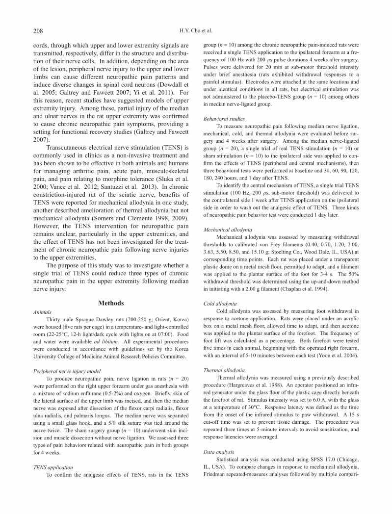
With regard to mechanical allodynia, significant decreases in the withdrawal threshold were observed at 4 days, 7 days, 10 days, 2 weeks, 3 weeks, and 4 weeks after median nerve ligation, compared to rats that underwent sham surgery (Fig. 2A). In the sham surgery group, the mechanical threshold in the ipsilateral side was temporarily reduced 2 days after surgery; however, it gradually recov-ered thereafter (Fig. 2A).
For cold allodynia, bilateral forefoot lifting did not significantly change in the sham surgery group. Nerve liga-tion significantly increased the foot-lifting frequency of the ipsilateral side, whereas the foot-lifting frequency in the contralateral side only showed a significant change at 4 days after surgery (Fig. 2C and D).
For thermal allodynia, the peak lifting latency of the ipsilateral side occurred on day 10 after injury, after which
a plateau was observed during 4 weeks in median nerve-ligated group (Fig. 2E). There were no significant differ-ences between the ipsilateral and contralateral sides in the sham surgery group (Fig. 2E and F).
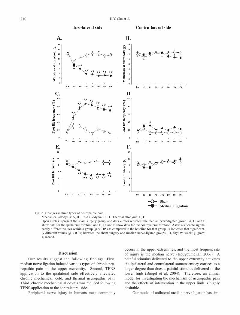
Effects of TENS application to the ipsilateral sideDecreased mechanical allodynia was observed within
60 minu of TENS application to the ipsilateral forearm, compared with placebo-TENS. This analgesic effect declined after about 120 min, although significant differ-ences remained between the two groups up to 180 min after the intervention (Fig. 3A).
Cold allodynia in the TENS group significantly decreased from 60 to 90 min after treatment. Interestingly, a significant difference between the two groups persisted at 120 min (Fig. 3C).
Notably, compared to baseline, the thermal allodynia pain threshold was significantly increased in the TENS group from 30 to 90 min after treatment, and a significant difference was found at 60 minutes compared to the pla-cebo-TENS group. Interestingly, we also observed tempo-rary pain inhibition at 30 minutes in the placebo-TENS group (Fig. 3E).
For the contralateral side, there were no significant changes in behavior for 1 day after intervention in either group (Fig. 3B, D and F).
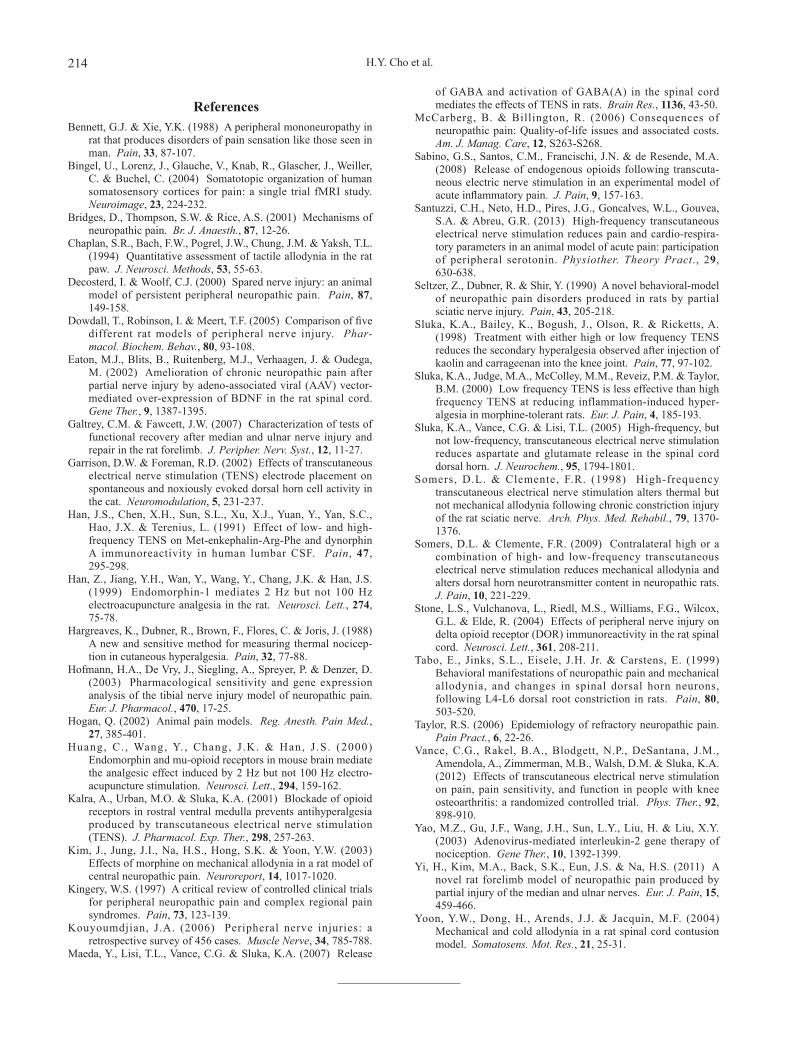
Effects of TENS application into contralateral sideA single trial application of TENS to the contralateral
side induced analgesia to mechanical stimulation in the ipsilateral side from 90 to 120 min after treatment (Fig. 4A). No significant differences were observed with cold or thermal allodynia in the ipsilateral side after TENS applica-tion to the contralateral side (Fig. 4B and C).
Fig. 1. Postural changes following median-nerve ligation. Photos show the postural changes following median-nerve ligation (A) and sham operation (B) in rats. The median
nerve-ligated group showed decreased weight bearing on the right side (ipsilateral forefoot) compared to the left side (contralateral forefoot). The sham surgery group distributed body weight equally on both forefeet.
H.Y. Cho et al.210
DiscussionOur results suggest the following findings: First,
median nerve ligation induced various types of chronic neu-ropathic pain in the upper extremity. Second, TENS application to the ipsilateral side effectively alleviated chronic mechanical, cold, and thermal neuropathic pain. Third, chronic mechanical allodynia was reduced following TENS application to the contralateral side.
Peripheral nerve injury in humans most commonly
occurs in the upper extremities, and the most frequent site of injury is the median nerve (Kouyoumdjian 2006). A painful stimulus delivered to the upper extremity activates the ipsilateral and contralateral somatosensory cortices to a larger degree than does a painful stimulus delivered to the lower limb (Bingel et al. 2004). Therefore, an animal model for investigating the mechanism of neuropathic pain and the effects of intervention in the upper limb is highly desirable.
Our model of unilateral median nerve ligation has sim-
Fig. 2. Changes in three types of neuropathic pain. Mechanical allodynia: A, B. Cold allodynia: C, D. Thermal allodynia: E, F. Open circles represent the sham surgery group, and dark circles represent the median nerve-ligated group. A, C, and E
show data for the ipsilateral forefoot, and B, D, and F show data for the contralateral forefoot. Asterisks denote signifi-cantly different values within a group (p < 0.05) as compared to the baseline for that group. # indicates that significant-ly different values (p < 0.05) between the sham surgery and median nerve-ligated groups. D, day; W, week; g, gram; s, second.

Upper-Limb TENS Reduces Neuropathic Pain 211
ilarities to carpal tunnel syndrome in human subjects. The results of this study show that mechanical, cold, and ther-mal allodynia were induced within 2 days after nerve liga-tion and persisted for 4 weeks. A gradual increase in pain intensity is the main symptom of neuropathic pain due to nerve compression. Our model therefore provides a reason-able representation of upper extremity neuropathic pain. Similar to the present study, partial injury of the median and ulnar nerves caused neuropathic pain symptoms, including
mechanical, thermal, and cold allodynia, that lasted 18 weeks (Yi et al. 2011). In that model, pain-related behav-iors appeared for 6-12 weeks, after which pain status gradu-ally recovered and reached its pre-nerve injury value after 18 weeks. However, the model transected two peripheral nerves in the forearm, which is uncommon in humans. In contrast, we performed single nerve ligation, which fre-quently occurs in humans and results in conditions like car-pal tunnel syndrome.
Fig. 3. Effects of ipsilateral TENS application on three types of neuropathic pain. Alterations in chronic mechanical, cold, and thermal allodynia on the ipsilateral (A, C, E) and contralateral sides (B, D, F).
Open circles represent the placebo-TENS group, and dark circles represent the TENS group. Asterisks denote signifi-cantly different values (p < 0.05) from the pre-application baseline value. # indicates significantly different values (p < 0.05) from the corresponding values in the placebo-TENS group. D, day; m, minute; g, gram; s, second.

H.Y. Cho et al.212
Neuropathic pain in our study persisted for at least 4 weeks. Similarly, neuropathic pain was present for 5 weeks after sciatic nerve ligation and was considered representa-tive of chronic neuropathy after 2 weeks (Yao et al. 2003). Symptoms manifesting 1 week after nerve injury were con-sidered characteristic of chronic neuropathic pain (Eaton et al. 2002). The development of chronic neuropathy, deter-mined by sensory nerve sprouting following nerve com-
pression injury, appeared after 1-2 weeks in various rat experimental systems of nerve damage (Bridges et al. 2001). Therefore, we consider that a 4-week time frame is reasonable for studying chronic neuropathic pain.
Peripheral nerve injuries reduce μ-opioid receptor expression in the dorsal horn of the spinal cord as well as δ-opioid receptor expression throughout the spinal cord. μ- and δ-opioid receptors are involved in pain inhibition via
Fig. 4. Effects of contralateral TENS application on three types of neuropathic pain. After treatment, rats were evaluated for allodynia in response to mechanical (A), cold (B), and thermal (C) stimuli.
Open circles indicate measured values for the limb contralateral to the side of nerve injury; dark circles represent results from the ipsilateral side of nerve ligation. Asterisks denote significantly different values ( p < 0.05) to baseline. D, day; m, minute; g, gram; s, second.

Upper-Limb TENS Reduces Neuropathic Pain 213
descending inhibitory regulation (Stone et al. 2004). Peripheral injuries of the upper limb increase activation of microglia and astrocytes at the C7-C8 levels of the spinal cord. These glial cells secrete inflammatory cytokines, such as tumor necrosis factor (TNF) and interleukin (IL)-6, which may also contribute to chronic neuropathic pain (Yi et al. 2011). Investigation of these mechanisms was beyond the scope of the present study, but is an important area for future work.
We found that TENS effectively reduced symptoms of chronic neuropathic pain in our rat model. The baseline threshold for mechanical pain was approximately 14 g, and this decreased to 3.0 g, approximately 21% of baseline, by 4 weeks after nerve injury. The maximum analgesic effect of TENS treatment was achieved after 90 min, with with-drawal threshold increased to 5.6 g. In a rat model of arthritis of the knee joint, in which disease is induced by carrageenan injection, applying TENS for 20 min after injection immediately decreased thermal hyperalgesia, and this effect lasted over 20 hours (Sluka et al. 1998). These differences of analgesic duration following TENS may be due to the different causes of pain between two models.
The analgesic effect of TENS is similar to that achieved with opioid agonists in the treatment of neuro-pathic pain (Kim et al. 2003). TENS increases the concen-trations of β endorphins and methionine-enkephalin in blood and cerebrospinal fluid (Han et al. 1991). In addition, treatment with a δ-opioid receptor antagonist in the rostro-ventromedial medulla blocks the analgesic effects of TENS (Kalra et al. 2001). Hyperalgesia in arthritic rats was decreased after TENS treatment, and this effect is blocked by the opioid receptor antagonist naltrexone (Sabino et al. 2008). Electrical stimulation of peripheral areas increases the levels of neuropeptides that interact with μ- and δ-opioid receptor subtypes in the central nervous system (Han et al. 1999; Huang et al. 2000). TENS effects may be mediated by increased expression and activation of opioid receptors in nerves and endogenous opioid peptides in blood, and/or cerebrospinal fluid.
TENS has been reported to reduce the expression and secretion of excitatory neurotransmitters, such as glutamate and aspartate, in the spinal cord (Sluka et al. 2005). The anti-hyperalgesic effects of TENS are inhibited by treat-ment with gamma-aminobutyric acid (GABA)-A receptor antagonist (Maeda et al. 2007). Similar to our findings, studies using the sciatic nerve ligation model have demon-strated that TENS treatment effectively reduced neuropathic pain and increased the number of GABA synaptosomes in the spinal cord dorsal horn (Somers and Clemente 2009). Thus, we assumed that the TENS-induced analgesia following median nerve ligation is at least partially due to changes in excitatory and inhibitory neurotransmitter expression levels in the spinal cord.
Interestingly, TENS to the contralateral side signifi-cantly decreased chronic mechanical allodynia in the ipsi-lateral side. TENS might induce analgesic effects by influ-
encing the central nervous system. In previous studies, TENS to the contralateral side led to reduced dorsal horn cell activity on the ipsilateral side (Garrison and Foreman 2002). In addition, application of TENS to the contralateral side reduced mechanical allodynia by increasing GABA levels in the dorsal horn, but did not affect thermal allo-dynia, while TENS application into the contralateral side decreased aspartate and glutamate levels and increased gly-cine levels in the spinal cord of chronic constriction-injured rats (Somers and Clemente 2009). The observed reduction in the ipsilateral pain following contralateral TENS in the present study may therefore be mediated by reduced cell activity and increased GABA release in the spinal cord. Of note, we found that contralateral TENS significantly decreased mechanical allodynia but not cold or thermal allodynia. This result might be due to the method used to measure mechanical allodynia, which is more sensitive than the methods for measuring cold and thermal allodynia (Tabo et al. 1999). Accordingly, the degree of improvement in mechanical allodynia following TENS applied to the con-tralateral side was relatively small as compared to the anal-gesic effect of TENS applied to the ipsilateral side. Similarly, minor analgesic effects might not have been detected by other measurement techniques.
Although our study demonstrated that a single trial application of TENS effectively attenuated chronic neuro-pathic pain following median nerve injury, there are some limitations. First, the effect of repeated TENS application on chronic neuropathic pain is not clear. Second, we did not assess the long-term follow-up effects of TENS. Third, we examined the impact of TENS in the early chronic phase, and this analgesic effect may not apply in later chronic phases.
In conclusion, the present study demonstrated that experimental median nerve ligation in rats leads to chronic neuropathic pain similar to that associated with carpal tunnel syndrome in humans. Treatment with TENS effectively reduced three manifestations of chronic neuro-pathic pain in this system, suggesting that TENS may be a valuable non-pharmacological, non-surgical modality for the clinical treatment of neuropathic pain. Moreover, TENS can also be applied on the contralateral side in patients with burns, severe allodynia, and wounds for whom electrode application to the injured side is impossible. Further studies are necessary to determine the optimal stimulation para-meters of TENS for managing chronic neuropathic pain in the upper extremity and to elucidate the underlying analgesic mechanisms.
AcknowledgementsThis study received the research grant (GCU-2013-M065)
from Gachon University in South Korea.
Conflict of InterestThe authors declare no conflict of interest.
H.Y. Cho et al.214
ReferencesBennett, G.J. & Xie, Y.K. (1988) A peripheral mononeuropathy in
rat that produces disorders of pain sensation like those seen in man. Pain, 33, 87-107.
Bingel, U., Lorenz, J., Glauche, V., Knab, R., Glascher, J., Weiller, C. & Buchel, C. (2004) Somatotopic organization of human somatosensory cortices for pain: a single trial fMRI study. Neuroimage, 23, 224-232.
Bridges, D., Thompson, S.W. & Rice, A.S. (2001) Mechanisms of neuropathic pain. Br. J. Anaesth., 87, 12-26.
Chaplan, S.R., Bach, F.W., Pogrel, J.W., Chung, J.M. & Yaksh, T.L. (1994) Quantitative assessment of tactile allodynia in the rat paw. J. Neurosci. Methods, 53, 55-63.
Decosterd, I. & Woolf, C.J. (2000) Spared nerve injury: an animal model of persistent peripheral neuropathic pain. Pain, 87, 149-158.
Dowdall, T., Robinson, I. & Meert, T.F. (2005) Comparison of five different rat models of peripheral nerve injury. Phar-macol. Biochem. Behav., 80, 93-108.
Eaton, M.J., Blits, B., Ruitenberg, M.J., Verhaagen, J. & Oudega, M. (2002) Amelioration of chronic neuropathic pain after partial nerve injury by adeno-associated viral (AAV) vector-mediated over-expression of BDNF in the rat spinal cord. Gene Ther., 9, 1387-1395.
Galtrey, C.M. & Fawcett, J.W. (2007) Characterization of tests of functional recovery after median and ulnar nerve injury and repair in the rat forelimb. J. Peripher. Nerv. Syst., 12, 11-27.
Garrison, D.W. & Foreman, R.D. (2002) Effects of transcutaneous electrical nerve stimulation (TENS) electrode placement on spontaneous and noxiously evoked dorsal horn cell activity in the cat. Neuromodulation, 5, 231-237.
Han, J.S., Chen, X.H., Sun, S.L., Xu, X.J., Yuan, Y., Yan, S.C., Hao, J.X. & Terenius, L. (1991) Effect of low- and high-frequency TENS on Met-enkephalin-Arg-Phe and dynorphin A immunoreactivity in human lumbar CSF. Pain, 47, 295-298.
Han, Z., Jiang, Y.H., Wan, Y., Wang, Y., Chang, J.K. & Han, J.S. (1999) Endomorphin-1 mediates 2 Hz but not 100 Hz electroacupuncture analgesia in the rat. Neurosci. Lett., 274, 75-78.
Hargreaves, K., Dubner, R., Brown, F., Flores, C. & Joris, J. (1988) A new and sensitive method for measuring thermal nocicep-tion in cutaneous hyperalgesia. Pain, 32, 77-88.
Hofmann, H.A., De Vry, J., Siegling, A., Spreyer, P. & Denzer, D. (2003) Pharmacological sensitivity and gene expression analysis of the tibial nerve injury model of neuropathic pain. Eur. J. Pharmacol., 470, 17-25.
Hogan, Q. (2002) Animal pain models. Reg. Anesth. Pain Med., 27, 385-401.
Huang, C. , Wang, Y. , Chang, J .K. & Han, J .S. (2000) Endomorphin and mu-opioid receptors in mouse brain mediate the analgesic effect induced by 2 Hz but not 100 Hz electro-acupuncture stimulation. Neurosci. Lett., 294, 159-162.
Kalra, A., Urban, M.O. & Sluka, K.A. (2001) Blockade of opioid receptors in rostral ventral medulla prevents antihyperalgesia produced by transcutaneous electrical nerve stimulation (TENS). J. Pharmacol. Exp. Ther., 298, 257-263.
Kim, J., Jung, J.I., Na, H.S., Hong, S.K. & Yoon, Y.W. (2003) Effects of morphine on mechanical allodynia in a rat model of central neuropathic pain. Neuroreport, 14, 1017-1020.
Kingery, W.S. (1997) A critical review of controlled clinical trials for peripheral neuropathic pain and complex regional pain syndromes. Pain, 73, 123-139.
Kouyoumdjian, J.A. (2006) Peripheral nerve injuries: a retrospective survey of 456 cases. Muscle Nerve, 34, 785-788.
Maeda, Y., Lisi, T.L., Vance, C.G. & Sluka, K.A. (2007) Release
of GABA and activation of GABA(A) in the spinal cord mediates the effects of TENS in rats. Brain Res., 1136, 43-50.
McCarberg, B. & Billington, R. (2006) Consequences of neuropathic pain: Quality-of-life issues and associated costs. Am. J. Manag. Care, 12, S263-S268.
Sabino, G.S., Santos, C.M., Francischi, J.N. & de Resende, M.A. (2008) Release of endogenous opioids following trans cuta-neous electric nerve stimulation in an experimental model of acute inflammatory pain. J. Pain, 9, 157-163.
Santuzzi, C.H., Neto, H.D., Pires, J.G., Goncalves, W.L., Gouvea, S.A. & Abreu, G.R. (2013) High-frequency transcutaneous electrical nerve stimulation reduces pain and cardio-respira-tory parameters in an animal model of acute pain: participation of peripheral serotonin. Physiother. Theory Pract., 29, 630-638.
Seltzer, Z., Dubner, R. & Shir, Y. (1990) A novel behavioral-model of neuropathic pain disorders produced in rats by partial sciatic nerve injury. Pain, 43, 205-218.
Sluka, K.A., Bailey, K., Bogush, J., Olson, R. & Ricketts, A. (1998) Treatment with either high or low frequency TENS reduces the secondary hyperalgesia observed after injection of kaolin and carrageenan into the knee joint. Pain, 77, 97-102.
Sluka, K.A., Judge, M.A., McColley, M.M., Reveiz, P.M. & Taylor, B.M. (2000) Low frequency TENS is less effective than high frequency TENS at reducing inflammation-induced hyper-algesia in morphine-tolerant rats. Eur. J. Pain, 4, 185-193.
Sluka, K.A., Vance, C.G. & Lisi, T.L. (2005) High-frequency, but not low-frequency, transcutaneous electrical nerve stimulation reduces aspartate and glutamate release in the spinal cord dorsal horn. J. Neurochem., 95, 1794-1801.
Somers, D.L. & Clemente, F.R. (1998) High-frequency transcutaneous electrical nerve stimulation alters thermal but not mechanical allodynia following chronic constriction injury of the rat sciatic nerve. Arch. Phys. Med. Rehabil., 79, 1370-1376.
Somers, D.L. & Clemente, F.R. (2009) Contralateral high or a combination of high- and low-frequency transcutaneous electrical nerve stimulation reduces mechanical allodynia and alters dorsal horn neurotransmitter content in neuropathic rats. J. Pain, 10, 221-229.
Stone, L.S., Vulchanova, L., Riedl, M.S., Williams, F.G., Wilcox, G.L. & Elde, R. (2004) Effects of peripheral nerve injury on delta opioid receptor (DOR) immunoreactivity in the rat spinal cord. Neurosci. Lett., 361, 208-211.
Tabo, E., Jinks, S.L., Eisele, J.H. Jr. & Carstens, E. (1999) Behavioral manifestations of neuropathic pain and mechanical allodynia, and changes in spinal dorsal horn neurons, follow ing L4-L6 dorsal root constriction in rats. Pain, 80, 503-520.
Taylor, R.S. (2006) Epidemiology of refractory neuropathic pain. Pain Pract., 6, 22-26.
Vance, C.G., Rakel, B.A., Blodgett, N.P., DeSantana, J.M., Amendola, A., Zimmerman, M.B., Walsh, D.M. & Sluka, K.A. (2012) Effects of transcutaneous electrical nerve stimulation on pain, pain sensitivity, and function in people with knee osteoarthritis: a randomized controlled trial. Phys. Ther., 92, 898-910.
Yao, M.Z., Gu, J.F., Wang, J.H., Sun, L.Y., Liu, H. & Liu, X.Y. (2003) Adenovirus-mediated interleukin-2 gene therapy of nociception. Gene Ther., 10, 1392-1399.
Yi, H., Kim, M.A., Back, S.K., Eun, J.S. & Na, H.S. (2011) A novel rat forelimb model of neuropathic pain produced by partial injury of the median and ulnar nerves. Eur. J. Pain, 15, 459-466.
Yoon, Y.W., Dong, H., Arends, J.J. & Jacquin, M.F. (2004) Mechanical and cold allodynia in a rat spinal cord contusion model. Somatosens. Mot. Res., 21, 25-31.