a simple portable ecg monitor with iot - ijirset simple.pdfa simple portable ecg monitor with iot...
TRANSCRIPT

ISSN(Online) : 2319-8753
ISSN (Print) : 2347-6710
International Journal of Innovative Research in Science, Engineering and Technology
(An ISO 3297: 2007 Certified Organization)
Website: www.ijirset.com Vol. 6, Issue 7, July 2017
Copyright to IJIRSET DOI:10.15680/IJIRSET.2017.0607283 13571
A Simple Portable ECG Monitor with IOT
R. H. Sayyed1, Maqdoom Farooqui2, A. R. Khan3 and Gulam Rabbani4
Asso. Prof, Department of Electronic Science, Abeda Inamdar Sr. College, Pune, India1
Principal, Dept. of Chemistry, Maulana Azad College, Dr. Rafiq Zakaria Campus, Auranagabad, India. 2
Head PG Dept. of computer Science, Maulana Azad College, Auranagabad, India. 3
Director, Dr. Rafiq Zakaria Centre for Higher Learning and Advanced Research, Auranagabad, India. 4
ABSTRACT: The Internet of Things (IOT) provides a proficient and new life to the healthcare field. The heart diseases results millions of death worldwide because of the increase in the aging population and the growing of healthcare costs. The Wireless Sensor Network and the cloud computing is becoming the popular strategy for the IOT era. One of the most useful techniques for recording of electrical activity of the heart over a period of time of the heart patient is the Electrocardiogram. In this, electrodes are placed on the chest of patient and variations in signals are recorded. The aim of work presented is to design and develop a simple 3-lead Portable ECG Monitor With IOT. The system is developed using an ARM Cortex M3 LPC1768 based biomedical parameter monitoring by supporting GSM/GPRS and internet together in the Wireless Sensor Network (WSN) under HTTP protocol to a database server containing clinic data, which can be accessed through a web application. The system is designed and implemented using embedded C-language. Digital Storage Oscilloscope is used to monitor ECG for real time and will display Heart Rates, cardiac cycle, and intervals of some critical components, which helps the physicians in heart disease diagnosis. The ECG system will be useful for remote monitoring of patient’s health.
KEYWORDS: Electrocardiogram, Hypertext Transfer Protocol,Wireless Sensor Network, Global System for Mobile Communication, General Packet Radio Service.
I. INTRODUCTION Electrocardiogram (abbreviated ECG/EKG) is a demonstration of electrical activity of the heart(cardiac) muscle as noted from the body surface, which is used in the analysis of the heart disease. This electrical activity is related to the impulse that is transportable through the heart which decides its rate and rhythm. For an ECG examination of a patient, the patient must be wired up with the chest electrodes. The ECG signal obtained represents the polarization and the depolarization of the cells throughout a heartbeat. The main objective of present work is to design and construct a portable Electrocardiogram (ECG) system to monitor patients who are at a distance and need monitoring for problems like heart attack. This portable ECG is monitoring system makes use of 3 electrodes mounted on the patients body and detects the electrical signals. These signals from electrodes are weak in strength, therefore an electronic circuit is employed to amplify this signal. Unwanted noise associated with the signal is removed using filters this results in enhancement of the signal to noise ratio. The ECG signal data is stored in the form of a time series and maintained in a data base and the data is sent over wireless communication to a monitoring system. At the monitoring system end the data is examined, and if it is out side standard limits a message is sent to notify the supervisor or doctor. Heart rate is one of the vital aspects for the different kinds of heart functioning disorder identification. The research work presented deals with design, implementation of portable ECG monitor with. A. ECG Measurement The electrical impulses within the heart represents a source of voltage, which set up a current flow in the torso and corresponding potentials on the skin. The potential distribution can be designed as if the heart were a time varying electric dipole.
ISSN(Online) : 2319-8753
ISSN (Print) : 2347-6710
International Journal of Innovative Research in Science, Engineering and Technology
(An ISO 3297: 2007 Certified Organization)
Website: www.ijirset.com Vol. 6, Issue 7, July 2017
Copyright to IJIRSET DOI:10.15680/IJIRSET.2017.0607283 13572
B. System Functionality The key task is to design simple "Portable ECG Monitor With IOT" to monitor ECG of a distant patient without electrical contact. It is the first electrocardiogram monitoring system to eliminate the wires and cables between patient's bedside instruments and remote monitor located at a distance at the diagnostic facility or a hospital. The signal captured by the electrodes, after initial conditioning, will be transmitted to the ARM Cortex M3 LPC 1768 and with the help of GSM / GPRS technology and internet together the data can be sent in the Wireless Sensor Network under HTTP protocol to a database server containing ECG data, which can be accessed through a web application and the system helps in giving early mobility to patients. The entire system is designed keeping in view the requirement of remote monitoring of the ECG data in real time for patients requiring intensive care, at the same time there are only three electrodes making it a wearable device.
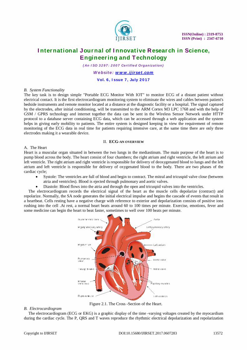
II. ECG-AN OVERVIEW A. The Heart Heart is a muscular organ situated in between the two lungs in the mediastinum. The main purpose of the heart is to pump blood across the body. The heart consist of four chambers; the right atrium and right ventricle, the left atrium and left ventricle. The right atrium and right ventricle is responsible for delivery of deoxygenated blood to lungs and the left atrium and left ventricle is responsible for delivery of oxygenated blood to the body. There are two phases of the cardiac cycle;
Systole: The ventricles are full of blood and begin to contract. The mitral and tricuspid valve close (between atria and ventricles). Blood is ejected through pulmonary and aortic valves.
Diastole: Blood flows into the atria and through the open and tricuspid valves into the ventricles. The electrocardiogram records the electrical signal of the heart as the muscle cells depolarize (contract) and
repolarize. Normally, the SA node generates the initial electrical impulse and begins the cascade of events that result in a heartbeat. Cells resting have a negative charge with reference to exterior and depolarization consists of positive ions rushing into the cell .At rest, a normal heart beats around 60 to 100 times per minute. Exercise, emotions, fever and some medicine can begin the heart to beat faster, sometimes to well over 100 beats per minute.
Figure 2.1. The Cross -Section of the Heart.
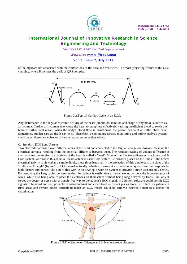
B. Electrocardiogram The electrocardiogram (ECG or EKG) is a graphic display of the time -varying voltages created by the myocardium
during the cardiac cycle. The P, QRS and T waves reproduce the rhythmic electrical depolarization and repolarization
ISSN(Online) : 2319-8753
ISSN (Print) : 2347-6710
International Journal of Innovative Research in Science, Engineering and Technology
(An ISO 3297: 2007 Certified Organization)
Website: www.ijirset.com Vol. 6, Issue 7, July 2017
Copyright to IJIRSET DOI:10.15680/IJIRSET.2017.0607283 13573
of the myocardium associated with the contractions of the atria and ventricles. The most projecting feature is the QRS complex, where R denotes the peak of QRS complex.
Figure 2.2.Typical Cardiac Cycle of an ECG. Any disturbance in the regular rhythmic activity of the heart (amplitude, duration and shape of rhythms) is known as arrhythmia. Cardiac arrhythmias may cause the heart to pump less effectively, causing insufficient blood to reach the brain a further vital organ. When the body's blood flow is insufficient, the person can faint or suffer chest pain. Sometimes, sudden cardiac death can exist. Therefore, a continuous cardiac monitoring and online analysis system could detect these rare episodes of cardiac arrhythmias as they obtain. C. Standard ECG Lead System Two electrodes arranged over different areas of the heart and connected to the Digital storage oscilloscope picks up the electrical currents, resulting from the potential difference between them. The resultant tracing of voltage difference at any two sites due to electrical activity of the heart is called a "lead". Most of the Electrocardiogram machines use12-Lead system, whereas in this paper a 3-lead system is used. Both feature 3 electrodes placed on the limbs. If the heart's electrical activity is viewed as a simple dipole, these three leads verify the projection of this dipole onto the sides of the 'Einthoven Triangle' (figure2.3). ECG signal is totally valuable, making it a conventional system used in hospitals by both doctors and nurses. The aim of this work is to develop a wireless system to provide a more user-friendly device. By removing the long cables between nodes, the patient is easily able to move around without the inconvenience of wires, while also being able to place the electrodes on themselves without being long delayed by leads. Similarly it serves the doctor or nurse with a trouble-free way to the patient’s ECG signal. In addition, software could permit ECG signals to be saved and sent possibly by using Internet and cloud to other distant places globally. In fact, for patients in rural areas and remote places difficult to reach an ECG record could be sent via electronic mail to a doctor for examination.
Figure 2.3.The Einthoven Triangle and 3- lead electrode placement.
ISSN(Online) : 2319-8753
ISSN (Print) : 2347-6710
International Journal of Innovative Research in Science, Engineering and Technology
(An ISO 3297: 2007 Certified Organization)
Website: www.ijirset.com Vol. 6, Issue 7, July 2017
Copyright to IJIRSET DOI:10.15680/IJIRSET.2017.0607283 13574
D. Common Artifacts
The electrical signals of heart received from the patient's body are of very low strength, typically of the order of 2.5mv and are mixed with noise. The instrumentation amplifier and further inverting amplifier amplify the low strength signals and non inverting amplifiers to maintain gain up to 1000. Three different types of noise are present in the signal.
1. DC electrode offset potential. 2. 50 Hz ac induced interference. 3. The electrodes pick up muscular noise.
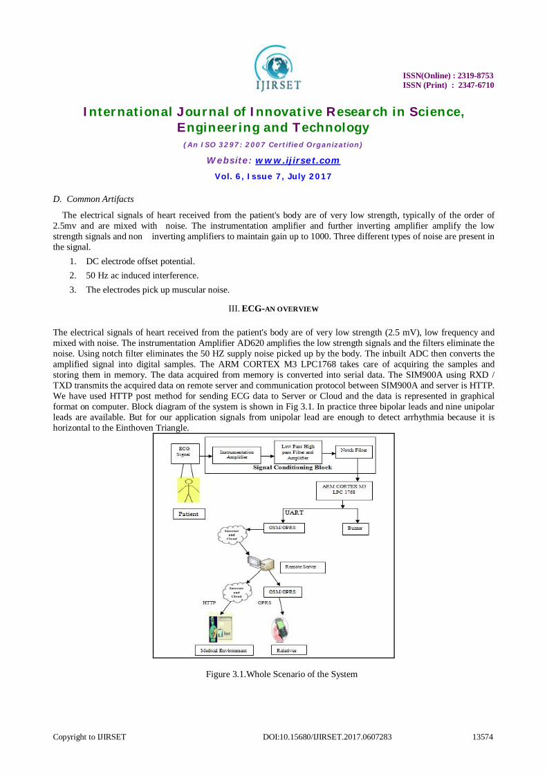
III. ECG-AN OVERVIEW The electrical signals of heart received from the patient's body are of very low strength (2.5 mV), low frequency and mixed with noise. The instrumentation Amplifier AD620 amplifies the low strength signals and the filters eliminate the noise. Using notch filter eliminates the 50 HZ supply noise picked up by the body. The inbuilt ADC then converts the amplified signal into digital samples. The ARM CORTEX M3 LPC1768 takes care of acquiring the samples and storing them in memory. The data acquired from memory is converted into serial data. The SIM900A using RXD / TXD transmits the acquired data on remote server and communication protocol between SIM900A and server is HTTP. We have used HTTP post method for sending ECG data to Server or Cloud and the data is represented in graphical format on computer. Block diagram of the system is shown in Fig 3.1. In practice three bipolar leads and nine unipolar leads are available. But for our application signals from unipolar lead are enough to detect arrhythmia because it is horizontal to the Einthoven Triangle.
Figure 3.1.Whole Scenario of the System
ISSN(Online) : 2319-8753
ISSN (Print) : 2347-6710
International Journal of Innovative Research in Science, Engineering and Technology
(An ISO 3297: 2007 Certified Organization)
Website: www.ijirset.com Vol. 6, Issue 7, July 2017
Copyright to IJIRSET DOI:10.15680/IJIRSET.2017.0607283 13575
IV. HARDWARE DESIGN A. Signal Conditioning Block To amplify low amplitude ECG (Electrocardiogram) signals this system uses variable gain amplifiers. Gain of the amplifiers is adjusted according to the specifications of the in built analog to digital converter. ECG signals have maximum voltage range of 2.5 mV. So the required gain of the system is
SignalECGofOutputADCofInput
InputOutputGain
1000 mV5.2V5.2Gain
This uses two stages to provide gain of 1000. It has been decided that the instrumentation amplifier provides gain of 7 and the inverting amplifier provides gain of 143. So the total gain provided by the system is
Gain = 7 (stage 1) *143 (stage 2) Total Gain = 1001
B. Instrumentation Amplifier This intended for low level signal amplification where low noise, low thermal and time drifts, high input impedance an accurate closed loop gain are required. Besides, high CMRR and high slew rate are desirable for better performance. As ECG signal is of very low amplitude, therefore an instrumentation amplifier is used to amplify it at initial level so that ECG signal should not get loaded. The two signals entering the differential amplifier are subtracted to cancel the common noise present in the signal. Because of instrumentation amplifier common noise gets cancel with advantage of strengthening the signal.
C. Filter
A low pass filter having cut off frequency 0.5 Hz is used here for removing noise and motion rate facts coming from the body. Formula: fc = 1 ∕ 2πRC
D. High Pass Filter and Amplifier
In order to block DC offset present in the signal, we have to use high pass filter to block DC. If DC is not getting blocked at this level it increases with gain of amplifiers connected after DC block, and induces noise. In order to provide gain of 1000 we have used two amplifiers one is inverting and the other is non-inverting.
E. Notch Filter
In order to reduce 50HZ power line frequency hum notch filter is especially essential. It should be highly precise at 50HZ. It is composed of one low pass filter to block and one high pass filter.
F. Microcontroller
For our system we are going to use ARM Cortex M3 LPC1768. This controller has an in built ADC for converting analog to digital converter operation. From microcontroller the digital samples are sent via UART to GPRS module. Programming of the microcontroller is done in embedded C language code. The LPC1768 processor is highly configurable enabling a wide range of application from those requiring memory protection and powerful trace technology to cost sensitive devices requiring minimal area.
ISSN(Online) : 2319-8753
ISSN (Print) : 2347-6710
International Journal of Innovative Research in Science, Engineering and Technology
(An ISO 3297: 2007 Certified Organization)
Website: www.ijirset.com Vol. 6, Issue 7, July 2017
Copyright to IJIRSET DOI:10.15680/IJIRSET.2017.0607283 13576
G. Analog to Digital converter
For converting analog signals of ECG into digital the LPC1768 has an in built 12-bit successive Approximation ADC which is multiplexed among 8 input pins. It has several features which are useful to us. It is very useful for digitizing the bipolar ECG signals. It gives parallel 12-bit output for further processing.
H. GPRS Communication
In this paper we are using SIM900A for GPRS communication.
I. Signal Display
In this system digital signals are captured through GPRS and stored on the server and displayed on the web browser and all over the world the doctor can access the ECG graph anywhere anytime.
J. Microcontroller and Interface
SIM900A uses GPRS to transmit the data on remote server. The communication protocol between SIM900A and server is HTTP. We have used HTTP post method for sending ECG data to server or cloud. Because of remote server the communication range is unlimited.
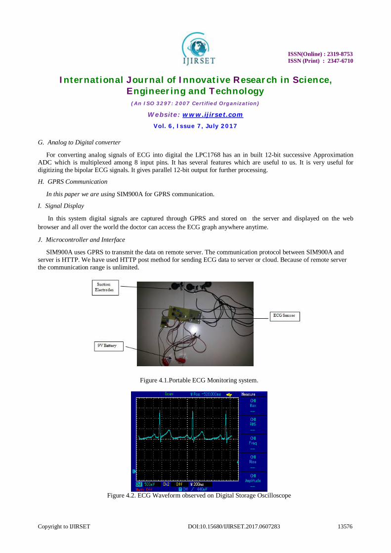
Figure 4.1.Portable ECG Monitoring system.
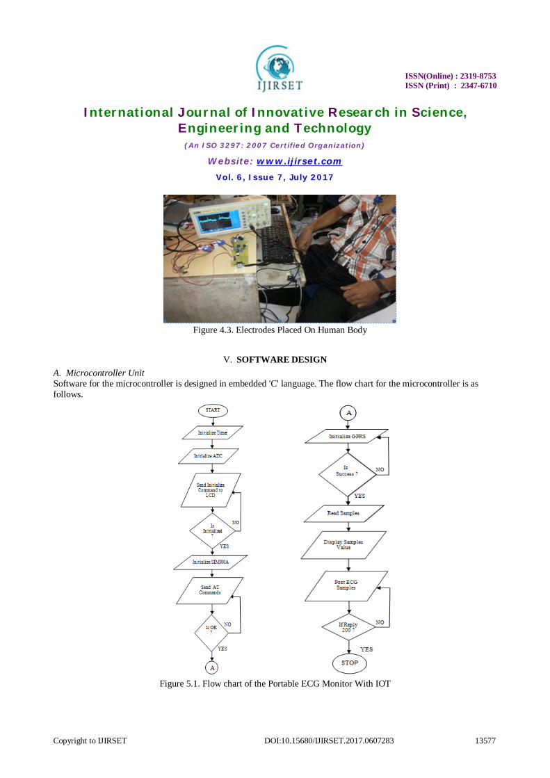
Figure 4.2. ECG Waveform observed on Digital Storage Oscilloscope
ISSN(Online) : 2319-8753
ISSN (Print) : 2347-6710
International Journal of Innovative Research in Science, Engineering and Technology
(An ISO 3297: 2007 Certified Organization)
Website: www.ijirset.com Vol. 6, Issue 7, July 2017
Copyright to IJIRSET DOI:10.15680/IJIRSET.2017.0607283 13577
Figure 4.3. Electrodes Placed On Human Body
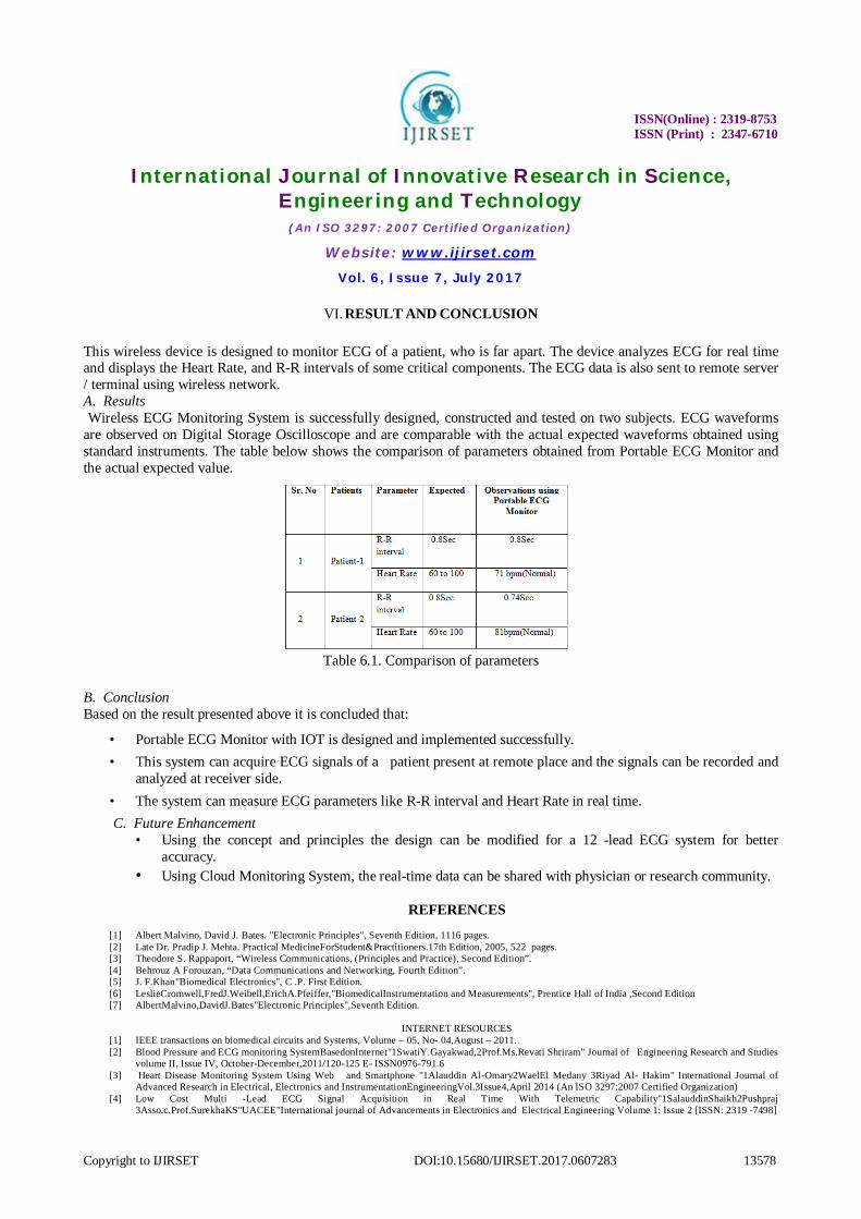
V. SOFTWARE DESIGN A. Microcontroller Unit Software for the microcontroller is designed in embedded 'C' language. The flow chart for the microcontroller is as follows.
Figure 5.1. Flow chart of the Portable ECG Monitor With IOT
ISSN(Online) : 2319-8753
ISSN (Print) : 2347-6710
International Journal of Innovative Research in Science, Engineering and Technology
(An ISO 3297: 2007 Certified Organization)
Website: www.ijirset.com Vol. 6, Issue 7, July 2017
Copyright to IJIRSET DOI:10.15680/IJIRSET.2017.0607283 13578
VI. RESULT AND CONCLUSION
This wireless device is designed to monitor ECG of a patient, who is far apart. The device analyzes ECG for real time and displays the Heart Rate, and R-R intervals of some critical components. The ECG data is also sent to remote server / terminal using wireless network. A. Results Wireless ECG Monitoring System is successfully designed, constructed and tested on two subjects. ECG waveforms are observed on Digital Storage Oscilloscope and are comparable with the actual expected waveforms obtained using standard instruments. The table below shows the comparison of parameters obtained from Portable ECG Monitor and the actual expected value.
Table 6.1. Comparison of parameters
B. Conclusion Based on the result presented above it is concluded that:
• Portable ECG Monitor with IOT is designed and implemented successfully. • This system can acquire ECG signals of a patient present at remote place and the signals can be recorded and
analyzed at receiver side. • The system can measure ECG parameters like R-R interval and Heart Rate in real time. C. Future Enhancement
• Using the concept and principles the design can be modified for a 12 -lead ECG system for better accuracy.
• Using Cloud Monitoring System, the real-time data can be shared with physician or research community.
REFERENCES
[1] Albert Malvino, David J. Bates. "Electronic Principles", Seventh Edition, 1116 pages. [2] Late Dr. Pradip J. Mehta. Practical MedicineForStudent&Practitioners.17th Edition, 2005, 522 pages. [3] Theodore S. Rappaport, “Wireless Communications, (Principles and Practice), Second Edition”. [4] Behrouz A Forouzan, “Data Communications and Networking, Fourth Edition”. [5] J. F.Khan"Biomedical Electronics", C .P. First Edition. [6] LeslieCromwell,FredJ.Weibell,ErichA.Pfeiffer,"BiomedicalInstrumentation and Measurements", Prentice Hall of India ,Second Edition [7] AlbertMalvino,DavidJ.Bates"Electronic Principles",Seventh Edition.
INTERNET RESOURCES
[1] IEEE transactions on biomedical circuits and Systems, Volume – 05, No- 04,August – 2011. [2] Blood Pressure and ECG monitoring SystemBasedonInternet"1SwatiY.Gayakwad,2Prof.Ms.Revati Shriram" Journal of Engineering Research and Studies
volume II, Issue IV, October-December,2011/120-125 E- ISSN0976-791.6 [3] Heart Disease Monitoring System Using Web and Smartphone "1Alauddin Al-Omary2WaelEl Medany 3Riyad Al- Hakim" International Journal of
Advanced Research in Electrical, Electronics and InstrumentationEngineeringVol.3Issue4,April 2014 (An ISO 3297:2007 Certified Organization) [4] Low Cost Multi -Lead ECG Signal Acquisition in Real Time With Telemetric Capability"1SalauddinShaikh2Pushpraj
3Asso.c.Prof.SurekhaKS"UACEE"International journal of Advancements in Electronics and Electrical Engineering Volume 1: Issue 2 [ISSN: 2319 -7498]