a robust algorithm for retinal blood vessel extraction
TRANSCRIPT

7/21/2019 A Robust Algorithm for Retinal Blood Vessel Extraction
http://slidepdf.com/reader/full/a-robust-algorithm-for-retinal-blood-vessel-extraction 1/9
ISSN(Online): 2320-9801
ISSN (Print): 2320-9798
International Journal of Innovative Research in Computer
and Communication Engineering
(An ISO 3297: 2007 Certified Organization)
Vol. 3, Issue 9, September 2015
Copyright to IJIRCCE DOI: 10.15680/IJIRCCE.2015. 0309001 7921
A Robust Algorithm for Retinal Blood Vessel
Extraction Nalan Karunanayake
1, Manaram Gnanasekera
2, N.D. Kodikara
3
Teaching Assistant, Dept. of Electrical and Computer Engineering, Sri Lanka Institute of Information Technology,
Sri Lanka1
Teaching Assistant, Dept. of Electrical and Computer Engineering, Sri Lanka Institute of Information Technology,
Sri Lanka2
Professor, Dept. of Computer Science, University of Colombo School of Computing, Sri Lanka3
ABSTRACT: Non-proliferative diabetic retinopathy (NDPR) detection is currently a highly interested research area.
Ophthalmologists detect NDPR by observing disorders in the vessel system. Therefore segmentation of the vessel
system will be an aid for ophthalmologist in order to detect an early retinopathy. In this proposed frame work a novel
method based on Gabo Filter and adaptive thresholding has been used. The results have been tested using sensitivity
and specificity and the values are 92.36% and 87.52% respectively.
KEYWORDS: Non-proliferative diabetic retinopathy, Vessel extraction, Gabor Filter, Local adaptive Thresholding,
Binarization.
I. INTRODUCTION
Diabetic retinopathy is an eye disease that occurs due to a complication of diabetes which usually results in severe
vision loss or permanent blindness. It occurs when high blood sugar levels damage the tiny blood vessels that nourish
the retina. High blood sugar levels can cause the blood vessels to narrow down and as a result the constant supply of
blood to the retina will reduce. There can also be leakages in blood vessels due to this issue. “Non-Proliferative
Diabetic Retinopathy” (NDPR) is the early state of the disease. During this period, tiny bulges occur in the vessel walls.
In order to monitor the early stages of retinopathy or “NDPR”, Ophthalmo logists need to closely monitor the blood
vessels in a retina. Hence, segmentation of blood vessels in a retinal image will ease the effort of the Ophthalmologists
when finding the early stages of this disease. This paper proposes a novel method of extracting the vessel system from
a retinal image.
II. R ELATED WORK
The green channel of the original fundus image has been used to obtain the traces of blood vessels and
morphological operations followed with enhancement, background exclusion and thresholding has been used to extract
the vessels in the approach taken by S. Joshi and P.T.Karule[1] . In “Vessel Segmentation in Retinal Images using
Graph-Theoretical Vessel Tracking” a vessel tracking technique based on seed points is used to extract the vesselsystem out of the retinal image [2]. Otsu thresholding and Medial Axis Skeletonization based method followed by
pruning has been used in the research done by L.Sukkaewet al [3]. Next a complex Gabor filter is used to enhance the
vessels and the result is further purified by using. entropicthresholding in the research done by P.C. Siddalingaswamy
and K.G. Prabhu [4]. D.onkaew and B.uyyanonvara in “Automatic Extraction of Retinal Vessels Based on Gradient
Orientation Analysis” [5] which has used a gradient orientation method to separate the vessel system.
7/21/2019 A Robust Algorithm for Retinal Blood Vessel Extraction
http://slidepdf.com/reader/full/a-robust-algorithm-for-retinal-blood-vessel-extraction 2/9
ISSN(Online): 2320-9801
ISSN (Print): 2320-9798
International Journal of Innovative Research in Computer
and Communication Engineering
(An ISO 3297: 2007 Certified Organization)
Vol. 3, Issue 9, September 2015
Copyright to IJIRCCE DOI: 10.15680/IJIRCCE.2015. 0309001 7922
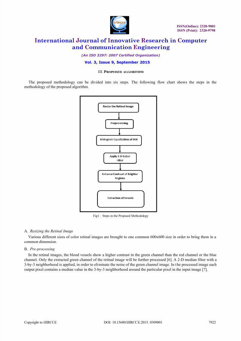
III. PROPOSED ALGORITHM
The proposed methodology can be divided into six steps. The following flow chart shows the steps in the
methodology of the proposed algorithm.
Fig1 : Steps in the Proposed Methodology
A. Resizing the Retinal Image
Various different sizes of color retinal images are brought to one common 600x600 size in order to bring them in a
common dimension.
B. Pre-processing
In the retinal images, the blood vessels show a higher contrast in the green channel than the red channel or the blue
channel. Only the extracted green channel of the retinal image will be further processed [6]. A 2-D median filter with a
3-by-3 neighborhood is applied, in order to eliminate the noise of the green channel image. In the processed image each
output pixel contains a median value in the 3-by-3 neighborhood around the particular pixel in the input image [7].

7/21/2019 A Robust Algorithm for Retinal Blood Vessel Extraction
http://slidepdf.com/reader/full/a-robust-algorithm-for-retinal-blood-vessel-extraction 3/9
ISSN(Online): 2320-9801
ISSN (Print): 2320-9798
International Journal of Innovative Research in Computer
and Communication Engineering
(An ISO 3297: 2007 Certified Organization)
Vol. 3, Issue 9, September 2015
Copyright to IJIRCCE DOI: 10.15680/IJIRCCE.2015. 0309001 7923
(a) (b)
(c) (d) Fig2 : Extraction of RGB Image: (a) input RGB Image (b) Red Channel Component (c) Green Channel Component (d) Blue Channel Component.
C.
Histogram equitation retinal ROI
The ROI (Region of Interest) in the fundus image is the retinal area (the area without the background). Histogram
equalization is an efficient image enhancement procedure to change the mean brightness of an image using its
histogram to change to the middle level of the permitted range [8]. In this proposed research, the histogram equalization
is performed to change the pixel intensities of the green channel to enhance the contrast of the retinal blood vessels in
the image.
The equalized histogram of the source image h(x) is represented as a x r -by-xc matrix and pixel intensities ranging
from grey level 0 to level L-1. Furthermore, cdf(x) is the cumulative density function of the green channel image and
cdf mincdf maxare the lowest and the highest integer values of the cdf(x).
eq. (1)
In the proposed algorithm, histogram equalization is only applied to the ROI of the retinal image in order to get
equalized distribution. This is done to reduce the variation of the intensity values of each pixel.
))1((min))xx(
(min))(()(
cr
L
cdf
cdf xcdf round xh

7/21/2019 A Robust Algorithm for Retinal Blood Vessel Extraction
http://slidepdf.com/reader/full/a-robust-algorithm-for-retinal-blood-vessel-extraction 4/9
ISSN(Online): 2320-9801
ISSN (Print): 2320-9798
International Journal of Innovative Research in Computer
and Communication Engineering
(An ISO 3297: 2007 Certified Organization)
Vol. 3, Issue 9, September 2015
Copyright to IJIRCCE DOI: 10.15680/IJIRCCE.2015. 0309001 7924
Fig3 : The Enhanced Green Channel using Histogram Equalization
(a)
(b)
Fig4: Green Channel Histogram (a) Before Equalization (b) After Equalization
D. Apply 2-D Gabor Filter
The 2-D Gabor filter is a linear filter, that has been widely used for low level oriented edge detection and extraction
of texture features for discrimination purposes in image processing and computer vision fields. Frequency
representation and orientation representation of the Gabor filter are identical to the human vision system. In the spatial
domain, a 2-D Gabor filter is a Gaussian kernel function modulated by a sinusoidal plane wave [9]. Enhancement of the
pixels of the blood vessels oriented along the various dimensions can be done due to the factor of directional selectivity
of the Gabor filter. The response of the Gabor filter is a complex number with real and imaginary parts that are
orthogonal and act as low level oriented edge discriminators [10] [11].
The equalized image is complimented (inverted) and Gabor filter is applied to highlight the blood vessel vascular
system, by ignoring the background noise. The filter has a real component as well as imaginary component expressing
orthogonal directions. The two components of real part and imaginary part can be formed into a complex number
represented by,
eq. (2)
Individually the real component represented by,
eq. (3)
xi
y x y x g 2exp
2exp),,,,:,(
2
222
x y x
y x g 2cos2
exp),,,,:,,(2
222

7/21/2019 A Robust Algorithm for Retinal Blood Vessel Extraction
http://slidepdf.com/reader/full/a-robust-algorithm-for-retinal-blood-vessel-extraction 5/9
ISSN(Online): 2320-9801
ISSN (Print): 2320-9798
International Journal of Innovative Research in Computer
and Communication Engineering
(An ISO 3297: 2007 Certified Organization)
Vol. 3, Issue 9, September 2015
Copyright to IJIRCCE DOI: 10.15680/IJIRCCE.2015. 0309001 7925
Imaginary component is given as,
eq. (4)
Where,
eq. (5)
eq. (7)
The Gabor filter depends on the a few parameters. The parameter θ exemplify the orientation of the filter. λ represents
wavelength of the sinusoidal function and ψ is the phase offset. σ is the variance of the Gaussian envelope. When σ
changes, Gabor filter with above parameters does not scale uniformly. Thus, it is better to use parameter =
instead
of λ. Where is the spatial aspect ratio which specifies the ellipticity of the support of the Gabor function. By
selectively changing the above parameters (,,) a clear response of vessels could be obtained.
(a) (b)
Fig5: Apply 2-D Gabor Filter (a) Inverted input image (b) Gabor Response image
E. Enhance Contrast of Brighter Regions
After applying the Gabor filter, further enhancement of the pixel contrast is done by using „raise to power‟ operator,which is an anamorphosis operator that can be applied to the grayscale images to enhance contrast of the brighter
regions.
The „raise to power‟ operator is an individual point process where each pixel intensity value is replaced according
to the basis value of the input image. The input image is raised to a certain value according to the mapping function.
The operator is defined as follows,
eq. (7)
Where, P(x,y) and Q(x,y) are the pixel intensity values of the input image and the processed image respectively. ‘c’
is the scaling factor and „r ‟ is a fixed value. The operator is also known as „gamma correction. It is a nonlinear method
for adjusting the overall luminance of an image [12].
x y x y x g 2sin
2exp),,,,:,,(
2
222
sincos y x x
cossin y x y
r y x P c y xQ ,,

7/21/2019 A Robust Algorithm for Retinal Blood Vessel Extraction
http://slidepdf.com/reader/full/a-robust-algorithm-for-retinal-blood-vessel-extraction 6/9
ISSN(Online): 2320-9801
ISSN (Print): 2320-9798
International Journal of Innovative Research in Computer
and Communication Engineering
(An ISO 3297: 2007 Certified Organization)
Vol. 3, Issue 9, September 2015
Copyright to IJIRCCE DOI: 10.15680/IJIRCCE.2015. 0309001 7926
Fig6: Enhance Contrast of Brighter Regions
F. Extraction of Vessels
The enhanced image after applying the „power to ratio‟ operator, an effective thresholding technique is required to
extract the blood vessels structure from the retinal image. At this stage global thresholding method cannot be applieddue to the various gray levels at various regions in the image. The processed image is a matrix with M rows and N
columns. Then window concept is used to convert the image into binary tone.
In the proposed methodology, the Sauvola‟s local binarization method is used to extrac t the vessel vascular system.
The local binarization method calculates a threshold value for each and every pixel in the window rather than using a
global threshold value. In Sauvola‟sbinarization method, the pixel threshold value T(x,y) is computed using the mean µ(x,y) and the standard deviation σ(x,y) of the pixels in a window size of MxN [13][14].
Sauvola‟s binarization method,
eq. (8)
Where R and K is the maximum standard deviation value and a bias value in the range of 0.2 to 0.5 respectively.
After the binarization process, it is necessary to remove the noise. In order to get a clear image, unconnected pixels
smaller than in size of 30 are removed.
Fig 7 : Final Segmented Image
1
,1,,
R
y xk y x y xT
7/21/2019 A Robust Algorithm for Retinal Blood Vessel Extraction
http://slidepdf.com/reader/full/a-robust-algorithm-for-retinal-blood-vessel-extraction 7/9
ISSN(Online): 2320-9801
ISSN (Print): 2320-9798
International Journal of Innovative Research in Computer
and Communication Engineering
(An ISO 3297: 2007 Certified Organization)
Vol. 3, Issue 9, September 2015
Copyright to IJIRCCE DOI: 10.15680/IJIRCCE.2015. 0309001 7927
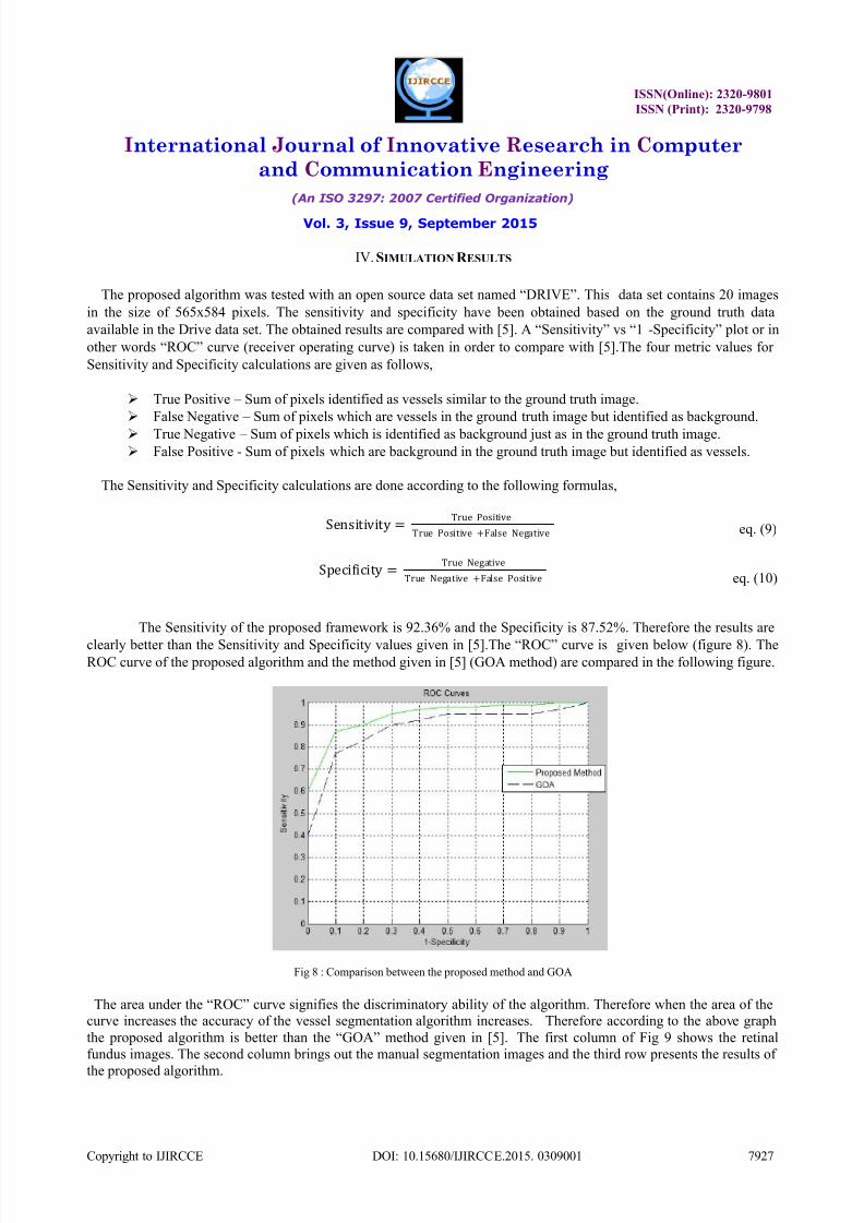
IV. SIMULATION R ESULTS
The proposed algorithm was tested with an open source data set named “DRIVE”. This data set contains 20 images
in the size of 565x584 pixels. The sensitivity and specificity have been obtained based on the ground truth data
available in the Drive data set. The obtained results are compared with [5]. A “Sensitivity” vs “1 -Specificity” plot or in
other words “ROC” curve (receiver operating curve) is taken in order to compare with [5].The four metric values for
Sensitivity and Specificity calculations are given as follows,
True Positive – Sum of pixels identified as vessels similar to the ground truth image.
False Negative – Sum of pixels which are vessels in the ground truth image but identified as background.
True Negative – Sum of pixels which is identified as background just as in the ground truth image.
False Positive - Sum of pixels which are background in the ground truth image but identified as vessels.
The Sensitivity and Specificity calculations are done according to the following formulas,
Sensitivity =True Positive
True Positive +False Negative
Specificity =True Negative
True Negative +False Positive
The Sensitivity of the proposed framework is 92.36% and the Specificity is 87.52%. Therefore the results are
clearly better than the Sensitivity and Specificity values given in [5].The “ROC” curve is given below (figure 8). TheROC curve of the proposed algorithm and the method given in [5] (GOA method) are compared in the following figure.
Fig 8 : Comparison between the proposed method and GOA
The area under the “ROC” curve signifies the discriminatory ability of the algorithm. Therefore when the area of thecurve increases the accuracy of the vessel segmentation algorithm increases. Therefore according to the above graph
the proposed algorithm is better than the “GOA” method given in [5]. The first column of Fig 9 shows the retinal
fundus images. The second column brings out the manual segmentation images and the third row presents the results of
the proposed algorithm.
eq. (9)
eq. (10)

7/21/2019 A Robust Algorithm for Retinal Blood Vessel Extraction
http://slidepdf.com/reader/full/a-robust-algorithm-for-retinal-blood-vessel-extraction 8/9
ISSN(Online): 2320-9801
ISSN (Print): 2320-9798
International Journal of Innovative Research in Computer
and Communication Engineering
(An ISO 3297: 2007 Certified Organization)
Vol. 3, Issue 9, September 2015
Copyright to IJIRCCE DOI: 10.15680/IJIRCCE.2015. 0309001 7928
(a) (b) (c)Fig 9 : Retinal Vessel Segmentation (a) Image from DRIVE Database (b) Manual Segmentation (c) Proposed Method Results
V. CONCLUSION
A method based on histogram equalization and Gabor filter to extract the retinal vessels in fundus images was
represented by the proposed work. The performance of the proposed methodology is evaluated on the DRIVE database.
The results were tested using sensitivity and specificity. They were more accurate than the method given in [5].
R EFERENCES
1. P. Shilpa Joshi, "Retinal Blood Vessel Segmentation," International Journal of Engineering and Innovative Technology (IJEIT), vol. 1, no. 3,
pp. 175-178, March 2012.
2. Suthit Rattathanapad,BunyaritUyyanonvara et al "Vessel Segmentation in Retinal Images using Graph-Theoretical Vessel Tracking,"Conference on Machine Vision Applications, pp. 548-551, 2011.
3.
Lassada Sukkaew, Bunyarit Uyyanonvara et al, "Automated Vessels Detection on Infant Retinal Images," ICCAS, pp. 321-325.4.
P. C. Siddalingaswamy, K. Gopalakrishna Prabhu, "Automatic detection of multiple oriented blood vessels in," J. Biomedical Science andEngineering, pp. 101-107, 2010.
5. Danu onkaew, Rashmi Turior et al, "Automatic Extraction of Retinal Vessels Based on Gradient Orientation Analysis," Eighth International
Joint Conference on Computer Science and Software Engineering (JCSSE), pp. 102-107, 2011.6. Florence Rossant, Maddalena Badellino et al, "A Morphological Approach for Vessel Segmentation in Eye Fundus Images, with Quantitative
Evaluation," Journal of Medical Imaging and Health Informatics, vol. 1, pp. 42-49, 2011.
7. Lim, Jae S, Two Dimentional Signal and Image Processing, Englewood Cliffs: Prentice Hall, 1990.8. Raju.A, Dwarakish et al, "A Comparative Analysis of Histogram Equalization based Techniques for Contrast Enhancment and Brightness
Preserving," International Journal of Signal Processing, Image Processing and Pattern Recognition , vol. 6, no. 5, pp. 353-366, 2013.9. Debmalya Bhattacharya, Jibanpriya Devi et al, "Brain Image Segmentation Technique Using Gabor filter parameter," American Journal of
Engineering Research (AJER), vol. 2, no. 9, pp. 127-132, 2013.
10. L.Siva Yamani, K.Asif et al, "A Noval Method for Extraction of Retinal Blood Vessels using Gabor Filter and Generalized Linear Model,"Global Journal of Trends in Engineering, vol. 2, no. 4, pp. 209-215, 2015.
11. Mandlenkosi Victor Gwetu, Jules-Raymond Tapamo et al, "Segmentation of retinal blood vessels using normalized Gabor filters and automatic
thresholding," South African Computer Journal, pp. 12-24, 2014.12. Alexander Wong, William Bishop, "Simultaneous Gamma Correction and Registration in the Frequency Domain," IPCV, pp. 141-151, 2007.
13.
T.Romen Singh, Sudipta Roy et al, "A New Local Adaptive Thresholding Technique in Binarization," International Journal of Computer
Science, vol. 8, no. 6, pp. 271-277, 2011.
14. Syed Saqib Bukhari, Faisal Shafait et al, "Foreground-background regions guided binarization of camera-captured document images,"
International Workshop on Camera Based Document Analysis and Recognition.

7/21/2019 A Robust Algorithm for Retinal Blood Vessel Extraction
http://slidepdf.com/reader/full/a-robust-algorithm-for-retinal-blood-vessel-extraction 9/9
ISSN(Online): 2320-9801
ISSN (Print): 2320-9798
International Journal of Innovative Research in Computer
and Communication Engineering
(An ISO 3297: 2007 Certified Organization)
Vol. 3, Issue 9, September 2015
Copyright to IJIRCCE DOI: 10.15680/IJIRCCE.2015. 0309001 7929
15. Pradeep.M.V, Fahimuddin Shaik et al, "Extraction of Retinal Blood Vessel by Combining Gabor filter and Generalized Linear model,"
International Journal of Advanced Technology in Engineering and Science, vol. 3, no. 1, pp. 846-855, March 2015.
BIOGRAPHY
Nalan Karunanayake is a Teaching Assistant in the Electrical and Computer Department, Sri Lanka Institute of
Information Technology. He received B.Eng(Hons) degree in 2014 from Sheffield Hallam University, United Kingdom
and B.Sc from University of Sri Jayewardenapura in 2015. His research interests are medical image processing, stereo
vision, point clouds and 3-D modeling.
Manaram Gnanasekera is a Teaching Assistant in the Electrical and Computer Department, Sri Lanka Institute of
Information Technology. He received B.Eng(Hons) degree in 2012 from Sheffield Hallam University, United Kingdomand currently reading for MSc from Sheffield Hallam University. His research interests are Computer Vision, Image
Processing, Simultaneous Localization and Mapping (SLAM).
N.D.Kodikara is Deputy Director, University of Colombo School of Computing. He received B.Sc. in Maths and
Statistics in 1978 from University of Colombo, M.Sc. in Computer Science in 1984 from Manchester,Ph.D. in
Computer Science from Manchester. His research interests are Computer Graphics, Virtual Reality, Image Processing,
Computer Vision.