a review on mucoadhesive drug delivery systems: a …
TRANSCRIPT

IJPCBS 2014, 4(2), 277-290 Dhutwalia Pooja et al. ISSN: 2249-9504
277
INTERNATIONAL JOURNAL OF PHARMACEUTICAL, CHEMICAL AND BIOLOGICAL SCIENCES Available online at www.ijpcbs.com
A REVIEW ON MUCOADHESIVE DRUG DELIVERY SYSTEMS: A
NOVEL APPROACH
Dhutwalia Pooja*, Kanwar Kapil, Anju, Amrit and Ankush
CT Institute of Pharmaceutical Sciences, Shahpur, P.O- Udopur, Near Lambra, Jalandhar -144020, Punjab, India.
INTRODUCTION For the systemic delivery of drugs via various pharmaceutical product of different dosage form, the oral route drug delivery has been known as the most widely utilized route of administration among all other routes1. Thus, oral controlled dosage forms have been developed for the past three decades, due to their considerable therapeutic advantages. However, this approach has not been suitable for those drugs which are absorbed only in a particular portion of gastrointestinal tract (GIT) or which are absorbed in various segment of the GIT to a different extent. Such drugs are characterized by a narrow absorption window due to the relatively short transit time of the gastrointestinal tract i.e. stomach and small intestine2. Thus, after only a short period of less than 6 h, the CR-DF has already left the upper gastrointestinal tract and the drug is released in nonabsorbing
distal segments of the gastrointestinal tract. This results in a short absorption phase that is often accompanied by lesser bioavailability3. Thus, the concept of mucosal adhesive or mucoadhesive was introduced in the early 1980’s, into the field of control drug delivery. Mucoadhesive drug delivery system are those delivery systems which utilizes the assets of bioadhesion of certain water –soluble polymer which on hydration become adhesive, thus can be used for targeting a drug or drug delivery system in particular region of the body for the extended period of time, not only for local targeting of drug but also for the better control of systemic drug delivery4. It prolongs the residence time of the dosage form at the site of application or absorption and facilitates an intimate contact of the dosage form with the underline absorption surface and thus contributes to improved and/or better therapeutic performance of the drug. In recent years many
Review Article
ABSTRACT Mucoadhesion is a field of current interest in the design of drug delivery systems. Mucoadhesion is commonly defined as the adhesion between two materials, at least one of which is a mucosal surface. Mucoadhesive drug delivery system may be designed to enable prolonged residence time of the dosage form at the site of application or absorption and facilitate an intimate contact of the dosage form with the underline absorption surface. Extending the residence time of a dosage form at a particular site and controlling the release of drug from the dosage form are useful especially for achieving controlled plasma level of the drug as well as improving bioavailability. Application of these dosage forms to mucosal surfaces may be of benefit to drug molecules not amenable to the oral route, such as those that undergo acid degradation or extensive first-pass metabolism. The present review describes mucoadhesion, mucoadhesive polymers and use of these polymers in designing different types of mucoadhesive gastrointestinal, nasal, ocular, vaginal and rectal drug delivery systems. The research on mucoadhesives, however, is still in its early stage, and further advances need to be made for the successful translation of the concept into practical application in controlled drug delivery. Keywords: Mucoadhesion, Bioadhesion, Mucoadhesive systems, Drugs delivery.

IJPCBS 2014, 4(2), 277-290 Dhutwalia Pooja et al. ISSN: 2249-9504
278
such mucoadhesive drug delivery systems have been developed for oral, buccal, nasal, gastrointestinal, rectal and vaginal routes for both systemic and local effects5. MECHANISMS OF MUCOADHESION The mechanism of adhesion of certain macro-molecules to the surface of a mucous tissue is not well understood yet. The mucoadhesive must spread over the substrate to initiate close contact and increase surface contact, promoting the diffusion of its chains within the mucus. Attraction and repulsion forces arise and, for a mucoadhesive to be successful, the attraction forces must dominate. Each step can be facilitated by the nature of the dosage form and how it is administered. For example, a partially hydrated polymer can be adsorbed by the substrate because of the attraction by the surface water Thus, the mechanism of mucoadhesion is generally divided in two steps, the contact stage and the consolida-tion stage (Figure 1). The first stage is characterized by the contact between the mucoadhesive and the mucous membrane, with spreading and swelling of the formulation, initiating its deep contact with the mucus layer. In some cases, such as for ocular or vaginal formulations, the delivery system is mechanically attached over the membrane. In other cases, the deposition is promoted by the aerodynamics of the organ to which the system is administered, such as for the nasal route. On the other hand, in the gastrointestinal tract direct formulation attachment over the mucous membrane is not feasible. Peristaltic motions can contribute to this contact, but there is little evidence in the literature showing appropriate adhesion. Additionally, an undesirable adhesion in the esophagus can occur. In these cases, mucoadhesion can be explained by peristalsis, the motion of organic fluids in the organ cavity, or by Brownian motion. If the particle approaches the mucous surface, it will come into contact with repulsive forces and attractive forces. Therefore, the particle must overcome this repulsive barrier. In the consolidation step (Figure 1), the mucoadhesive materials are activated by the presence of moisture. Moisture plasticizes the system, allowing the mucoadhesive molecules to break free and to link up by weak van der Waals and hydrogen bonds. Essentially, there are two theories explaining the consolidation step: the diffusion theory and the dehydration theory. According to diffusion theory, the mucoadhesive molecules and the glycoproteins of the mucus mutually interact by means of interpenetration of their chains and the building of secondary bonds.
For this to take place the mucoadhesive device has features favoring both chemical and mechanical interactions. For example, molecules with hydrogen bonds building groups (–OH, –COOH), with an anionic surface charge, high molecular weight, flexible chains and surface-active properties, which induct its spread throughout the mucus layer, can present mucoadhesive properties. According to dehydration theory, materials that are able to readily gelify in an aqueous environment, when placed in contact with the mucus can cause its dehydration due to the difference of osmotic pressure. The difference in concentration gradient draws the water into the formulation until the osmotic balance is reached. This process leads to the mixture of formulation and mucus and can thus increase contact time with the mucous membrane. Therefore, it is the water motion that leads to the consolidation of the adhesive bond, and not the interpenetration of macromolecular chains. However, the dehydration theory is not applicable for solid formulations or highly hydrated forms6. MUCOADHESION THEORIES Although the chemical and physical basis of muco-adhesion are not yet well understood, there are six classical theories adapted from studies on the performance of several materials and polymer-polymer adhesion which explain the phenomenon. Electronic theory Electronic theory is based on the premise that both mucoadhesive and biological materials possess opposing electrical charges. Thus, when both materials come into contact, they transfer electrons leading to the building of a double electronic layer at the interface, where the attractive forces within this electronic double layer determines the mucoadhesive strength. Adsorption theory According to the adsorption theory, the mucoadhesive device adheres to the mucus by secondary chemical interactions, such as in van der Waals and hydrogen bonds, electrostatic attraction or hydrophobic interactions. For example, hydrogen bonds are the prevalent interfacial forces in polymers containing carboxyl groups. Such forces have been considered the most important in the adhesive interaction phenomenon because, although they are individually weak, a great number of interactions can result in an intense global adhesion.

IJPCBS 2014, 4(2), 277-290 Dhutwalia Pooja et al. ISSN: 2249-9504
279
Wetting theory The wetting theory applies to liquid systems which present affinity to the surface in order to spread over it. This affinity can be found by using measuring techniques such as the contact angle. The general rule states that the lower the contact angle then the greater the affinity. The contact angle should be equal or close to zero to provide adequate spreadability. The spreadability coefficient, SAB, can be calculated from the difference between the surface energies γB and γA and the interfacial energy γAB, as indicated in equation (1)
SAB= γB- γA –γAB……………………………… (1) The greater the individual surface energy of mucus and device in relation to the interfacial energy, the greater the adhesion work, WA, i.e. the greater the energy needed to separate the two phases7.
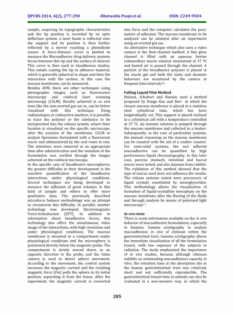
WA= γB+γA –γAB……………………………………… (2) Diffusion theory Diffusion theory describes the interpenetration of both polymer and mucin chains to a sufficient depth to create a semi-permanent adhesive bond (Figure 4). It is believed that the adhesion force increases with the degree of penetration of the polymer chains. This penetration rate depends on the diffusion coefficient, flexibility and nature of the mucoadhesive chains, mobility and contact time . According to the literature, the depth of in-terpenetration required to produce an efficient bioadhesive bond lies in the range 0.2-0.5 μm. This interpenetration depth of polymer and mucin chains can be estimated by equation 3:
l= (tDb) 1/2 ………………………………………………..(3) where t is the contact time, and Db is the diffusion coefficient of the mucoadhesive material in the mucus. The adhesion strength for a polymer is reached when the depth of penetration is approximately equivalent to the polymer chain size . In order for diffusion to occur, it is important that the components involved have good mutual solubility, that is, both the bioadhesive and the mucus have similar chemical structures. The greater the structural similarity, the better the mucoadhesive bond8. Fracture theory This is perhaps the most-used theory in studies on the mechanical measurement of mucoadhesion. It analyses the force required to separate two surfaces after adhesion is established . This force, sm, is frequently calculated in tests of resistance to
rupture by the ratio of the maximal detachment force, Fm, and the total surface area, A0, involved in the adhesive interaction (equation 4):
Sm =Fm/Ao………………………………………………………..(4) In a single component uniform system, the fracture force, sj, which is equivalent to the maximal rupture tensile strength, sm, is proportional to the fracture energy (gc), for Young’s module (E) and to the critical breaking length (c) for the fracture site, as described in equation 5:
Sf=(gcE/C)1/2…………………………………(5) Fracture energy (gc) can be obtained from the reversible adhesion work, Wr (energy required to produce new fractured surfaces), and the irreversible adhesion work, Wi (work of plastic deformation provoked by the removal of a proof tip until the disruption of the adhesive bond), and both values are expressed as units of fracture surface (Af).
Gc=Wr+Wi………………………………………………………………(6) The elastic module of the system (E) is related to the stress (s) and to the shear (e) by Hooke’s law: A criticism of this analysis is that the system under investigation must have known physical dimensions and should be constituted by a single and uniform material. In virtue of this, the relationship obtained cannot be applied to analyze the fracture site of a multiple component bioa-dhesive. In this case, the equation should be expanded to accommodate elastic dimensions and modules for each component. Besides, it must be considered that a failure of adhesion will occur at the bioadhesive interface. However, it has been demonstrated that the rupture rarely occurs at the surface, but near it or at the weakest point, which can be the interface itself, the mucus layer or the hydrated region of the mucus, as illustrated in Figure 5 . Since the fracture theory is concerned only with the force required to separate the parts, it does not take into account the interpenetration or diffusion of polymer chains. Consequently, it is appropriate for use in the calculations for rigid or semi-rigid bioadhesive materials, in which the polymer chains do not penetrate into the mucus layer9.
Mechanical theory Mechanical theory considers adhesion to be due to the filling of the irregularities on a rough surface by a mucoadhesive liquid. Moreover, such roughness increases the interfacial area available to interactions thereby aiding dissipating energy

IJPCBS 2014, 4(2), 277-290 Dhutwalia Pooja et al. ISSN: 2249-9504
280
and can be considered the most important phenomenon of the process. It is unlikely that the mucoadhesion process is the same for all cases and therefore it cannot be described by a single theory. In fact, all theories are relevant to identify the important process variables.The mechanisms governing mucoadhesion are also determined by the intrinsic properties of the formulation and by the environment in which it is applied. Intrinsic factors of the polymer are related to its molecular weight, concentration and chain flexibility. For linear polymers, mucoadhesion increases with molecular weight, but the same relationship does not hold for non-linear polymers. It has been shown that more concentrated mucoadhesive dispersions are retained on the mucous mem-brane for longer periods, as in the case of systems formed by in situ gelification. After application, such systems spread easily, since they present rheological properties of a liquid, but gelify as they come into contact the absorption site, thus preventing their rapid removal. Chain flexibility is critical to consolidate the interpenetration between formulation and mucus. Environment-related factors include pH, initial contact time, swelling and physiological variations. The pH can influence the formation of ionizable groups in polymers as well as the formation of charges on the mucus surface. Contact time between mucoadhesive and mucus layer determines the extent of chain interpenetration. Super-hydration of the system can lead to build up of mucilage without adhesion. The thickness of the mucus layer can vary from 50 to 450 μm in the stomach to less than 1μm in the oral cavity. Other physiolo-gical variations can also occur with diseases. None of these mechanisms or theories alone can explain the mucoadhesion which occurs in an array of different situations. However, the understanding of these mechanisms in each instance can help toward the development of new mucoadhesive products10. MUCOADHESIVE MATERIALS The first study presenting the use of a mucoadhesive material was conducted by Nagai, and proposed an improved treatment for stomatitis by using adhesive tablets. Additionally, an increase in the systemic bioavailability of insulin was observed in the form of bioadhesive powder after nasal administration in dogs. Thereafter, bioadhesive materials have been used as absorption promoters for several administration routes. Earlier experiments were also done with known polymers available on the market, such as polyacrylic
acids. Currently, the latest research is seeking to develop materials that direct the formulation more specifically to the action site and that can offer other functions besides mucoadhesion such as control over permeation within epithelial tissues, and inactivation of enzymes which can compromise release system action.
First generation mucoadhesive materials These materials are natural or synthetic hydrophilic molecules containing numerous organic functions that generate hydrogen bonds such as carboxyl, hydroxyl and amino groups, which do not adhere specifically onto several surfaces. The very first use of mucoadhesive was as denture fixers and the most known examples are carbomers, chitosans, alginates and cellulose derivatives. They can be incorporated into solid formulations, such as tablets, transdermal adhesives and microparticles, and into semisolid formulations including gels, ointments, pastes and suppositories. These polymers can be subdivided into three classes: cationic, anionic and nonionic. Cationic molecules can interact with the mucus surface, since it is negatively charged at physiological pH. Mucoadhesion of cationic polymers such as chitosan, occurs because of the electrostatic interactions of their amino groups with the sialic groups of mucin in the mucus layer. Chitosan is a semi-synthetic polymer obtained by the deacetylation of chitin and has been extensively investigated as a drug delivery mucoadhesive system. Studies have demonstrated that chitosan can promote the absorption of hydrophilic molecules by the structural reorganization of the proteins associated to the intercellular junctions. In contrast, synthetic polymers derived from polyacrylic acid (carbomers) are negatively charged but are also mucoadhesive. In this case, mucoadhesion results from physical-chemical processes, such as hydrophobic interactions, hydrogen and van der Waals bonds, which are controlled by pH and ionic composition. Polyacrylic acid hydrogels have been extensively studied as mucoadhesive systems. Their chains are flexible and have non-abrasive characteristics when in the partially hydrated state, which decreases the tissue damage caused by friction when they come into contact. The majority of polyacrylic acid derivatives are not water soluble, such as polycarbophil, but form viscous gels when hydrated. Other examples of anionic polymers are carboxymethylcellulose and alginates. The alginates, negatively charged polysaccharides, are

IJPCBS 2014, 4(2), 277-290 Dhutwalia Pooja et al. ISSN: 2249-9504
281
widely used in the production of microparticles and are frequently reported as polyanionic mucoadhesive polymers11. Nonionic polymers, including hydroxypropylme-thylcellulose, hydroxyethylcellulose and methylcellulose, present weaker mucoadhesion force compared to anionic polymers.
Second generation mucoadhesive materials Studies on novel mucoadhesive systems involve the use of multifunctional materials. An ideal polymer should exhibit the ability to incorporate both hydrophilic and lipophilic drugs, show mucoadhesive properties in its solid and liquid forms, inhibit local enzymes or promote absorption, be specific for a particular cellular area or site, stimulate endocytosis and finally to have a broad safety range . These novel multifunctional mucoadhesive systems are classified as second generation polymers. They are an alternative to non-specific bioadhesives because they bind or adhere to specific chemical structures on the cell or mucus surface. Good examples of these molecules are lectins, invasins, fimbrial proteins, antibodies , and those obtained by the addition of thiol groups to known molecules. Lectins are immunogenic vegetal glycoproteins that specifically recognize sugar molecules. They are able to non-covalently bind to glycosilated components of the cellular membrane but not of the mucus, and adhesion can therefore be called cytoadhesion. Through the transmission of a cellular signal, this specific bond can result not only in bioadhesion but also in cellular internalization by different lysosomal and non-lysosomal mechanisms (Lehr, 2000). The most commonly found lectins are those isolated from Abrus precatroius, Agaricus bisporus, Anguilla anguilla, Arachis hypogaea, Pandeiraea simplicifolia, and Bauhinia purpurea. Bacterial invasins are proteins from the membrane of Yersinia pseudotuberculosis that stimulate fagocytosis at cellular membrane through linkage with integrin receptors . Bacterial fimbrial proteins are able to adhere to the epithelial surface of erythrocytes. This adhesion is related to the pathogenicity of the bacteria. Bacterial adhesive factors can be an efficient mechanism of improving adhesion of mucoadhesive agents used in release systems . Antibodies can be produced against selected mo-lecules present on the mucus surface. Due to their high specificity, antibodies can be a rational choice as polymeric ligand in the development of site-specific mucoadhesives. This strategy can be
useful for instance, in drugs targeting tumor tissues12.
METHODS OF ANALYZING MUCOADHESION No technology has still been developed specifically to analyze mucoadhesion. Most of the tests available were adapted from other preexisting techniques but are useful and necessary for selecting the promising candidates as mucoadhesives as well as in elucidating their mechanisms of action.
In vitro and ex vivo tests In vitro/ex vivo tests are important in the develop-ment of a controlled release bioadhesive system because they contribute to studies of permeation, release, compatibility, mechanical and physical stability, superficial interaction between formulation and mucous membrane and strength of the bioadhesive bond. These tests can simulate a number of administration routes including oral, buccal, periodontal, nasal, gastrointestinal, vaginal and rectal. The in vitro and ex vivo tests most prevalent in the literature are reported below.
Techniques utilizing gut sac of rats The everted gut sac technique is an example of an ex vivo method. It has been used since 1954 to study intestinal transport. Applied this method on mucoadhesion assays. It is easy to reproduce and can be performed in almost all laboratories. Figure 6 schematically represents the technique. A segment of intestinal tissue is removed from the rat, everted, and one of its ends sutured and filled with saline. The sacs are introduced into tubes containing the system under analysis at known concentrations, stirred, incubated and then removed. The percent adhesion rate of the release system onto the sac is determined by subtracting the residual mass from the initial mass. Other techniques use non-everted gut sac filled rats’ intestines with liposome suspensions. The sacs were sealed and incubated in saline. After a stipulated time, the number of liposomes adhered before (N0) and after (Ns) incubation was assessed with a coulter counter and the percent mucoadhesive was expressed by equation 7.
%adhesive={No-Ns/ No }* 100…………………(7) Tests measuring mucoadhesive strength Most in vitro/ex vivo methodologies found in the literature are based on the evaluation of mucoadhesive strength, that is, the force required to break the binding between the model membrane and the mucoadhesive. Depending on the direction in which the mucoadhesive is separated from the substrate, is it possible to

IJPCBS 2014, 4(2), 277-290 Dhutwalia Pooja et al. ISSN: 2249-9504
282
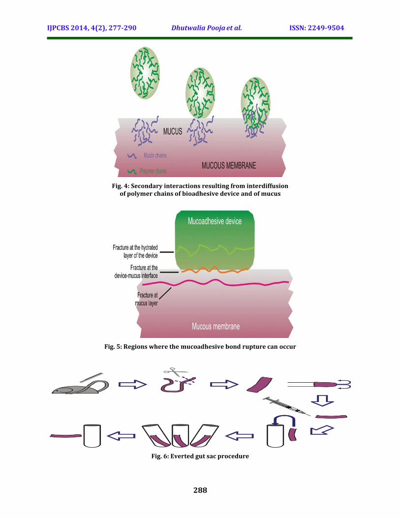
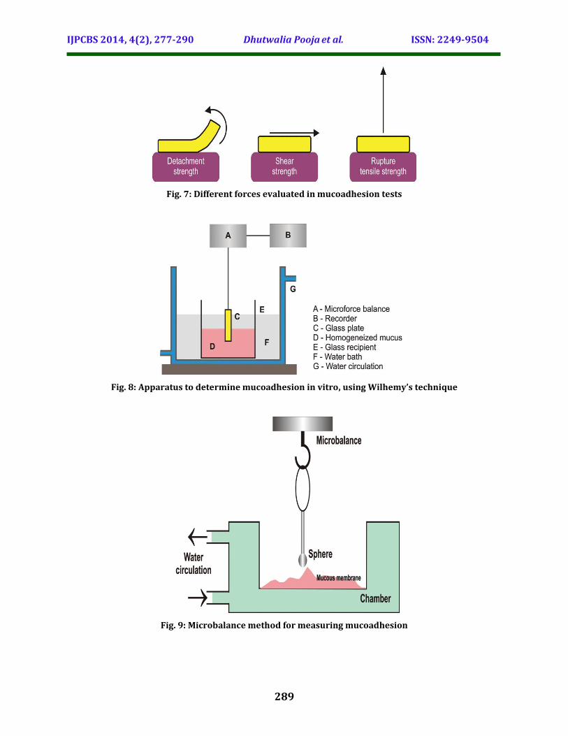
obtain the detachment, shear, and rupture tensile strengths as indicated in Figure 7. The force most frequently evaluated in such tests is rupture tensile strength. Generally, the equipment used is a texture analyzer (Figure 8) or a universal testing machine. In this test, the force required to remove the formulation from a model membrane is measured, which can be a disc composed of mucin, a piece of animal mucous membrane, generally porcine nasal mucus or intestinal mucus from rats . Based on results, a force-distance curve can be plotted which yields the force required to detach the mucin disc from the surface with the formulation, the tensile work (area under the curve during the detachment process), the peak force and the deformation to failure . This method is more frequently used to analyze solid systems like microspheres, although there are also studies on semi-solid materials. In addition to rupture tensile strength, the texture analyzer can also, as inferred by its name, evaluate the texture of the formulations and assess other mechanical properties of the system. A mobile arm containing an analytical probe forces down into a sample held in a flask placed on the equipment’s platform. Speed rate, time and depth are preset. From the resulting force-time and force-distance plots, it is possible to calculate the hardness (force required to reach a given deformation), compressibility (work required to deform the product during the compression), and adhesiveness (work required to overcome the attraction forces between the surfaces of sample and probe). Using this technique, it is possible to perform a previous evaluation of the material’s adhesive capacity, evidencing mucoadhesion properties. Mucoadhesion strength can also be measured in terms of shear strength. This test measures the force required to separate two parallel glass slides covered with the polymer and with a mucus film. This can also be done using Wilhemy’s model (Figure 9), in which a glass plate is suspended by a microforce balance and immersed in a sample of mucus under controlled temperature. The force required to pull the plate out of the sample is then measured under constant experimental conditions. Although measures taken by this method are reproducible, the technique involves no biological tissue and therefore does not provide a realistic simulation of biological conditions. Wilhemy’s plate technique, or the microforce balance technique, can also be modified in order to measure the specific adhesion force of microparticles. This involves the use of a microtensiometer and a microforce balance (Figure 10) and is specific, yielding both
contact angle and surface tension. The mucous membrane is placed in a small mobile chamber with both pH and physiological temperature controlled. A unique microsphere is attached by a thread to the stationary microbalance. The chamber with the mucous membrane is raised until it comes into contact with the microsphere and, after contact time, is lowered back to the initial position. Following the trajectory, and with the aid of software, results can be obtained for several parameters such as fracture strength, deformation and rupture tensile strength, from a load versus deformation curve, as shown in Figure 11 . The microforce balance is not indicated for microspheres smaller than 300 μm, but has the advantage of simulating physiological conditions and providing results at a more microscopic level, besides being more reproducible and sensitive13.
Rheological methods This category of methods are all carried out in vitro and were first proposed by Hassan and Gallo, who used viscosimetric assays to macroscopically analyze the formulation-mucin interaction. From this test, it is possible to obtain the mucoadhesion force by monitoring the viscosimetric changes of the system constituted by the mixture of the polymer chosen and mucin. The energy of the physical and chemical bonds of the mucin-polymer interaction can be transformed into mechanical energy or work. This work, which causes the rearrangements of the macromolecules, is the basis of the change in viscosity. A way to analyze the coefficient of viscosity of a hydrophilic dispersion containing mucin plus the mucoadhesive polymer is through the contribution of each component, which results in equation 8:
ηt = ηm + ηp + ηb………………………………… (8) where ηt is the coefficient of viscosity of the system, and ηm and ηp are the coefficients of viscosity of mucin and bioadhesive polymer, respectively. The bioadhesion component, ηb, can be obtained from equation 9, resulting in equation 9:
ηb = ηt – ηm – ηp…………………………………… (9) For equations 9 and 10 to be valid, all components should be measured at the same concentration, temperature, time and shear gradient. The bioadhesion force, F, is determined by equation 10:
F = ηbs……………………………………………….. (10) where σ is the shear gradient.

IJPCBS 2014, 4(2), 277-290 Dhutwalia Pooja et al. ISSN: 2249-9504
283
The main disadvantage of this method is the breakdown of the polymer and mucin network under continuous flow. To avoid this problem, the method was adapted using oscillatory rheology ; Hägerström, 2003). Based on the same assumption that the rheological response of polymer-mucin mixture should be greater than the contributions from the gel and isolated mucin, a parameter called rheological synergism can be obtained. This method is more advantageous than the original, since oscillatory rheology is a non-destructive technique and simultaneously measures viscosity and elastic behavior and can be used to determine mucoadhesion between polymers and mucin .The evaluation of rheological synergism can be done through two types of oscillatory assays: stress sweep and frequency sweep. In stress sweep, the elastic (G´) and viscous (G´´) moduli are obtained under constant frequency. This is used to investigate the influence of stress on the dynamic modulus, which should be obtained in the linear viscoelastic region, that is, the region where the material response is characteristic for its microstructure. Above this region, the structure is destroyed. The magnitude of the moduli is a qualitative indication of the system structure. Three situations can be found for polymeric dispersion: G’>> G” for a chemically interconnected system, G’>G” for chains with secondary bonds, and G’≤ G” for dispersions with physically-bound molecules. The quantitative measure of rheological synergism (ΔG’) can be calculated either in relation to G’or G” , as shown in equation 11.
ΔG=Gmixture - [Gpolymer + Gmucin ]……………. (11) In frequency sweep, stress is maintained constant. The structure of the system can remain intact during the assay if it is conducted in the linear viscoelastic region. Under constant stress and at low frequencies, better structured systems present greater elastic modulus than viscous modulus and both are independent of frequency. On a log-log graph, they are represented by a constant straight line. For less organized systems, dynamic moduli are dependent on the frequency and a slope is observed. This test enables analysis of the dynamic viscoelastic parameters corresponding to the same frequency as a function of polymer or mucin concentration, yielding the rheological behavior in relation to the concentration of the system constituents. Reveals an alternative parameter of rheological synergism, called relative rheological synergism parameter, and with which it is possible to
quantitatively compare the force of polymer-mucin mixture with the isolated polymer: where DG´ is the rheological synergism, given by the difference between elastic modulus of the mixture (DG´mixture) and the elastic modulus of the polymer (G´p). However, DG´relative has the disadvantage of a negative limit up to -1, while the positive values run to infinity. Therefore, the magnitude of positive values cannot be compared with that of negative values. Thus, a new relative parameter was proposed called the logarithmic relation of elastic module (log G´), which is given by the ratio between elastic modulus of the mixture (Gmix) and the elastic modulus of the polymer (G´p ), as indicated in equation 12. logG= log ( Gmix/ Gp ) …………………………………………..(12) This parameter offers the advantage that both po-sitive and negative values have the same magnitude, and are therefore comparable. For instance, the value 1 means that G´ of the mixture is 10-fold greater than that of the isolated polymer. Rheological tests are performed totally in vitro and consequently are conducted in combination with the rupture tensile strength test, most frequently used in studies on mucoadhesion. The experimental conditions of both tests differ and there are cases in which the techniques are complementary. Rheology measures the mechanical properties of the system, i.e., the resistance against flow and deformation, assessing the changes the system undergoes in the presence of mucin. However, rheology does not provide any direct information on what occurs at the interface, because the two phases – mucin and polymer – are mixed together prior to the experiment. In the rupture tensile strength test, the interface is artificially created. Even with this difference, when the mucin-polymer produces rheological synergism, a corresponding structure organization is observed at the mucoadhesive interface. The rupture tensile strength test can be applied to solids and semi-solids, while rheology is applicable to semi-solids and liquids. Experimental conditions are critical in the rupture tensile strength test and there are several variables (sample layer, hydration, time of hydration, sample load, time of loading, detach-ment rate, etc.), which should be optimized and set in order to produce reproducible results. The reproducibility of rheological measures is reasonably good, since the measures are taken on already balanced mixtures; composition, pH, and temperature can be carefully controlled and therefore fewer repetitions are necessary to obtain statistically significant data. Thus, it can be

IJPCBS 2014, 4(2), 277-290 Dhutwalia Pooja et al. ISSN: 2249-9504
284
concluded that both methods contribute to different extents toward explaining the mucoadhesive phenomenon, depending on the mucoadhesion mechanism involved, system type, polymer used, etc14. Tests analyzing molecular interactions involved in mucoadhesion The general problem arising from methods that show the adhesion force and from the rheological methods is that the mucoadhesive response is seen macroscopically while the interactions occur at a microscopic level. The use of low frequency dielectric spectroscopy represents an attempt to study gel-mucus interactions near the molecular level. It evaluates the possible physicochemical interactions between molecules and glycoproteins of the mucus at the interface, which is considered the step preceding the formation of bonds during the mucoadhesion process. This technique involves the study of material response to the application of an electrical field. A sinusoidal voltage is applied throughout the sample and the response is measured in function of the frequency. From the responses, the impedance or permittivity of the sample is obtained and the property of charges changing in the system can be determined . This technique can provide information about the compatibility between mucus and mucoadhesive system by means of the evaluation of the movement of the charged particles. This compatibility is achieved according to the ease with which the particle crosses the barrier between the gel and mucous membrane. The dielectric measures reveal information about the gel and the mucous membrane separately, and about the interface between them. Since the mucoadhesion process can be a conse-quence of interactions between the mucus layer and the mucoadhesive polymer, it is highly dependent upon the molecular structure, including its charge. It is also well known that glycoproteins molecules, which form the mucus structure, are negative at physiological pH. By means of zeta potential, it is possible to understand the polymer-mucin electrostatic interactions . The zeta potential of dispersion is defined as the potential between the liquid superficial layer surrounding the dispersed particle and the remaining solution volume. It is a measure of the net surface charge of particles in a dispersed system. In this test, the mucin particles are suspended in an appropriate buffer and mixed with a solution of the polymer. If the addition of the polymer changes the zeta potential value of
the mucin particles, this can suggest greater affinity between polymer and mucin particles. Another technique being applied to evaluate mo-lecular interactions is the optical biosensor, or resonant mirror biosensor technique. Sigurdsson, Loftsson and Lehr used this technique to measure the interaction between glycoproteins of the mucus and different polymers. It allows the monitoring of any interaction between two unknown molecules in real time, since one of them can be immobilized with covalent or non-covalent on the system surface while the other remains in solution at the surface. The molecules in solution, when binding to the immobilized molecules, alter the refraction index of the medium and this change is detected by the screening of a laser beam. The results of this study suggested the need for a clearer definition of mucoadhesion, because they called into question the polymers that are swelling dependent and undergo in situ gelification, because they do not seem to interact with glycoproteins, although they are called mucoadhesives. Another test using the same principle, the Biacore test, was applied for the analysis of mucoadhesion. This test is based on the passage of a mucin suspension through a sensor containing the immobilized polymer. When a mucin particle binds to the polymer at the sensor, both the solute concentration and the refraction index on this surface undergo changes, where the interaction is quantitatively evaluated and reproduced on a diagram. The sensor is a chip with a glass surface covered in a fine gold layer, where functional groups are introduced and the polymer is attached15. Imaging methods Optical microscopes offer insufficient resolution for studying effects at a molecular level. For such investigations, a resolution at micro- or nanometric level is needed. Electronic microscopy gives a larger view, but the environmental conditions in which the sample must be submitted are far from the physiological conditions. For instance, the samples are analyzed in a vacuum chamber and generally are covered with a metallic film to avoid changes caused by the electronic .Atomic force microscopy (AFM) is a relatively new technique that overcomes such restrictions, because it can be used under any environmental conditions, in air, liquids or vacuum. It enlarges
more than 109-fold, which enables visualization of
isolated atoms and offers a tridimensional image of the surface. The equipment has a support combined with a probe perpendicularly attached to it. This tip moves toward a plane parallel to the
IJPCBS 2014, 4(2), 277-290 Dhutwalia Pooja et al. ISSN: 2249-9504
285
sample, acquiring its topographic characteristics and the tip position is recorded by an optic deflection system: a laser beam is reflected onto the support and its position is then further reflected by a mirror reaching a photodiode sensor. A force-distance curve is plotted to measure the Mucoadhesive drug delivery systems forces between this tip and the surface of interest. This curve is then used in bioadhesion studies. This entails coating the tip in adhesive material, which is generally spherical in shape and then the interaction with the surface, in this case the mucous membrane, can be measured. Besides AFM, there are other techniques using photographic images, such as fluorescence microscopy and confocal laser scanning microscopy (CSLM). Results achieved in ex vivo tests like the non-everted gut sac te, can be better visualized with this technique. Using radioisotopes or radioactive markers, it is possible to trace the polymer or the substance to be incorporated into the release system, where their location is visualized on the specific microscope, after the excision of the membrane. CSLM to analyze liposomes formulated with a fluorescent tracer and administered by the oral route in rats. The intestines were removed at an appropriate time after administration and the retention of the formulation was verified through the images achieved on the confocal microscope. In the specific case of bioadhesive microspheres, the greater difficulty in their development is the sensitive quantification of the bioadhesive interactions under physiological conditions. Several techniques are being developed to measure the adhesion of great volumes in this kind of sample and others to offer more qualitative data. The previously described microforce balance methodology was an attempt to circumvent this difficulty. In parallel, another technology was developed, Electromagnetic Force-transduction (EFT). In addition to information about bioadhesive forces, this technology also offers the simultaneous video image of the interactions, with high resolution and under physiological conditions. The mucous membrane is mounted in a compartment under physiological conditions and the microsphere is positioned directly below the magnetic probe. The compartment is slowly moved down, in an opposite direction to the probe, and the video camera is used to detect sphere movement. According to the movement, the control system increases the magnetic current and the resulting magnetic force (Fm) pulls the sphere to its initial position, separating it from the tissue. After the experiment, the magnetic current is converted
into force and the computer calculates the para-meters of adhesion. The mucous membrane to be analyzed can be attained after an experiment using an everted gut sac. An alternative technique which also uses a video camera is the flow-channel method. A fine glass channel is filled with an aqueous bovine submaxillary mucin solution maintained at 37 ºC and humid air is passed through the channel. A particle of the bioadhesive polymer is placed in the mucin gel and both the static and dynamic behaviors are monitored by the camera at frequent time intervals16. Falling Liquid Film Method Nielsen, Schubert and Hansen used a method proposed by Rango Rao and Buri in which the chosen mucous membrane is placed in a stainless steel cylindrical tube, which has been longitudinally cut. This support is placed inclined in a cylindrical cell with a temperature controlled at 37 ºC. An isotonic solution is pumped through the mucous membrane and collected in a beaker. Subsequently, in the case of particulate systems, the amount remaining on the mucous membrane can be counted with the aid of a coulter counter. For semi-solid systems, the non adhered mucoadhesive can be quantified by high performance liquid chromatography. In this later case, porcine stomach, intestinal and buccal mucus were tested, and also jejunum from rabbits. The validation of this method showed that the type of mucus used does not influence the results. The release systems tested were precursors of liquid crystals constituted by monoglycerides. This methodology allows the visualization of formation of liquid-crystalline mesophase on the mucous membrane after the flowing of the fluids and through analysis by means of polarized light microscopy17. In vivo tests There is scant information available on the in vivo behavior of mucoadhesive formulations, especially in humans. Gamma scintigraphy to analyze mucoadhesion in vivo of chitosan within the gastrointestinal tract. Gamma scintigraphy allows the immediate visualization of all the formulation transit, with low exposure of the subjects to radiation. The study emphasized the importance of in vivo studies, because although chitosan exhibits an outstanding mucoadhesion capacity in vitro, the retention time at the absorption site in the human gastrointestinal tract was relatively short and not sufficiently reproducible. The gastrointestinal transit time in animals can also be evaluated in a non-invasive way, in which the

IJPCBS 2014, 4(2), 277-290 Dhutwalia Pooja et al. ISSN: 2249-9504
286
release systems can be formulated with opaque radioisotopes and signals can be followed by X-rays, without affecting normal gastrointestinal motility18. CONCLUSIONS Studies on mucoadhesive systems have focused on a broad array of aspects. It is a growth area whose goal is the development of new devices and more “intelligent” polymers, as well as the creation of new methodologies that can better elucidate the mucoadhesion phenomenon. With the great influx of new molecules stemming from drug research, mucoadhesive systems may play an increasing role in the development of new pharmaceuticals. REFERENCES 1. Ahuja A, Khar RK and Ali J. Mucoadhesive
drug delivery systems. Drug Dev Ind Pharm. 1997;23(5):489-515.
2. Andrews GP and Jones DS. Rheological characterization of bioadhesive binary polymeric systems designed as platforms for drug delivery implants. Biomacromol. 2006;7:899-906.
3. Andrews GP, Laverty TP and Jones DS. Mucoadhesive polymeric platforms for controlled drug delivery. Eur J Pharm Biopharm. 2008;71(3):505-518.
4. Bocataj M, Vovk T, Kerec M, Dimnik AD, Grabnar I and Mrhar A. The correlation between zeta potential and mucoadhesion strength on pig vesical mucosa. Biol Pharm Bull. 2003;26(5):743-746.
5. Bravo-Osuna I, Vauthier C, Farabollini A, Palmieri GF and Ponchel G. Mucoadhesion mechanism of chitosan and thiolated chitosan-poly(isobutiyl cyanoacrylate) core-shell nanoparticles. Biomaterials. 2007;28(13):2233-2243.
6. Bromberg L, Temchenko M, Alakhov V and Hatton TA. Bioadhesive properties and rheology of polyether-modified poly(acrylic acid) hydrogels. Int J Pharm. 2004;282(1):45-60.
7. Bruschi ML and Freitas O. Oral bioadhesive drug delivery systems. Drug Ind. Pharm. 2005;31(3):293-310.
8. Bruschi ML, Jones DS, Panzeri H, Gremião MPD, Freitas O and Lara Ehg. Semisolid systems containing propolis for the treatment of periodontal disease: in vitro release kinetics, syringeability, rheological, textural, and mucoadhesive properties. J Pharm Sci. 2007;96(8):2074-2089.
9. Bruschi ML, Jones DS, Panzeri H, Gremião MPD, Freitas O and Lara Ehg. Development and characterization of precursor of liquid crystalline phase with propolis-containing microparticles for use in the treatment of periodontal disease. Drug Develop Ind Pharm. 2008;34(3):267-278.
10. Callens C, Ceulemans J, Ludwig A, Foreman P and Remon JP. Rheological study on mucoadhesivity of some nasal powder formulations. Eur J Pharm Biopharm. 2003;55(3):323-328.
11. Ceulemans J, Vinckier I and Ludwig A. The use of xantan gum in an ophthalmic liquid dosage form: rheological characterization of the interaction with mucin. J Pharm Sci. 2002;91(4):1117-1127.
12. Chowdary CPR and Rao YS. Mucoadhesive microspheres for controlled drug delivery. Biol Pharm Bull. 2004;27(11):1717-1724.
13. Cleary J, Bromberg L and Magner E. Adhesion of Polyether-Modified Poly(acrylic acid) to Mucin. Langmuir. 2004;20(22):9755-9762.
14. Formariz TP, Urban MCC, Silva Junior AA, Gremião MPD and Oliveira AG. Microemulsões e fases líquidas cristalinas como sistemas de liberação de fármacos. Rev Bras Ciênc Farm. 2005;41(3):301-313.
15. Goto T, Morishita M, Kavimandan NJ, Takayama K and Peppas NA. Gastrointestinal transit and mucoadhesive characteristics of complexation hydrogels in rats. J Pharm Sci. 2006;95(2):462-469.
16. Grabovac V, Guggi D and Bernkop-Schnürch A. Comparison of the mucoadhesive properties of varios polymers. Adv Drug Del Rev. 2005;57(11):1713-1723.

IJPCBS 2014, 4(2), 277-290 Dhutwalia Pooja et al. ISSN: 2249-9504
287
Fig. 1: The two steps of the mucoadhesion process
Fig. 2: Dehydration theory of mucoadhesion
Fig. 3: Schematic diagram showing influence of contact angle
between device and mucous membrane on bioadhesion
IJPCBS 2014, 4(2), 277-290 Dhutwalia Pooja et al. ISSN: 2249-9504
288
Fig. 4: Secondary interactions resulting from interdiffusion of polymer chains of bioadhesive device and of mucus
Fig. 5: Regions where the mucoadhesive bond rupture can occur
Fig. 6: Everted gut sac procedure
IJPCBS 2014, 4(2), 277-290 Dhutwalia Pooja et al. ISSN: 2249-9504
289
Fig. 7: Different forces evaluated in mucoadhesion tests
Fig. 8: Apparatus to determine mucoadhesion in vitro, using Wilhemy’s technique
Fig. 9: Microbalance method for measuring mucoadhesion

IJPCBS 2014, 4(2), 277-290 Dhutwalia Pooja et al. ISSN: 2249-9504
290
Fig. 10: Constituents of AFM and the adaptations made for measuring the adhesive force between polymer and mucus surface
Fig. 11: Elements of EFT