a rent that let meningitis for lease - cpachennai.com filežneck stiffness, positive kernig’s and...
TRANSCRIPT

A RENT THAT LET MENINGITIS FOR LEASE
DR.V.AKILA SIVAKUMAR
(DNB PG)
BRS HOSPITAL,CHENNAI.
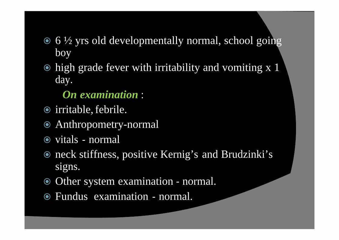
ž 6 ½ yrs old developmentally normal, school going boy
ž high grade fever with irritability and vomiting x 1 day.
On examination :
ž irritable, febrile.
ž Anthropometry-normal
ž vitals - normal
ž neck stiffness, positive Kernig’s and Brudzinki’ssigns.
ž Other system examination - normal.
ž Fundus examination - normal.

ž Investigations:
ž CBC- polymorphic leucocytosis (TC-14,600); Hb & platelets-normal,peripheral smear study-normal.
ž LFT & RFT-Normal.
ž CT Brain – Normal study.
ž CSF analysis- sugar-low; protein-normal,
cells- polymorphs predominant.
Gram stain-Gram positive cocci in clusters .
Treatment:
Meningitic dose of Ceftriaxone (Responded very well).

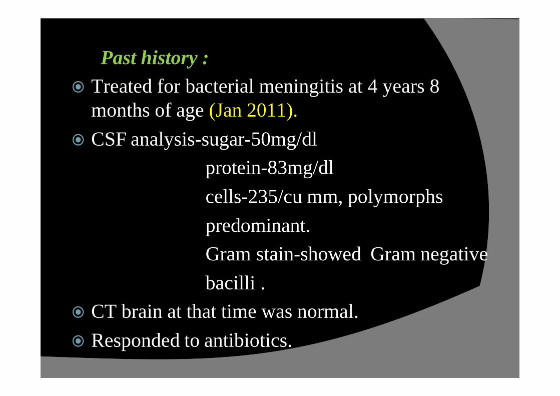
Past history :
ž Treated for bacterial meningitis at 4 years 8 months of age (Jan 2011).
ž CSF analysis-sugar-50mg/dl
protein-83mg/dl
cells-235/cu mm, polymorphs
predominant.
Gram stain-showed Gram negative
bacilli .
ž CT brain at that time was normal.
ž Responded to antibiotics.
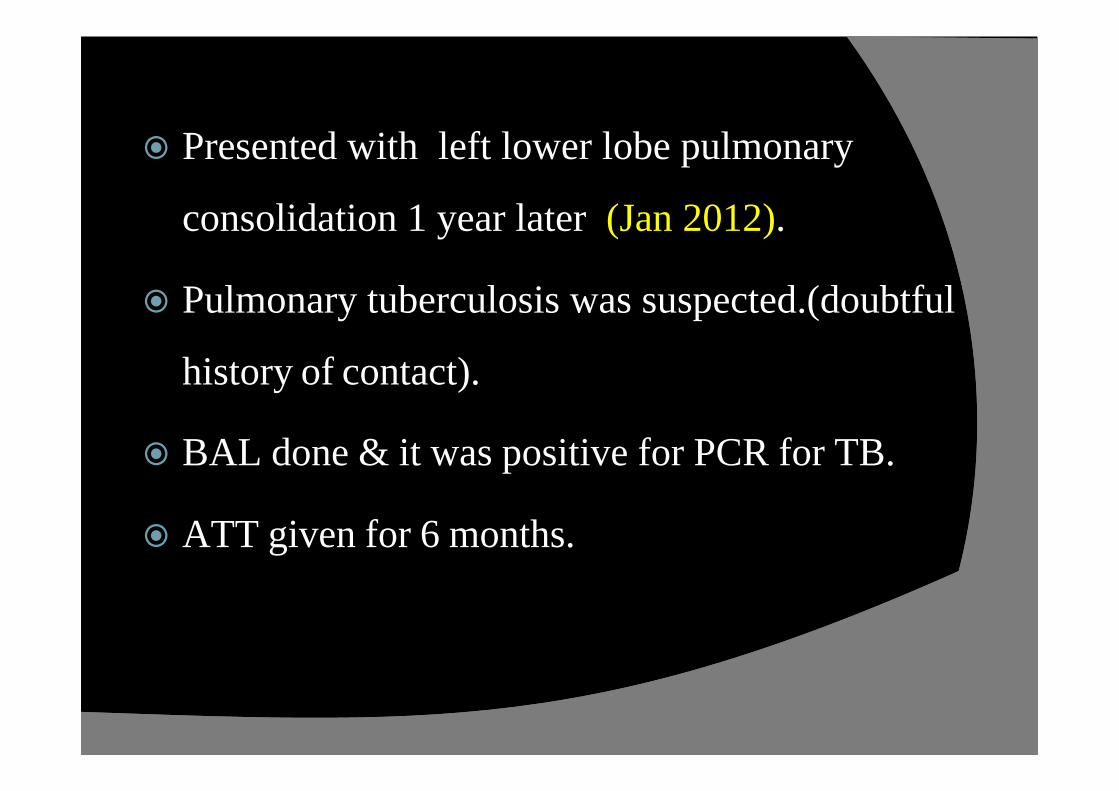
ž Presented with left lower lobe pulmonary
consolidation 1 year later (Jan 2012).
ž Pulmonary tuberculosis was suspected.(doubtful
history of contact).
ž BAL done & it was positive for PCR for TB.
ž ATT given for 6 months.
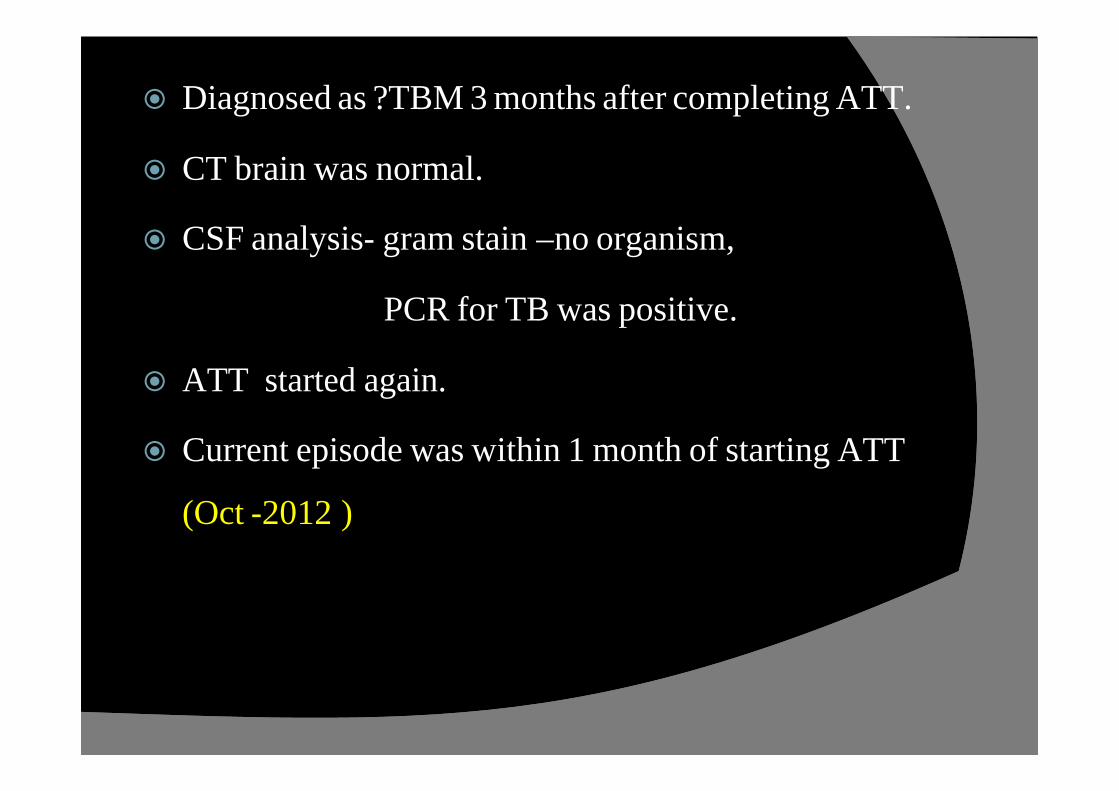
ž Diagnosed as ?TBM 3 months after completing ATT.
ž CT brain was normal.
ž CSF analysis- gram stain –no organism,
PCR for TB was positive.
ž ATT started again.
ž Current episode was within 1 month of starting ATT
(Oct -2012 )

ž No H/O head trauma or surgery.
ž Immunoglobulin levels - normal
ž HIV Elisa - negative.
ž CBC - normal during all episodes

MOTHER’s Major concern…….
ž Persistent rhinorrhoea since 4 years of age.
ž “typical allergic salute’’ present
ž treated as allergic rhinitis frequently .
ž on probing - -> he had nasal discharge without allergic symptoms also .
ž Nasal examn – clear non - sticky discharge from the right nostril
?CSF rhinorrhoea

ž Nasal secretion analysis:
ž Clear, sugar – 70 mg/dl.
ž CSF cisternogram:
Ø A tiny rent measuring 1.8mm in right fovea ethmoidalis & cribriform plate junction.
Ø Another tiny rent of 0.8 mm size seen in right fovea ethmoidalis.
Ø Cotton ball kept in the right nasal cavity became soaked with contrast –suggestive of CSF leak on right side.
Ø Bilateral sino nasal polyposis.

Further course of this episode
ž Diagnostic nasal endoscopy done and CSF leak from right middle meatus was confirmed (left nostril normal)
ž Endoscopic CSF leak repair (using temporalis fascia as graft) done by ENT surgeon
ž Discharged on the 5th post operative day.
ž Condition at discharge – afebrile, no vomiting / headache

FOLLOW UP
ž No recurrence of symptoms or signs of
meningitis.
ž Needs further follow up for :
ØRecurrence of meningitis
ØAudiological evaluation
ØDetailed immunological workup

RECURRENT MENINGITIS
ž Indicates pyogenic meningitis occurring on two or more occasions after an intervening period of full convalescence.
ž Etiology
v anatomical abnormalities- congenital / acquired
v immunodeficiencies - congenital / acquired
vchronic parameningeal infections.
PRESENCE OF A MINOR ANTIBODY DEFICIENCY SHOULDNOT PRECLUDE THE SEARCH FOR A CRANIAL DEFECT!

2008;21:519-537
APPROACH TO
A CASE OF RECURRENT MENINGITIS

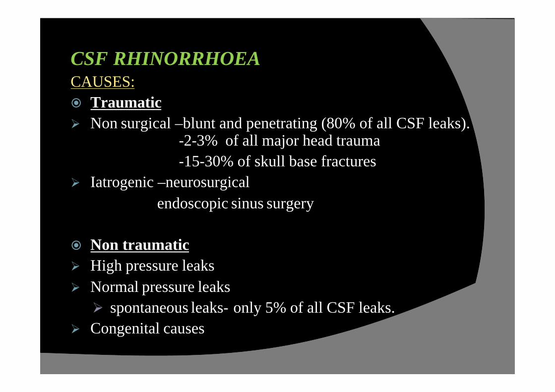
CSF RHINORRHOEACAUSES:
ž Traumatic
Ø Non surgical –blunt and penetrating (80% of all CSF leaks). -2-3% of all major head trauma
-15-30% of skull base fractures
Ø Iatrogenic –neurosurgical
endoscopic sinus surgery
ž Non traumatic
Ø High pressure leaks
Ø Normal pressure leaks
Ø spontaneous leaks- only 5% of all CSF leaks.
Ø Congenital causes

HISTORY:
ž Typical history - clear, watery discharge from a
single nare
ž An increase in postnasal drip while in the supine
position.
ž Older child may complain of a salty taste in his or
her mouth.
ž In the case of headaches, the child may state that the
headache resolves when the leak occurs.

PHYSICAL EXAMINATION:
ž Mostly unremarkable.
ž Maneuvers :
Ø Ask the child lean forward and strain. This raises ICP
and may elicit a leak.
Ø Another way to raise ICP is to compress both jugular
veins.
Rhinorrhoea is typically clear but may be mixed with
blood (look for ‘Ring sign’ or ‘Halo sign’).
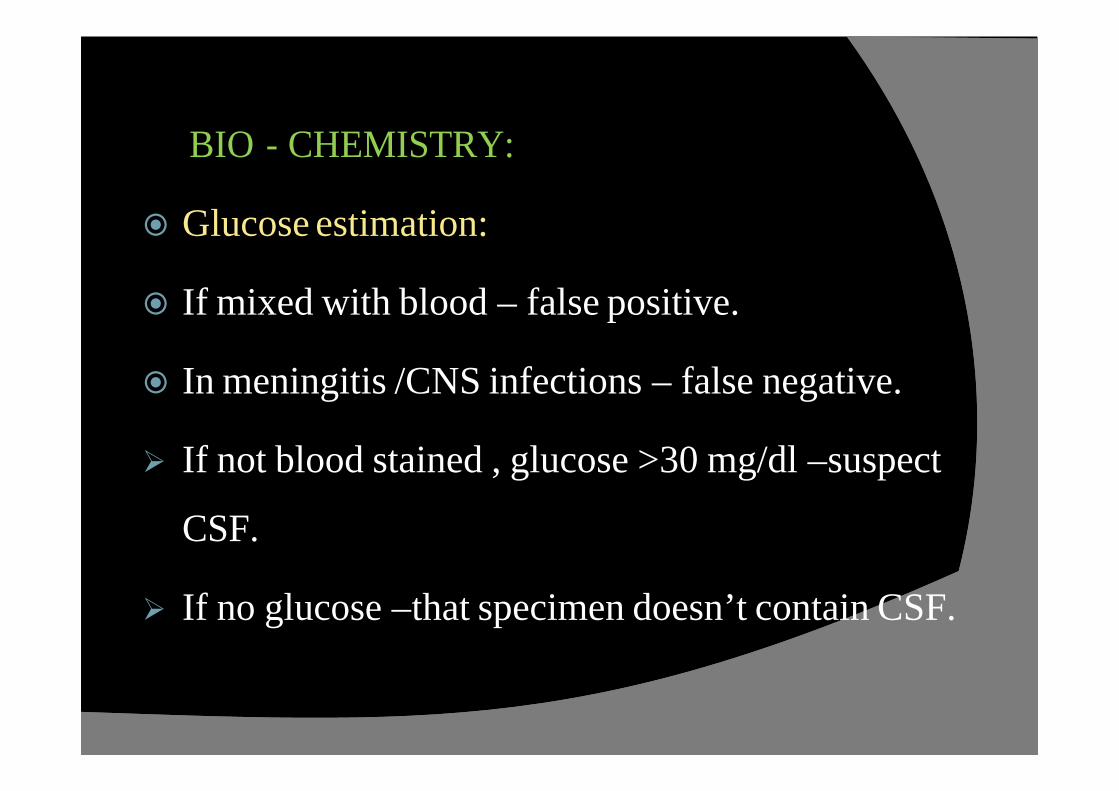
BIO - CHEMISTRY:
ž Glucose estimation:
ž If mixed with blood – false positive.
ž In meningitis /CNS infections – false negative.
Ø If not blood stained , glucose >30 mg/dl –suspect
CSF.
Ø If no glucose –that specimen doesn’t contain CSF.

ž Beta-2-transferrin estimation:
Ø almost exclusively found in the CSF
Ø blood or nasal secretion does not disturb the test .
Ø not present in blood, nasal mucus, tears or mucosal
discharge.
Ø Sensitivity of near 100% and specificity of about
95 %.
ž (Ref: CSF Rhinorrhea ,Grand Rounds Presentation, UTMB, Dept. of Otolaryngology)
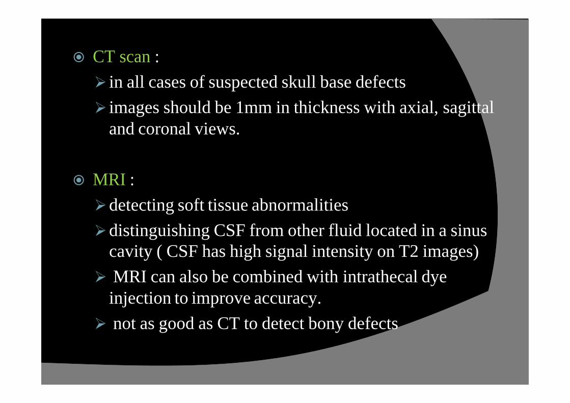
ž CT scan :
Ø in all cases of suspected skull base defects
Ø images should be 1mm in thickness with axial, sagittaland coronal views.
ž MRI :
Ødetecting soft tissue abnormalities
Ødistinguishing CSF from other fluid located in a sinus cavity ( CSF has high signal intensity on T2 images)
Ø MRI can also be combined with intrathecal dye injection to improve accuracy.
Ø not as good as CT to detect bony defects
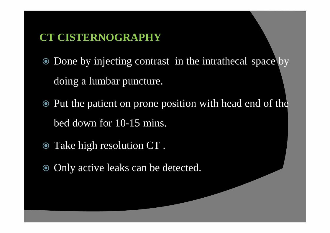
CT CISTERNOGRAPHY
ž Done by injecting contrast in the intrathecal space by
doing a lumbar puncture.
ž Put the patient on prone position with head end of the
bed down for 10-15 mins.
ž Take high resolution CT .
ž Only active leaks can be detected.
TREATMENT
ž The majority of traumatic CSF leaks respond well
to conservative management.
ž Spontaneous leaks tend to require surgical
correction.
ž The presence of a CSF leak increases the risk of
meningitis 10-fold.

MEDICAL:
ž Bed rest for 7-10 days with the head of bed at 15-30
degrees .
ž Advised not to strain,cough
ž 75-80% of all traumatic CSF leaks will resolve.
ž The use of antibiotics in the treatment of CSF rhinorrhea
remains controversial.
ž When there is increased ICP, diuretic use should be
considered –acetazolamide.
If the leak fails to responds after 5-7 days of conservative management
ž Consider is the use of a lumbar drain
ž Continuous drainage is recommended .
ž The rate of drainage should be about 10-15cc/hr.
ž Side effects such as headaches, nausea, and emesis.

SURGERYž The surgical management of CSF rhinorrhea :Ø intracranial approachØ extracranial approach
ž The defects can be repaired by primary closure with or without the use of grafts.
ž Grafting material - cartilage, bone (septum, mastoid tip,
middle turbinate), mucoperichondrium, septal mucosa,
turbinate mucosa and/or bone, fascia (temporalis, fascia lata),
abdominal fat, and pedicled septal or turbinate flaps.

Post operative care:
ž should be placed on bed rest with the head of the bed set at 15-30 degrees for 3-5 days.
ž The patient’s blood pressure should be maintained at a normal level.
ž The patient should also be placed on stool softeners to prevent straining,
ž Instructed not to cough, blow his or her nose, and avoid any heavy lifting.
ž Some surgeons will utilize a lumbar drain post-operatively.

SIMILAR CASE REPORTS
ž CSF rhinorrhoea and recurrent meningitis –Dennis G Pappas et al. (Clinical infectious diseases,Vol 17, no 3,sep 1993)
ž CSF Rhinorrhea and Recurrent Meningitis Caused by Transethmoidal Meningoencephaloceles - Parul Garg,VinitaRathi,Satish K. Bhargava,Anju Aggarwal
ž Remediable Recurrent Meningitis--K. Rajeshwari,AjaySharma (Indian pediatrics- Vol 32-April 1995)

ACKNOWLEDGEMENTS
ž Dr. D. Vijayasekaran
ž Dr. N. Mahesh
ž Dr. Balakumar
ž Dr. S.Ramesh