a randomized controlled study to evaluate the...
TRANSCRIPT
Copyright © 2018 Infusion Nurses Society. Unauthorized reproduction of this article is prohibited.
VOLUME 41 | NUMBER 2 | MARCH/APRIL 2018 Copyright © 2018 Infusion Nurses Society 131
The Art and Science of Infusion Nursing
Short peripheral catheter (SPC) insertion is a common procedure in hospitals and is the most frequently used method for drug, fluid, and blood product administration.1,2 It is estimated that up to 85% of
patients in acute care hospitals require infusion therapy, with 70% of patients needing an SPC.3,4
However, the procedure of SPC insertion is not risk free. Skin integrity is breached when the skin is punctured with a needle to allow the insertion of an SPC into a vein.5
A common complication is phlebitis, which causes pain, interrupts infusion therapy, and necessitates insertion of a new catheter. Phlebitis may also compromise subsequent vascular access and lead to bloodstream infections.6,7 For these reasons, prevention and early detection of phlebitis is important. This study aimed to compare the effective-ness of 2 treatment methods in reducing the incidence of SPC-related phlebitis. The 2 treatment methods differed in terms of the cleansing solution used before insertion and dressing material used after removal.
BACKGROUND
Phlebitis is defined as an “inflammation of the vein; which may be accompanied by pain, erythema, edema, streak formation and/or palpable cord.”8(pS153) Phlebitis can occur while the catheter is in situ and up to 96 hours after remov-al of the catheter.9 According to Lamb and Dougherty,10 the common classifications of phlebitis are:
• Mechanicalphlebitis—associatedwithcathetersize,insertionsite,insertiontechniques,andmethodsofcatheterandjointstabilization;
• Chemicalphlebitis—associatedwithinfusionofhyper-osmolarfluidsgreaterthan600mOsmperliterand/orsolutionsandmedicationswithapH<5 and >9; and
ABSTRACTShort peripheral catheter (SPC)-related phlebitis can lead to bloodstream infections and affect patients’ quality of life. Arandomizedtrialwascarriedouttoevaluatetheeffectivenessof2treatmentmethodsinreducingtheincidenceofSPC-related phlebitis. The 2 treatment methods differed in terms of the cleansing solution used before insertion and dressing material used after removal. The results demonstrated that the type of cleansing solution and postremoval dressing material did not make a difference in the incidence of phlebitis. Strict adherence to aseptic techniques and prompt removal of the SPC remained the cornerstone in the prevention of phlebitis.Key words: 2% chlorhexidine, 70% isopropyl alcohol, phlebitis, short peripheral catheter, visual infusion phlebitis score
Authors’ Affiliation: Singapore General Hospital, Singapore (Ms Gunasegaran, Miss See, Ms Leong, Ms Yuan, and Ms Ang).
Nanthakumahrie Gunasegaran, MSc, BHS, is currently a nurse cli-nician. In addition to her master of science degree in clinical lead-ership, she holds an advanced diploma in medical/surgical nursing, as well as postgraduate certificates in domiciliary nursing and clin-ical nursing. Min Ting Alicia See, BSc (Hons), is currently a senior staff nurse. Siew Teing Leong, MHSM, BHS, is currently a senior nurse manager. She also holds an advanced diploma in critical care. Long Xia Yuan, BN, is currently a senior nurse manager. She also holds an advanced diploma in gerontology nursing. Shin Yuh Ang, MBA, BSc (Hons), is currently the assistant director of nursing. She also holds an advanced diploma in medical/surgical nursing.
This study was supported by FY 2013/14 Singapore General Hospital Research Grant SRG/C1/04/2014.
The authors have no conflicts of interest to disclose.
Corresponding Author: Nanthakumahrie Gunasegaran, MSc, BHS, Singapore General Hospital, Outram Road, Singapore 169608 ([email protected]).
A Randomized Controlled Study to Evaluate the Effectiveness of 2 Treatment Methods in Reducing Incidence of Short Peripheral Catheter-Related Phlebitis
Nanthakumahrie Gunasegaran, MSc, BHS Min Ting Alicia See, BSc (Hons) Siew Teing Leong, MHSM, BHS Long Xia Yuan, BN Shin Yuh Ang, MBA, BSc (Hons)
DOI: 10.1097/NAN.0000000000000271
Copyright © 2018 Infusion Nurses Society. Unauthorized reproduction of this article is prohibited.
132 Copyright © 2018 Infusion Nurses Society Journal of Infusion Nursing
• Bacterialphlebitis—associatedwithdeficitsinskinantisep-sis,catheterhandlingandstabilization,anddressingmaterials.
Most studies in the literature have focused on the pre-vention and reduction of bacterial infections associated with the use of central venous catheters.11-13 However, a study by Pujol et al3 reported that the incidence rate of bloodstream infections associated with short-term SPCs (0.19 cases/1000 patient days) was similar to that associated with central venous catheters (0.18 cases/1000 patient days). An inte-grative literature review conducted by Hadaway14 estimated that as many as 10 000 cases of Staphylococcus aureus bacte-remia from SPCs occur annually in the United States.
The primary responsibility for the care of SPCs usually lies with nurses. This is because nurses are the most fre-quent health care personnel to insert SPCs, administer intravenous (IV) medications, and change IV administration sets and dressings.15 Therefore, nurses are tasked as the most strategic group of health care personnel to prevent the occurrence of phlebitis.
Skin antisepsis or cleansing of the catheter site is regarded as a critically important measure in the preven-tion of SPC infections.16 Strict adherence to hand washing and aseptic techniques in caring for SPCs is a preventive strategy against contamination of the catheter site and IV administration sets, and helps eradicate the presence of microorganisms at the catheter site, which can migrate into and around the catheter and cause bacterial phlebitis.
Preinsertion Skin AntisepsisRecent studies have suggested the superiority of chlorhex-idine gluconate in the removal of skin microorganisms.17-20 The use of 2% aqueous chlorhexidine was associated with a lower incidence of SPC infections in studies by Small et al21 and Adams et al.22 Other studies demonstrated that itsuse was more effective than 70% isopropyl alcohol in the reduction of the number of skin microorganisms and risk of subsequent SPC contaminations.22,23
Postremoval Phlebitis and DressingBeing an inflammation process, phlebitis may also occur after IV infusion has ceased and the SPC has been removed.14 There are limited studies that have reported on the incidence of phlebitis after removal. In a more recent study,24 it was reported that 1.8% of patients expe-rienced postinfusion phlebitis at 48 hours after catheter removal. Although rare, there were also reports of postin-fusion phlebitis among patients in this current study site.
There is also a dearth of studies that investigated the impact of spray-on film dressings (transparent vapor per-meable spray) on postinfusion phlebitis.
Significance of Current StudyIn the tertiary hospital where this study was conducted, standard practice involves the use of 70% isopropyl alcohol
swabs for preinsertion skin antisepsis and the application of adhesive bandages to the wound site following removal of the catheter. Institutional guidelines currently stipulate that catheters can be kept in situ for up to 96 hours in the absence of signs and symptoms of infection. Given recent evidence on the benefits of chlorhexidine gluconate and increased patient requests for spray-on film dressings, it is of interest to evaluate the use of these 2 products on the incidence of SPC-related phlebitis.
METHODS
Study Design and ParticipantsArandomizedcontrolledtrialwascarriedout.Participantswere recruited from an 87-bed medical ward in an acute tertiary hospital in Singapore. Recruitment and data collec-tion took place between April 2014 and July 2015.
Inclusion criteria involved patients:
• Requiring an SPC in an upper limb• Ranging in age between 21 and 99 years
Exclusion criteria involved patients who were:
• Criticallyill• Allergic to alcohol and/or chlorhexidine• Admittedforupperlimb(s)phlebitisand/orcellulitis• Known to have dermatological issues
Randomization and InterventionsCluster randomization using a computer-generated tableof random numbers was used to assign allocation of treat-ment method to each week during the data collection period. Specifically, all patients admitted to the ward in the particular week received the same treatment method.
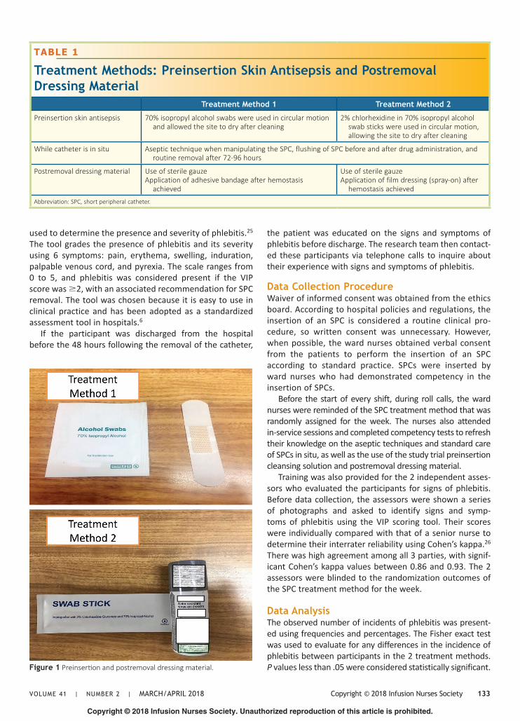
Cluster randomization was deemed the most practicalapproach to prevent any forms of treatment contamination given that the study took place in a busy, large, acute medical ward setting. Each participant was recruited only once during the period of data collection. If the participant had more than 1 SPC inserted in the upper limb(s), only 1 site of insertion was monitored and followed up on (Table 1, Figure 1).
Sample SizeBased on an estimated incidence of phlebitis of 2% in treat-ment method 2 versus 5% in treatment method 1, a total samplesizeof926participants(1:1allocationratio)wasneed-ed to attain 80% power at 5% level of significance. Accounting for a 10% dropout rate, 1020 participants were recruited.
Outcomes and Follow-upThe primary outcome was signs of phlebitis during the course of infusion therapy or within 48 hours after remov-al. The participants were followed and evaluated for signs of phlebitis on a daily basis, once every shift, by an inde-pendent assessor for the duration of the time the SPC was in situ. The visual infusion phlebitis (VIP) score tool was
Copyright © 2018 Infusion Nurses Society. Unauthorized reproduction of this article is prohibited.
VOLUME 41 | NUMBER 2 | MARCH/APRIL 2018 Copyright © 2018 Infusion Nurses Society 133
used to determine the presence and severity of phlebitis.25 The tool grades the presence of phlebitis and its severity using 6 symptoms: pain, erythema, swelling, induration, palpable venous cord, and pyrexia. The scale ranges from 0 to 5, and phlebitis was considered present if the VIP score was ≥2, with an associated recommendation for SPC removal. The tool was chosen because it is easy to use in clinical practice andhas been adopted as a standardizedassessment tool in hospitals.6
If the participant was discharged from the hospital before the 48 hours following the removal of the catheter,
the patient was educated on the signs and symptoms of phlebitis before discharge. The research team then contact-ed these participants via telephone calls to inquire about their experience with signs and symptoms of phlebitis.
Data Collection ProcedureWaiver of informed consent was obtained from the ethics board. According to hospital policies and regulations, the insertion of an SPC is considered a routine clinical pro-cedure, so written consent was unnecessary. However, when possible, the ward nurses obtained verbal consent from the patients to perform the insertion of an SPC according to standard practice. SPCs were inserted by ward nurses who had demonstrated competency in the insertion of SPCs.
Before the start of every shift, during roll calls, the ward nurses were reminded of the SPC treatment method that was randomly assigned for the week. The nurses also attended in-service sessions and completed competency tests to refresh their knowledge on the aseptic techniques and standard care of SPCs in situ, as well as the use of the study trial preinsertion cleansing solution and postremoval dressing material.
Training was also provided for the 2 independent asses-sors who evaluated the participants for signs of phlebitis. Before data collection, the assessors were shown a series of photographs and asked to identify signs and symp-toms of phlebitis using the VIP scoring tool. Their scores were individually compared with that of a senior nurse to determine their interrater reliability using Cohen’s kappa.26 There was high agreement among all 3 parties, with signif-icant Cohen’s kappa values between 0.86 and 0.93. The 2 assessorswereblindedtotherandomizationoutcomesofthe SPC treatment method for the week.
Data AnalysisThe observed number of incidents of phlebitis was present-ed using frequencies and percentages. The Fisher exact test was used to evaluate for any differences in the incidence of phlebitis between participants in the 2 treatment methods. P values less than .05 were considered statistically significant.
TABLE 1
Treatment Methods: Preinsertion Skin Antisepsis and Postremoval Dressing Material
Treatment Method 1 Treatment Method 2
Preinsertion skin antisepsis 70% isopropyl alcohol swabs were used in circular motion and allowed the site to dry after cleaning
2% chlorhexidine in 70% isopropyl alcohol swab sticks were used in circular motion, allowing the site to dry after cleaning
While catheter is in situ Aseptic technique when manipulating the SPC, flushing of SPC before and after drug administration, and routine removal after 72-96 hours
Postremoval dressing material Use of sterile gauzeApplication of adhesive bandage after hemostasis
achieved
Use of sterile gauzeApplication of film dressing (spray-on) after
hemostasis achieved
Abbreviation: SPC, short peripheral catheter.
Figure 1 Preinsertion and postremoval dressing material.
Copyright © 2018 Infusion Nurses Society. Unauthorized reproduction of this article is prohibited.
134 Copyright © 2018 Infusion Nurses Society Journal of Infusion Nursing
RESULTS
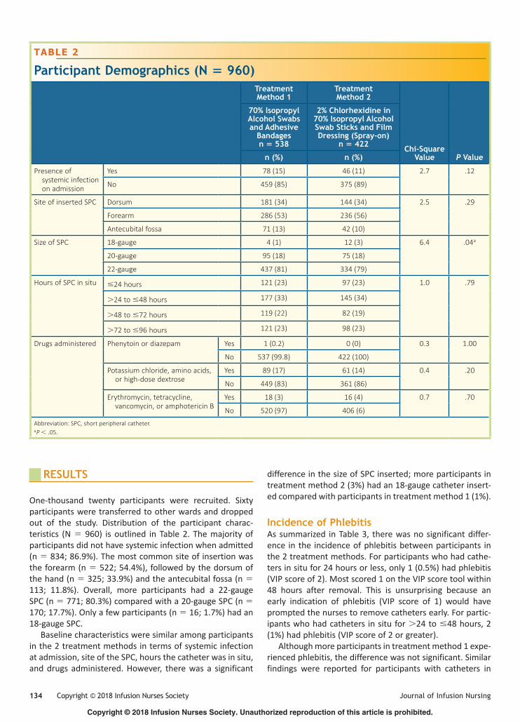
One-thousand twenty participants were recruited. Sixtyparticipants were transferred to other wards and dropped out of the study. Distribution of the participant charac-teristics (N = 960) is outlined in Table 2. The majority of participants did not have systemic infection when admitted (n = 834; 86.9%). The most common site of insertion was the forearm (n = 522; 54.4%), followed by the dorsum of the hand (n = 325; 33.9%) and the antecubital fossa (n = 113; 11.8%). Overall, more participants had a 22-gaugeSPC (n = 771; 80.3%) compared with a 20-gauge SPC (n = 170;17.7%).Onlyafewparticipants(n= 16; 1.7%) had an 18-gauge SPC.
Baseline characteristics were similar among participants in the 2 treatment methods in terms of systemic infection at admission, site of the SPC, hours the catheter was in situ, and drugs administered. However, there was a significant
differenceinthesizeofSPCinserted;moreparticipantsintreatment method 2 (3%) had an 18-gauge catheter insert-ed compared with participants in treatment method 1 (1%).
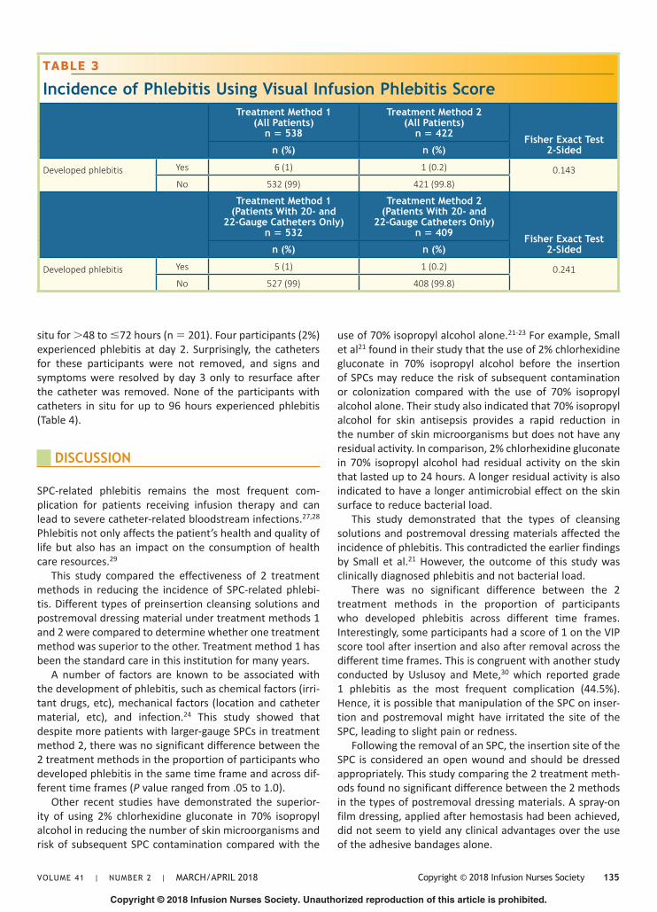
Incidence of PhlebitisAssummarized inTable3, therewasnosignificantdiffer-ence in the incidence of phlebitis between participants in the 2 treatment methods. For participants who had cathe-ters in situ for 24 hours or less, only 1 (0.5%) had phlebitis (VIP score of 2). Most scored 1 on the VIP score tool within 48 hours after removal. This is unsurprising because an early indication of phlebitis (VIP score of 1) would have prompted the nurses to remove catheters early. For partic-ipants who had catheters in situ for >24 to ≤48 hours, 2 (1%) had phlebitis (VIP score of 2 or greater).
Although more participants in treatment method 1 expe-rienced phlebitis, the difference was not significant. Similar findings were reported for participants with catheters in
TABLE 2
Participant Demographics (N = 960)Treatment Method 1
Treatment Method 2
Chi-Square Value P Value
70% Isopropyl Alcohol Swabs and Adhesive
Bandagesn = 538
2% Chlorhexidine in 70% Isopropyl Alcohol Swab Sticks and Film Dressing (Spray-on)
n = 422
n (%) n (%)
Presence of systemic infection on admission
Yes 78 (15) 46 (11) 2.7 .12
No 459 (85) 375 (89)
Site of inserted SPC Dorsum 181 (34) 144 (34) 2.5 .29
Forearm 286 (53) 236 (56)
Antecubital fossa 71 (13) 42 (10)
Size of SPC 18-gauge 4 (1) 12 (3) 6.4 .04a
20-gauge 95 (18) 75 (18)
22-gauge 437 (81) 334 (79)
Hours of SPC in situ ≤24 hours 121 (23) 97 (23) 1.0 .79
>24 to ≤48 hours 177 (33) 145 (34)
>48 to ≤72 hours 119 (22) 82 (19)
>72 to ≤96 hours 121 (23) 98 (23)
Drugs administered Phenytoin or diazepam Yes 1 (0.2) 0 (0) 0.3 1.00
No 537 (99.8) 422 (100)
Potassium chloride, amino acids, or high-dose dextrose
Yes 89 (17) 61 (14) 0.4 .20
No 449 (83) 361 (86)
Erythromycin, tetracycline, vancomycin, or amphotericin B
Yes 18 (3) 16 (4) 0.7 .70
No 520 (97) 406 (6)
Abbreviation: SPC, short peripheral catheter.aP < .05.
Copyright © 2018 Infusion Nurses Society. Unauthorized reproduction of this article is prohibited.
VOLUME 41 | NUMBER 2 | MARCH/APRIL 2018 Copyright © 2018 Infusion Nurses Society 135
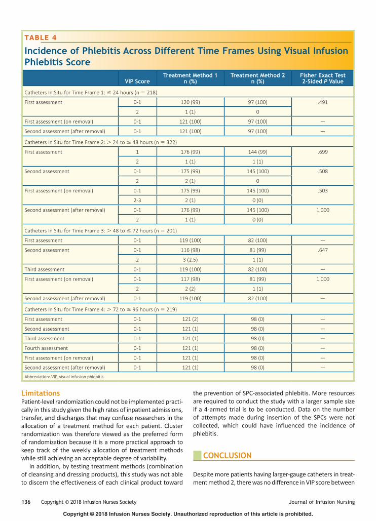
situ for >48 to ≤72 hours (n = 201). Four participants (2%) experienced phlebitis at day 2. Surprisingly, the catheters for these participants were not removed, and signs and symptoms were resolved by day 3 only to resurface after the catheter was removed. None of the participants with catheters in situ for up to 96 hours experienced phlebitis (Table 4).
DISCUSSION
SPC-related phlebitis remains the most frequent com-plication for patients receiving infusion therapy and can lead to severe catheter-related bloodstream infections.27,28 Phlebitis not only affects the patient’s health and quality of life but also has an impact on the consumption of health care resources.29
This study compared the effectiveness of 2 treatment methods in reducing the incidence of SPC-related phlebi-tis. Different types of preinsertion cleansing solutions and postremoval dressing material under treatment methods 1 and 2 were compared to determine whether one treatment method was superior to the other. Treatment method 1 has been the standard care in this institution for many years.
A number of factors are known to be associated with the development of phlebitis, such as chemical factors (irri-tant drugs, etc), mechanical factors (location and catheter material, etc), and infection.24 This study showed that despite more patients with larger-gauge SPCs in treatment method 2, there was no significant difference between the 2 treatment methods in the proportion of participants who developed phlebitis in the same time frame and across dif-ferent time frames (P value ranged from .05 to 1.0).
Other recent studies have demonstrated the superior-ity of using 2% chlorhexidine gluconate in 70% isopropyl alcohol in reducing the number of skin microorganisms and risk of subsequent SPC contamination compared with the
TABLE 3
Incidence of Phlebitis Using Visual Infusion Phlebitis ScoreTreatment Method 1
(All Patients)n = 538
Treatment Method 2 (All Patients)
n = 422 Fisher Exact Test 2-Sidedn (%) n (%)
Developed phlebitis Yes 6 (1) 1 (0.2) 0.143No 532 (99) 421 (99.8)
Treatment Method 1 (Patients With 20- and
22-Gauge Catheters Only)n = 532
Treatment Method 2 (Patients With 20- and
22-Gauge Catheters Only)n = 409 Fisher Exact Test
2-Sidedn (%) n (%)
Developed phlebitis Yes 5 (1) 1 (0.2) 0.241No 527 (99) 408 (99.8)
use of 70% isopropyl alcohol alone.21-23 For example, Small et al21 found in their study that the use of 2% chlorhexidine gluconate in 70% isopropyl alcohol before the insertion of SPCs may reduce the risk of subsequent contamination or colonization compared with the use of 70% isopropylalcohol alone. Their study also indicated that 70% isopropyl alcohol for skin antisepsis provides a rapid reduction in the number of skin microorganisms but does not have any residual activity. In comparison, 2% chlorhexidine gluconate in 70% isopropyl alcohol had residual activity on the skin that lasted up to 24 hours. A longer residual activity is also indicated to have a longer antimicrobial effect on the skin surface to reduce bacterial load.
This study demonstrated that the types of cleansing solutions and postremoval dressing materials affected the incidence of phlebitis. This contradicted the earlier findings by Small et al.21 However, the outcome of this study was clinically diagnosed phlebitis and not bacterial load.
There was no significant difference between the 2 treatment methods in the proportion of participants who developed phlebitis across different time frames. Interestingly, some participants had a score of 1 on the VIP score tool after insertion and also after removal across the different time frames. This is congruent with another study conducted by Uslusoy and Mete,30 which reported grade 1 phlebitis as the most frequent complication (44.5%). Hence, it is possible that manipulation of the SPC on inser-tion and postremoval might have irritated the site of the SPC, leading to slight pain or redness.
Following the removal of an SPC, the insertion site of the SPC is considered an open wound and should be dressed appropriately. This study comparing the 2 treatment meth-ods found no significant difference between the 2 methods in the types of postremoval dressing materials. A spray-on film dressing, applied after hemostasis had been achieved, did not seem to yield any clinical advantages over the use of the adhesive bandages alone.
Copyright © 2018 Infusion Nurses Society. Unauthorized reproduction of this article is prohibited.
136 Copyright © 2018 Infusion Nurses Society Journal of Infusion Nursing
TABLE 4
Incidence of Phlebitis Across Different Time Frames Using Visual Infusion Phlebitis Score
VIP ScoreTreatment Method 1
n (%)Treatment Method 2
n (%)Fisher Exact Test 2-Sided P Value
Catheters In Situ for Time Frame 1: ≤ 24 hours (n = 218)
First assessment 0-1 120 (99) 97 (100) .491
2 1 (1) 0
First assessment (on removal) 0-1 121 (100) 97 (100) —
Second assessment (after removal) 0-1 121 (100) 97 (100) —
Catheters In Situ for Time Frame 2: > 24 to ≤ 48 hours (n = 322)
First assessment 1 176 (99) 144 (99) .699
2 1 (1) 1 (1)
Second assessment 0-1 175 (99) 145 (100) .508
2 2 (1) 0
First assessment (on removal) 0-1 175 (99) 145 (100) .503
2-3 2 (1) 0 (0)
Second assessment (after removal) 0-1 176 (99) 145 (100) 1.000
2 1 (1) 0 (0)
Catheters In Situ for Time Frame 3: > 48 to ≤ 72 hours (n = 201)
First assessment 0-1 119 (100) 82 (100) —
Second assessment 0-1 116 (98) 81 (99) .647
2 3 (2.5) 1 (1)
Third assessment 0-1 119 (100) 82 (100) —
First assessment (on removal) 0-1 117 (98) 81 (99) 1.000
2 2 (2) 1 (1)
Second assessment (after removal) 0-1 119 (100) 82 (100) —
Catheters In Situ for Time Frame 4: > 72 to ≤ 96 hours (n = 219)
First assessment 0-1 121 (2) 98 (0) —
Second assessment 0-1 121 (1) 98 (0) —
Third assessment 0-1 121 (1) 98 (0) —
Fourth assessment 0-1 121 (1) 98 (0) —
First assessment (on removal) 0-1 121 (1) 98 (0) —
Second assessment (after removal) 0-1 121 (1) 98 (0) —
Abbreviation: VIP, visual infusion phlebitis.
LimitationsPatient-levelrandomizationcouldnotbeimplementedpracti-cally in this study given the high rates of inpatient admissions, transfer, and discharges that may confuse researchers in the allocation of a treatment method for each patient. Cluster randomizationwas thereforeviewedas thepreferred formofrandomizationbecauseitisamorepracticalapproachtokeep track of the weekly allocation of treatment methods while still achieving an acceptable degree of variability.
In addition, by testing treatment methods (combination of cleansing and dressing products), this study was not able to discern the effectiveness of each clinical product toward
the prevention of SPC-associated phlebitis. More resources arerequiredtoconductthestudywithalargersamplesizeif a 4-armed trial is to be conducted. Data on the number of attempts made during insertion of the SPCs were not collected, which could have influenced the incidence of phlebitis.
CONCLUSION
Despite more patients having larger-gauge catheters in treat-ment method 2, there was no difference in VIP score between
Copyright © 2018 Infusion Nurses Society. Unauthorized reproduction of this article is prohibited.
VOLUME 41 | NUMBER 2 | MARCH/APRIL 2018 Copyright © 2018 Infusion Nurses Society 137
the participants from the 2 treatment methods across the different time frames. The results of the study demonstrate that the types of cleansing solutions and postremoval dress-ing materials did not affect the incidence of phlebitis. Strict adherence to aseptic techniques, such as compliance with hand hygiene practices and adherence to universal infection prevention measures, continues to remain the cornerstone in the prevention of bacterial phlebitis.
Relevance to Clinical PracticePhlebitis can put patients’ safety at risk. This study has shown that safe clinical practices during the insertion and care management of SPCs are more important than the use of different (superior) products.
REFERENCES
1. Dychter SS, Gold DA, Carson D, Haller M. Intravenous therapy: a review of complications and economic considerations of peripheral access. J Infus Nurs. 2012;35(2):84-91.
2. Ho KHM, Cheung DSK. Guidelines on timing in replacing peripheral intravenous catheters. J Clin Nurs. 2012;21(11-12):1499-1506.
3. Pujol M, Hornero A, Saballs M, et al. Clinical epidemiology and out-comes of peripheral venous catheter-related bloodstream infections at a university-affiliated hospital. J Hosp Infect. 2007;67(1):22-29.
4. O’Grady N, AlexanderM, Burns L, Dellinger E, et al. Guidelines forthe prevention of intravascular catheter-related infections. https://www.cdc.gov/infectioncontrol/guidelines/pdf/bsi/bsi-guidelines-H.pdf. Published 2011. Accessed November 28, 2017.
5. Morris W, Tay MH. Strategies for preventing peripheral intravenous catheter infection. Br J Nurs. 2008;17(9):14-21.
6. Ray-Barruel G, Polit DF, Murfield JE, Rickard CM. Infusion phlebi-tis assessment measures: a systematic review. J Eval Clin Pract. 2014;20(2):191-202.
7. Enes SMS,Opitz SP, FaroARMC,PedreiraMLG. Phlebitis associatedwith peripheral intravenous catheters in adults admitted to hos-pital in the Western Brazilian Amazon. J School Nurs. 2016;50(2): 261-269.
8. Gorski L, Hadaway L, Hagle ME, McGoldrick M, Orr M, DoellmanD. Infusion therapy standards of practice. J Infus Nurs. 2016;39 (suppl 1):S1-S159.
9. Washington GT, Barrett R. Peripheral phlebitis: a point-prevalence study. J Infus Nurs. 2012;35(4):252-258.
10. Lamb J, Dougherty I. Local and systemic complications of intravenous therapy. In: Dougherty L, Lamb J, eds. Intravenous Therapy in Nursing Practice. Malden, MA: Blackwell; 2008:167-196.
11. Marschall J, Mermel LA, Fakih M, et al. Strategies to prevent central line-associated bloodstream infections in acute care hospitals: 2014 update. Infect Control. 2014;35(suppl S2):S89-S107.
12. Karpanen TJ, Casey AL, Whitehouse T, Nightingale P, Das I, Elliott TSJ. Clinical evaluation of a chlorhexidine intravascular catheter gel dressing on short-term central venous catheters. Am J Infect Control. 2016;44(1):54-60.
13. Salgueiro-Oliveria A, Parreira P, Veiga P. Incidence of phlebitis inpatients with peripheral intravenous catheters: the influence of some risk factors. Aust J Adv Nurs. 2012;30(2):32-39.
14. Hadaway L. Short peripheral intravenous catheters and infections. J Infus Nurs. 2012;35(4):230-240.
15. Scales K. Intravenous therapy: a guide to good practice. Br J Nurs. 2008;17(19):4-12.
16. Mimoz O, Chopra V, Timsit JF. What’s new in catheter-relatedinfection: skin cleansing and skin antisepsis. Intensive Care Med. 2016;42(11):1784-1786.
17. Milstone AM, Passaretti CL, Trish MP. Chlorhexidine: expanding the armamentarium for infection control and prevention. Clin Infect Dis. 2008;46(2):274-281.
18. Petlin A, Schallom M, Prentice D, et al. Chlorhexidine gluconate bath-ing to reduce methicillin-resistant staphylococcus aureus acquisition. Crit Care Nurse. 2014;34(5):17-25.
19. Bleasdale SC, Trick WE, Gonzalez IM, Lyles RD, Hayden MK,Weinstein RA. Effectiveness of chlorhexidine bathing to reduce catheter-associated bloodstream infections in medical intensive care unit patients. Arch Intern Med. 2007;167(19):2073-2079.
20. Girard R, Comby C, Jacques D. Alcoholic povidone-iodine or chlorhexidine-based antiseptic for the prevention of central venous catheter-related infections: in-use comparison. J Infect Public Health. 2012;5(1):35-42.
21. Small H, Adams D, Casey AL, Crosby CT, Lambert PA, Elliott T. Efficacy of adding 2% (w/v) chlorhexidine gluconate to 70% (v/v) isopropyl alcohol for skin disinfection prior to peripheral venous cannulation. Infect Control Hosp Epidemiol. 2008;29(10):963-965.
22. Adams A, Quayum M, Worthington T, et al. Evaluation of a 2% chlor-hexidine gluconate in 70% isopropyl alcohol skin disinfectant. J Hosp Infection. 2005;61(1):287-290.
23. McLellan E, Townsend R, Parsons HK. Evaluation of ChloraPrep (2% chlorhexidine gluconate in 70% isopropyl alcohol) for skin antisepsis in preparation for blood culture collection. J Infect. 2008;57(6):459-463.
24. Webster J, McGrail M, Marsh N, Wallis MC, Ray-Barruel G, Rickard CM. Postinfusion phlebitis: incidence and risk factors. Nurs Res Pract. 2015:691934. http://dx.doi.org/10.1155/2015/691934. Accessed November 29, 2017.
25. GallantP,SchultzAA.Evaluationofavisualinfusionphlebitisscalefordetermining appropriate discontinuation of peripheral intravenous catheters. J Infus Nurs. 2006;29(6):338-345.
26. McHugh ML. Interrater reliability: the kappa statistic. Biochem Med. 2012;22(3):276-282.
27. Cicolini G, Manzoli L, Simonetti V, et al. Phlebitis risk varies byperipheral venous catheter site and increases after 96 hours: a large multi-centre prospective study. J Adv Nurs. 2014;70(11): 2539-2549.
28. RocaGM,BertoloCB,LopezPT,etal.Assessingtheinfluenceofriskfactors on rates and dynamics of peripheral vein phlebitis: an obser-vational cohort study. Med Clin (Barc). 2012;139(5):185-191.
29. Kaur P, Thakur R, Kaur S, Bhalla A. Assessment of risk factors of phle-bitis amongst intravenous cannulated patients. Nurs Midwifery Res J. 2011;7(3):106-114.
30. Uslusoy E, Mete S. Predisposing factors to phlebitis in patients with peripheral intravenous catheters: a descriptive study. J Am Acad Nurse Pract. 2008;20(4):172-180.