a r p d the digestive system of diaphorina citri and
TRANSCRIPT
ARTHROPODS IN RELATION TO PLANT DISEASES
The Digestive System of Diaphorina citri and Bactericera cockerelli(Hemiptera: Psyllidae)
J. M. CICERO,1,2 J. K. BROWN,1 P. D. ROBERTS,3 AND P. A. STANSLY3
Ann. Entomol. Soc. Am. 102(4): 650Ð665 (2009)
ABSTRACT The psyllids Diaphorina citri (Kuwayama) and Bactericera cockerelli (Sulc) (Hemiptera:Psyllidae) are vectors ofCandidatusLiberibacter spp., bacterial agents of serious agricultural diseases. Therapidly expanding geographical distributions of these diseases dictate increasing urgency for their control.Therefore, it is important to gain a full understanding of the psyllid digestive system in which thevectorÐpathogen interactions begin. Their midgut is looped so that the foregutÐmidgut and midgutÐhindgut transitional regions are grafted together to form a composite tube within a Þlter chamber sheath.Unwanted sap components could thus be extracted directly into the hindgut, bypassing digestion. Theesophageal lumen enters the chamber axially to become the inner midgut lumen. The upper half of thismidgut section is bulbous while the lower half is tubular. The tube lumen exits the chamber to becomethe external midgut lumen, which loops through the hemocoel and reenters the chamber, becoming theinner hindgut lumen. The inner hindgut tracks the adherent inner midgut in an antiparallel direction. Thecomposite tube is helically wound and undergoes one hairpin turn. The inner hindgut straps diagonallyacross the bulb and then exits the chamber next to the esophagus as the outer hindgut to anus. The sourceof honeydew, whether Þltrate, midgut waste, or both, is questioned. Paired, spherical, primary salivaryglands each have a digitate accessory gland and a lateral duct that leads to the stylets. The accessory glandlumen is lined exclusively with intima, whereas the primary gland apical cell membranes are indicated tobe more complex.
KEY WORDS Psyllidae, alimentary canal, Þlter chamber, huanglongbing, Liberibacter
Citrus greening disease or huanglongbing (HLB) is con-sidered the most threatening disease of citrus (Citrusspp.) worldwide, and it is the major limiting factor incitrus production in Asia and Africa (Capoor et al. 1974,Lallemand et al. 1986, da Graca 1991, Bove 2006, Brown-ing et al. 2006). According to Roistacher (1996), affectedcitrus trees often survive only 5Ð8 yr and produce un-usable fruit.
The disease is widespread in Asia, Africa, and theSaudi Arabian Peninsula. It was reported in July 2004in Sao Paulo State, Brazil (Coletta-Filho et al. 2004,Teixeira et al. 2005), in the Caribbean Basin, and forthe Þrst time in the United States in south Miami-DadeCounty, FL, in August 2005 (Halbert 1998, 2005; Hal-bert and Manjunath 1994; Halbert et al. 2000; Halbertand Nunez 2004).
Three species of phloem-limited bacteria are rec-ognized: Candidatus Liberibacter asiaticus, Candida-tus L. africanus, and Candidatus L. americanus (Teix-eira et al. 2005). To date only Ca. L. asiaticus has beenidentiÞed in Florida (Halbert 2005), however, futureintroductions of additional species ofCa.Liberibacterand of the psyllid vector are considered likely.
The establishment of Ca. Liberibacter asiaticus inFlorida has greatly increased the signiÞcance of thepresence of the psyllid Diaphorina citri (Kuwayama)there (Halbert 1998) and elsewhere and underscoresthe urgent need for basic knowledge about vectorÐpathogen interactions that is now essential for devis-ing effective management strategies to deter or blockvector-mediated pathogen transmission.
The psyllidBactericera cockerelli(Sulc), a relative ofD. citri, is a major pest of potato (Solanum spp.),tomato (Solanum spp.), and other fruiting vegetablesand is the vector of a newly described disease oftomato and potato caused by Ca. Liberibacter psyl-laurous (Hansen et al. 2008). However, most aspectsof acquisition and transmission ofCa.L. psyllaurous bythe psyllid are unstudied (Hansen et al. 2008).
Concise descriptions of psyllid vector digestive sys-tems are needed to understand the transmission path-way of psyllid-borne pathogens, from ingestion, topassage through the consecutive internal organs, andduring transmission to the subsequent host plant. Theonly information available regarding the organizationof the psyllid digestive system comes from classicalstudies conducted 80Ð90 yr ago [Dufor (1833) forPsylla ficus,Witlaczil (1885) for Psylla buxi (L.) andTrioza urticae (L.), Macloskie (1886) and Packard1898 for Psyllopsis sp., Saunders (1921) for Psylla maliSchmidberger]. BrittainÕs copy (Brittain 1923) of
1 Plant Sciences Department, P.O. Box 210036, University of Ari-zona, Tucson, AZ 85721.
2 Corresponding author, e-mail: [email protected] University of Florida/IFAS, SFREC, 2686 State Rd./29 N., Im-
mokalee FL 34142.
0013-8746/09/0650Ð0665$04.00/0 � 2009 Entomological Society of America

SaundersÕ drawing of the P. mali gut made its way intothe secondary literatureas representativeof the family(Pesson 1951, Goodchild 1966, Weber 1968, Bielenin1993). Saunders noted an eccentric lobe on the sali-vary gland that he did not identify. More recently,Ullman and McLean (1986) studied Psylla pyricolaForster with modern techniques but concentrated onsensilla of the oral region only.
The purpose of this study was to elucidate the con-struction of the digestive system of D. citri and B.cockerelli so that its components can be recognizedand identiÞed at the ultrastructural level during trans-mission pathway studies. Particular emphasis wasplaced on the alimentary canal component known asthe “Þlter chamber,” because itwasanticipated tohavegreat structural complexity and to be the anterior-most organ that lacks an inner cuticle. If the canalanterior to it is fully lined with a pathogen-impene-trable cuticle, then the Þlter chamber would be theÞrst organ of opportunity encountered in the earlieststages of Ca. Liberibacter transmission.
Materials and Methods
Adult D. citri were collected at Southwest FloridaResearch and Education Center, University of Florida,in Immokalee, FL, from colonies reared onCitrus spp.orMurraya paniculata (L.) (mock orange jasmine) orfrom the Þeld on Citrus spp. Adult B. cockerelli werecollected from infested tomato plants grown in agreenhouse in southern Arizona.
Psyllids were dissected (n � �60 each) in a weaksolution of toluidine blue and 0.1 M Na-K phosphatesaline buffer (PBS), pH 7.75, to remove the alimentarycanal and salivary glands. Gentle pressure was exertedunder a dissecting microscope to break extirpatedÞlter organs open and study their internal conduitsystem. For scanning and transmission electron mi-croscopy (SEM and TEM, respectively), extirpatedorgans were transferred to 4% formaldehyde, 0.5%glutaraldehyde in PBS for a 2-h Þxation; rinsed twotimes 15 min in PBS; and dehydrated in a gradedethanol series. Organs relegated to TEM and serialreconstruction were embedded in LR White resin(25% resin:75% ethanol for 1 h, 75% resin:25% ethanolfor 1 h, 2� 100% for 1 h, polymerized overnight at55�C), and sectioned and stained with uranyl acetate/lead citrate. Organs relegated to SEM were mountedwhole or freeze fractured with a razor blade on a metalblock in liquid nitrogen and then critical point-driedand mounted.
Once the conÞgurations and external boundaries ofthese organs were determined, whole specimens wereprocessed for TEM by gluing to a thumbtack in amicro-watchglass, submerging them in PBS, removingair bubbles with Aerosol OT [sodium 1,2-bis (2-eth-ylhexoxycarbonyl) ethane sulfonate], rinsing, andßooding with Þxative. Specimens were then openedwith separate, transverse cuts across the vertex, ter-minal abdominal segment, and meso-metathoracicjuncture by using microscissors. Specimens were Þxedovernight, rinsed, dehydrated, and embedded in LR
White. Blocks were cut with an LKB Ultratome 5 andviewed under JOEL 100CX and Philips CM12 trans-mission electron microscopes.
Results
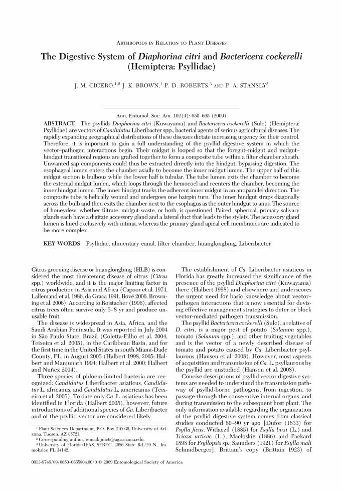
Alimentary Canal. General Anatomical Organiza-tion. The alimentary canal of D. citri and B. cockerellishowed no differences in gross anatomical organiza-tion (Fig. 2B and C). Both were modiÞed from thegeneral insect canal (Fig. 1A) as follows. The midgut(ventriculus) was looped so that its anterior and pos-terior ends were in proximity to each other and to theesophagus (Fig. 1B and C, a) and hindgut (Fig. 1B andC, b), which, by their enclosure inside a sheath(coined by Kershaw 1913) (Fig. 1B and C, c), formedthe Þlter chamber complex. Within the complex, themidgut component (Fig. 1B and C, d), and what isherein provisionally (see Discussion) termed the in-ner hindgut component (Fig. 1B and C, e), were lon-gitudinally adherent to each other, forming a com-posite tube. The inner midgut section was divided intotwo parts, an upper bulbous section and a lower tu-bular section. The external midgut was divided intotwo parts also, an inßated anterior part (Fig. 1C, f) anda tubular posterior part (Fig. 1B and C, g), the latterproviding the looping back to the point of reentry intothe Þlter chamber. Three ventricular sections areidentiÞed in this basic design, termed V1 (the innermidgut), V2 (the outer, inßated midgut section), andV3 (the outer, tubular, midgut section) following theordinal level diagram devised by Snodgrass (1935, p.384, Þg. 209A) (Fig. 1D). The lumen of all three is thesame continuous lumen that starts at the mouth andends at the anus.Orientation. The hemocoelic positions of the ali-
mentary canal components were the same for bothspecies, as follows. The esophagus extended from themouth posteriorly through the thorax, whereas thehindgut extended from the anus anteriorly throughthe abdomen. The distal extremities met at the baseof the abdomen where they were attached to theanatomical apex of the Þlter chamber. Although con-Þned to the abdominal base by these ectodermaltubes, the Þlter chamber was free to twist and turnwithout regard to dorsoventral or leftÐright orientation.
The abdominal base was walled anteriorly by thethoracic musculature and posteriorly by adipose tissueand gonads in the abdomen, loosely conÞning themidgut loop to this region also. Tracheal attachmentsconÞned the midgut loop and its appendages (Fig. 1E,q and F, p) to this region, too; but the location ofattachments were variable from specimen to speci-men, allowing for some latitude in the orientation ofthe loop. In some specimens, the loop was twisted andconvoluted within the base of the abdomen, whereasin others, sections of the loop were located deeper intothe thorax and abdomen.
When extirpated alimentary canals were allowed tospread in the dissection buffer, they settled into afree-ßoating conÞguration with the least tension be-tween its components. This conÞguration was used
July 2009 CICERO ET AL.: PSYLLID DIGESTIVE SYSTEMS 651
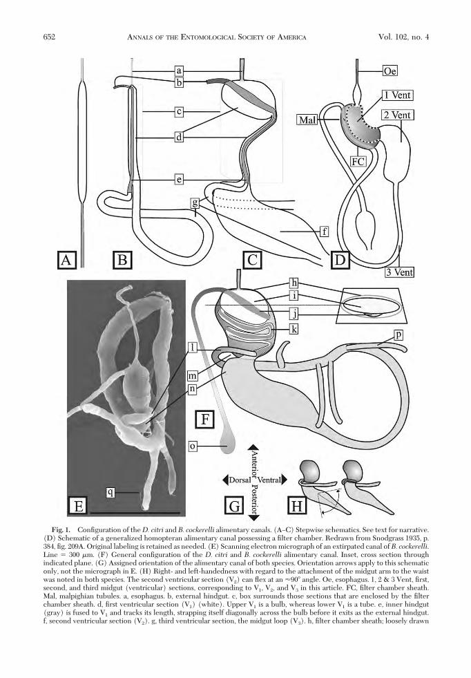
Fig. 1. ConÞguration of theD. citri and B. cockerelli alimentary canals. (AÐC) Stepwise schematics. See text for narrative.(D) Schematic of a generalized homopteran alimentary canal possessing a Þlter chamber. Redrawn from Snodgrass 1935, p.384, Þg. 209A. Original labeling is retained as needed. (E) Scanning electron micrograph of an extirpated canal ofB. cockerelli.Line � 300 �m. (F) General conÞguration of the D. citri and B. cockerelli alimentary canal. Inset, cross section throughindicated plane. (G) Assigned orientation of the alimentary canal of both species. Orientation arrows apply to this schematiconly, not the micrograph in E. (H) Right- and left-handedness with regard to the attachment of the midgut arm to the waistwas noted in both species. The second ventricular section (V2) can ßex at an �90� angle. Oe, esophagus. 1, 2 & 3 Vent, Þrst,second, and third midgut (ventricular) sections, corresponding to V1, V2, and V3 in this article. FC, Þlter chamber sheath.Mal, malpighian tubules. a, esophagus. b, external hindgut. c, box surrounds those sections that are enclosed by the Þlterchamber sheath. d, Þrst ventricular section (V1) (white). Upper V1 is a bulb, whereas lower V1 is a tube. e, inner hindgut(gray) is fused to V1 and tracks its length, strapping itself diagonally across the bulb before it exits as the external hindgut.f, second ventricular section (V2). g, third ventricular section, the midgut loop (V3). h, Þlter chamber sheath; loosely drawn
652 ANNALS OF THE ENTOMOLOGICAL SOCIETY OF AMERICA Vol. 102, no. 4
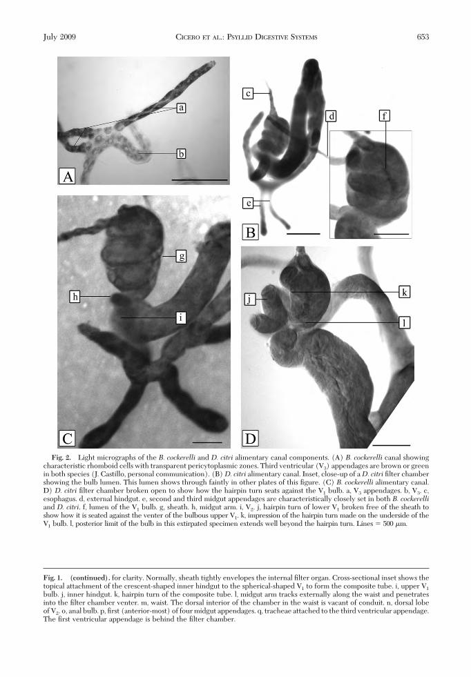
Fig. 1. (continued). for clarity. Normally, sheath tightly envelopes the internal Þlter organ. Cross-sectional inset shows thetopical attachment of the crescent-shaped inner hindgut to the spherical-shaped V1 to form the composite tube. i, upper V1
bulb. j, inner hindgut. k, hairpin turn of the composite tube. l, midgut arm tracks externally along the waist and penetratesinto the Þlter chamber venter. m, waist. The dorsal interior of the chamber in the waist is vacant of conduit. n, dorsal lobeof V2. o, anal bulb. p, Þrst (anterior-most) of four midgut appendages. q, tracheae attached to the third ventricular appendage.The Þrst ventricular appendage is behind the Þlter chamber.
Fig. 2. Light micrographs of the B. cockerelli and D. citri alimentary canal components. (A) B. cockerelli canal showingcharacteristic rhomboid cells with transparent pericytoplasmic zones. Third ventricular (V3) appendages are brown or greenin both species (J. Castillo, personal communication). (B)D. citri alimentary canal. Inset, close-up of aD. citri Þlter chambershowing the bulb lumen. This lumen shows through faintly in other plates of this Þgure. (C) B. cockerelli alimentary canal.D) D. citri Þlter chamber broken open to show how the hairpin turn seats against the V1 bulb. a, V3 appendages. b, V3. c,esophagus. d, external hindgut. e, second and third midgut appendages are characteristically closely set in both B. cockerelliand D. citri. f, lumen of the V1 bulb. g, sheath. h, midgut arm. i, V2. j, hairpin turn of lower V1 broken free of the sheath toshow how it is seated against the venter of the bulbous upper V1. k, impression of the hairpin turn made on the underside of theV1 bulb. l, posterior limit of the bulb in this extirpated specimen extends well beyond the hairpin turn. Lines � 500 �m.
July 2009 CICERO ET AL.: PSYLLID DIGESTIVE SYSTEMS 653

to establish convention for references to orientation(Fig. 1G). In situ orientation in live specimens wasdistorted from the latter conÞguration, due to pres-sure exerted by other neighboring organ systemsand endoskeletal features. Also, left- and right-handedness was discovered in the attachment of thedistal midgut (Fig. 1H). Therefore, only anterior,posterior, dorsal and ventral aspects can be deÞnedby this approach.Detailed Anatomical Organization. The following
detail was determined to be true for both speciesexcept as noted. The sheath (Fig. 1F, h) was a thinenvelope tightly enclosing the internal Þlter organs,with apparently no openings that would allow fordirect continuity between the blood and the interior.No clues were found to indicate which embryologicaltissue type the sheath is derived from or composed of.The upper V1, located in the anterior one third of theÞlter chamber, was a more or less hemispherical bulbwith a diagonally ßat posterior face (Fig. 1 F, i). LowerV1 was a narrow, helically coiled tube with a singlehairpin turn (Fig. 1F, k) that extended posteriorly forthe rest of the Þlter chamberÕs length. In some extir-pated specimens, the bulb was expanded further pos-terior than the hairpin turn (compare Fig. 1F, i withFig. 2D, l). A conspicuous constriction in the contourof the alimentary canal, herein referred to as thewaist(Fig. 1F, m), was present where V1 left the Þlterchamber as V2, in a ventroposterior direction, at an�110� angle to the long axis of the Þlter chamber. V2
was elongate, bulbous, with a strong dorsal lobe (Fig.1F, n), and its radius gradually decreased as it nearedV3, the midgut loop. Most notable in extirpated spec-imens was the settling of the distal section of the V3
loop (the midgut arm, Fig. 1E and F, l) into a hori-zontal attitude because of its histological attachmentto the point of reentry. It always crossed the full widthof the waist, closely tracking the latterÕs left or right
transverse constriction, before entering into the ven-troposterior left or right side of the Þlter chamber(Fig. 1H).
The continuous lumen of the midgut arm reenteredthe Þlter chamber at the waist to become the lumenof the inner hindgut. The inner hindgut joined imme-diately in adherence with lower V1, and, as a compos-ite tube, both pressed against the venter of the waist.The waist dorsal to this was vacant of tissue allowingthe Þlter chamber to ßex at approximately a 90� angleabout V2 (Fig. 1H).
The inner hindgut, in adherence to V1, tracked thecoiled shape of V1 in an antiparallel direction and thencrossed upper V1 diagonally (Fig. 1F, j) and exited thechamber apex next to the esophagus. Relative to thelong axis of the variably positioned Þlter chamber,the esophagus met the Þlter chamber axially, whereasthe external hindgut left the Þlter chamber tangen-tially, in line with the inner hindgut that preceded it.The external hindgut continued posteriorly, ending asa bulb at the anus (Fig. 1F, o).
Four �0.3-mm-long appendages extended from V3
in the last third of its length (Fig. 1F, p). The basal-most appendage pointed ventrally, the second andthird pointed posteriorly, and the fourth pointed lat-erally. The attachments of the second and third wereseparated by �1� their basal girth, whereas the othertwo were spaced further apart. Numerous dissectionsshowed that the apices of these appendages were sitesfor transient attachment/detachment/reattachmentof tracheae (Fig. 1E, q).
In both species, the unstained Þlter chamber wascolorless and hyaline. Cells of V2 and V3 were opaquewhite, whereas those of the V3 appendages were darkgreen or brown (J. Castillo, personal communication).Cells of V2, V3, and V3 appendages had a transparentpericytoplasm, giving each cell a rhomboid appear-ance (Fig. 2A, a and b).
Fig. 3. SEM of a B. cockerelli Þlter chamber with a rip in the sheath, showing internal helical conduits and connectivematerial that seems to hold the coils in place. (a) Rip in sheath. (b) Connective tissue. (c) Space lateral to contact of helicalcoils. Line � 20 �m.
654 ANNALS OF THE ENTOMOLOGICAL SOCIETY OF AMERICA Vol. 102, no. 4
Internal Complexity. The sheath of both speciescould be peeled away to expose an interstitial spacebetween the composite tube coils, indicating that thesheath was a separate component from the internalconduit system (Fig. 3). SEM of theB. cockerelli cham-ber indicated that the space between conduit loopswas laced with a web of material that seemed to holdthem in place. The contents of the interstitial chamberspace, be it gas, ßuid, mucilage, or some other material,were not determined.
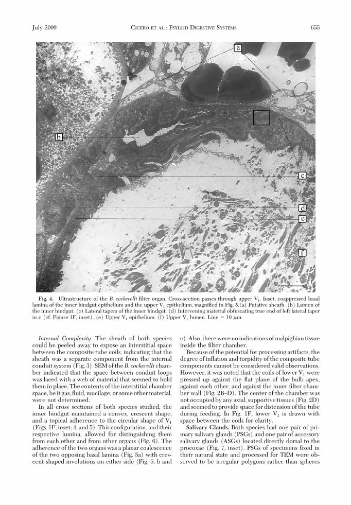
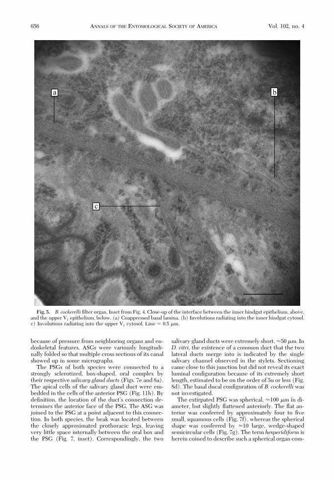
In all cross sections of both species studied, theinner hindgut maintained a convex, crescent shape,and a topical adherence to the circular shape of V1
(Figs. 1F, inset; 4, and 5). This conÞguration, and theirrespective lumina, allowed for distinguishing themfrom each other and from other organs (Fig. 6). Theadherence of the two organs was a planar coalescenceof the two opposing basal lamina (Fig. 5a) with cres-cent-shaped involutions on either side (Fig. 5, b and
c).Also, therewereno indicationsofmalpighian tissueinside the Þlter chamber.
Because of the potential for processing artifacts, thedegree of inßation and torpidity of the composite tubecomponents cannot be considered valid observations.However, it was noted that the coils of lower V1 werepressed up against the ßat plane of the bulb apex,against each other, and against the inner Þlter cham-ber wall (Fig. 2BÐD). The center of the chamber wasnot occupied by any axial, supportive tissues (Fig. 2D)and seemed to provide space for distension of the tubeduring feeding. In Fig. 1F, lower V1 is drawn withspace between the coils for clarity.Salivary Glands. Both species had one pair of pri-
mary salivary glands (PSGs) and one pair of accessorysalivary glands (ASGs) located directly dorsal to theprocoxae (Fig. 7, inset). PSGs of specimens Þxed intheir natural state and processed for TEM were ob-served to be irregular polygons rather than spheres
Fig. 4. Ultrastructure of the B. cockerelli Þlter organ. Cross-section passes through upper V1. Inset, coappressed basallamina of the inner hindgut epithelium and the upper V1 epithelium, magniÞed in Fig. 5.(a) Putative sheath. (b) Lumen ofthe inner hindgut. (c) Lateral tapers of the inner hindgut. (d) Intervening material obfuscating true end of left lateral taperin c (cf. Figure 1F, inset). (e) Upper V1 epithelium. (f) Upper V1 lumen. Line � 10 �m.
July 2009 CICERO ET AL.: PSYLLID DIGESTIVE SYSTEMS 655
because of pressure from neighboring organs and en-doskeletal features. ASGs were variously longitudi-nally folded so that multiple cross sections of its canalshowed up in some micrographs.
The PSGs of both species were connected to astrongly sclerotized, box-shaped, oral complex bytheir respective salivary gland ducts (Figs. 7e and 8a).The apical cells of the salivary gland duct were em-bedded in the cells of the anterior PSG (Fig. 11h). BydeÞnition, the location of the ductÕs connection de-termines the anterior face of the PSG. The ASG wasjoined to the PSG at a point adjacent to this connec-tion. In both species, the beak was located betweenthe closely approximated prothoracic legs, leavingvery little space internally between the oral box andthe PSG (Fig. 7, inset). Correspondingly, the two
salivary gland ducts were extremely short, �50 �m. InD. citri, the existence of a common duct that the twolateral ducts merge into is indicated by the singlesalivary channel observed in the stylets. Sectioningcame close to this junction but did not reveal its exactluminal conÞguration because of its extremely shortlength, estimated to be on the order of 5u or less (Fig.8d). The basal ducal conÞguration of B. cockerelliwasnot investigated.
The extirpated PSG was spherical, �100 �m in di-ameter, but slightly ßattened anteriorly. The ßat an-terior was conferred by approximately four to Þvesmall, squamous cells (Fig. 7f), whereas the sphericalshape was conferred by �10 large, wedge-shapedsemicircular cells (Fig. 7g). The term hesperidiform isherein coined to describe such a spherical organ com-
Fig. 5. B. cockerelli Þlter organ. Inset from Fig. 4. Close-up of the interface between the inner hindgut epithelium, above,and the upper V1 epithelium, below. (a) Coappressed basal lamina. (b) Involutions radiating into the inner hindgut cytosol.c) Involutions radiating into the upper V1 cytosol. Line � 0.5 �m.
656 ANNALS OF THE ENTOMOLOGICAL SOCIETY OF AMERICA Vol. 102, no. 4

posed of wedge-shaped cells resembling sections ofCitrus spp. fruit.Lightmicroscopical examinationof alldissections of both species revealed a funnel-shapedpattern to the PSGs, indicated in TEM also (Fig. 11,
inset). Apicomesal cells had strongly staining materialin their cytoplasm, apicolateral cells had weakly stain-ing material in their cytoplasm, but basal cells did notstain appreciably. TEM corroborated these observa-
Fig. 6. B. cockerellimale gonad. This is the only other bulbous organ that might be confused with the Þlter chamber. Line �4 �m.
Fig. 7. Block face of embedded D. citri oral complex, ventral view. Inset, circle represents the procoxal cavity. The PSGis located directly dorsal to it. Rectangle surrounds the stylet bundle. (a) PSG. (b) ASG slightly out of focus. (c) Stylet. (d)Anteclypeus. (e) External salivary duct. (f) Anterior squamous cells. (g) Posterior wedge-shaped cells, giving the PSG a“hesperidiform” or Citrus-like appearance. Line � 100 �m.
July 2009 CICERO ET AL.: PSYLLID DIGESTIVE SYSTEMS 657

tions: Apicomesal PSG cells contained full, or nearlyfull, electron opaque spherules (Figs. 9a, 10a, and 11a),and apicolateral cells contained what seemed to beremnants of spherules (Figs. 9c and 11, b and d). Athird cell type, adjacent to these two types, had elec-tron transparent spherules (Figs. 9b and 11c). Basalcells had no indication of secretory capacity (Fig.11e). Storage material in full spherules was highlyorganized into concentric arrays of densely packedsubunits that were beadshaped in cross section (Fig.
10) and columnar in longitudinal section. Differentdegrees of mobilization, whether importation or ex-portation, of storage material were apparent fromspherule to spherule (Fig. 10a). Further characteriza-tion of this transport system is underway.
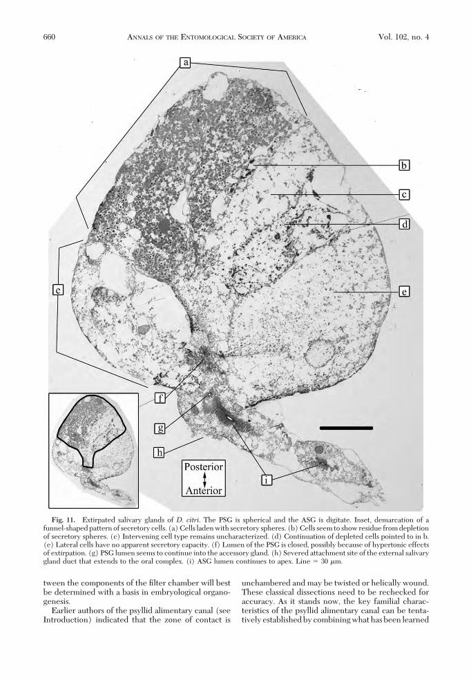
The ASG was a short, stout, digitate, slightly clavateappendage of the PSG, about as long as the PSG di-ameter in both species (Fig. 11). The connection ofthe ASG to the PSG was not by a long thin, tether,instead the ASG was broadly connected to the PSG
Fig. 8. Cross section of embedded D. citri oral complex, through the block in Fig. 7. (a) Left and right lateral salivarygland ducts. (b) Salivary gland duct cells. (c) Salivary syringe muscle complex. (d) Intima lined lumen in the location expectedfor a very short common duct. It is expected that the lateral ducts merge into a common duct in this area. Line � 100 �m.
Fig. 9. D. citri. Transverse, mesal cross section of a PSG from the block in Fig. 7. (a) Laden cell. (b) Uncharacterizedcell type. (c) Partially depleted cell. Line � 10 �m.
658 ANNALS OF THE ENTOMOLOGICAL SOCIETY OF AMERICA Vol. 102, no. 4

because its basal cells were embedded in the squa-mous cells of the anterior PSG (Fig. 11g). The ASGtherefore lifted directly out with the PSG during ex-tirpations. The internal canal of the ASG was linedwith cuticle from base to apex (Figs. 11i and 12b). Thisintima, in turn, was surrounded by a dense layer oflamellae radiating perpendicularly into the cytosol(Fig. 12c). Punctate material occurred at the base ofeach lamella (Fig. 12d). The junction of the ASGlumen and the PSG lumen (Figs. 11f and 13) under-went closure during processing and could only begiven limited characterization. At least two apicalmembrane morphologies are evidentÑone morphol-ogy with a cuticular intima and lamellae radiatingperpendicularly into the cytosol from it, and the othermorphology without such features.
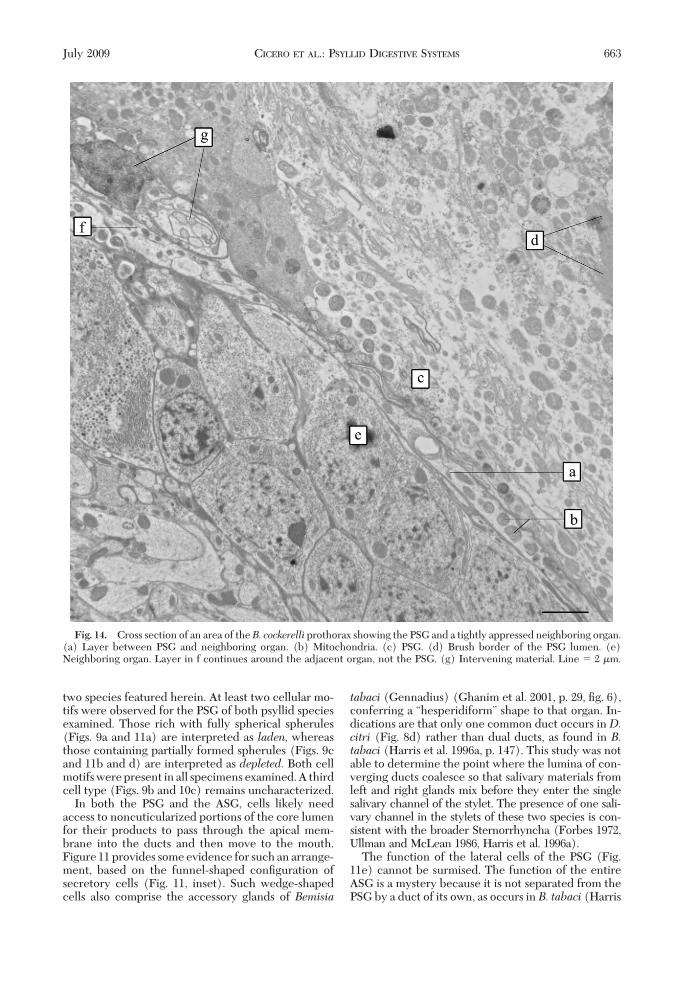
An enveloping sheath was not found in extirpated,plastic embedded PSGs of either species. In wholemounts of plastic embedded B. cockerelli, TEM re-vealed that a neighboring organ was invested by anarrow layer with numerous mitochondria thatseemed to be part of the PSG where the two weretightly appressed (Fig. 14).
Discussion
Alimentary Canal. The midgut is looped amongmany Sternorryncha so that the esophagusÐmidgut
transitional region is in contact with the midgutÐhind-gut transitional region (Fig. 1B). The conclusion ofclassical histologists that this “zone of contact” facili-tates removal of excess water and nutrients (“Þltra-tion,” Goodchild 1963, 1966) is still considered to be anaccurate interpretation by insect physiologists (Chap-man 1998). The conÞgurations of the tubes in thiszone, generally esophagus, midgut, malpighian tu-bules and hindgut are, taxonomically, very diverse,and the means by which contact is maintained (e.g.,adherence, fusion, and ligation) is largely unknown.They may be chambered within the primitive epithe-lial walls of the midgutÐhindgut transitional region, asin whiteßies (Cicero et al. 1995), within a sheath, orunchambered. We propose that when chambered, theinner complex of tubes is called the “Þlter organ,” andthe entire conglomerate structure (chamber walls �Þlter organ) is called the “Þlter chamber.”
The makeup, whether cellular or noncellular, andorigin, both organismic and phylogenetic, of the Þlterchamber sheath remains a mystery among Sternor-ryncha. Ponsen (1979, p. 19) addressed the wide di-versity of sheathed and unsheathed Þlter organssketched in the classical literature, but was not able todraw any conclusions in these regards. Because of theconfusion in the literatureas to theactual constructionof Þltering devices within various taxa, most need to bereexamined with modern techniques. Homology be-
Fig. 10. D. citri. Cross section of an embedded PSG spherule from a laden cell in the block in Fig. 7. (a) Spherule isundergoing either storage or mobilization of secretory material in this area. Line � 0.1 �m.
July 2009 CICERO ET AL.: PSYLLID DIGESTIVE SYSTEMS 659
tween the components of the Þlter chamber will bestbe determined with a basis in embryological organo-genesis.
Earlier authors of the psyllid alimentary canal (seeIntroduction) indicated that the zone of contact is
unchambered and may be twisted or helically wound.These classical dissections need to be rechecked foraccuracy. As it stands now, the key familial charac-teristics of the psyllid alimentary canal can be tenta-tively established by combining what has been learned
Fig. 11. Extirpated salivary glands of D. citri. The PSG is spherical and the ASG is digitate. Inset, demarcation of afunnel-shaped pattern of secretory cells. (a) Cells laden with secretory spheres. (b) Cells seem to show residue from depletionof secretory spheres. (c) Intervening cell type remains uncharacterized. (d) Continuation of depleted cells pointed to in b.(e) Lateral cells have no apparent secretory capacity. (f) Lumen of the PSG is closed, possibly because of hypertonic effectsof extirpation. (g) PSG lumen seems to continue into the accessory gland. (h) Severed attachment site of the external salivarygland duct that extends to the oral complex. (i) ASG lumen continues to apex. Line � 30 �m.
660 ANNALS OF THE ENTOMOLOGICAL SOCIETY OF AMERICA Vol. 102, no. 4
here for D. citri and B. cockerelli with the classicalworks, and comparing them with other Sternorryncha.These key features include a relatively short midgutloop with four short, stout, well-spaced appendages,and a short, sheathed, tightly wound zone of contactwith a trailing hindgut that is routed to the anus fromthe anterior end, next to the point of entry of theesophagus, rather than the posterior end (e.g., Snodgrass1935, p. 384, Þg. 209B). Also, the midgut may have aconspicuously swollen section immediately anal to thezone of contact (Fig. 1C, f and 1D, 2 Vent), beforeattenuating to a uniform girth along the rest of the loop(Fig. 1B and C, g).
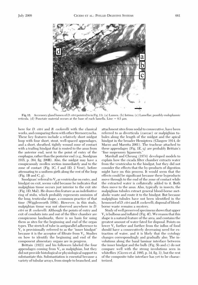
SnodgrassÕ referral to V3 as ventricular on entry, andhindgut on exit, seems valid because he indicates thatmalpighian tissue occurs just interior to the exit site(Fig. 1D, Mal). He draws this feature as an indeÞnitivering of stubs, which probably represents omission ofthe long, tenticular shape, a common practice of thattime (Wigglesworth 1956). However, in this study,malpighian tissue was not observed anywhere in D.citri or B. cockerelli. Although the points of entry andexit of conduits into and out of the Þlter chamber areconspicuous landmarks, there is no basis for usingthem as sites for the beginning and end of the organtypes. The stretch of tube in antiparallel adherence toV1 is provisionally referred to as the “inner hindgut”because it is the acceptor of Þltrate from V1. Studieson how to identify the beginning and end of thecomponent alimentary organs are in progress.
Brittain (1923) and his followers labeled the fourappendages coming from V3 as malpighian, but theydid not provide histological or physiological support tosubstantiate this. Substantiation is essential because avariety of tubular arrays, from simple to branched, and
attachment sites from nodal to consecutive, have beenreferred to as diverticula (caecae) or malpighian tu-bules along the length of the midgut and the apicalhindgut in the broader Hemiptera (Glasgow 1914, deMarzo and Marotta 2001). The tracheae attached tothese appendages (Fig. 1E, q) are probably BrittainÕs“Þne suspensory ligaments.”
Marshall and Cheung (1974) developed models toexplain how the cicada Þlter chamber extracts waterfrom the ventriculus to the hindgut, but they did notconsider the effects that the by-products of digestionmight have on this process. It would seem that theeffects could be signiÞcant because these byproductsmove through to the end of the zone of contact whilethe extracted water is collaterally added to it. Boththen move to the anus. Also, typically in insects, themalpighian tubules extract general blood-borne met-abolic waste and route it to the hindgut. But becausemalpighian tubules have not been identiÞed in thehemocoel ofD.citriandB. cockerelli,disposal of blood-borne waste remains a mystery.
Study of well preserved specimens shows that upperV1 is bulbous and inßated (Fig. 4f). We reason that thisshape is a natural feature of the area, and contains thegreatest amount of water fated for disposal. Points onlower V1 further and further from the inßux of foodshould have a consecutively decreasing need for ex-traction of water, and it is likely that the cytologychanges correspondingly and gradually also. The in-volutions along the basal laminar interface betweenthe inner hindgut and the bulb (Fig. 5b and c) do notcompare well with the strong involutions seen inwhiteßies (Cicero et al. 1995, p. 34, Þg. 3), but the restof the composite tube interface has yet to be charac-terized.
Fig. 12. Accessory gland lumen ofD. citripointed to in Fig. 11i. (a) Lumen. (b) Intima. (c) Lamellae, possibly endoplasmicreticula. (d) Punctate material occurs at the base of each lamella. Line � 0.5 �m.
July 2009 CICERO ET AL.: PSYLLID DIGESTIVE SYSTEMS 661

These interpretations are based on the classicalview that ßuids are conÞned to the conduit system.However, the content and function of the interstitialspace of the Þlter chamber surrounding the compositetube is not known. Therefore, the interstitial spacecould serve as a possible pool for holding excess waterthat also could pass directly into the inner hindgut.The opaque white color of V2 and V3 is interpreted tobe due to the accumulation of stored food inside theirlumina. The hyaline color of the Þlter chamber would,by that same reasoning, be due to its function as a hubfor ßow-through of materials involved in the Þltrationprocess.
Nothing is known of the origin of honeydew inpsyllids. The extraction process could occur along thecomposite tube, or honeydew could be the residuefrom midgut digestion that reenters the chamber andmingles with adsorbed material from V1 as it moves tothe exit point, or both.Salivary Glands. Drawings of the P. mali salivary
gland (Saunders 1921) and the P. buxi and T. urticaesalivary glands (Witlaczil 1885), show a spherical pat-tern of crescent-shaped cells arranged about a nodewhich represents the authorsÕ view of the lumen lead-ing to the salivary duct. Saunders (1921, plate 3, Þg. 22,
and text) referred to the organ as the primary salivarygland, and noticed a secondary, cylindrical compo-nent emerging from the base, where the salivary ductattaches, but stopped short of deÞning it as the ac-cessory salivary gland. For our purposes, this organ iscalled “accessory” in accordance with classical authorsof other taxa who labeled it as such because of itseccentric, subordinate location and smaller size withrespect to a larger PSG. Studies of theD. citriPSG (Xuet al. 1988) indicate an enveloping sheath to bepresent that harbors bacteria-like organisms. We havelocated this layer and identify it as belonging to aneighboring organ (Fig. 14).
It is generally accepted that the primary role ofsalivary glands is to produce secretory products forfeeding, including the salivary sheath (Miles 1999).But unequivocal conÞrmation is needed to assert thatthe glands are the sole source of these products, andwhether the PSG, ASG, or a combinatorial mix ofsecretions from both is involved in each salivary func-tion in the various taxa concerned. Stylet sheaths aresecreted by other hemipterans including aphids, cer-copids, whiteßies (Valerio 1989, Freeman et al. 2001,Takemura et al. 2006), and certain psyllids (Brennanet al. 2001), but they have not yet been reported in the
Fig. 13. D. citri. Close-up of the core PSG luminal complex pointed to in Fig. 11f. It is apparently a hub that iscontinuous with the accessory gland lumen and the salivary gland duct that leads to the mouth. At least two apicalmembrane morphologies occur in this area, but because of luminal closure, the associated cells and their positions inthe luminal continuity could not be determined. (a) Intima lined lumen. (b) Lamellae, cf. Fig. 12c. (c) A second apicalmembrane morphology. Line � 0.5 �m.
662 ANNALS OF THE ENTOMOLOGICAL SOCIETY OF AMERICA Vol. 102, no. 4
two species featured herein. At least two cellular mo-tifs were observed for the PSG of both psyllid speciesexamined. Those rich with fully spherical spherules(Figs. 9a and 11a) are interpreted as laden, whereasthose containing partially formed spherules (Figs. 9cand 11b and d) are interpreted as depleted. Both cellmotifs were present in all specimens examined. A thirdcell type (Figs. 9b and 10c) remains uncharacterized.
In both the PSG and the ASG, cells likely needaccess to noncuticularized portions of the core lumenfor their products to pass through the apical mem-brane into the ducts and then move to the mouth.Figure 11 provides some evidence for such an arrange-ment, based on the funnel-shaped conÞguration ofsecretory cells (Fig. 11, inset). Such wedge-shapedcells also comprise the accessory glands of Bemisia
tabaci (Gennadius) (Ghanim et al. 2001, p. 29, Þg. 6),conferring a “hesperidiform” shape to that organ. In-dications are that only one common duct occurs inD.citri (Fig. 8d) rather than dual ducts, as found in B.tabaci (Harris et al. 1996a, p. 147). This study was notable to determine the point where the lumina of con-verging ducts coalesce so that salivary materials fromleft and right glands mix before they enter the singlesalivary channel of the stylet. The presence of one sali-vary channel in the stylets of these two species is con-sistent with the broader Sternorrhyncha (Forbes 1972,Ullman and McLean 1986, Harris et al. 1996a).
The function of the lateral cells of the PSG (Fig.11e) cannot be surmised. The function of the entireASG is a mystery because it is not separated from thePSG by a duct of its own, as occurs in B. tabaci (Harris
Fig. 14. Cross section of an area of theB. cockerelli prothorax showing the PSG and a tightly appressed neighboring organ.(a) Layer between PSG and neighboring organ. (b) Mitochondria. (c) PSG. (d) Brush border of the PSG lumen. (e)Neighboring organ. Layer in f continues around the adjacent organ, not the PSG. (g) Intervening material. Line � 2 �m.
July 2009 CICERO ET AL.: PSYLLID DIGESTIVE SYSTEMS 663

et al. 1996a,b); in addition, all of its cells seem isolatedfrom the lumen by an intima (Figs. 11i and 12b). Thebasally punctate lamellae (Fig. 12 c and d) could beendoplasmic reticula. Their relationship to the meta-bolic processes of the ASG is currently under inves-tigation.
Acknowledgments
We acknowledge advice and Þeld and laboratory assis-tance from Jose Castillo, Barry Kostyk, Rosa Muchovej,Jawaad Qureshi, Mujaddad Rehman, Robert Riefer, andGregory Walker and the University of Arizona Spectroscopyand Imaging Facility staff.
References Cited
Bielenin, I. 1993. Filter chamber of Homoptera with par-ticular reference to Coccoidea. Przel. Zool. 37: 181Ð186.
Bove, J. M. 2006. Huanglongbing: a destructive, newly-emerging, century-old disease of citrus. J. Plant Pathol. 88:7Ð37.
Brennan, E. B., S. A. Weinbaum, and K. Pinney. 2001. Anew technique for studying the stylet tracks of ho-mopteran insects in hand-sectioned plant tissue usinglight or epißuorescence microscopy. Biotechnol. Histo-chem. 76: 59Ð66.
Brittain, W. H. 1923. The morphology and synonymy ofPsyllia mali Schmidberger. Proc. Acadian Entomol. Soc.8: 23Ð42.
Browning,H.W.,C.C.Childers,P.A.Stansly, J.Pena, andM.E.Rogers. 2006. Florida citrus pest management guide: soft-bodied insects attacking foliage and fruit. University of Flor-ida Institute of Food and Agricultural Sciences Extension.(http://edis.ifas.uß.edu/BODY_CG004).
Capoor, S. P., D. G. Rao, and S. M. Viswanath. 1974. Green-ing disease of citrus in the Deccan trap country and itsrelationship with the vector,Diaphorina citriKuwayama,pp. 43Ð49. In L. G. Weathers and M. Cohen [eds.], Pro-ceedings of the 6th Conference of the International Or-ganization of Citrus Virologists, 1972, Swaziland. Univer-sity of California, Berkeley, CA.
Chapman, R. F. 1998. The insects: structure and function,4th ed. Cambridge University Press, New York.
Cicero, J. M., E. Hiebert, and S. E. Webb. 1995. The ali-mentary canal of Bemisia tabaci and Trialeurodes abutilo-nea (Homoptera, Sternorrhynchi): histology, ultrastruc-ture and correlations to function. Zoomorphology 115:31Ð39.
Coletta-Filho,H.D.,M.L.P. N. Targon,M. A. Takita, J. D.DeNegri, J. Pompeu, Jr., M. A. Machado, do A. M. Amaral,and G. W. Muller. 2004. First report of the causal agentof Huanglongbing (“Candidatus Liberibacter asiaticus”)in Brazil. Plant Dis. 88: 1382.
da Graca, J. V. 1991. Citrus greening disease. Annu. Rev.Phytopathol. 19: 109Ð136.
de Marzo, L., and S. Marotta. 2001. Anatomy of the alimen-tary canal in female Coccoidea: an iconographic review.Boll. Zool. Agric. Bachic. Ser. II 33: 77Ð83.
Dufor, M. L. 1833. Recherches anatomiques et physi-ologiques sur les Hemipteres, accompagnees de consid-erations relatives a lÕhistoire naturelle et a la classiÞcationde ces insects. Mem. Acad. Imp. Sci. Saint-Petersbourg,France.
Forbes, A. R. 1972. Innervation of the stylets of the pearpsylla, Psylla pyricola (Homoptera: Psyllidae), and thegreenhouse whiteßy, Trialeurodes vaporariorum (Ho-
moptera: Aleyrodidae). J. Entomol. Soc. Br. Columbia 69:27Ð30.
Freeman, T. P., J. S. Buckner, D. R. Nelson, C.-C. Chu, andT. J. Henneberry. 2001. Stylet penetration by Bemisiaargentifolii (Homoptera: Aleyrodidae) into host leaf tis-sue. Ann. Entomol. Soc. Am. 94: 761Ð768.
Ghanim, M., R. C. Rosell, L. R. Campbell, H. Czosnek, andJ. K. Brown. 2001. Digestive, salivary, and reproductiveorgans of Bemisia tabaci (Gennadius) (Hemiptera: Aley-rodidae) B type. J. Morphol. 248: 22Ð40.
Glasgow, H. 1914. The gastric caeca and the caecal bacteriaof the Heteroptera. Biol. Bull. 26: 101Ð170.
Goodchild, A.J.P. 1963. Some new observations on the in-testinal structures concerned with water disposal in sap-sucking Hemiptera. Trans. R. Entomol. Soc. Lond. 115:217Ð237.
Goodchild, A.J.P. 1966. Evolution of the alimentary canal inthe Hemiptera. Biol. Rev. 41: 97Ð140.
Halbert, S. E. 1998. Asian citrus psyllidÑA serious potentialexotic pest of Florida citrus. (http://www.ifas.uß.edu/entweb/DCITRI.htm).
Halbert, S. E. 2005. Pest alert: citrus greening/huanglong-bing. (http://www.doacs.state.ß.us/pi/chrp/greening/citrusgreeningalert.html).
Halbert, S. E., and K. L. Manjunath. 2004. Asian citrus psyl-lids (Sternorrhyncha: Psyllidae) and greening disease ofcitrus: a literature review and assessment of risk in Flor-ida. Florida. Entomol. 87: 330Ð353.
Halbert, S. E., and C. A. Nunez. 2004. Distribution of theAsian citrus psyllid, Diaphorina citri Kuwayama (Ster-norrhyncha: Psyllidae) in the Caribbean basin. Florida.Entomol. 87: 401Ð402.
Halbert, S. E., X. Sunand, and W. N. Dixon. 2000. Asiancitrus psyllid and citrus greening disease. Citrus Ind. 91:22Ð24.
Hansen, A .K., T. Trumble, R. Stouthamer, and T.D. Paine.2008. A new Huanglongbing species, “CandidatusLiberibacter psyllaurous,” found to infect tomato andpotato, is vectored by the psyllid Bactericera cockerelli(Sulc). Appl. Environ. Microbiol. 74: 5862Ð5865.
Harris, K. F., Z. Pesic-van Esbroeck, and J. E. Duffus. 1996a.Morphology of the sweet potato whiteßy, Bemisia tabaci(Homoptera, Aleyrodidae) relative to virus transmission.Zoomorphology 116: 143Ð156.
Harris, K. F., Z. Pesic-van Esbroeck, and J. E. Duffus. 1996b.Anatomy of a virus vector, pp. 289Ð318. InD. Gerling andR. T. Mayer [eds.], Bemisia 1995: taxonomy, biology,damage control and management. Intercept Ltd., An-dover, United Kingdom.
Kershaw, J.G.C. 1913. Anatomical notes on a membracid.Ann. Soc. Entomol. Belg. 57: 191Ð201.
Lallemand, J., A. Fos, and J. M. Bove. 1986. Transmission dela bacterie associee a la forme africaine de la maladie du“greeningÕ par le psylle asiatique Diaphorina citri Ku-wayama. Fruits 41: 341Ð343.
Macloskie, G. 1886. Witlaczil on Psyllidae. Am. Nat. 20: 283Ð287.
Marshall,A.T., andW.W.K.Cheung. 1974. Studies on waterand ion transport in homopteran insects: ultrastructureand cytochemistry of the cicadoid and cercopoid mal-pighian tubules and Þlter chamber. Tissue Cell 6: 153Ð171.
Miles, P. W. 1999. Aphid saliva. Biol. Rev. (Camb.) 74: 41Ð85.Packard, A. S. 1898. A text-book of entomology, including
the anatomy, physiology, embryology and metamorpho-ses of insects, for use in agricultural and technical schoolsand colleges as well as by the working entomologist.Macmillan, New York.
664 ANNALS OF THE ENTOMOLOGICAL SOCIETY OF AMERICA Vol. 102, no. 4
Pesson, P. 1951. Ordre des Homopteres (Homoptera,Leach, 1815). Traite Zool. Anat. Syst. Biol. 10: 1390Ð1656.
Ponsen, M. B. 1979. The digestive system of Subsaltusaphisornata (Homoptera: Aphididae). Mededel. Landbou-whogesch. Wagening. 79: 1Ð30.
Roistacher, C. N. 1996. The economics of living with citrusdiseases: huanglongbing (greening) in Thailand, pp. 279Ð285. In J. V. da Graca, P. Moreno, and R. K. Yokomi [eds.],Proceedings of the 13th Conference of the InternationalOrganization of Citrus Virologists, 1995. University ofCalifornia, Riverside, CA.
Saunders, L. G. 1921. The anatomy of Psyllia mali Schmid-berger. Ph.D. dissertation, McGill University, Montreal,QC, Canada.
Snodgrass R. E. 1935. Principles of insect morphology.McGraw-Hill, New York.
Takemura, M., Y. Kuwahara, and R. Nishida. 2006. Feedingresponses of an oligophagous bean aphid, Megoura cras-sicauda, to primary and secondary substances in Viciaangustifolia. Entomol. Exp. Appl. 121: 51Ð57.
Teixeira, D. C., C. Saillard, S. Jagoueix-Eveillard, J. L. Danet,A. J. Ayres, and J. M. Bove. 2005. “Candidatus Liberib-acter americanus” associated with citrus huanglongbing
(greening disease) in Sao Paulo State, Brazil. Inter. J. Syst.Evol. Microbiol. 55: 1857Ð1862.
Ullman,D.E., andD.L.McLean. 1986. Anterior alimentarycanal of the pear psylla, Psylla pyricola Foerster (Ho-moptera, Psyllidae). J. Morphol. 189: 89Ð98.
Valerio, J.R. 1989. Detection of salivary sheaths and feedingmarks of Zulia entreriana (Berg 1879) (Homoptera: Cer-copidae). An. Soc. Entomol. Bras. 18(Suppl): 169Ð174.
Weber, H. 1968. Biologie der Hemipteren, eine Naturge-schichte der Schnabelkerfe, reprint of 1930 edition.Asher, Amsterdam, The Netherlands.
Wigglesworth, V. B. 1956. Insect physiology, 5th ed. Wiley,New York.
Witlaczil, E. 1885. Die anatomie der psylliden. Zeits. Wis-senschaftliche Zool. 42: 569Ð638.
Xu, C. F., Y. H. Xia, K. B. Li, and C. Ke. 1988. Further studyof the transmission of citrus Huanglungbin by a psyllid,Diaphorina citri Kuwayama, pp. 243Ð248. In L. W. Trim-mer, S. M. Garnsey, and L. Navarro [eds.], Proceedingsof the 10th Conference of the International Organizationof Citrus Virologist, 1986, Valencia, Spain. University ofCalifornia, Riverside, CA.
Received 11 February 2009; accepted 4 May 2009.
July 2009 CICERO ET AL.: PSYLLID DIGESTIVE SYSTEMS 665