a protein therapeutic modality founded on molecular...
TRANSCRIPT

A protein therapeutic modality foundedon molecular regulationChapman M. Wrighta, R. Clay Wrighta, James R. Eshlemanb, and Marc Ostermeiera,1
aDepartment of Chemical and Biomolecular Engineering, Johns Hopkins University, Baltimore, MD 21218; and bDepartments of Pathology and Oncology,Sol Goldman Pancreatic Cancer Research Center, Johns Hopkins University School of Medicine, Baltimore, MD 21231
Edited by David Baker, University of Washington, Seattle, WA, and approved August 19, 2011 (received for review February 18, 2011)
The exquisite specificity of proteins is a key feature driving theirapplication to anticancer therapies. The therapeutic potential ofanother fundamental property of proteins, their ability to be regu-lated by molecular cues in their environment, is unknown. Here,we describe a synthetic biology strategy for designing proteintherapeutics that autonomously activate a therapeutic function inresponse to a specific cancer marker of choice.We demonstrate thisapproach by creating a prodrug-activating enzyme that selectivelykills human cancer cells that accumulate the marker hypoxia-indu-cible factor 1α. This property arises primarily through increased cel-lular accumulation of the enzyme in the presence of the marker.Our strategy offers a platform for the development of inherentlyselective protein therapeutics for cancer and other diseases.
directed evolution ∣ protein engineering ∣ protein switch ∣enzyme/prodrug therapy
The attractiveness of proteins as therapeutics stems in partfrom the exquisite specificity by which they execute diverse
functions—e.g., they catalyze exactly the right reaction or inhibitexactly the right cell receptor. Proteins possess another funda-mental property central to life whose therapeutic potential hasnot been exploited—the ability to be regulated at the proteinlevel by molecular signals. Prevailing approaches to cancer pro-tein therapeutics focus on cancer marker recognition/modulationand downstream effects of that modulation (1). Such an approachis limiting because the therapeutic mechanism is restricted tothose that naturally arise from modulation of the cancer marker.Furthermore, this approach precludes the use of cancer markersfor which modulators cannot be found or for which modulationdoes not produce a therapeutic effect (2). In addition, manypotential protein therapies lack the desired selective cancer celltargeting. The ability to link recognition of any cancer markerwith activation of any desired therapeutic function would enor-mously expand the number of possible protein therapeutics. Hereis where the therapeutic potential of regulation can be realized.
One approach to establishing unique regulatory relationshipsis to build proteins that function as switches—proteins whosecellular level of activity is modulated through interactions withan input signal such as a protein or small molecule. Our switchdesign strategy views all proteins as an extensive parts list fromwhich switches can be built using domains with the prerequisiteinput and output functions of the desired switch. The design chal-lenge for this approach is how to fuse the input (i.e., signalrecognizing) and output domains (i.e., the function to be modu-lated) such that the input domain regulates the output domain’sfunction. We have explored a directed evolution approach to thisgeneral design problem in which libraries of random insertionsof one domain into the other are subjected to selections andscreens designed to identify library members with switching be-havior (3–5). Our approach extensively explores the geometricspace of how two protein domains can be fused through insertionof one domain into another (3, 4). For example, we have identi-fied switch proteins with maltose-activated β-lactamase activityfrom libraries of circular permuted β-lactamase genes insertedinto the gene encoding maltose-binding protein (MBP) (4, 5).
Many of these switches function as allosteric enzymes withmaltose binding to the MBP domain inducing conformationalchanges in the β-lactamase domain that affect its catalytic activity(4, 6). For other switches, maltose binding does not affect thespecific activity of the protein but instead increases the accumu-lation of the switch protein in the cell (7).
Here, we propose a synthetic biology strategy for designingprotein therapeutics that link activation of a chosen therapeuticfunction to a specific cancer marker of choice. We demonstratethis strategy by creating a protein switch that renders human co-lon and breast cancer cells susceptible to the prodrug 5-fluorocy-tosine (5FC) in response to the cancer marker hypoxia-induciblefactor 1α (HIF-1a).
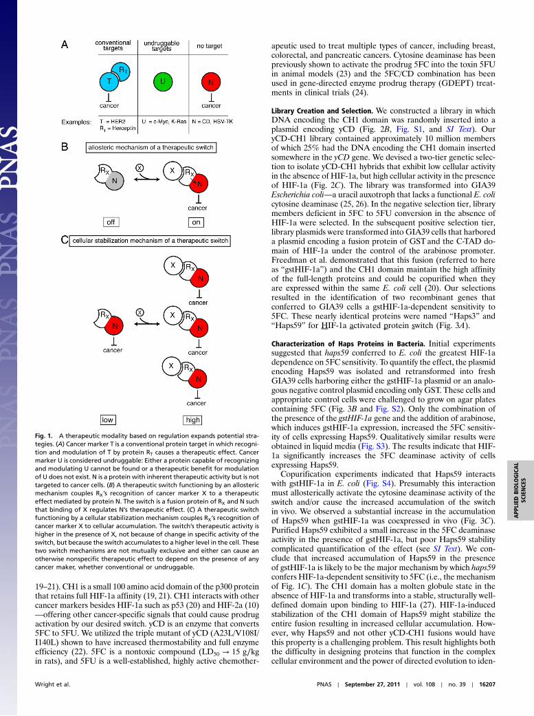
Results and DiscussionA Protein Therapeutic Modality Based on Regulation. Our approachgreatly expands the number of possible therapeutic strategies byproviding a platform for how a cancer marker of choice can beused to trigger any therapeutic function of choice (Fig. 1). Centralto this strategy is the development of a protein switch that couplesrecognition of the cancer marker to a therapeutic function. Suchcoupling could arise via newly established allosteric interactions(Fig. 1B) or through increased cellular accumulation in thepresence of the marker (Fig. 1C). We sought to demonstrate thisstrategy by linking the unrelated functions of recognition oftumor-marker HIF-1a and the enzymatic production of the che-motherapeutic 5-fluorouracil (5FU) from the prodrug 5FC bycytosine deaminase (Fig. 2A). HIF-1a accumulates to a high levelin many solid tumors, including breast, prostate, colorectal, andpancreatic cancers (8–10) but is virtually undetectable in normal,well-oxygenated tissues (11–13). Cancer cells that contain highlevels of HIF-1a are able to survive under extreme conditions,are resistant to therapy, and have a greater potential for metas-tasis (10, 14–18). Thus, the linking of HIF-1a levels to 5FU pro-duction could in theory attack the most aggressive tumors, whilehaving limited or no effect on well-oxygenated, normal cells. Sucha strategy, like any strategy that utilizes an intracellular target,faces the challenge of efficient delivery of the therapeutic geneor protein to the cancer cell.
We constructed our switch using the CH1 domain from thehuman p300 protein as the HIF-1a recognition input domainand yeast cytosine deaminase (yCD) as the prodrug activationoutput domain (Fig. 2A). The p300/CBP protein complex bindsHIF-1a in the cytoplasm and translocates it to the nucleus (13,
Author contributions: C.M.W., J.R.E., and M.O. designed research; C.M.W. and R.C.W.performed research; C.M.W., R.C.W., J.R.E., and M.O. analyzed data; and C.M.W. andM.O. wrote the paper.
Conflict of interest statement: M.O. and C.M.W. declare a conflict of interest in the form ofa patent application.
This article is a PNAS Direct Submission.1To whom correspondence may be addressed at: Johns Hopkins University Department ofChemical and Biomolecular Engineering, 3400 North Charles Street, Baltimore, MD 21218.E-mail: [email protected].
This article contains supporting information online at www.pnas.org/lookup/suppl/doi:10.1073/pnas.1102803108/-/DCSupplemental.
16206–16211 ∣ PNAS ∣ September 27, 2011 ∣ vol. 108 ∣ no. 39 www.pnas.org/cgi/doi/10.1073/pnas.1102803108
19–21). CH1 is a small 100 amino acid domain of the p300 proteinthat retains full HIF-1a affinity (19, 21). CH1 interacts with othercancer markers besides HIF-1a such as p53 (20) and HIF-2a (10)—offering other cancer-specific signals that could cause prodrugactivation by our desired switch. yCD is an enzyme that converts5FC to 5FU. We utilized the triple mutant of yCD (A23L/V108I/I140L) shown to have increased thermostability and full enzymeefficiency (22). 5FC is a nontoxic compound (LD50 → 15 g∕kgin rats), and 5FU is a well-established, highly active chemother-
apeutic used to treat multiple types of cancer, including breast,colorectal, and pancreatic cancers. Cytosine deaminase has beenpreviously shown to activate the prodrug 5FC into the toxin 5FUin animal models (23) and the 5FC/CD combination has beenused in gene-directed enzyme prodrug therapy (GDEPT) treat-ments in clinical trials (24).
Library Creation and Selection. We constructed a library in whichDNA encoding the CH1 domain was randomly inserted into aplasmid encoding yCD (Fig. 2B, Fig. S1, and SI Text). OuryCD-CH1 library contained approximately 10 million membersof which 25% had the DNA encoding the CH1 domain insertedsomewhere in the yCD gene. We devised a two-tier genetic selec-tion to isolate yCD-CH1 hybrids that exhibit low cellular activityin the absence of HIF-1a, but high cellular activity in the presenceof HIF-1a (Fig. 2C). The library was transformed into GIA39Escherichia coli—a uracil auxotroph that lacks a functional E. colicytosine deaminase (25, 26). In the negative selection tier, librarymembers deficient in 5FC to 5FU conversion in the absence ofHIF-1a were selected. In the subsequent positive selection tier,library plasmids were transformed into GIA39 cells that harboreda plasmid encoding a fusion protein of GST and the C-TAD do-main of HIF-1a under the control of the arabinose promoter.Freedman et al. demonstrated that this fusion (referred to hereas “gstHIF-1a”) and the CH1 domain maintain the high affinityof the full-length proteins and could be copurified when theyare expressed within the same E. coli cell (20). Our selectionsresulted in the identification of two recombinant genes thatconferred to GIA39 cells a gstHIF-1a-dependent sensitivity to5FC. These nearly identical proteins were named “Haps3” and“Haps59” for HIF-1a activated protein switch (Fig. 3A).
Characterization of Haps Proteins in Bacteria. Initial experimentssuggested that haps59 conferred to E. coli the greatest HIF-1adependence on 5FC sensitivity. To quantify the effect, the plasmidencoding Haps59 was isolated and retransformed into freshGIA39 cells harboring either the gstHIF-1a plasmid or an analo-gous negative control plasmid encoding only GST. These cells andappropriate control cells were challenged to grow on agar platescontaining 5FC (Fig. 3B and Fig. S2). Only the combination ofthe presence of the gstHIF-1a gene and the addition of arabinose,which induces gstHIF-1a expression, increased the 5FC sensitiv-ity of cells expressing Haps59. Qualitatively similar results wereobtained in liquid media (Fig. S3). The results indicate that HIF-1a significantly increases the 5FC deaminase activity of cellsexpressing Haps59.
Copurification experiments indicated that Haps59 interactswith gstHIF-1a in E. coli (Fig. S4). Presumably this interactionmust allosterically activate the cytosine deaminase activity of theswitch and/or cause the increased accumulation of the switchin vivo. We observed a substantial increase in the accumulationof Haps59 when gstHIF-1a was coexpressed in vivo (Fig. 3C).Purified Haps59 exhibited a small increase in the 5FC deaminaseactivity in the presence of gstHIF-1a, but poor Haps59 stabilitycomplicated quantification of the effect (see SI Text). We con-clude that increased accumulation of Haps59 in the presenceof gstHIF-1a is likely to be the major mechanism by which haps59confers HIF-1a-dependent sensitivity to 5FC (i.e., the mechanismof Fig. 1C). The CH1 domain has a molten globule state in theabsence of HIF-1a and transforms into a stable, structurally well-defined domain upon binding to HIF-1a (27). HIF-1a-inducedstabilization of the CH1 domain of Haps59 might stabilize theentire fusion resulting in increased cellular accumulation. How-ever, why Haps59 and not other yCD-CH1 fusions would havethis property is a challenging problem. This result highlights boththe difficulty in designing proteins that function in the complexcellular environment and the power of directed evolution to iden-
Fig. 1. A therapeutic modality based on regulation expands potential stra-tegies. (A) Cancer marker T is a conventional protein target in which recogni-tion and modulation of T by protein RT causes a therapeutic effect. Cancermarker U is considered undruggable: Either a protein capable of recognizingand modulating U cannot be found or a therapeutic benefit for modulationof U does not exist. N is a protein with inherent therapeutic activity but is nottargeted to cancer cells. (B) A therapeutic switch functioning by an allostericmechanism couples RX’s recognition of cancer marker X to a therapeuticeffect mediated by protein N. The switch is a fusion protein of RX and N suchthat binding of X regulates N’s therapeutic effect. (C) A therapeutic switchfunctioning by a cellular stabilization mechanism couples RX’s recognition ofcancer marker X to cellular accumulation. The switch’s therapeutic activity ishigher in the presence of X, not because of change in specific activity of theswitch, but because the switch accumulates to a higher level in the cell. Thesetwo switch mechanisms are not mutually exclusive and either can cause anotherwise nonspecific therapeutic effect to depend on the presence of anycancer maker, whether conventional or undruggable.
Wright et al. PNAS ∣ September 27, 2011 ∣ vol. 108 ∣ no. 39 ∣ 16207
APP
LIED
BIOLO
GICAL
SCIENCE
S

tify proteins that provide the desired cellular phenotype when theappropriate genetic selection is applied.
Haps59 Increases the 5FC Sensitivity of Cancer Cells Expressing HIF-1a.We next examined whether Haps59 would function in human can-cer cells and confer sensitivity to 5FC selectively under conditionsthat cause HIF-1a to accumulate. Colorectal solid tumors accu-mulate high levels of HIF-1a (8–10). RKO is a colorectal cancercell line that is known to accumulate high levels of HIF-1a inhypoxia (28) and is sensitive to 5FU in culture (29). 5FC washighly toxic to stable cell lines of RKO expressing yCD (LC50 ¼∼15 μM) but nontoxic up to approximately 2 mM to stable celllines constructed with the empty vector control (Fig. S5). Wechose the CMV promoter to control expression of Haps59 andyCD because hypoxic conditions do not change the level of ex-pression from this promoter (30). Initial experiments used theaddition of Co2þ to the media to cause HIF-1a accumulation.Co2þ disrupts the degradation pathway of HIF-1a, allowing theprotein to accumulate in the cytoplasm of the cell (31) (Fig. 4A).In the presence of Co2þ, Haps59-expressing RKO cell lines were10-fold more sensitive to 5FC (Fig. 4B). Hypoxic growth condi-
tions (1% O2) also caused a marked increase in the 5FC sensi-tivity of these cells (Fig. 4B). Neither hypoxic growth conditionsnor the addition of Co2þ caused 5FC sensitivity to the empty vec-tor control cells or altered the 5FC sensitivity of cells expressingyCD (Fig. 4B and Fig. S5). Additionally, neither the presence ofCo2þ nor hypoxic conditions were inherently toxic to Haps59-expressing RKO cells (Fig. S6A).
Analogous experiments with MCF7 breast cancer cells illu-strated the generality of the approach (Fig. 4D and E). Mirroringresults with E. coli cells, Haps59 accumulated at a higher level inthe presence of HIF-1a in both RKO and MCF7 cells (Fig. 4 Cand F), whereas yCD did not (Fig. S7A). This finding furthersupports the hypothesis that the increase in 5FC sensitivity resultsfrom a Haps59-HIF-1a interaction that causes increased accumu-lation of Haps59. This interaction was substantiated in RKOcells by coimmunoprecipitation experiments (Fig. S7B) and is thedesigned input signal to activate 5FU production. We confirmedthe production of 5FU in Haps59-expressing RKO cells andfound the production to be 2.5-fold higher in cells exposed toCo2þ (Fig. S6B). These results suggest that Haps59 functions as
Fig. 2. Protein switch concept, creation, and isolation. (A) The desired switch is a fusion protein between a HIF-1a binding domain (CH1) and yeast cytosinedeaminase (yCD) that increases the cellular conversion of 5FC to 5FU in the presence of HIF-1a. (B) Library creation. A plasmid containing the yCD gene israndomly linearized and ligated to a collection of circularly permuted CH1 genes (cpCH1; see Fig. S1), resulting in a library of random insertions of cpCH1 intothe plasmid. (C) Two-tiered genetic selection for yCD-CH1 hybrids that possess cytosine deaminase activity only in the presence of HIF-1a.
Fig. 3. Characterization of Haps59 in E. coli. (A) Switches contain a complete, noncircularly permuted CH1 domain (green) flanked by short linker sequences ofthe indicated sequences (black) inserted after amino acid 8 of yCD (red). The black numbers indicate the corresponding amino acid numbers in the wild-typeyCD protein. (B) Growth of GIA39 cells expressing MBP, yCD, or Haps59 and either GST or gstHIF-1a on minimal media agar plates as a function of 5FCconcentration. (Top) In the absence of arabinose and (Bottom) in the presence of arabinose to induce GST and gstHIF-1a expression. (C) Accumulation ofyCD and Haps59 in GIA39 E. coli in the absence and presence of coexpressed GST or gstHIF-1a as detected by Western blot with anti-yCD antibodies.
16208 ∣ www.pnas.org/cgi/doi/10.1073/pnas.1102803108 Wright et al.

designed—increasing 5FU production in cancer cells in responseto HIF-1a.
Haps59’s Potential as a Cancer Therapeutic. HIF-1a is a molecularsignature of cancer cells that can survive under extreme condi-tions, are resistant to therapy, and have a greater potential formetastasis (14–18). A treatment method that could specificallytarget these cells could significantly improve the effectiveness ofcancer treatments when used in combination with traditionaltherapies. Haps59 is designed to establish a direct relationshipbetween HIF-1a levels and the intracellular production of a che-motherapeutic drug. Haps59’s therapeutic potential is derivedfrom this unique regulatory property. Our establishment of thisregulation results in a complex protein that autonomously “deci-des” whether its drug-producing capability should be activated.
Hypoxic regions within a solid tumor can vary from 10–80%(32), and aberrant HIF-1a accumulation as a result of both gain-of-function and loss-of-function mechanisms has been observedexperimentally and clinically in normoxic conditions (10). Thus, ahigh percentage of solid tumor cells can potentially activateHaps59, and non-Haps59 expressing tumor cells would be suscep-tible to the strong 5FU bystander effect (33, 34). Haps59 exploitsan intracellular cancer marker for this activation. Prevailing stra-tegies for protein cancer therapeutics target more-readily acces-sible extracellular cancer markers such as membrane-bound cellreceptors. However, its been estimated that <10% of the humangenome codes for cell surface proteins, limiting the available tar-gets for therapy (2). There is a great need to develop effectivetargeting strategies, and this might be accomplished throughintracellular cancer markers. Such strategies will face deliverychallenges: Either the therapeutic protein or its correspondinggene must be efficiently delivered inside the cell. However, ourplacement of the selectivity at the protein level overcomes seriouslimitations of existing gene therapy approaches that require se-lective targeting of gene delivery to malignant cells. For example,
the combination of specific delivery of a cytosine deaminase geneto cancer cells followed by 5FC administration is an example ofGDEPT (33, 34). One major limitation to existing CD/5FCGDEPT strategies is the poor transmission efficiency of the CDgene using current viral vectors because of the need for trans-mission specifically to cancer cells (35). Our strategy overcomesthis limitation by moving the specificity from the transductionallevel to the protein level, allowing efficient means of gene deliv-ery to be used regardless of cell-type specificity. In addition, ourapproach is complementary to both transcriptional (36) andtransductional targeting and might be combined with these ap-proaches to afford a double or triple layer of specificity: at thegene delivery level, at the transcription level, and at the proteinlevel. Recent advances in protein delivery offer a potential routetoward achieving efficient delivery without gene therapy (37, 38).
Nature chose proteins as a molecule of choice to carry outa wide array of specific, intricately regulated functions. Our in-creasing ability to emulate nature and design proteins capableof complex, carefully regulated functions offers an attractive pathto achieving the effective and selective therapeutics we seek. Byconnecting disparate functions, protein switches expand thescope of potential therapeutic strategies in a manner that inher-ently encompasses specificity.
MethodsLibrary Creation and Selection. Creation of the yCD-CH1 hybrid library isdescribed in detail in SI Text. For selections, yCD-CH1 library plasmid DNAwas isolated from an aliquot of DH5α E. coli cells, and 25 ng of the purifiedplasmid was used to transform GIA39 E. coli cells (Coli Genetic Stock Center#5594) by electroporation. The transformed cells were plated on LB agarcontaining 50 μg∕mL chloramphenicol and then recovered using a sweepbuffer (1× M9 salts containing 2% glucose and 15% glycerol) and storedin aliquots at −80 °C. These cells were first plated on 24.5 × 24.5 cm minimalmedia plates (1× nitrogen base, 1× yeast synthetic dropout without uracil,2% glucose, and 20 g∕L select agar) containing 75 μg∕mL 5FC, 1 μg∕mLuracil, 1 mM IPTG, and 50 μg∕mL chloramphenicol. Cells were plated at a
Fig. 4. Characterization of Haps59 in human cancer cells. (A–C) RKO colorectal cancer cells; (D–F) MCF7 breast cancer cells. (A and D) Co2þ causes accumulationof HIF-1a in cells expressing Haps59, yCD, or the empty vector control as detected byWestern blot with anti-HIF-1a antibodies. (B and E) Both Co2þ (orange solidsquares) and exposure to hypoxic growth conditions (red solid squares) increase the 5FC sensitivity of Haps59-expressing cells compared to Haps59-expressingcells not exposed to those conditions (open blue squares). The 5FC sensitivities of cells expressing yCD (solid diamonds) and EV control (solid triangle) are shownfor comparison (see Fig. S5 for additional controls). Each point represents the mean from three different clones of each cell line, and each clone was tested inthree separate experiments (error bars, SD, N ¼ 9). (C and F) Haps59 accumulates at higher levels in cells when HIF-1a is present. In these experiments a FLAGepitopewas appended to the N terminus of Haps59 (FLAG-Haps59) for switch detection byWestern blot. Cells were cultured under normoxic (N), hypoxic (H), ornormoxic conditions with the addition of 100 μM cobalt (Co2þ). Detection of β-actin served as a loading control.
Wright et al. PNAS ∣ September 27, 2011 ∣ vol. 108 ∣ no. 39 ∣ 16209
APP
LIED
BIOLO
GICAL
SCIENCE
S

concentration of approximately 500;000 cfu∕plate, with cfu defined on mini-mal media plates without 5FC but with 1 μg∕mL uracil. Colonies on theseplates were recovered into sweep buffer and replated at the same cfu densityon the same type of plate media except the 5FC concentration was loweredto 50 μg∕mL. Library members surviving this second negative selection platewere recovered into sweep buffer, aliquoted, and stored at −80 °C. Libraryplasmid DNA was isolated from these samples and used for the positiveselection.
GIA39 cells harboring a gstHIF-1a plasmid were cotransformed withlibrary plasmids from the negative selection and plated on LB media contain-ing 100 μg∕mL ampicillin and 50 μg∕mL chloramphenicol. CotransformedGIA39 cells were recovered from the plate using sweep buffer, aliquoted,and stored as described above. These cells were plated on 24.5 × 24.5 cmminimal media plates supplemented with 25 μg∕mL cytosine, 1 mM IPTG,0.15% arabinose, 50 μg∕mL chloramphenicol, and 100 μg∕mL ampicillin ata density of approximately 500;000 cfu∕plate. A total of 99 colonies formedon this selection plate, all of which were screened by colony PCR for pDIM-C8plasmids that contained the CH1 insert within the yCD gene. This wasperformed using primers that anneal outside the yCD gene followed bygel electrophoresis to observe the size of the PCR product. About 20% ofthese colonies harbored library members that contained a CH1 insert withinthe yCD gene, and these members were sequenced. Eight members of the20 sequenced were in-frame, of which Haps3 appeared twice and Haps59appeared three times. In-frame members were then individually replatedon selection media to ensure their switching behavior in the presence ofgstHIF-1a. Only Haps3 and Haps59 behaved as switches after replating.
5FC Sensitivity Assays with GIA39 Cells on Solid Media. Fresh GIA39 cell linesharboring a pDIM-C8 plasmid for expression of MBP (negative control), yCD(positive control), or Haps59 were cotransformed with either the gstHIF-1aplasmid or an analogous negative control plasmid encoding only GST werecreated. These six cell lines were cultured to midlog phase (0.3 OD) in minimalmedia and then serially diluted with minimal media 3.3-fold in 96-wellformat. Minimal media consisted of 1× nitrogen base, 1× yeast syntheticdropout without uracil, 2% glucose, 5 μg∕mL uracil, 100 μg∕mL of ampicillin,and 50 μg∕mL chloramphenicol. One microliter of each cell line dilution wasspotted on minimal media plates (OmniTray, 86 × 128 mm, Nunc) containing1mM IPTG, 20 g∕L select agar, and different amounts of 5FC (0, 100, 200, 300,500, and 700 μM). The media either omitted arabinose or contained 0.05%arabinose to induce GSTand gstHIF-1a. The plates were incubated for 24–36 hat 37 °C, and the results of these experiments can be seen in Fig. 3 and Fig. S2.
Haps59 Accumulation Studies in E. coli. Four GIA39 cell lines (expressing yCD +GST, yCD + gstHIF-1a, Haps59 + GST, or Haps59 + gstHIF-1a) were cultured in25 mL of minimal media containing 1× nitrogen base, 1× yeast syntheticdropout without uracil, 2% glucose, 10 μg∕mL uracil, 50 μg∕mL chloramphe-nicol, and 100 μg∕mL ampicillin overnight at 37 °C. These cultures werediluted into eight separate fresh minimal media flasks to compare the addi-tion or omission of arabinose on yCD and Haps59 accumulation. These eightcultures were grown to an OD of 0.2, at which point IPTG (to 1 mM) wasadded to all cultures and 0.05% arabinose was added to half of the flasksto express GST or gstHIF-1a. The cultures were then incubated at 37 °C for12–16 h. An equal amount of cells were aliquoted based on final OD mea-surements for lysis. The bacterial cells (680million cells per sample) were lysedusing BugBuster™ (Novagen) following the manufacturer’s protocol. Equalamounts of the cell lysates were separated on a 4–12% Bis-Tris NuPAGE gels(Invitrogen) and then transferred to PVDF membrane (BioRad). The Westernprotocol is described in SI Text.
5FC Sensitivity Experiments in RKO and MCF7 Cells Expressing yCD and ProteinSwitches. RKO cells stably expressing an empty vector (one clone), yCD (threeclones), or Haps59 (three clones) were used to seed 96-well plates, 1,500 cellsper well, in 100 μL ofMEMmedia supplemented with 10% fetal bovine serumand 1% antibiotic and antimycotic (ABAM) (all from Gibco). MCF7 cells stablyexpressing an empty vector (one clone), yCD (three clones), or Haps59 (threeclones) were used to seed 96-well plates, 3,500 cells per well, in 100 μL ofDMEM media supplemented with 10% fetal bovine serum and 1% ABAM
(all from Gibco). After 24 h, 100 μL of MEM (RKO) or DMEM (MCF7) mediacontaining 5FC (0–2 mM) was added to these cells. These experiments werecarried out for 6 d with one media change after 3 d. After 6 d, the cellswere lysed by rinsing twice with 100 μL of PBS and then incubated in100 μL of 0.1 % SDS in water. The cells were incubated at 37 °C for 2 h inthe SDS solution for complete lysis. To detect dsDNA, SYBR green (Invitrogen)was added to a final concentration of 0.075% to lysed cells and the fluores-cence for each well was recorded at 520 nm after excitation at 485 nm. Thepercent survival was calculated using the equation ðAÞsample∕ðAÞcontrol × 100,where ðAÞsample is the fluorescence of sample wells with 5FC and ðAÞcontrol isthe fluorescence of the control well lacking 5FC. The results displayed in Fig. 4represent the mean with experiments performed on three separate days(N ¼ 6 for empty vector (EV), N ¼ 9 for yCD, and N ¼ 9 for Haps59). Error barsare the standard deviation from the mean.
To induce HIF-1a accumulation, MEM or DMEM media containing 75 μMCoCl2 was used. The presence of Co2þ had little effect on the growth of theRKO cells, regardless of whether or not the cells had been transfected with agene (Fig. S6). Alternatively, a hypoxic environment was induced by incuba-tion of the cancer cells in a MIC-101 hypoxia chamber (Billups–Rothenberg).The chamber was purged with three 3-min purges of 1% O2 gas containing5% CO2 and 94% N2 over 2 h. A 3-min purge was then used every 24 h tomaintain a hypoxic environment for the 6 d. Incubation at 1%O2 tension alsohad no effect on cell growth compared to the parental cell lines grown in thesame conditions (Fig. S6). After 6 d (with one media change after 3 d), thecells were lysed and the percent survival was calculated using the same pro-tocol as described above. Hypoxic environment experiments were repeatedon separate days (N ¼ 6 for all), and error was calculated by standard devia-tion from the mean.
HIF-1a Dependent Accumulation of Haps59 in RKO and MCF7 Cancer Cells. yCDantibodies cross-reacted with many human proteins (Fig. S7A). To circumventthis problem, a FLAG™ tag (Sigma-Aldrich) was appended to the N terminusof Haps59 for protein switch accumulation experiments. RKO or MCF7 cellsexpressing an EV or Haps59 with the N-terminal FLAG™ tag (FLAG-Haps59)were incubated in normoxia (with and without 100 μM CoCl2), or hypoxia(1% O2) conditions for 12–18 h. After exposure, cell cultures were incubatedat 4 °C for 30 min and then treated with radioimmunoprecipitation assaybuffer (Sigma) containing protease inhibitor cocktail (Sigma). Cells werescrapped from the flask and placed in a 1.5-mL tube and incubated on icefor 30 min. Cell debris was removed by centrifugation (20;000 × g for30 min, 4 °C) and the supernatant was transferred to a fresh 1.5-mL tube.Protein concentrations of the whole cell lysates were determined usingthe detergent compatible protein assay (BioRad). A total of 50 μg of eachlysate were separated on 4–12% Bis-Tris NuPAGE gels (Invitrogen) and thentransferred to PVDF membrane (BioRad). The membrane was blocked with3% nonfat milk for 30 min.
Primary anti-FLAG™-HRP conjugated antibodies (Sigma) were dilutedinto the SignalBoost™ Immunoreaction Enhancer buffer (EMD Biosciences)according to the manufacturer’s protocol and incubated at room tempera-ture for 1 h. The membrane was then washed and ECL was visualized usinga Universal Hood II and QuanityOne software (BioRad); the results can beseen in Fig. 4 C and F. After detection of FLAG™, the membrane was strippedand then reprobed with beta-actin-HRP conjugated antibodies (Abcam) toverify protein-loading levels. For HIF-1a detection, 50 μg of the same sampleswere separated on 4–12% Bis-Tris NuPAGE gels and transferred to PVDFmembrane. The membrane was blocked with 3% nonfat milk for 30 min.Primary antibodies for HIF-1a were diluted 1∶1;000 into blocking bufferand incubated at 4 °C overnight. The membrane was then washed, followedby the addition of mouse-HRP conjugated secondary antibodies, and ECL wasvisualized as described above.
ACKNOWLEDGMENTS. The authors thank Sharon Gerecht, Phillip Cole, andDenis Wirtz for comments on the manuscript and Gregg L. Semenza andHirohiko Kamiyama for helpful discussions. This work was supported by Na-tional Institutes of Health Grants R01GM066972 (to M.O.) and R01CA130938(to J.R.E.).
1. Leader B, Baca QJ, Golan DE (2008) Protein therapeutics: A summary and pharmaco-
logical classification. Nat Rev Drug Discov 7:21–39.
2. Verdine GL, Walensky LD (2007) The challenge of drugging undruggable targets in
cancer: Lessons learned from targeting BCL-2 family members. Clin Cancer Res
13:7264–7270.
3. Guntas G, Ostermeier M (2004) Creation of an allosteric enzyme by domain insertion.
J Mol Biol 336:263–273.
4. Guntas G, Mitchell SF, Ostermeier M (2004) A molecular switch created by in vitro
recombination of nonhomologous genes. Chem Biol 11:1483–1487.
5. Guntas G, Mansell TJ, Kim JR, Ostermeier M (2005) Directed evolution of protein
switches and their application to the creation of ligand-binding proteins. Proc Natl
Acad Sci USA 102:11224–11229.
6. Wright CM, Majumdar A, Tolman JR, Ostermeier M (2010) NMR characterization of
an engineered domain fusion between maltose binding protein and TEM1 beta-
16210 ∣ www.pnas.org/cgi/doi/10.1073/pnas.1102803108 Wright et al.

lactamase provides insight into its structure and allosteric mechanism. Proteins78:1423–1430.
7. Sohka T, et al. (2009) An externally tunable bacterial band-pass filter. Proc Natl AcadSci USA 106:10135–10140.
8. Zhong H, et al. (1999) Overexpression of hypoxia-inducible factor 1alpha in commonhuman cancers and their metastases. Cancer Res 59:5830–5835.
9. Mabjeesh NJ, Amir S (2007) Hypoxia-inducible factor (HIF) in human tumorigenesis.Histol Histopathol 22:559–572.
10. Semenza GL (2010) Defining the role of hypoxia-inducible factor 1 in cancer biologyand therapeutics. Oncogene 29:625–634.
11. Huang LE, Gu J, Schau M, Bunn HF (1998) Regulation of hypoxia-inducible factor1alpha is mediated by an O2-dependent degradation domain via the ubiquitin-proteasome pathway. Proc Natl Acad Sci USA 95:7987–7992.
12. Yu AY, et al. (1998) Temporal, spatial, and oxygen-regulated expression of hypoxia-inducible factor-1 in the lung. Am J Physiol 275:L818–826.
13. Semenza GL (2004) Hydroxylation of HIF-1: Oxygen sensing at the molecular level.Physiology (Bethesda) 19:176–182.
14. Sun HC, et al. (2007) Expression of hypoxia-inducible factor-1a and associated proteinsin pancreatic ductal adenocarcinoma and their impact on prognosis. Int J Oncol30:1359–1367.
15. Dales JP, et al. (2005) Overexpression of hypoxia-inducible factor HIF-1alpha predictsearly relapse in breast cancer: Retrospective study in a series of 745 patients. Int JCancer 116:734–739.
16. Akakura N, et al. (2001) Constitutive expression of hypoxia-inducible factor-1alpharenders pancreatic cancer cells resistant to apoptosis induced by hypoxia and nutrientdeprivation. Cancer Res 61:6548–6554.
17. Khandrika L, et al. (2009) Hypoxia-associated p38 mitogen-activated protein kinase-mediated androgen receptor activation and increased HIF-1alpha levels contribute toemergence of an aggressive phenotype in prostate cancer. Oncogene 28:1248–1260.
18. Liao D, Corle C, Seagroves TN, Johnson RS (2007) Hypoxia-inducible factor-1alpha is akey regulator of metastasis in a transgenic model of cancer initiation and progression.Cancer Res 67:563–572.
19. Kung AL, Wang S, Klco JM, Kaelin WG, Livingston DM (2000) Suppression of tumorgrowth through disruption of hypoxia-inducible transcription. Nat Med 6:1335–1340.
20. Freedman SJ, et al. (2002) Structural basis for recruitment of CBP/p300 by hypoxia-inducible factor-1 alpha. Proc Natl Acad Sci USA 99:5367–5372.
21. Freedman SJ, et al. (2003) Structural basis for negative regulation of hypoxia-induciblefactor-1alpha by CITED2. Nat Struct Biol 10:504–512.
22. Korkegian A, Black ME, Baker D, Stoddard BL (2005) Computational thermostabiliza-tion of an enzyme. Science 308:857–860.
23. Kievit E, et al. (1999) Superiority of yeast over bacterial cytosine deaminase for en-zyme/prodrug gene therapy in colon cancer xenografts. Cancer Res 59:1417–1421.
24. Crystal RG, et al. (1997) Phase I study of direct administration of a replication deficientadenovirus vector containing the E. coli cytosine deaminase gene to metastatic coloncarcinoma of the liver in association with the oral administration of the pro-drug5-fluorocytosine. Hum Gene Ther 8:985–1001.
25. Mahan SD, Ireton GC, Stoddard BL, Black ME (2004) Alanine-scanning mutagenesisreveals a cytosine deaminase mutant with altered substrate preference. Biochemistry43:8957–8964.
26. Mahan SD, Ireton GC, Knoeber C, Stoddard BL, Black ME (2004) Random mutagenesisand selection of Escherichia coli cytosine deaminase for cancer gene therapy. ProteinEng Des Sel 17:625–633.
27. Dial R, Sun ZY, Freedman SJ (2003) Three conformational states of the p300 CH1domain define its functional properties. Biochemistry 42:9937–9945.
28. Dang DT, et al. (2006) Hypoxia-inducible factor-1alpha promotes nonhypoxia-mediated proliferation in colon cancer cells and xenografts. Cancer Res 66:1684–1936.
29. Thant AA, et al. (2008) Role of caspases in 5-FU and selenium-induced growth inhibi-tion of colorectal cancer cells. Anticancer Res 28:3579–3592.
30. Shibata T, Giaccia AJ, Brown JM (2000) Development of a hypoxia-responsive vectorfor tumor-specific gene therapy. Gene Ther 7:493–498.
31. Epstein AC, et al. (2001) Celegans EGL-9 and mammalian homologs define a family ofdioxygenases that regulate HIF by prolyl hydroxylation. Cell 107:43–54.
32. Zhao D, Ran S, Constantinescu A, Hahn EW, Mason RP (2003) Tumor oxygen dynamics:Correlation of in vivo MRI with histological findings. Neoplasia 5:308–318.
33. Greco O, Dachs GU (2001) Gene directed enzyme/prodrug therapy of cancer: Historicalappraisal and future prospectives. J Cell Physiol 187:22–36.
34. Russell PJ, Khatri A (2006) Novel gene-directed enzyme prodrug therapies againstprostate cancer. Expert Opin Investig Drugs 15:947–961.
35. Schepelmann S, Springer CJ (2006) Viral vectors for gene-directed enzyme prodrugtherapy. Curr Gene Ther 6:647–670.
36. Marignol L, et al. (2009) Hypoxia response element-driven cytosine deaminase/5-fluor-ocytosine gene therapy system: A highly effective approach to overcome the dynamicsof tumour hypoxia and enhance the radiosensitivity of prostate cancer cells in vitro.J Gene Med 11:169–179.
37. Yan M, et al. (2010) A novel intracellular protein delivery platform based on single-protein nanocapsules. Nat Nanotechnol 5:48–53.
38. Cronican JJ, et al. (2010) Potent delivery of functional proteins intoMammalian cells invitro and in vivo using a supercharged protein. ACS Chem Biol 5:747–752.
Wright et al. PNAS ∣ September 27, 2011 ∣ vol. 108 ∣ no. 39 ∣ 16211
APP
LIED
BIOLO
GICAL
SCIENCE
S

Supporting InformationWright et al. 10.1073/pnas.1102803108SI TextGeneral.All chemicals were purchased from Sigma-Aldrich unlessotherwise noted. All cell culture materials were purchased fromGibco (Invitrogen), and cell lines were purchased from AmericanType Culture Collection (ATCC). For tissue culture, RKO cellswere grown in MEM media, containing 10% FBS and 1% anti-biotic and antimycotic (ABAM) (Gibco). GIA39 Escherichia colicells (Coli Genetic Stock Center# 5594) were purchased from theE. coli genetic stock center. Antibodies for Western blots (yeastcytosine deaminase: Ab35251 and beta-Actin: Ab20272) werepurchased from Abcam. The FLAG™ M2 (F3165) and FLAG™-HRP conjugated (A8592) antibodies were purchased fromSigma, and the HIF-1a antibody (BDB 610958) was purchasedfrom BD Biosciences (Fisher Scientific). All antibodies were usedaccording to the manufacturers’ instructions.
Plasmids and Genes. Yeast cytosine deaminase was cloned fromyeast genomic DNA using the primers: 5′-ttataaggatccatggtga-cagggggaatggcaag and 5′-ttataaactagtctactcaccaatatcttcaaaccaatcwith the NcoI and SpeI restriction sites underlined, and insertedinto the pDIM-C8 plasmid, which contains a tac promoter andconfers chloramphenicol resistance (1–3). Three thermostabiliz-ing mutations (A23L, V108I, and I140L) (4) were incorporatedusing QuikChange (Stratagene) following the manufacturer’sprotocol. This thermostable version of the gene is referred toas “yCD” throughout SI Text for simplicity and the plasmid isreferred to as pDIM-yCD. The DNA encoding the C-TAD do-main of HIF-1a (amino acids 786–826) and the CH1 domainof the human p300 protein (amino acids 334–420) were orderedfrom IDTwith E. coli codon optimization. The gstHIF-1a plasmid(for gstHIF-1a expression) was constructed using the GSTfusion tag from the pGEX-6P-1 plasmid (GE Healthcare) andthe araC gene and the arabinose promoter from the pBAD plasmid(Invitrogen) and conferred ampicillin resistance. The DNA encod-ing C-TAD domain of HIF-1a was fused to the DNA encodingGSTas described by Freedman et al. (5) to form “gstHIF-1a.” ThegstHIF-1a fusion is inducible by the addition of arabinose to themedia. DNA encoding human codon optimized yCD, Haps3, andHaps59 were purchased from GenScript and cloned into thepcDNA 3.1(+) plasmid with neomycin resistance (Invitrogen).
Library Creation. Purified pDIM-yCD plasmid was digested withdilute concentrations of DNaseI as described (1–3). Singly cutplasmids were isolated using gel electrophoresis and purifiedusing Qiagen’s gel extraction kit following the manufacturer’s in-structions. Isolated singly cut plasmids were repaired and bluntedusing T4 DNA polymerase (0.5 U∕mg) and T4 DNA Ligase(150 U∕mg) both from New England Biolabs. The repaired,linear DNA was isolated using gel electrophoresis and used in aligation reaction with DNA encoding CH1 domain inserts. Threetypes of CH1 domain inserts were prepared. Two inserts aredescribed as direct inserts and used as an unaltered CH1 genewith appended DNA that encoded peptide linkers. The appendedlinkers encoded a glycine on the N terminus and either a GGSpeptide linker (“3-mer”) or a GGGGS (“5-mer”) peptide linkeron the C terminus. The third CH1 domain insert was preparedusing the circular permutation method shown in Fig. S1. The geneencoding the CH1 domain had a piece of DNA appended codingfor a ðGSGGGÞ3 linker that joined together the N and C terminiof the CH1 domain. The appended CH1 gene was cyclized anddigested with a nonspecific nuclease to create random circularpermutations of this gene. To accomplish this, the CH1 DNA
was excised from its plasmid using BamHI sites located withinthe linker region and the ends of the gene ligated together underdilute DNA concentrations to favor intramolecular ligation overintermolecular ligation. A standard cyclization reaction diluted5 μg of the excised CH1 gene into 500–600 μL of 1× T4 DNAligase buffer (New England Biolabs) to a DNA concentrationof approximately 8–10 ng∕μL. The dilute CH1 genes were cy-clized using T4 DNA ligase (20 units∕μL) at room temperature(RT) for 1 h. Cyclized CH1 genes were isolated using gel electro-phoresis. S1 nuclease was added (2.5 U∕μg) to purified, cyclizedDNA to make a variety of single double-stranded breaks withinthe CH1 DNA. The singly cut DNA was isolated using gel elec-trophoresis and repaired as described above in the digestion ofpDIM-yCD. To create the yCD-CH1 hybrid library, a ligation re-action was performed with the randomly linearized pDIM-yCDplasmid DNA and a 5-fold higher amount (molar basis) of a1∶1∶1mixture of the three types of CH1 inserts. A typical ligationreaction mixture included 500 ng of plasmid DNA, approximately200 ng of CH1 inserts, and 5% PEG in 1× T4 ligase buffer.Ligated plasmids were electroporated into DH5α E. coli cellsand the transformation mixture plated on LB agar containing50 μg∕mL chloramphenicol in a 24.5 × 24.5 cm Bio-Assay dish(Nunc, Thermo Fisher Scientific). The number of transformantswas 9.6 × 10⁶ of which approximately 25% contained insert DNA,as estimated by gel electrophoresis of plasmid DNA isolated fromthe library.
5-fluorocytosine (5FC) Sensitivity Assays with GIA39 Cells in LiquidMedia.GIA39 cells harboring a pDIM-C8 plasmid for expressionof maltose-binding protein (MBP) (negative control), yCD(positive control), Haps3, or Haps59 were cotransformed witheither the gstHIF-1a plasmid or an analogous negative controlplasmid encoding only GST. These eight GIA39 cell lines wereused for 5FC sensitivity experiments. In 96-well format, 1-mLcultures of the eight GIA39 cell lines described above were grownin minimal media at 37 °C with shaking. Minimal media consistedof 1× nitrogen base, 1× yeast synthetic dropout without uracil,2% glucose, 5FC (varied from 0 to 1 mM), 1.5 μg∕mL uracil,1 mM IPTG, 100 μg∕mL of ampicillin, 50 μg∕mL chlorampheni-col, and with or without 0.15% arabinose (to express gstHIF-1aand GST). The bacteria cells were cultured for 40–48 h at 37 °Cafter which the OD at 600 nm was measured using a SPECTRA-max Plus 96-well plate reader (Molecular Devices) and analyzedusing Softmax Pro software (Molecular Devices). The relativecell densities were calculated using the equation ðAÞsample∕ðAÞcontrol × 100, in which ðAÞsample is the absorbance of a samplewell containing 5FC and ðAÞcontrol is the absorbance of the controlwell lacking 5FC. Experiments performed with GIA39 celllines expressing yCD and Haps proteins were repeated on threeseparate days in duplicate (N ¼ 6). Control experiments per-formed with GIA39 cells expressing MBP were repeated twicein duplicate on separate days (N ¼ 4). Error was calculated bystandard deviation from the mean.
Western Blot for Accumulated yCD and Haps59 in GIA39 Cells. Themembrane containing transferred cell lysates was blocked with3% nonfat milk for 30 min. To observe the expression of Haps59and yCD, primary antibodies for yCD were diluted into blockingbuffer according to the manufacturer’s protocol and incubatedat RT for 1 h. The membrane was then washed, followed by theaddition of sheep-HRP conjugated secondary antibodies (BethylLaboratories) using the Snap ID protein detection system
Wright et al. www.pnas.org/cgi/doi/10.1073/pnas.1102803108 1 of 7

(Millipore) following the manufacturer’s protocol. ECL wasvisualized using a Universal Hood II and QuanityOne software(BioRad).
Purification of yCD, Haps3, and Haps59.After failed attempts to pur-ify the protein switches using a His tag and Ni2þ affinity, the genesencoding yCD, Haps3, and Haps59 were cloned into the pGEX-6P-1 plasmid for fusion to the affinity tag GST for purification(GE Healthcare). Separate 500-mL cultures of DH5α bacteriacells harboring each plasmid were grown in the presence ofampicillin at 37 °C until an OD of 0.4–0.6, at which point proteinwas induced by the addition of 1 mM IPTG for 3 h. Harvestedcells were resuspended in lysis buffer (50 mM Tris buffer, pH 7.5,containing 150 mM NaCl, 0.1 mM EDTA) containing proteaseinhibitor cocktail (Sigma). Cells were lysed using a FrenchPress and the insoluble material pelleted by centrifugation at15;000 × g for 45 min at 4 °C. Cleared lysate was loaded onto a5 mL GSTrap FF column using an AKTA purifier FPLC (GEHealthcare). The GSTrap column was washed with four columnvolumes of lysis buffer to clear unbounded proteins. The fusionprotein was eluted using 50 mM Tris pH 8.0, containing 10 mMreduced glutathione. Alternatively, an in-column cleavage wasperformed on bound GST fusions using PreScission Protease(GE Healthcare) at 80 U∕mg. Storage buffer (25 mM Tris buffer,pH 7.5, containing 50 mM NaCl) was used to elute the cleavedprotein from the GSTrap column. Purity was judged to be >95%using SDS-PAGE and protein concentrations were determinedusing A280 and the calculated extinction coefficient for each en-zyme. Purification of the gstHIF-1a fusion was performed exactlythe same as above, except the fusion was not cleaved with Pre-Scission Protease. GST-HIF-1a was eluted from the GSTrap col-umn in elution buffer (50 mM Tris buffer, pH 8.0, containing10 mM reduced glutathione) and dialyzed into storage buffer(25 mM Tris buffer, pH 7.5, containing 50 mM NaCl and 10%glycerol) then frozen at −20 °C.
5FC Activity Assay. The cytosine deaminase activity assays for yCDand the Haps proteins were performed on purified samples thatwere never frozen and within two weeks of purification. The 5FCactivity assay was based on similar assays reported by Mahan andcoworkers (6, 7). To determine the cytosine deaminase activity,yCD or Haps3 or Haps59 were added to a final concentration of100 nM in a reaction tube containing 50 mM Tris buffer, pH 7.5.The mixture was incubated at 37 °C for 5 min and then 5FC wasadded at various concentrations (50 μM–1 mM). For experimentsin the presence of 2.5 μM purified gstHIF-1a, both the proteinswitch and gstHIF-1a were incubated at 37 °C for 5 min beforethe addition of 5FC. Aliquots (50 μL) were removed from thereaction mixture at various time points (10, 20, 30, 60, 120, 180,and 240 s) and mixed with 0.1 N HCl. Measurements at 290 nmfor 5FC and 255 nm for 5-fluorouracil (5FU) were recordedand inserted in the following equations to determine the concen-trations of 5FC and 5FU: ½5FC� ¼ 0.119A290 − 0.025A255 and½5FU� ¼ 0.105A255 − 0.049A290 as described elsewhere (6). Alter-natively, CD activity was monitored using the decrease in absor-bance at 235 nm, where 5FC absorbs. The production of 5FUwas calculated using the decrease in absorbance at 235 nm overtime and the extinction coefficient of 5FC. Kinetic parameterswere determined using double reciprocal plots. Our calculatedcatalytic efficiency for triple mutant yCD at 37 °C was4.98 × 104 E−1 M−1 s−1, which is consistent with other reports(8, 9). Although both switches exhibited cytosine deaminase ac-tivity and produced absorbance spectra changes consistent withthe production of 5FU, the measured catalytic activities wereinconsistent from purification to purification and decreasedmarkedly over the period of a day. In general, the two proteinswitches’ catalytic activity in the presence of gstHIF-1a rangedfrom 30–85% of that measured for yCD. The rate of deamination
for both protein switches was always the same or higher (up toseveralfold) when measured in the presence of purifiedgstHIF-1a. Analogous experiments with GST tags attached tothe protein switches resulted in similar outcomes.
Copurification Experiments in E. coli. Copurification experimentswere performed using GIA39 cells harboring the gstHIF-1aplasmid and either pDIM-Haps3 or pDIM-Haps59 (i.e., versionsof the switches lacking the GST tag). A 500-mL culture wasincubated at 37 °C until the OD reached 0.4–0.6. At this point,arabinose (0.15%) was added to the culture to express gstHIF-1a,and IPTG (1 mM) was added to express the protein switch. After3 h at 37 °C, the cells were lysed and passed over a GSTrap col-umn using a AKTA purifier FPLC (GE Healthcare). Unboundproteins were eluted by the passage of four column volumes oflysis buffer over the GSTrap column. Protein complexes wereeluted using 50 mM Tris buffer, pH 8.0, containing 10 mMreduced glutathione and separated using SDS-PAGE. Becausethe switch proteins and gstHIF-1a have similar molecular weights(28 kDa and 31 kDa, respectively), an aliquot was treated with20 U of PreScission Protease (GE Healthcare) to cleave offthe GST tag of gstHIF-1a. The sample was subsequently passedover the GSTrap column again to remove the majority of freeGST from the sample (Fig. S4).
5FC Experiments in Parental RKO and MCF7 Cells. RKO and MCF7cells were acquired from the ATCC and genotyped before use.For controls, parental RKO cells were used to seed 96-well plates,1,500 cells per well, in 100 μL of MEM media supplementedwith 10% fetal bovine serum and 1% ABAM (all from Gibco).Parental MCF7 cells were used to seed 96-well plates, 3,500 cellsper well, in 100 μL of DMEM media supplemented with 10%fetal bovine serum and 1% ABAM (all from Gibco). After 24 h,the media was changed with 100 μL of MEM (RKO) or DMEM(MCF7) media containing either 5FC (0–20 mM) or 5FU(0–2 mM). Cells were incubated for 3 to 6 d and then lysed.The cells were lysed by rinsing twice with 100 μL of PBS and thenincubated in 100 μL of 0.1% SDS in water. The cells were incu-bated at 37 °C for 2 h in the SDS solution for complete lysis. Todetect dsDNA, SYBR® green (Invitrogen) was added to a finalconcentration of 0.075% to lysed cells and the fluorescencefor each well was recorded at 520 nm after excitation at 485 nm.The percent survival was calculated using the equation ðAÞsample∕ðAÞcontrol × 100, where ðAÞsample is the fluorescence of the samplewells with 5FC and ðAÞcontrol is the absorbance of the control welllacking 5FC and 5FU. Results shown in Fig. S5 A and D are themean of experiments performed on three different days. Errorbars are the standard deviation from the mean.
Creation of RKO and MCF7 Stable Cell Lines.Genes encoding humancodon optimized yCD, Haps3, and Haps59 were cloned into thepcDNA 3.1 (+) plasmid (Invitrogen) under the control of a CMVpromoter and containing neomycin resistance. Transfectionswere performed using Lipofectamine 2000 following the manu-facturer’s protocol in 6-well plates. A bulk selection using G418(Geneticin) (Gibco) at a concentration of 0.8 mg∕mL was per-formed 24 h after the transfection. Cells that survived the bulkselection were separated into one cell per well in 96-well platesfor further selection in the presence of Geneticin. Single cells thatgrew into colonies in the presence of Geneticin were examinedfor their 5FC sensitivity by the addition 800 μM 5FC for yCDclones and 75 μM Co2þ and 800 μM 5FC for protein switchclones. Clones of RKO-yCD, RKO-Haps3, and RKO-Haps59that were sensitive to 5FC containing media (i.e., functionallyconfirmed) were also confirmed genetically. Genomic DNA iso-lated from these clones was probed by PCR with primers thatannealed outside of the multiple cloning site of the pcDNA3.1(+) plasmid. Sequencing of the correct size PCR product con-
Wright et al. www.pnas.org/cgi/doi/10.1073/pnas.1102803108 2 of 7

firmed the successful creation of the desired RKO cell lines. Forthe MCF7 cells, a gene encoding GFP was linked to the genesencoding yCD and Haps59 via a T2A peptide linker. The T2Alinker is self-cleaving, yields two separate proteins (GFP + yCDor GFP + Haps59), with a short C-terminal peptide on GFP andonly a single amino acid on the N terminus of the yCD or Haps59proteins (10). The tandem genes (GFP-T2A-yCD or GFP-T2A-Haps59) were cloned into the pcDNA 3.1 (+) plasmid as wellas the gene encoding GFP as an empty vector control. The sametransfection protocol used for RKO cells was used for MCF7cells. A bulk selection using G418 at a concentration of0.6 mg∕mL was performed 24 h after the transfection. Cells thatsurvived the bulk selection were separated into one cell per wellin 96-well plates using GFP as a reporter and further selection inthe presence of Geneticin. Single cells that grew into colonieswere characterized functionally and genetically as describedabove for RKO cells. Sequencing of MCF7-yCD and MCF7-Haps59 clones confirmed the successful creation of the stable celllines.
5FU Production in RKO Cells. We confirmed that RKO cells expres-sing Haps59 generate 5FU and that HIF-1a accumulation in-creased the amount of 5FU produced. We used an activity assayssimilar to that used by others to demonstrate the conversion of5FC to 5FU by yCD in human cell lysates (11, 12). RKO cellsexpressing an empty vector, yCD or Haps59, were grown to con-fluency in four T-25 flasks in MEMmedia, with two flasks growingRKO-Haps59 cells. One RKO-Haps59 flask was exposed to100 μMCo2þ for >24 h. Cells in all four flasks were collected andresuspended in 400 μL of PBS, pH 7.4 and then lysed using twofreeze–thaw cycles. RKO-Haps59 cells that had been exposed to100 μMCo2þ were resuspended in PBS containing 100 μM Co2þ.After the lysis, cell debris was pelleted by centrifugation ofsamples for 5 min (20;000 × g, 4 °C) and protein concentrationswere determined using a DC protein assay (BioRad). Clearedlysates (to a final concentration of 0.2 mg∕mL) were added toa PBS solution containing 50 μM 5FC and incubated at 37 °Cfor 16–18 h.
The Analytical Pharmacology Core at Johns Hopkins Univer-sity School of Medicine performed analysis of these samples. 5FUconcentrations were quantified using an analytic assay based onreversed-phase HPLC with tandem mass spectrometric detection(13). The calibration curve and quality control samples were pre-pared in PBS, pH 7.4. Samples were quantified over the assayrange of 50 to 5;000 ng∕mL. Results can be seen in Fig. S6.
Western Blots for the Effect of Co2þ on HIF-1a (Fig. 4 A and D) andHaps59 (Fig. S7A) Accumulation. Mammalian cell whole cell lysateswere prepared after incubation with or without 150 μMCoCl2 for4 h. After exposure, cell cultures were incubated at 4 °C for30 min and then treated with radioimmunoprecipitation assay(RIPA) buffer (Sigma) containing protease inhibitor cocktail(Sigma). Cells were scrapped from the flask and placed in a1.5-mL tube and incubated on ice for 30 min. Cell debris wasremoved by centrifugation (20;000 × g for 30 min) and the super-natant was transferred to a fresh 1.5-mL tube. Protein concentra-tions of the whole cell lysates were determined using the DCprotein assay (BioRad). A total of 50 μg of each lysate were se-parated on 4–12% Bis-Tris NuPAGE gels (Invitrogen) and thentransferred to PVDF membrane (BioRad). The membrane wasblocked with 3% nonfat milk for 30 min.
Primary antibodies for HIF-1a were diluted into blockingbuffer according to the manufacturer’s protocol and incubatedat 4 °C overnight. The membrane was then washed, followedby the addition of mouse-HRP conjugated secondary antibodies(BioRad) using the Snap ID protein detection system (Millipore)following the manufacturer’s protocol. ECL was visualized usinga Universal Hood II and QuanityOne software (BioRad). After
detection of HIF-1a, the membrane was stripped and then re-probed with beta-actin-HRP conjugated antibodies (Abcam) toverify protein-loading levels.
To observe the expression of Haps59 and yCD in these sameRKO lysates, primary antibodies for yCD were diluted intoblocking buffer according to the manufacturer’s protocol and in-cubated at RT for 1 h. The membrane was then washed, followedby the addition of sheep-HRP conjugated secondary antibodies(Bethyl Laboratories) using the Snap ID protein detection system(Millipore) following the manufacturer’s protocol. ECL wasvisualized using a Universal Hood II and QuanityOne software(BioRad). After initial protein screen, the membrane wasstripped and then reprobed with beta-actin-HRP conjugated anti-bodies (Abcam) to verify protein-loading levels (Fig. S7).
Coimmunoprecipitation (coIP) Experiments. yCD antibodies wereshown to cross-react with many mammalian proteins (Fig. S7A).To circumvent this problem, a FLAG tag (Sigma-Aldrich) wasadded to the N terminus of Haps59 for coIP experiments. CoIPexperiments were performed on lysates of RKO cells transientlyexpressing an empty vector, yCD or FLAG-Haps59 (Haps59with an N-terminal FLAG tag) after overnight incubation with150 μM Co2þ. These cells were lysed using RIPA buffer (Sigma)containing protease inhibitor cocktail (Sigma), and then primaryantibodies (anti-HIF-1a or FLAG-M2) were added at a concen-tration of 1.5 μg antibody per 100 μL cell lysates. The mixture wasincubated at 4 °C overnight with light mixing. After incubationwith the primary antibody, the coIP complexes were purifiedusing PureProteome Protein G Magnetic Beads (Millipore) fol-lowing the manufacturer’s protocol. Protein samples were elutedin 70 μL of denaturing sample buffer. An additional negativecontrol was performed using the TATA-binding protein (TBP)antibody, which should have no affinity for HIF-1a or FLAG-Haps59. The TBP antibody (Abcam) was added to the FLAG-Haps59 lysate, and this sample was treated exactly the same asthe other samples.
For Western blot analysis, 20 μL of each eluted complex sam-ple was separated on 4–12% Bis-Tris NuPAGE gels (Invitrogen)and then transferred to PVDF membrane (BioRad). The mem-brane was blocked with 3% nonfat milk for 30 min. Primary anti-bodies against yCD or FLAG-M2 were diluted according to themanufacturer’s protocol and incubated at room temperature for1 h. Primary antibodies for HIF-1a were diluted into blockingbuffer according to the manufacturer’s protocol and incubated at4 °C overnight. The membrane was then washed followed by theaddition of mouse-HRP conjugated secondary antibody (FLAG-M2) or sheep-HRP conjugated secondary (yCD) using the SnapID protein detection system (Millipore) following the manufac-turer’s protocol. ECL was visualized using a Universal Hood IIand QuanityOne software (BioRad). To confirm the identity ofthe FLAG-Haps59 band, the membrane initially probed withyCD antibodies was stripped and then reprobed with anti-FLAGantibodies as described above.
Amino Acid Sequence for Haps Proteins. Haps3. MVTGGMASGD-PEKRKLIQQQLVLLLHAHKCQRREQANGEVRQCNLPH-CRTMKNVLNHMTHCQSGKSCQVAHCASSRQIISHWKN-CTRHDCPVCLPLKNAGGSKWDQKGMDIAYEEALLGYK-EGGVPIGGCLINNKDGSVLGRGHNMRFQKGSATLHGE-ISTLENCGRLEGKVYKDTTLYTTLSPCDMCTGAIIMYGI-PRCVIGENVNFKSKGEKYLQTRGHEVVVVDDERCKKL-MKQFIDERPQDWFEDIGE
Haps59. MVTGGMASDPEKRKLIQQQLVLLLHAHKCQR-REQANGEVRQCNLPHCRTMKNVLNHMTHCQSGKSCQ-VAHCASSRQIISHWKNCTRHDCPVCLPLKNAGGWDQKG-MDIAYEEALLGYKEGGVPIGGCLINNKDGSVLGRGHNM-RFQKGSATLHGEISTLENCGRLEGKVYKDTTLYTTLSPCD-
Wright et al. www.pnas.org/cgi/doi/10.1073/pnas.1102803108 3 of 7

MCTGAIIMYGIPRCVIGENVNFKSKGEKYLQTR-GHEVVVVDDERCKKLMKQFIDERPQDWFEDIGE
SI Acknowledgments This research was supported by the Analy-tical Pharmacology Core of the Sidney Kimmel ComprehensiveCancer Center at Johns Hopkins (National Institutes of HealthGrants P30 CA006973 and S10 RR026824). These experimentswere made possible by Grant UL1 RR 025005 from the National
Center for Research Resources (NCRR), a component of theNIH, and NIH Roadmap for Medical Research—P30CA006973 (funding from the NCI to W.G.N.); S10 RR026824(funding from the NCRR to M.A.R.); UL1 RR025005 (fundingfrom the NCRR to D.E.F.). The paper’s contents are solely theresponsibility of the authors and do not necessarily represent theofficial view of NCRR or NIH.
1. Guntas G, Mitchell SF, Ostermeier M (2004) A molecular switch created by in vitrorecombination of nonhomologous genes. Chem Biol 11:1483–1487.
2. Guntas G, Mansell TJ, Kim JR, Ostermeier M (2005) Directed evolution of proteinswitches and their application to the creation of ligand-binding proteins. Proc NatlAcad Sci USA 102:11224–11229.
3. Wright CM, Majumdar A, Tolman JR, Ostermeier M (2010) NMR characterization of anengineered domain fusion betweenmaltose binding protein and TEM1 betalactamaseprovides insight into its structure and allosteric mechanism. Proteins 78:1423–1430.
4. Korkegian A, Black ME, Baker D, Stoddard BL (2005). Computational thermostabiliza-tion of an enzyme. Science 308:857–860.
5. Freedman SJ, et al. (2002) Structural basis for recruitment of CBP/p300 by hypoxia-inducible factor-1 alpha. Proc Natl Acad Sci USA 99:5367–5372.
6. Mahan SD, Ireton GC, Stoddard BL, Black ME (2004) Alanine-scanning mutagenesisreveals a cytosine deaminase mutant with altered substrate preference. Biochemistry43:8957–8964.
7. Mahan SD, Ireton GC, Knoeber C, Stoddard BL, Black ME (2004) Random mutagenesisand selection of Escherichia coli cytosine deaminase for cancer gene therapy. ProteinEng Des Sel 17:625–633.
8. Kievit E, et al. (1999) Superiority of yeast over bacterial cytosine deaminase for en-
zyme/prodrug gene therapy in colon cancer xenografts. Cancer Res 59:1417–1421.
9. Stolworthy TS, et al. (2008). Yeast cytosine deaminase mutants with increased thermo-
stability impart sensitivity to 5-fluorocytosine. J Mol Biol 377:854–869.
10. de Felipe P, Martín V, Cortés ML, Ryan M, Izquierdo M. (1999) Use of the 2A sequence
from foot-and-mouth disease virus in the generation of retroviral vectors for gene
therapy. Gene Ther 6:198–208.
11. Khatri A, et al. (2006) Combination of cytosine deaminase with uracil phosphoribosyl
transferase leads to local and distant bystander effects against RM1 prostate cancer in
mice. J Gene Med 8:1086–1096.
12. Gopinath P, Ghosh SS (2008) Implication of functional activity for determining ther-
apeutic efficacy of suicide genes in vitro. Biotechnol Lett 30:1913–1921.
13. Xu Y, Grem JL (2003) Liquid chromatography-mass spectrometry method for the
analysis of the anti-cancer agent capecitabine and its nucleosidemetabolites in human
plasma. J Chromatogr B Analyt Technol Biomed Life Sci 783:273–285.
Fig. S1. Schematic showing domain insertion method used to create the yCD-CH1 hybrid library. The CH1 domain inserts (cpCH1, CH1-3-mer, and CH1-5-mer)were mixed in an equimolar ratio before they were used in the ligation mixture with singly cut pDIM-yCD plasmids.
Wright et al. www.pnas.org/cgi/doi/10.1073/pnas.1102803108 4 of 7

Fig. S2. Growth of GIA39 cells expressingMBP, yCD, or Haps59 and either GSTor gstHIF-1a on minimal media agar plates as a function of 5FC concentration. Ais in the absence of arabinose and B is in the presence of 0.05% arabinose to express GST or gstHIF-1a. (C) Example “full” plates showing all dilutions thatexhibited growth. Both plates contained 300 μM 5FC and either omitted arabinose (Left) or contained 0.05% arabinose (Right). Threefold less arabinose wasused in these experiments compared to the liquid media experiments to lessen the burden of high expression from the arabinose promoter. Dilutions 3 and 4from all plates were used to create Fig. 3B andA and B of this figure. The control lines (MBP and yCD) were unaffected by the expression of either GSTor gstHIF-1a. 5FC toxicity in cells expressing Haps3 increased more with gstHIF-1a than GST, whereas only gstHIF-1a increased the 5FC toxicity for cells expressing Haps59.
Fig. S3. Growth of GIA39 cells expressing MBP, yCD, or Haps59 and either GST or gstHIF-1a in minimal liquid media as a function of 5FC concentration. Mediaeither omitted (open symbols) or contained (solid symbols) 0.15% arabinose to compare the effects of coexpressing GSTor gstHIF-1a. (A) GIA39 cells expressingcontrol proteins (MBP + GST, circles; MBP + gstHIF-1a, triangles; yCD + GST, diamonds; yCD + gstHIF-1a, squares). GIA39 cells expressing (B) Haps3 or (C) Haps59with either GST (circles) or gstHIF-1a (triangles) demonstrate that gstHIF-1a increases the 5FC toxicity of cells expressing these switches. For all graphs, errorbars, SD (N ¼ 6). Experiments with yCD (A) revealed that both GST and gstHIF-1a induction by arabinose caused a small but equal increase in 5FC toxicity. Thiseffect likely arises from the added burden of high expression from the arabinose promoter to cells that are coping with near lethal levels of 5FC. In contrast,gstHIF-1a expression increased the 5FC toxicity of cells expressing Haps3 (B) and Haps59 (C) to a much greater extent than did GSTexpression. We attribute theincrease in cell density observed at sublethal 5FC concentrations for cells expressing yCD, Haps3, or Haps59 to a stress response that allows growth to higherdensities in the minimal media because the increase is not observed when MBP is expressed.
Wright et al. www.pnas.org/cgi/doi/10.1073/pnas.1102803108 5 of 7

Fig. S4. Copurification of Haps3 and Haps59 with gstHIF-1a as visualized with Coomassie blue stain. GIA39 cells harboring the gstHIF-1a plasmid were used toexpress gstHIF-1a and either Haps3 or Haps59. The lysates were passed over a GSTrap column. Lanes 2 and 4 show the eluted proteins purified from cellsexpressing gstHIF-1a and either Haps3 (lane 2) or Haps59 (lane 4). In both of these lanes a heavy band corresponding to the molecular weight of gstHIF-1a (which is present in excess) and lighter, faster migrating band corresponding to the Haps protein was observed. To remove the majority of the gstHIF-1a, an aliquot of each purified protein mixture was treated with PreScission protease to cleave the GST tag off of gstHIF-1a, then passed over a GSTtrapcolumn to remove most of the cleaved GST protein. The flow through from this experiment was loaded in lane 3 (Haps3) and lane 5 (Haps59).
Fig. S5. 5FC and 5FU sensitivity experiments in RKO and MCF7 cells. (A) Parental RKO cells treated with 5FU (solid blue diamonds; ♦) or 5FC (blue circles; ○).Each point represents the mean of six experiments performed on three separate days. Error bars, SD (N ¼ 6). (B) Positive and negative control experiments instable RKO cell lines. The 5FC sensitivity of RKO cells stably expressing yCD (red, solid symbols) or an empty vector control (blue, open symbols) that wereincubated in normoxic conditions in the absence (squares) or presence (circles) of 100 μM Co2þ or in 1% O2 (diamonds). Error bars, SD (N ¼ 6). (C) Haps3-expressing RKO cells incubated in normoxic conditions show a small increase in sensitivity to 5FC in the presence of 100 μM Co2þ (solid red squares, ▪) overcells in the absence Co2þ (open blue squares,□). Error bars, SD (N ¼ 4). Controls from RKO cells expressing yCD or EV (B) are shown in gray for comparison. (D)Parental MCF7 cells treated with 5FU (solid blue diamonds;♦) or 5FC (blue circles;○). Each point represents the mean of nine experiments performed on threeseparate days. Error bars, SD (N ¼ 9). (E) Positive and negative control experiments in stable MCF7 cell lines. The 5FC sensitivity of MCF7 cells stably expressingyCD (red, solid symbols) or an empty vector control (blue, open symbols) that were incubated in normoxic conditions in the absence (squares) or presence(circles) of 100 μM Co2þ or in 1% O2 (diamonds). Error bars, SD (N ¼ 9).
Wright et al. www.pnas.org/cgi/doi/10.1073/pnas.1102803108 6 of 7

Fig. S6. (A) The relative growth of various RKO cells under experimental conditions. The presence of Co2þ or 1% O2 did not have an effect on the growth ofRKO cells expressing Haps59 or RKO cells expressing an EV when compared to the parental RKO cells. (B) 5FU production by RKO cells. Lysates from RKO cellsexpressing an empty vector (RKO-EV) or Haps59 (RKO-59; either with or without Co2þ) were added to a solution containing 5FC. 5FU production was measuredby using an analytic assay based on reversed-phase HPLC with tandemmass spectrometric detection (9). Expression of yCD instead of Haps59 resulted in 10-foldmore 5FU production, which corresponds to complete conversion 5FC. We speculate that the instability of Haps59, especially after cell lysis, could contribute tothe large difference in production of 5FU compared to yCD lysates.
Fig. S7. (A) The effect of Co2þ on Haps59 and yCD accumulation in RKO cells stably expressing these proteins. Haps59 and yCD were detected byWestern blotusing anti-yCD antibodies. An EV was used as a negative control. The cross-reaction of the yCD antibodies with many human proteins complicated Haps59detection, but the results suggest that Haps59 may accumulate to a higher level when the cells were grown under conditions that cause HIF-1a to accumulate.yCD accumulation was unaffected by HIF-1a accumulation. (B) Haps59 and HIF-1a interact in RKO cells. In these experiments, a FLAG epitope was appended tothe N terminus of Haps59 (FLAG-Haps59) for switch immunoprecipitation and detection. Western blot (WB) analysis of coIP reactions with lysates of RKO cellsexpressing FLAG-Haps59, yCD, or an EV control show that HIF-1a is precipitated with anti-FLAG antibodies only in FLAG-Haps59-expressing cells (Top) and thatFLAG-Haps59 is precipitated by anti-HIF-1a antibodies (Bottom). The far right lane (*) shows that coimmunoprecipitations of lysates of FLAG-Haps59-expressingcells using TATA-binding protein antibody [a negative control instead of the IP antibodies (Left)] did not result in detection of HIF-1a or FLAG-Haps59.
Wright et al. www.pnas.org/cgi/doi/10.1073/pnas.1102803108 7 of 7