a novel transmembrane protein recruits numb to the plasma ... · of numb. we report here the...
TRANSCRIPT

A Novel Transmembrane Protein Recruits Numb to the plasma Membrane
during Asymmetric Cell Division
Hanjuan Qin1, Anthony Percival-Smith2, Chengjun Li1, Christina Y. H. Jia1, Greg Gloor1, and
Shawn S.-C. Li1,*
Departments of Biochemistry1 and Biology2, University of Western Ontario,
London, Ontario N6A 5C1, Canada
*Correspondence: Shawn S.-C. Li
Email: [email protected] Running Head: Membrane recruitment of Numb by NIP
Copyright 2003 by The American Society for Biochemistry and Molecular Biology, Inc.
JBC Papers in Press. Published on December 11, 2003 as Manuscript M311733200 by guest on M
ay 10, 2020http://w
ww
.jbc.org/D
ownloaded from
2
ABSTRACT
Numb, an evolutionarily conserved cell fate-determining factor, plays a pivotal role in the
development of Drosophila and vertebrate nervous systems. Despite lacking a transmembrane
(TM) segment, Numb is associated with the cell membrane during the asymmetric cell division
of Drosophila neural precursor cells and is selectively partitioned to one of the two progeny cells
from a binary cell division. Numb contains an N-terminal phosphotyrosine-binding (PTB)
domain that is essential for both the asymmetric localization and the fate-specification function
of Numb. We report here the isolation and characterization of a novel PTB-binding, Numb-
interacting protein (NIP). NIP is a multipass transmembrane protein that contains two PTB-
binding, NxxF motifs required for the interaction with Numb. In dividing Drosophila
neuroblasts, NIP is colocalized to the cell membrane with Numb in a basal cortical crescent.
Expression of NIP in Cos-7 cells recruited Numb from the cytosol to the plasma membrane. This
recruitment of Numb to membrane by NIP was dependent on the presence of at least one NxxF
site. In Drosophila Schneider 2 cells, NIP and Numb were colocalized at the plasma membrane.
Inhibition of NIP expression by RNA interference released Numb to the cytosol. These results
suggest that a direct protein-protein interaction between NIP and Numb is necessary and
sufficient for the recruitment of Numb to the plasma membrane. Recruitment of Numb to a basal
cortical crescent in a dividing neuroblast is essential for Numb to function as an intrinsic cell- fate
determinant.
by guest on May 10, 2020
http://ww
w.jbc.org/
Dow
nloaded from

3
INTRODUCTION
Asymmetric cell division, which can involve extrinsic and/or intrinsic factors, is a
fundamental mechanism of generating cell diversity during the development of complex
organisms (1, 2). Extrinsic factors such as Delta and its receptor Notch (3, 4), function in cell-
cell communication to specify the fate of cells (5-7). Asymmetric determinants are intrinsic
factors that are selectively segregated into one of the two daughter cells when a cell divides (2,
8). Consequently, the sibling cell that inherits the asymmetric determinants adopts a different
fate from the one that doesn’t. Numb is a member of a growing family of proteins, which
include also Prospero, Miranda, Inscuteable, and Partner of Numb (PON) (9-14), that act as
intrinsic determinants in asymmetric cell division. These proteins were identified through their
requirement in the development of Drosophila peripheral and central nervous systems. The
external sensory organ in Drospophila is composed of two outer (hair and socket) cells and three
inner (sheath, glial and neuron) cells, which are derived from a single sensory organ precursor
(SOP) through three consecutive asymmetric cell divisions. Numb is selectively partitioned to
one of the two daughter cells at each binary division (15). Numb is also required for the
development of the central nervous system (16-18). During delaminating from the
neuroectoderm and asymmetric division of a neuroblast (NB), Numb, Prospero and several other
proteins are co- localized in a basal cortical crescent. These proteins are partitioned to the basal
daughter cell or the ganglion mother cell (GMC) that will divide once more, generating two
neurons or a neuron and a glial cell. The apical daughter to which the proteins were not
partitioned to maintains the neuroblast characteristics and is capable of undergoing several
additional rounds of cell division (18).
by guest on May 10, 2020
http://ww
w.jbc.org/
Dow
nloaded from

4
The amino acid sequence of Numb suggests that it may function as an adaptor protein
capable of mediating protein-protein interactions (19). The N-terminal portion of Numb contains
a phosphotyrosine-binding (PTB) domain, a protein-protein interaction module important in
receptor tyrosine kinase signaling (20, 21). The C-terminal part of the protein contains a proline-
rich region that can potentially interact with proteins containing the SH3 domain (22). Genetic
and biochemical evidence from Drosophila suggest that Numb may interact with Notch to
antagonize its function such that the Notch-mediated cell-cell interaction is asymmetric (17, 23).
Similarly, the mammalian Numb homologue, mNumb, physically interacts with mouse Notch1
(24), and negatively regulates Notch1 activity by promoting its ubiquitination and degradation
(25).
In addition to Notch, Numb interacts through its PTB domain with PON, Numb-associated
kinase (NAK), and ligand of Numb X (LNX) (14, 26, 27). PON is an adaptor protein required for
the basal localization of Numb during the asymmetric division of a neuroblast along the apical-
basal axis (14). NAK is proposed to be a Numb-associated Ser/Thr kinase that negatively
regulates Numb function (26). LNX is a unique protein containing a ring finger and multiple
PDZ domains that acts as an E3 ligase for the ubiquitination and degradation of mNumb (28).
The identification of this array of binding partners for Numb reflects the promiscuous nature of
its PTB domain. Unlike the majority of PTB domains that require tyrosine phosphorylation for
binding, the Numb PTB domain is capable of binding unphosphorylated sequences, some of
which are devoid of Tyr residues (29-31). For example, the Numb PTB-binding site in NAK
contains an NMSF motif (26) whereas the highly conserved mNumb PTB domain binds to an
unphosphorylated NPAY-containing sequence in LNX (27, 28).
Although our understanding of the mechanism of Numb function as an intrinsic cell- fate
by guest on May 10, 2020
http://ww
w.jbc.org/
Dow
nloaded from

5
determinant has advanced tremendously with the identification of these Numb-binding proteins,
a fundamental question remains unanswered. How is the cytoplasmic protein Numb recruited to
the cell membrane in a basally localized crescent within a neuroblast, which is a prerequisite for
the specific partitioning of Numb into one of two daughter cells at cell division? Since Numb
lacks a transmembrane segment, its recruitment to the membrane would necessarily involve one
of several possible mechanisms – by binding to a membrane lipid(s), by directly or indirectly
interacting with a membrane protein, or by engaging a protein transporting machinery (32, 33).
Asymmetric localization of Numb in neuroblasts requires the PTB domain (34). The pivotal role
of the PTB domain is further underscored by the observation that it is indispensable not only for
Numb localization during SOP and NB division, but also for the specification of sibling cell fates
(34-36).
The broad specificity of the Numb PTB domain suggests that additional binding proteins
may exist that localizes Numb to the plasma membrane. To search for these proteins, we
screened a Drosophila embryonic cDNA library using an isolated Numb PTB domain as a probe,
and identified a binding protein that we designated Numb-interacting protein (NIP). NIP is an
intrinsic membrane protein that interacts specifically with Numb in vitro and in vivo, and is
required for the membrane recruitment of Numb. Proteins homologous to Drosophila NIP were
identified in various other species ranging from Anopheles to Homo sapiens, suggesting that the
function of NIP is evolutionarily conserved.
MATERIALS AND METHODS
Expression Library Screening and Cloning of NIP- The Drosophila Numb PTB domain was
purified from E.coli as described (31). Purified PTB domain was labeled with biotin using the
by guest on May 10, 2020
http://ww
w.jbc.org/
Dow
nloaded from
6
biotin-LC-NHS reagent (Pierce) and then used to screen a 22-24 hour Drosophila embryonic
cDNA library constructed with the λEXlox vector (Novagen) following the manufacturer’s
protocols. Clones displaying positive binding to biotin-PTB were identified using streptavadin
conjugated alkaline phosphatase (Bio-Rad) followed by a phosphatase reaction using NBT/BCIP
as substrates. The plasmids were isolated for DNA sequencing.
From 500,000 phage plaques in the primary screening, a total of 9 positives were identified,
of which four displayed extensive overlaps with one another and were apparently derived from a
single gene. The longest open reading frame (ORF) from these four clones encodes a 190-residue
fragment of a protein. This sequence was used to search the Drosophila expression sequence tag
(EST) database and several EST clones were retrieved. The longest EST clone (LD14488,
Invitrogen) contained an ORF that encoded a protein of 474 amino acids, and the coding
sequence was PCR-amplified and subcloned into a pOT2 vector (Invitrogen).
Constructs and Antibodies-Full- length NIP cDNA was subcloned by PCR amplification into
pEGFP, pFLAG-CMV2, or pcDNA3 vectors as required. Fragments encoding various truncated
forms of NIP, eg., NIP-N(residues 1-106), -N1(residues 1-148), -N1/2(residues 1-382), -
N2(363-474aa), and -C(368-474aa) were generated by PCR and subcloned into pFLAG-CMV2.
Full- length Drosophila Numb (556 aa) was subcloned into a pcDNA3 or a pEGFP vectors.
Numb and various fragments of Numb, eg., Nb-N (residues 1-76), Nb-PTB (residues 58-205),
Nb-C (residues), were cloned respectively into a pGEX4T2 (Amersham-Pharmacia) vector and
expressed as GST-fusion in E.coli.
A rabbit polyclonal antibody against Drosophila Numb were raised using a peptide
containing the C-terminal 16 residues of Numb. Anti-NIP antibody was generated similarly
using a peptide (CLPNPPVERIRDMDHW) derived from the protein’s C-terminus. Where
by guest on May 10, 2020
http://ww
w.jbc.org/
Dow
nloaded from

7
necessary, these antibodies were affinity purified and labeled with either FITC or NHS-
rhodamine (Pierce). Texas red- and FITC-conjugated anti-rabbit secondary antibodies were
purchased from Jackson ImmunoResearch Laboratories, Inc. Anti-digoxigenin-AP was from
Roche, propidium iodide from Sigma, and TO-PRO-3 from Molecular Probes. A mouse anti-
FLAG monoclonal antibody was purchased from Sigma.
Northern Blot and in situ Hybridization-Total RNA from the embryonic, larval, pupal or adult
stage of Drosophila was extracted using the TRZol reagent (Invitrogen, 37). A total of 12.5µg
RNA was separated by electrophoresis on a formaldehyde agarose gel and transferred overnight
to a Hybond-N membrane (Amersham Biosciences). The blot was then cross- linked and
hybridized at 42 oC to RNA probe (101 bases, corresponding to nucleotides 426-527 of the NIP
cDNA) labeled with [α32P] dCTP. Digoxigenin- labeled RNA probes specific for the full- length
NIP and Numb were prepared by in vitro transcription with T7 or SP6 RNA polymerase with
random priming according to manufacture’s recommendations (Boehringer Mannheim).
Embryos were collected, fixed in 4% formaldehyde, hybridized to the probe at 55 oC. After
exposing the embryo to anti-Digoxigenin-AP for 2 hours, a color reaction was initiated by
incubating with NBT/BCIP.
Cell Culture, GST Pull-down and Immunoprecipitation- Human embryonic kidney (HEK) 293
and Cos-7 cells were cultured at 37 oC in Dulbecco’s modified Eagle’s medium (DMEM)
containing 10% fetal bovine serum (FBS) and appropriate antibiotics. For transient transfection,
cells were allowed to grow to 50-70% confluence in 10 cm dishes, and ~ 5 µg DNA was added
with lipofectAMINE (Life Technologies, Inc.) in serum-free medium. GST pull-down and co-
immunoprecipitation experiments were carried out essentially as described (38).
by guest on May 10, 2020
http://ww
w.jbc.org/
Dow
nloaded from

8
Peptide Synthesis and Fluorescence Polarization-Peptides containing the NERF or the NKGF
motif were synthesized on an A431 peptide synthesizer (Applied Biosystems) using standard
Fmoc chemistry. Fluorescein-NHS (Molecular Probes) was coupled on-resin to the N-terminus
of the peptide in the presence of TEA. The labeled peptide was cleaved off the resin using TFA
and purified on HPLC using a Luna C18 column (Phenomonnex). Fluorescence polarization
measurements were conducted using established procedures as previously reported (38).
Immunofluorescence Microscopy-To observe the localization of NIP in Drosophila, embryos
were collected from 3.5 to 4.5 h at 25 oC after egg-laying, dechorionated in 3% bleach, and fixed
in 4% formadehyde in phosphate buffered saline (PBS). The embryos were then blocked for
nonspecific reaction using 1% skim milk for 30 min at RT and exposed to the primary antibody
for 2 hours. This is followed by a treatment with FITC-conjugated anti-rabbit secondary antibody
(used at 1:50 dilution in PBS) for 1 hour at RT. The nuclei of cells were stained with propidium
iodide (PI). Confocal images were recorded on a Zeiss confocal microscope (LSM410).
To observe a direct interaction between NIP and Numb in cells, Cos-7 cells were transfected
with pEGFP Numb in the presence or absence of pFLAG-NIP or a mutant. Mouse anti-FLAG
monoclonal antibody and Texas Red -conjugated anti-mouse secondary antibody (Jackson
ImmunoRes. Labs, Inc.) were used to detect NIP in cells. The LSM 410 software was used for
image analysis. Reported images were processed using Adobe Photoshop.
RNAi-S2 cells were maintained in Schneider’s Drosophila medium (Invitrogen) with 10%
FBS (Sigma) at 23 oC. The RNAi technique was performed according to Clemens et al (39).
Full- length NIP (1990bp) single-strand RNA was prepared by in vitro transcription with T7 or
SP6 RNA polymerase.
by guest on May 10, 2020
http://ww
w.jbc.org/
Dow
nloaded from

9
Briefly, cells were diluted to a density of 1 x 10⁶ cells/ml in Drosophila expression system
(DES) serum-free medium (Invitrogen) and plated in 6-well culture dishes with cover slids.
Double-stranded (ds) RNA was added directly to the medium. After 1 hour of incubation at RT,
2ml of Schneider’s Drosophila medium with FBS was added and incubated for another 72 hours.
For Western blots, cells were harvested and pelleted by centrifugation at 1000 x g. Cell pellets
were lysed in 40 µl Laemmli buffer and 20 µl of lysate was used on a 12% SDS-PAGE. Western
blot analyses were performed as described above. Anti-NIP and anti-Numb antibodies were used
at 1:500 dilution respectively, while anti-β-tublin was applied at 1:5000 dilution.
RESULTS
Isolation of a Drosophila cDNA Encoding a Numb-interacting Protein- Biotinylated Numb
PTB domain was used to screen a 22-24 hr Drosophila embryonic cDNA library. From 500,000
primaries, we obtained four clones of overlapping sequences, representing the partial cDNA of
the gene CG4482-PA (40). The EST clone LD14488 (Invitrogen) was used to isolate the full-
length cDNA, which encodes a protein of 474 amino acids with a predicated molecular weight of
53,348 daltons (Fig. 1A). The protein was named Numb-interaction protein (NIP). The NIP gene
maps to the region 35B7-35B8 on chromosome 2L in the Drosophila genome (40). Sequence
analyses by SMART (41, 42) suggest that NIP is a membrane protein with six readily identifiable
transmembrane (TM) segments (Fig. 1A). Another potential TM segment located at residues
156-171 (Fig. 1A) has a relatively lower average hydrophobicity compared to those of the other
TM segments. When this segment is included, NIP has an overall structure of a 7TM receptor
(43). In addition, two candidate Numb PTB domain-binding sites, NERF (residues 134-137) and
NKGF (residues 363-366), were found between TM segments 3 and 4 and at the C-terminal
by guest on May 10, 2020
http://ww
w.jbc.org/
Dow
nloaded from

10
portion of the protein, respectively (Fig. 1A).
Exhaustive homology-based sequence searches against protein databases retrieved several
proteins in various species ranging from Anopheles gambiae to Homo Sapiens that share
sequence similarity with Drosophila NIP. Significant sequence similarity was detected at the N-
terminal halves of the proteins tha t include the predicted transmembrane segments, while the C-
terminal halves were less conserved (Fig. 1B). Of the two candidate Numb PTB-binding sites,
the first, represented by a degenerated sequence of F/Y-N-E-x-F-x-W/I, where x represents an
undefined amino acid, is conserved in all species examined, whereas the second site, typified by
the sequence E-N-K/R-G-F-Q, was shared only by the Anopheles and Drosophila NIP proteins
(Fig. 1B). Interestingly, two mouse NIP (mNIP) variants were identified, of which only one
(mNIP2) displays significant sequence identity (70-93%) to the rat (rNIP) or human counterpart
(hNIP). All mammalian NIPs, except for hNIP, are truncated at the C-terminus relative to
Drosophila NIP.
Identification NIP Transcripts in Whole Drosophila Embryos and the NIP Protein in
Embryonic Lysate- The accumulation of NIP transcripts was investigated by Northern blot
analysis. Total RNA was extracted from Drosophila embryos (0-16 hours), larvae, pupae, and
adults, and hybridized to 32P-labeled NIP anti-sense RNA. As shown in Fig. 2A, a transcript of
approximately 2.0 kb was detected in embryos, larvae, and adult samples. The size of the
transcript corresponds to the length of the cDNA LD14488 (1990 bp). The NIP transcript was
most abundant in embryos, adults and pupae, and was barely detectable in larvae. This pattern of
NIP transcript accumulation, particularly the low level observed at the larvae stage, is
reminiscent of that of Numb (9).
by guest on May 10, 2020
http://ww
w.jbc.org/
Dow
nloaded from

11
Polyclonal antibodies to the NIP protein were generated using a synthetic peptide
corresponding to the C-terminal 16 residues of the predicted protein sequence. A Western blot of
Drosophila embryonic lysate revealed a band at 53 kDa, which agrees with the expected
molecular weight of 53,348 daltons.
Characterization of a Numb-NIP Interaction in vitro and in vivo- GST pull-down
experiments were carried out to examine whether Numb and NIP can interact with each other in
vitro. Specifically, GST-NIP immobilized on glutathione beads was used to precipitate Numb
from lysate of human embryonic kidney (HEK) 293 cells transfected with Numb cDNA. As seen
in Fig. 3A, GST-NIP was capable of bringing down Numb from cell lysate, whereas the control
GST failed to do so. In a reciprocal experiment, GST-Numb was found to precipitate NIP from
293 cells (Fig. 3B). These results indicate that NIP and Numb interact specifically with each
other in vitro.
To ascertain that this interaction occurs in vivo, we used the Drosophila lysate in a co-
immunoprecipitation (co-IP) assay. However, due to the similar size of NIP and immunoglobulin
heavy chain, it proved difficult to obtain conclusive results. An alternative strategy was therefore
taken using FLAG-tagged NIP. Specifically, 293 cells were co-transfected with expressing
constructs for Numb and FLAG-NIP, and the cell lysate was subjected to immunoprecipitation
using either a mouse anti-FLAG antibody, a rabbit anti-Numb antibody or non-specific IgG. As
shown in Fig. 3C, Numb was detected in anti-FLAG (NIP) immunoprecipitates, but not in those
using control IgG. Conversely, NIP was observed to co-IP with Numb (Fig. 3D). These data
demonstrate that a Numb-NIP interaction can occur in the physiological setting of a cell.
by guest on May 10, 2020
http://ww
w.jbc.org/
Dow
nloaded from

12
A PTB Domain-NxxF Motif Interaction Mediates the Association of Numb with NIP-
Although NIP was cloned as a PTB-binding protein, it is necessary to verify whether other
regions in Numb contribute to the Numb-NIP interaction. To this end, three truncation constructs
corresponding respectively to the N-terminus (Nb-N, residues 1-76), the PTB domain (Nb-PTB,
residues 58-205), and the C-terminal region (Nb-C, residues 205-556) of Numb were generated
and cloned into a pGEX4T2 vector (Fig. 4A). The Numb fragments expressed as GST fusion
were used to bring down NIP from 293 cells. Only Nb-PTB was able to precipitate NIP from 293
cells, whereas the other two fragments of Numb and the control GST failed to do so (Fig. 4B).
To determine the regions in NIP responsible for Numb-binding, a series of truncations were
made that included none, one, or both of the predicted binding sites for the Numb PTB domain
(Fig. 4D). These truncated NIP mutants were expressed in 293 cells, one at a time, in conjunction
with full- length Numb. Co-IP experiments were then performed to assess their ability to bind
Numb. Only fragments NIP-N1, NIP-N2, and NIP-N1/2, which contain either one or both of the
NxxF motifs, were capable of binding Numb. In contrast, the N- and C-terminal fragments (NIP-
N and NIP-C), which lack the PTB-binding motifs, did not exhibit appreciable binding (Fig. 4E).
It is clear from these experiments that the PTB domain in Numb and the two NxxF-motifs in
NIP are essential for the Numb-NIP protein-protein interaction. To ensure a direct involvement
of the NxxF motifs in Numb-binding, two peptides derived respectively from the NERF and the
NKGF sites were synthesized and labeled with fluorescein. The affinity of purified Numb PTB
domain for each of these peptides was then measured by fluorescence polarization. As shown in
Fig. 5A and B, both pep-NERF, representing the first predicted binding site, and pep-NKGF,
corresponding to the second binding site, displayed strong binding to the Numb PTB domain
with dissociation constants (Kd) of 2.32 and 0.77 µM, respectively, for the corresponding
by guest on May 10, 2020
http://ww
w.jbc.org/
Dow
nloaded from

13
protein-peptide complexes. These values fall in the same range as for other known Numb PTB
domain-peptide complexes (31).
The NERF and NKGF motifs are similar to the NSMF binding motif for Numb in NAK
(26). The Asn and Phe residues in the NMSF motif were shown to be essential for the Numb-
NAK interaction (31). To determine whether the same residues in the NKGF sequence play an
important role in Numb PTB-binding, we performed Ala-scanning substitutions on pep-NKGF
using the SPOT technique of multiple peptide synthesis (44). Screening of these peptide
analogues for binding to purified Numb PTB domain demonstrated that most residues in pep-
NKGF are non-essential for binding as each of them could be replaced by an Ala without
compromising binding affinity. However, substitution of the Phe residue by an Ala resulted in a
drastic decrease in PTB-binding compared to the original peptide, and a change of Asn to Ala
completely abolished binding (Fig. 5B).
To explore the effect of mutations in the NxxF motifs of the NIP protein, we constructed two
NIP single mutants, which contain Ala substitutions at either Asn134 (of the NERF site, mutant
N1A) or Asn363 (of the NKGF site, mutant N2A), and a double mutant (N1N2/AA) bearing
both mutations. Each of the mutants was co-expressed (in FLAG tag) with Numb in 293 cells,
and their interaction was assayed by co-IP experiments. Both single mutants, N1A and N2A,
retained significant affinities for Numb (Fig. 5C). In contrast, the double mutants N1N2/AA
displayed no appreciable binding under the same conditions. These results suggest that the two
NxxF motifs in NIP mediate a direct interaction with Numb. Furthermore, the role of these two
motifs may be redundant, eg., the presence of a single motif is sufficient for Numb PTB-binding.
Co-localization of NIP and Numb during Mitosis of Neural Precursor Cells- Given that
Numb is an essential protein for asymmetric cell division in the Drosophila nervous system
by guest on May 10, 2020
http://ww
w.jbc.org/
Dow
nloaded from
14
development and that NIP interacts directly with Numb, it is likely that NIP may play a part in
asymmetric cell division. To explore this possibility, embryos at stage 10 were stained with a
rabbit anti-NIP antibody to reveal NIP localization in dividing neuroblasts and with propidium
iodide (in red) to distinguish the nuclei. NIP was seen to form a crescent at the basal cortex of a
neuroblast at prophase, which was maintained through metaphase to telophase (Fig. 6A- D). At
telophase, NIP was found predominantly in the small, basal daughter cell (GMC), although a
discernable crescent is still maintained on the basal membrane of the apical daughter cell where
it contacts the GMC (Fig. 6D). The specificity of the anti-NIP antibody was demonstrated in a
negative staining using the secondary antibody alone (Fig. 6K). This pattern of asymmetric
localization for NIP during the division of a neuroblast is similar to that of Numb except at
prophase (33). To investigate whether these two proteins are colocalized , embryos (stage 10)
were co-stained with FITC-conjugated rabbit anti-NIP (green) and rhodamine- labeled anti-Numb
(red) antibodies. As shown in Fig. 6 E to J, NIP and Numb exhibited essentially identical staining
patterns in neuroblasts during mitosis. Indeed, the two proteins were found to colocalize not only
in neuroblasts at metaphase (Fig. 6E-G) but also in the two daughter cells at telophase (Fig.6H-
J). These results indicate that NIP and Numb may be functionally coupled during the asymmetric
division of Drosophila neuroblasts.
NIP is both Sufficient and Necessary for the Recruitment of Numb from the Cytosol to the
Plasma Membrane- Since NIP is a membrane protein that interacts specifically and directly with
Numb in vitro and in vivo, we were interested in determining whether NIP can localize Numb to
the plasma membrane. We transiently expressed NIP and Numb either singularly or in
combination in Cos-7 cells. NIP was found exclusively in the plasma membrane as expected
(Fig. 7A). The Numb protein, on the other hand, resided in the cytosol when expressed alone in
by guest on May 10, 2020
http://ww
w.jbc.org/
Dow
nloaded from

15
Cos-7 cells (Fig. 7B). Co-expression of NIP with Numb in the same cells, however, recruited
Numb to the plasma membrane (Fig. 7C-E). The recruitment of Numb was likely through a
direct interaction of the two proteins. This idea was confirmed using the N1N2/AA double
mutant of NIP. Although the mutant protein localized to the plasma membrane like wild-type
NIP, it failed to recruit Numb to the membrane when they were co-expressed in the same cell
(Fig. 7F-H). This result demonstrated that the membrane recruitment of Numb was dependent on
the NxxF sites of NIP.
To investigate whether NIP is necessary for the membrane association of Numb in
Drosophila S2 cells, we used an RNA interference strategy to examine the effect of depleting
NIP expression on Numb localization. We first established the localization profiles of
endogenous NIP and Numb in S2 cells. Both NIP and Numb localized to the plasma membrane
of S2 cells (Fig. 7I-K). Treatment of these cells with NIP-specific, double-stranded RNA resulted
in silencing of NIP expression in a majority of cells (Fig. 7L). In the cells that lack NIP
expression, Numb was found in the cytosol, whereas it stayed on the membrane in the cell that
contained NIP (Fig. 7 M & N). It should be noted that, since only a small amount of dsRNA was
used, the RNAi-treated cells appeared healthy. The NIP RNAi- induced redistribution of Numb to
the cytosol is seen also in Fig. 7O where a cell was co-stained with labeled anti-NIP (green) and
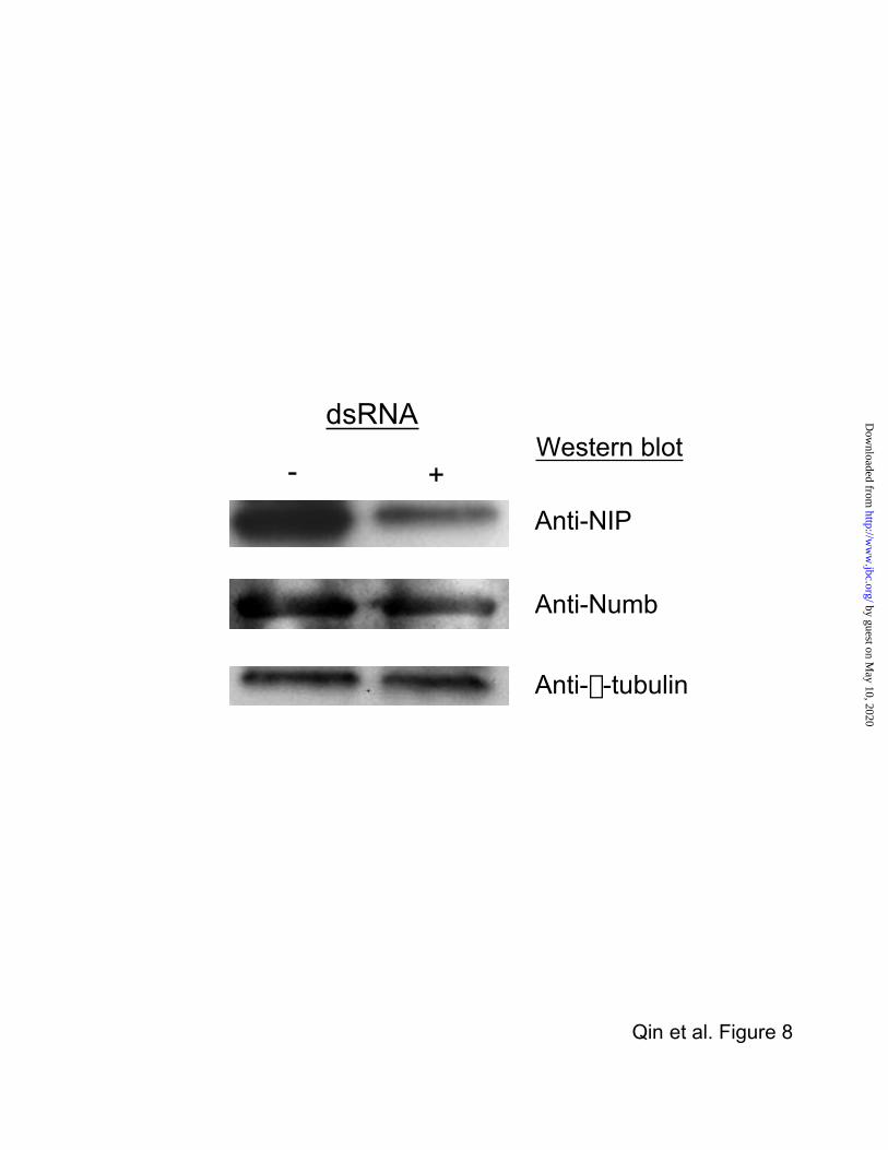
anti-Numb (red) antibodies. The RNAi- induced knockdown of NIP was also seen in
corresponding Western blots. As shown in Fig. 8, expression of NIP was reduced by
approximately 85% in dsRNA-treated cells, while the level of Numb is maintained the same in
both treated and control S2 cells. Collectively, these data demonstrate that NIP is necessary for
maintaining the membrane association of endogenous Numb in S2 cells.
by guest on May 10, 2020
http://ww
w.jbc.org/
Dow
nloaded from

16
DISCUSSION
NIP is a Membrane Anchor for Numb- A central question in asymmetric cell division is how
intrinsic cell- fate determinants are asymmetrically localized during mitosis. Numb, PON,
Prospero and Miranda proteins form a basal cortical crescent at prophase when a neuroblast
divides along the apical-basal axis and delaminates from the neuroectodermal epithelium (18,
45). Asymmetric localization of determinants is thought to occur in two distinct steps –
translocation to the plasma membrane and the formation of distinct cortical crescents (45).
Numb does not contain a transmembrane segment or a lipid anchor, and therefore, its recruitment
to the plasma membrane may be mediated by a lipid or/and a protein component of the
membrane. The N-terminal region of Numb, including the PTB domain, was shown to be
necessary for the membrane recruitment and asymmetric localization of Numb (34). Although
the PTB domain was originally identified as a protein module that interacts with NPxpY-
containing sequences (pY denotes a phosphotyrosine), some members of the family, such as the
Shc and Dab1 PTB domains, are also capable of binding to acidic lipids (46, 47) and are
therefore, implicated in membrane localization. However, the Numb PTB domain does not bind
specifically to these lipids (Li, data not shown).
The identification of the Numb PTB domain-binding protein NIP provides a plausible
mechanism for the membrane localization of Numb. NIP is predicted to be a membrane protein
with seven potential TM segments, and is localized to the plasma membrane in Cos-7 cells and
in Drosophila S2 cells. Numb can be recruited to, and anchored on, the plasma membrane via a
direct protein-protein interaction with NIP. Indeed, a direct Numb-NIP protein-protein
interaction is mediated by the Numb PTB domain and the two NxxF sites of NIP. In Drosophila
neuroblasts, NIP colocalizes with Numb in basal cortical crescents. In Drosophila embryonic S2
by guest on May 10, 2020
http://ww
w.jbc.org/
Dow
nloaded from

17
cells, the membrane association of Numb is dependent on NIP, because RNAi-mediated
depletion of NIP protein resulted in Numb relocating to the cytosol. Thus, NIP appears both
necessary and sufficient for the recruitment and retention of Numb in the plasma membrane.
Interestingly, a 76-residue fragment upstream of the PTB domain was localized to the neuroblast
membrane when expressed in Drosophila (34). Since this fragment does not interact with NIP in
vitro, it is likely that it may interact indirectly with NIP or that other mechanisms of Numb
membrane-localization exist.
NIP and Asymmetric Localization of Determinants- Does NIP play a role in the formation of
basal Numb crescents in neuroblasts? Although additional work, such as the generation of NIP
mutant flies, is needed to provide a definitive answer to this question, it is tempting to speculate
that NIP may play a part in the asymmetric segregation of Numb. Two lines of evidence support
this idea. First, NIP itself is asymmetrically localized in delaminating neuroblasts and forms a
basal cortical crescent at mitosis. Second, the NIP crescent and the Numb crescent overlay from
prophase through telophase of a cell cycle. It is possible that the formation of a NIP crescent
induced the formation of the Numb crescent by a direct protein-protein interaction.
How is the NIP crescent formed in a neuroblast? The same mechanism that controls the
localization of other basal determinants may also direct the localization of NIP (2). For instance,
Inscuteable is essential for asymmetric Numb localization to the basal cortex in mitotic
neuroblasts (13, 48). Interestingly, Inscuteable itself is localized to the apical membrane, an
event that is dependent on Bazooka, the Drosophila homologue of C. elegans Par3 (49). In
Drosophila neuroblasts and epithelial cells, Bazooka localizes to the apical membrane and forms
a complex with the Drosophila Par-6 homologue and the atypical protein kinase C (aPKC)
homologue, DaPKC (50, 51). Bazooka not only provides an apical cue for the correct
by guest on May 10, 2020
http://ww
w.jbc.org/
Dow
nloaded from

18
localization of Inscuteable (49), together with Par-6 and DaPKC, it also directs the localization
of basal determinants such as Numb, PON, Miranda, and Prospero (50, 51). In mammalian cells,
an analogous complex is formed among mPar3, mPar-6 and aPKC (52-54).
While it is not fully understood how the apical complex of Bazooka/Par-6/DaPKC is
anchored to the apical membrane, a possible mechanism is through interactions with membrane
proteins such as Crumbs, an integral membrane protein capable of binding to Par-6 through its
PDZ domain and thereby recruiting Par-6 to the plasma membrane (54). It is likely that NIP may
anchor the basal protein complex of PON, Numb and Miranda through binding to the Numb PTB
domain in a fashion akin to the role of Crumbs in the membrane recruitment of Par-6 and the
apical complex.
How does an apical complex direct basal segregation of determinants? The missing link
between the establishment of cell polarity and asymmetric localization of determinants was
recently found in the tumor suppressor, lethal giant larvae (Lgl). Lgl was identified as a substrate
of aPKC (55, 56), and phosphorylation of Lgl by the apically localized aPKC results in its
inactivation and dissociation from the apical crescent (55, 57). Restricted Lgl activity at the basal
crescent appears to be essential for the basal segregation of Miranda and PON (55). It remains to
be seen whether basal localization of NIP in a neuroblast is controlled by a similar mechanism.
An Evolutionarily Conserved Role for NIP?- The importance of NIP in asymmetric cell
division is reflected in the presence of proteins with a high degree of amino acid similarity in
other species. Although only one of the two Numb PTB-binding sites identified in Drosophila
NIP is conserved in mammalian NIP proteins, our studies indicated that the conserved NExF
motif alone was sufficient for mediating a physical interaction between NIP and the Numb PTB
domain. This same motif is also capable of mediating an interaction between mammalian Numb
by guest on May 10, 2020
http://ww
w.jbc.org/
Dow
nloaded from

19
and human NIP (data not shown). It is therefore possible that mammalian NIP proteins play a
role in regulating mNumb localization and function.
by guest on May 10, 2020
http://ww
w.jbc.org/
Dow
nloaded from

20
ACKNOWLEDGEMENT
We thank Cindy Ho for technical assistance. This work was supported by operating grants from
the Canadian Institutes of Health Research and from the National Cancer Institute of Canada (to
SSCL). SSCL is a scientist of NCIC with funds made available through the Canadian Cancer
Society.
by guest on May 10, 2020
http://ww
w.jbc.org/
Dow
nloaded from

21
REFERENCES
1. Horvitz, H.R. and Herskowitz, I. (1992) Cell 68, 237-458.
2. Jan, Y.N. and Jan, L.Y. (1998) Nature 392, 775-778.
3. Kidd, S., Kelley, M.R., and Young, M.W. (1986) Mol. Cell Biol. 6, 3094-3108.
4. Weinmaster, G., Roberts. V.J., and Lemke, G (1991) Development 113, 199-280.
5. Fortini, M.E., Rebay, I., Caron, L.A., and Artavanis-Tsakonas, S (1993) Nature 365, 555-
557.
6. Artavanis-Tsakonas, S., Rand, M.D., and Lake, R.J. (1999) Science 284, 770-776.
7. Wakamatsu, Y., Maynard, T.M., and Weston, J.A. (2000) Development 127, 2811-2821.
8. Lu, B., Jan, L.Y., and Jan, Y.N. (1998) Curr. Opin. Genet. Dev. 8, 392-399.
9. Uemura, T., Shepherd, S., Ackerman, L., Jan, L.Y., and Jan, Y.N. (1989) Cell 58, 349-360.
10. Hirata, J., Nakagoshi, H., Nabeshima, Y., and Matsuzaki, F. (1995) Nature 377, 627-630.
11. Shen, C.P., Jan, L.Y., and Jan, Y.N.(1997) Cell 90, 449-458.
12. Ikeshima-Kataoka, H., Skeath, J.B., Nabeshima, Y., Doe, C.Q., and Matsuzaki, F. (1997)
Nature 390, 625-629.
13. Kraut, R., Chia, W., Jan, L.Y., Jan, Y.N., and Knoblich, J.A. (1996) Nature 383, 50-55.
14. Lu, B., Rothenberg, M., Jan, L.Y., and Jan, Y.N. (1998) Cell 95, 225-235.
15. Jan, Y.N. and Jan, L.Y. (2001) Nat. Rev. Neurosci. 2, 772-779.
16. Spana, E.P., Kopczynski, C., Goodman. C.S., and Doe, C.Q. (1995) Development 121,
3489-3494.
17. Spana, E.P. and Doe, C.Q. (1996) Neuron 17, 21-26.
18. Knoblich, J.A. (2001) Nat. Rev. Mol. Cell Biol. 2, 11-20.
19. Pawson, T. and Scott, J.D. (1997) Science 278, 2075-2080.
by guest on May 10, 2020
http://ww
w.jbc.org/
Dow
nloaded from

22
20. Kavanaugh, W.M., and Williams, L.T. (1994) Science 266, 1862-1865.
21. Blaikie, P., Immanuel, D., Wu, J., Li, N., Yajnik, V., and Margolis, B. (1994) J. Biol. Chem.
269, 32031-432034.
22. Ren, R., Mayer, B.J., Cicchetti, P., and Baltimore, D. (1993) Science 259, 1157-1161.
23. Frise, E., Knoblich. J.A., Younger-Shepherd, S., Jan, L.Y., and Jan, Y.N. (1996) Proc. Natl.
Acad. Sci. USA. 93, 11925-11932.
24. Zhong, W., Feder, J.N., Jiang, M.M., Jan, L.Y., and Jan, Y.N. (1996) Neuron 17, 43-53.
25. McGill, M.A. and McGlade, C.J. (2003) J. Biol. Chem. 278, 23196-23203.
26. Chien, C.T., Wang, S., Rothenberg, M., Jan, L.Y., and Jan, Y.N.(1998) Mol. Cell. Biol. 18,
598-607.
27. Dho, S.E., Jacob, S., Wolting, C.D., French, M.B., Rohrschbeider, L.R., and McGlade, C.J.
(1998) J. Biol. Chem. 273, 9179-9187.
28. Nie, J., McGill, M.A., Dermer, M., Dho, S.E., Wolting, C.D., and McGlade, C.J. (2002)
EMBO J. 21, 93-102.
29. Li, S.-C., Songyang, Z., Vincent, S.J., Zwahlen, C., Wiley,S., Cantley, L., Kay, L.E.,
Forman-Kay, J., and Pawson, T. (1997) Proc. Natl. Acad. Sci. USA 94, 7204-7209.
30. Li, S.-C., Zwahlen, C., Vincent, S.J., McGlade, C.J., Kay, L.E., Pawson, T., and Forman-
Kay, J.D. (1998) Nat. Struct. Biol. 5, 1075-1083.
31. Zwahlen, C., Li, S.-C., Kay, L.E., Pawson, T., and Forman-Kay, J.D. (2000) EMBO J. 19,
1505-1515.
32. Knoblich, J.A., Jan, L.Y., and Jan, Y.N. (1994) Cell 76, 477-491.
33. Knoblich, J.A., Jan, L.Y, and Jan, Y.N. (1995) Nature 377, 624-627.
by guest on May 10, 2020
http://ww
w.jbc.org/
Dow
nloaded from
23
34. Knoblich, J.A., Jan, L.Y., and Jan, Y.N. (1997) Proc. Natl. Acad. Sci. USA 94, 13005-
13010.
35. Guo, M., Jan, L., and Jan, Y.N. (1996) Neuron 17, 27-41.
36. Yaich, L., Ooi, J., Park, M., Borg, J.P., Landry, C., Bodmer, R., and Margolis, B. (1998) J.
Biol. Chem. 273, 10381-10388.
37. Qin, H., Ishiwata,T., and Asano, G. (2001) J. Pathol. 195, 604-608.
39. Li, C., Jia, C.Y., Han, V.K., and Li, S.S-C. (2003) J. Biol. Chem. 278, 3852-3859.
39. Clemens, J.C., Worby,C.A., Simonson-Leff, N., Muda, M., Maehama,T., Hemmings,B.A.,
and Dixon, J.E. (2000) Proc. Natl. Acad. Sci. USA 97, 6499-6503.
40. Adams, M.D. et al. (2000) Science 287, 2185-2195.
41. Schultz, J., Copley, R.R., Doerks, T., Ponting, C.P., and Bork, P. (2000) Nucleic Acids Res.
28, 231-234.
42. Letunic, I., Goodstadt, L., Dickens, N.J., Doerks, T., Schultz, J., Mott, R., Ciccarelli, F.,
Copley, R.R., Ponting, C.P., and Bork, P. (2002) Nucleic Acids Res. 30, 242-244.
43. Kilpatrick, G.L., Dautzenberg, F.M., Martin, G.R., and Eglin, R.M. (1999) Treands
Pharmacol. Sci. 20, 294-301.
44. Frank R. (2002) J. Immunol. Methods 267, 13-26.
45. Schaefer, M. and Knoblich, J.A. (2001) Exp. Cell Res. 271, 66-74.
46. Zhou, M.M., Ravichandran, K.S., Olejniczak, E.F., Petros, A.M., Meadows, R.P., Sattler,
M., Harlan, J.E., Wade, W.S., Burakoff, S.J., and Fesik, S.W. (1995) Nature 378, 584-592.
47. Howell, B.W., Lanier, L. M., Frank, R., Gertler, F. B., and Cooper, J.A. (1999) Mol. Cell
Biol. 19, 5179-5188.
48. Yu, F., Morin, X., Cai, Y., Yang, X., and Chia, W. (2000) Cell 100, 399-409.
by guest on May 10, 2020
http://ww
w.jbc.org/
Dow
nloaded from

24
49. Schober, M., Schaefer, M., and Knoblich, J.A. (1999) Nature 402, 548-551.
50. Petronczki, M. and Knoblich, J.A. (2001) Nat. Cell Biol. 3, 43-49.
51. Wodarz, A., Ramrath, A., Grimm, A., and Knust, E. (2000) J. Cell Biol. 150, 1361-1374.
52. Lin, D., Edwards, A.S., Fawcett, J.P., Mbamalu, G., Scott, J.D., and Pawson, T. (2000) Nat.
Cell Biol. 2, 540-547.
53. Etienne-Manneville, S., and Hall, A. (2001) Cell 106, 489-498.
54. Hurd, T.W., Gao, L., Roh, M.H., Macara, I.G., and Margolis, B. (2003) Nat. Cell Biol. 5,
137-142.
55. Betschinger, J., Mechtler, K., and Knoblich, J.A. (2003) Nature 422, 326-330.
56. Plant, P.J., Fawcett, J.P., Lin, D.C., Holdorf, A.D., Binns, K., Kulkarni, S., and Pawson, T.
(2003) Nat. Cell Biol. 5, 301-308.
57. Yamanaka, T., Horikoshi, Y., Sugiyama, Y., Ishiyama, C., Suzuki, A., Hirose, T., Iwamatsu,
A., Shinohara, A., and Ohno, S. (2003) Curr. Biol. 13, 734-743.
58. Kyte, J. and Doolittle, R. (1982) J. Mol. Biol. 157, 105-132.
by guest on May 10, 2020
http://ww
w.jbc.org/
Dow
nloaded from
25
Figure Legends
Figure 1. The nip gene encodes a multi-spanning membrane protein. (A) cDNA sequence of
the nip gene and the deduced protein (NIP) sequence. A Kyte-Dolittle (58) hydropathy plot
identifies six potential transmembrane (TM) segments (underlined). A potential seventh TM
segment is distinguished by italics. The two PTB-binding, NxxF motifs are identified in bold
type. (B) Alignment of Drosophila NIP with potential homologues from various other species
identified using homology-based sequence search. Conserved residues are color-coded. Red
indicates high consensus (greater or equal to 90%) while blue denotes low consensus (equal or
less than 50%). An YNExFxW motif shared conserved in all NIP proteins is identified by a
rectangle box.
Figure 2. Identification of nip mRNA and NIP protein in Drosophila. (A) Northern blot
analysis of NIP mRNA at different developmental stages: embryonic, larval, pupal, and adult.
(B) Western blot analysis of NIP protein in lysate of embryos collected from stage 0 through 16.
Figure 3. Characterization of a NIP-Numb interaction. (A) Bacterial expressed and purified
GST-NIP was used to pull down Numb from HEK 293 cells while an equal amount of GST
protein was used as a control. (B) GST-Numb pulls down NIP from 293 cell lysate. (C) 293 cells
co-expressing Numb and FLAG-tagged NIP was subjected to imminoprecipitation by antibodies
against either Numb or FLAG or using a non-specific IgG. Western blots were performed using a
rabbit anti-Numb antibody. WCL, whole cell lysate, 20% of what was used in co-IP was loaded.
(D) NIP was detected in anti-Numb immunoprecipitates. Note that FLAG-NIP co-migrated with
the immunoglobulin heavy chain.
by guest on May 10, 2020
http://ww
w.jbc.org/
Dow
nloaded from

26
Figure 4. Mapping regions in Numb and NIP that mediate their interaction. (A) Constructs of
truncated Numb proteins in GST-fusion used in (B). (B) FLAG-NIP expressed in 293 cells were
subjected to binding using GST or various GST-Numb truncation mutants. (C) Coomassie-blue
staining showing equal application of GST or GST-fusion in B. (D) NIP truncation constructs in
a pFLAG-CMV2 vector. N1, N2 denote the NERF and the NKGF sites (shown as rectangles),
respectively. (E) Co-immunoprecipitation of Numb and various truncation mutants of NIP
(tagged with FLAG) from 293 cells. A monoclonal anti-FLAG antibody was used for
immunoprecipitation while a rabbit anti-Numb antibody was employed for Western blot. (F, G)
Western blots of cell lysate to demonstrate equal amounts of proteins used in all lanes. Note that
a non-specific band at approximately 50 kDa was seen in all lanes of (F).
Figure 5. The Numb PTB domain interacts specifically with the two NxxF motifs in NIP. (A)
Binding of peptides pep-NERF and pep-NKGF to the Numb PTB domain. The amino acid
sequences are: PDIDYNERFTWEG for pep-NERF, corresponding to residues 129-141 of NIP;
and SGGVENKGFQSD for pep-NKGF, corresponding to residues 358-369 of NIP. Each
peptide was labeled with fluorescein at the N-terminus. Incremental amounts of purified Numb
PTB domain (as GST fusion) were added to the labeled peptide to generate the corresponding
binding curve on a Beacon 2000 fluorescence spectrometer (Panvera, CA). No appreciable
binding was observed when GST alone was used (not shown). (B) An Ala-scanning SPOT-array
of pep-NKGF was screened for binding to purified GST-Numb PTB domain. Sequence of the
original peptide is shown above the spots. The first spot represents the original (wild-type)
sequence, whereas, subsequent spots represent peptides with one of the amino acids shown
by guest on May 10, 2020
http://ww
w.jbc.org/
Dow
nloaded from
27
replaced by an Ala. Bright (fluorescent) spots indicate positive binding, while dim or dark spots
signal weak or negative binding. (C) Binding of NIP, NIP single mutants N1A and N2A, and a
double mutant N1N2/AA to Numb. HEK 293 cells co-expressing Numb and FLAG-NIP or a
mutant were subjected to immunoprecipitation using a mouse anti-FLAG antibody. The presence
of Numb in the precipitates was verified in a Western blot using a rabbit anti-Numb antibody.
The lysate lane contained 20% of the sample used for immunoprecipitation.
Figure 6. Co-localization of NIP and Numb during mitosis of Drosophila neuroblasts.
Wild-type embryos at stage 10 were stained using a rabbit anti-NIP antibody and propidium
iodide (for DNA) to reveal NIP protein localization at various phases during the mitosis of a
neuroblast: (A, B) prophase, (C) metaphase, and (D) telophase. To examine colocalization of
NIP and Numb, metaphase neuroblasts were stained with a FITC-conjugated anti-NIP antibody
(E). The same cells was co-stained with an anti-Numb antibody labeled with rohdamine (F) to
reveal colocalization of NIP and Numb (G). Colocalization of the two proteins was also observed
in telophase neuroblasts (H, I, J) where both NIP and Numb were found mainly in the smaller
GMC and in the daughter neuroblast as a basal cortical crescent. (K) Negative staining of
embryos using FITC-conjugated secondary antibody alone. For clarity, cell boundaries were
delineated by broken circles. The apical side is up in all panels.
Figure 7. NIP recruits Numb to the plasma membrane in vitro and in vivo. Confocal
microscopic images of Cos-7 cells expressing NIP or/and Numb and S2 cells with and without
the application of interference dsRNA. Cos-7 cells were made to transiently express either NIP
or Numb and were stained for proteins with an antibody against NIP (A) or Numb (B), visualized
by guest on May 10, 2020
http://ww
w.jbc.org/
Dow
nloaded from

28
by a FITC-conjugated anti-rabbit secondary antibody. DNA is in red. Cos-7 cells co-expressing
Numb-GFP and FLAG-NIP (C, D, E) or FLAG-NIP-N1N2/AA double mutant (F, G, H) were
stained with a Texas Red-conjugated anti-mouse secondary antibody to reveal its subcellular
localization (in red) relative to Numb (in green). Endogenous NIP and Numb proteins in S2 cells
were stained respectively with FITC-labeled anti-NIP antibody (green) and rohdamine- labeled
anti-Numb antibody (red) (I-K). S2 cells treated with NIP dsRNA (L-N) are stained for NIP and
Numb as in I-K. An arrow indicates cells that have incorporated the dsRNA and contained no
detectable NIP (L) while maintaining normal levels of Numb expression. Note that Numb is
localized to the cytosol in the RNAi-treated cells but not in the control cell (M-O).
Figure 8. Knockdown of NIP expression in S2 cells by RNAi. Lysate of S2 cells treated or not
with NIP-specific dsRNA was subjected to SDS-PAGE and Western blotted respectively using
antibodies against NIP, Numb or β-tublin. The level of NIP expression was approximately 15%
of that seen in control cells.
by guest on May 10, 2020
http://ww
w.jbc.org/
Dow
nloaded from

Qin et al. Figure 1A
1 tgaacacgaaaagttaataacaattattttgtatgccggtttcttcttgacaagtttgat61 cccaaaaccccgacatcagcaacagcaaccagcatgaaaggctggttcgacgcgtttcga1 M K G W F D A F R121 gacgatgggggaccaacgctatattccttctcaaatcgaacacccgtaaccggagatgtc10 D D G G P T L Y S F S N R T P V T G D V181 tcaatcgtagccgtctcagtgctatttgccacattctatgtagcatttttagtcatcttt30 S I V A V S V L F A T F Y V A F L V I F241 ccgggtgtcagaaaacagaaattcacaacgttttcgacagtcacattgagcctttttgtg50 P G V R K Q K F T T F S T V T L S L F V301 ggtctagtcatattaatcactcgcctgggatccgcgtggcatgtggcacatgcaactatc70 G L V I L I T R L G S A W H V A H A T I361 atcgcgccatacaaagccttctcacgcgagaagctcccagcgcgcatcggcacccacatt90 I A P Y K A F S R E K L P A R I G T H I421 ggcctcatgcatgtcaatgtgacgctgacggcgattcccattggaaactggacaccgccg110 G L M H V N V T L T A I P I G N W T P P481 gatatagactacaacgaacgcttcacctgggagggagccaatgacatgagtgccaactat130 D I D Y N E R F T W E G A N D M S A N Y541 cgtcacgccctccagcggggtctcccctttcccatcctgaccgttgccgaatactttagc150 R H A L Q R G L P F P I L T V A E Y F S601 ctgggccgtgagggattctcgtggggtggacaataccgggctgcgggatatttcgcgagc170 L G R E G F S W G G Q Y R A A G Y F A S661 ataatgctctgggcctcgctggcctcgtggctgctgatgaacctgctgctgatagcggta190 I M L W A S L A S W L L M N L L L I A V721 ccccgatatggggcctatatgaaggctttgacaggtgccttgctggtctgcaccacggtg210 P R Y G A Y M K A L T G A L L V C T T V781 ggctatcattgcctgctcccgaagaggcctttgtccattcacctcgaaggcggacgcttg230 G Y H C L L P K R P L S I H L E G G R L841 gagtttcatttcggatggtgctactggctggtcttggtggcaggcattctctgctttatt250 E F H F G W C Y W L V L V A G I L C F I901 gcgggagttctgatctccattattgacttggtttggccgcacaccttctccactgtgctg270 A G V L I S I I D L V W P H T F S T V L961 gaggtatactacggcacgccatacgatcgccatgtgattctggaggagtctagtgatgtg290 E V Y Y G T P Y D R H V I L E E S S D V1021 cgttatcgaaagccgcgcaacagtcgcagcttagaggatccacctggactgggctcccgc310 R Y R K P R N S R S L E D P P G L G S R1081 atcctgcgccgcctcagctccaaggcacgtgacttgcaacctggcacggcaccgcgtcgc330 I L R R L S S K A R D L Q P G T A P R R1141 gatagtccagccggagtgtccagcagcggtggagttgaaaacaagggcttccagtcggat350 D S P A G V S S S G G V E N K G F Q S D1201 gcaccaaagagtccttggagatatcccttccgaaggtcgcagcaaatggcgcagcagcag370 A P K S P W R Y P F R R S Q Q M A Q Q Q1261 cattcgcatccactgcatcagcatccgttgcagcagcatcatcagcaccaccagcaccac390 H S H P L H Q H P L Q Q H H Q H H Q H H1321 catcagcagcagctgcagtttgtgggcggtccggtggtgcagcatcccatgcatcatatg410 H Q Q Q L Q F V G G P V V Q H P M H H M1381 cagcgcaccatgtcgcaggattcaggatccagcattgcctcggcagccgtgcaaatctcc430 Q R T M S Q D S G S S I A S A A V Q I S1441 ccgctgcacaagcatgctttggcgcggatgttgcctaatcccccagtggagcgtattcgt450 P L H K H A L A R M L P N P P V E R I R1501 gacatggatcactggtgattgccattggacaaaccataaaaatcgactatttttattggg470 D M D H W1561 cttacataactcttgactaattggaaataattttagaggaaatcttaaaacaaaacattt1621 tgtgataatttcttaatttaaacaaaacaaatcaacacaaattaggatgaaaaatatttt1681 ataattgaccagatcacatactttgataatataacataaaaaacgaaaacgatgttattc1741 ttaactgtacttttaattaaattattttaaatgttaagctaaccatgtaataacaatcga1801 cgaatcaattttagatataagaatgttttttagtgatgtttatttgtgataagacaatcc1861 tgccacacacgcacagagaatcgacacatacaggagcgagaaattatggcaaaaaatcaa1921 tgtttatttctaaagtaatttattatttgtaatttgaattcaatgccagtgtctataacg1981 aataagttga
by guest on May 10, 2020
http://ww
w.jbc.org/
Dow
nloaded from


Em
bryo
Lar
vae
Pupae
Adult
A
58
48.5
NIP
(kDa)
B
Qin et al. Figure 2
NIP
by guest on May 10, 2020http://www.jbc.org/Downloaded from

IgG a-Num
b
IgGW
CL
Numb
(WB: anti-Numb)
a-FLA
G (NIP
)
IP IP
a-FLA
G (NIP
)C D
NIP WCL
GSTNum
b
WCL
GST
Numb NIP
A B
Qin et al. Figure 3
a-Num
b
WCL
54 -
(kDa)
NIPH -
(WB: anti-FLAG)
by guest on May 10, 2020http://www.jbc.org/Downloaded from

GST
Nb-N
Nb-PTB
Nb-C
WCL
NIP
30-
66-
PTB1
58 205
556Numb
1 76
205 556
Nb-N
Nb-PTB
Nb-C
A
B
C
D
E
F
(kDa)
NERF (N1)
NKGF (N2)
NIP-N
NIP-N1
NIP-N1/2
NIP-N2
NIP-C
1 106
1148
1382
474351
474368
NIP-N
NIP-N
1
NIP-N
1/2
NIP-N
2NIP
-C
(IP: a-FLAG)
Numb
(WB: a-FLAG)
10-
73-
(kDa)
G Numb
Qin et al., Figure 4
by guest on May 10, 2020http://www.jbc.org/Downloaded from

0 10 20 30 40 50 600
10
20
30
40
[PTB] (mM)
Polarization (
DmP)
0 10 20 30 40 50 600
50
100
150
200
[PTB] (mM)
Polarization (
DmP)Pep-NERF Pep-NKGF
A
WCL
IP: a-FLAG
IgGNIP NIP
-N1A
NIP-N
2 A
NIP-N
1N2/AA
Numb
a-Numb IP
H -
C
Qin et al, Figure 5
S G G V E N K G F Q S D
B
WB: anti-Numb
by guest on May 10, 2020
http://ww
w.jbc.org/
Dow
nloaded from

E F G
H I J
Qin et al, Figure 6
NIP
NIP
Numb
Numb
Merge
Merge
B C DA
K
by guest on May 10, 2020
http://ww
w.jbc.org/
Dow
nloaded from

A B
C D E
F G
Qin et al. Figure 7
I J K
L M N
H
RNAi RNAi RNAi
NIP Numb
NIP Numb Merge
NIP-NN/AA Numb Merge
NIP Numb Merge
NIP Numb Merge
RNAi
O
by guest on May 10, 2020
http://ww
w.jbc.org/
Dow
nloaded from
dsRNA
Anti-NIP
Anti-Numb
Anti-b-tubulin
- +Western blot
Qin et al. Figure 8
by guest on May 10, 2020
http://ww
w.jbc.org/
Dow
nloaded from

Shawn S-C. LiHanjuan Qin, Anthony Percival-Smith, Chengjun Li, Christina Y.H. Jia, Greg Gloor and
asymmetric cell divisionA novel transmembrane protein recruits numb to the plasmic membrane in
published online December 11, 2003J. Biol. Chem.
10.1074/jbc.M311733200Access the most updated version of this article at doi:
Alerts:
When a correction for this article is posted•
When this article is cited•
to choose from all of JBC's e-mail alertsClick here
by guest on May 10, 2020
http://ww
w.jbc.org/
Dow
nloaded from