a novel restriction endonuclease glai for rapid and highly
TRANSCRIPT
ChemicalScience
EDGE ARTICLE
Ope
n A
cces
s A
rtic
le. P
ublis
hed
on 1
1 D
ecem
ber
2017
. Dow
nloa
ded
on 2
/6/2
022
2:40
:11
PM.
Thi
s ar
ticle
is li
cens
ed u
nder
a C
reat
ive
Com
mon
s A
ttrib
utio
n 3.
0 U
npor
ted
Lic
ence
.
View Article OnlineView Journal | View Issue
A novel restrictio
Key Laboratory of Applied Surface and Collo
Laboratory of Analytical Chemistry for Life
Chemistry and Chemical Engineering, Sha
Shaanxi Province, P. R. China. E-mail: liuch
† Electronic supplementary informa10.1039/c7sc04975g
Cite this: Chem. Sci., 2018, 9, 1344
Received 20th November 2017Accepted 9th December 2017
DOI: 10.1039/c7sc04975g
rsc.li/chemical-science
1344 | Chem. Sci., 2018, 9, 1344–1351
n endonuclease GlaI for rapid andhighly sensitive detection of DNA methylationcoupled with isothermal exponential amplificationreaction†
Yueying Sun, Yuanyuan Sun, Weimin Tian, Chenghui Liu, * Kejian Gaoand Zhengping Li *
Sensitive and accurate detection of site-specific DNA methylation is of critical significance for early
diagnosis of human diseases, especially cancers. Herein, for the first time we employ a novel
methylation-dependent restriction endonuclease GlaI to detect site-specific DNA methylation in a highly
specific and sensitive way by coupling with isothermal exponential amplification reaction (EXPAR). GlaI
can only cut the methylated target site with excellent selectivity but leave the unmethylated DNA intact.
Then the newly exposed end fragments of methylated DNA can trigger EXPAR for highly efficient signal
amplification while the intact unmethylated DNA will not initiate EXPAR at all. As such, only the
methylated DNA is quantitatively and faithfully reflected by the real-time fluorescence signal of the GlaI–
EXPAR system, and the potential false positive interference from unmethylated DNA can be effectively
eliminated. Therefore, by integrating the unique features of GlaI for highly specific methylation
discrimination and EXPAR for rapid and powerful signal amplification, the elegant GlaI–EXPAR assay
allows the direct quantification of methylated DNA with ultrahigh sensitivity and accuracy. The detection
limit of methylated DNA target has been pushed down to the aM level and the whole detection process
of GlaI–EXPAR can be accomplished within a short time of 2 h. More importantly, ultrahigh specificity is
achieved and as low as 0.01% methylated DNA can be clearly identified in the presence of a large excess
of unmethylated DNA. This GlaI–EXPAR is also demonstrated to be capable of determining site-specific
DNA methylations in real genomic DNA samples. Sharing the distinct advantages of ultrahigh sensitivity,
outstanding specificity and facile operation, this new GlaI–EXPAR strategy may provide a robust and
reliable platform for the detection of site-specific DNA methylations with low abundances.
Introduction
DNA methylation at the C5 position of cytosine in CpG dinu-cleotides is the most frequent and important epigenetic modi-cation mechanism in mammalian cells, which plays criticalroles in the regulation of gene expression, genomic imprinting,and X-chromosome inactivation.1,2 Aberrant CpG methylationstates, especially those that occur in the gene promoter regions,have been proved to be closely associated with the developmentof many human diseases including cancers.3 The CpG hypo-methylation in the promoter of oncogenes or the hyper-methylation in the promoter of tumor-suppressor, have beensuggested to be crucial drivers for cancer progression.4 So the
id Chemistry, Ministry of Education, Key
Science of Shaanxi Province, School of
anxi Normal University, Xi’an 710062,
@snnu.edu.cn; [email protected]
tion (ESI) available. See DOI:
alterations of DNA methylation may serve as importantbiomarkers for early diagnosis, classication as well as prog-nosis of cancers.5–7 In this regard, the highly sensitive andaccurate quantication of site-specic DNA methylation will beof great signicance for biomedical studies.8
Generally, in real clinical samples, the tumor-derived meth-ylated DNA is only present in rather low quantities (less than0.1%) with the majority of the DNA being derived from normalcells.9 So how to effectively discriminate DNA target witha specic methylation site from a large pool of unmethylatedDNA is the most critical issue for the detection of DNA meth-ylation. According to the employed methylation discriminationmechanisms, the conventional methylation assays can bemainly classied into two categories, namely, the bisulteconversion (BC)-assisted approaches and the methylation-sensitive restriction endonuclease (MSRE) digestion-basedprotocols. BC is a chemical method that can convert cytosines(C) into uracils (U) but leave the methylated cytosinesunchanged with the treatment of sodium bisulte.3,10,11 In such
This journal is © The Royal Society of Chemistry 2018

Edge Article Chemical Science
Ope
n A
cces
s A
rtic
le. P
ublis
hed
on 1
1 D
ecem
ber
2017
. Dow
nloa
ded
on 2
/6/2
022
2:40
:11
PM.
Thi
s ar
ticle
is li
cens
ed u
nder
a C
reat
ive
Com
mon
s A
ttrib
utio
n 3.
0 U
npor
ted
Lic
ence
.View Article Online
cases, the detection of methylated cytosine sites from theunmethylated counterparts can be fullled by discriminatingthe resulting base differences (methylated cytosine vs. uracil)between the DNA targets. Although widely applied as the gold-standard methylation assays, the BC-assisted protocols sufferheavily from cumbersome and time-consuming procedures,which need multiple steps including target denaturation withNaOH, treatment with bisulte, desulfonation process and DNApurication under stringent experimental conditions. Moreseriously, the BC-based chemical treatment of DNA targets willlead to inevitable DNA degradation, incomplete conversion aswell as DNA loss during the multi-step operations, which mayresult in batch-to-batch inconsistency and nally misleadingresults for methylation analysis,12 particularly for the targetswith very low levels of methylation.
MSREs (e.g., HpaII, AciI and BsoFI) are a group of enzymesthat can identify and cleave DNA at the unmethylated cytosinesite while the methylated DNA will not be digested and thusremain intact.13 The MSRE digestion-based methylation assaysare typically based on the determination of uncleavedmethylatedDNA by using appropriate DNA amplication strategies with theassumption that the unmethylated DNA is completely cleavedand thus cannot be amplied.14–17 Compared with BC treatment,the MSRE digestion-based methylation assays are performedunder mild conditions with rapid and simple operation. Never-theless, since the methylated DNA targets only constitute a verysmall percentage of the overall DNA pool in most cases, evena trivial portion of incomplete cutting of the unmethylated DNAwill lead to signicant false positive interference for the quanti-cation of the low-abundance methylated targets.
Totally different from MSREs, GlaI, a newly discoveredmethylation-dependent DNA restriction endonuclease, canspecically recognize and cleave DNA with methylated cytosineswhile cannot cut DNA with unmethylated cytosines.18,19 Inspiredby the fact that the GlaI enzyme exhibits ultrahigh specicitytowards the digestion of methylated DNAs with simple opera-tion, we believe that if the GlaI-cleaved fragments of methylatedDNA can be specically and rapidly determined against theundigested DNAs, the low-abundance DNA methylation can bemore faithfully quantied with high accuracy and reliabilityeven when the unmethylated DNAs outnumber it massively.Based on this premise, herein, we wish to develop an elegantGlaI-assisted DNA methylation assay with high sensitivity,specicity and accuracy.
In this regard, besides for the GlaI-assisted specicdiscrimination between methylated and unmethylated cyto-sines in DNA targets, how to achieve high sensitivity is anotherkey issue for the detection of rare DNA methylation in limitedgenomic samples. In order to obtain enough sensitivity formethylation analysis, DNA amplication protocols are generallyrequired by coupling with either BC treatment or restrictionendonuclease digestion.14,20,21 Among them, the various adaptedpolymerase chain reaction (PCR) protocols, such as the meth-ylation specic PCR,22 PCR-based single-nucleotide primerextension,23 end-specic PCR (ESPCR)24 and so on, are the mostwidely employed methods for DNA methylation analysis.However, the PCR-based methods have some inevitable
This journal is © The Royal Society of Chemistry 2018
limitations. First, specic PCR relies heavily on the stringentprimer/template design and precisely optimized thermalcycling. If not fully optimized, the frequent false-positive resultsof PCR may lead to uncertain results. Second, most of the PCR-based methylation assays need to be combined with furtherpost-PCR detection steps or even additional signal amplica-tion routes, making the assay procedures rather complicated. Asan alternative of PCR, ligase chain reaction (LCR) has beendeveloped recently by our group and others for site-specicmethylation analysis with high sensitivity and specicity,25–27
but precisely controlled thermal cycles similar to PCR are stillindispensable. To tackle such drawbacks associated with thethermal cycling PCR and LCR, several isothermal amplicationmethods, such as rolling circle amplication (RCA)28–30 andstrand displacement amplication (SDA),31,32 are proposed todetect DNA methylation under isothermal conditions, whicheffectively avoid the optimization of thermal cycles. Neverthe-less, both RCA and SDA need long reaction time up to severalhours, sophisticated probe design, and show relatively lowsensitivity for the detection of low-abundance methylated DNA.
It is worth nothing that compared with conventionalisothermal amplication protocols such as RCA and SDA, theisothermal exponential amplication reaction (EXPAR) sharesseveral distinct advantages of rapidness, simple design andultrahigh sensitivity. EXPAR can achieve 106 to 109-fold expo-nential amplication of target nucleic acids within minutes33 byusing only one simple DNA template. Fascinatingly, we noticethat the GlaI-cleaved methylated DNA end fragments are wellsuited to serve as triggers to efficiently initiate EXPAR, while theintact unmethylated DNA aer GlaI treatment will not beamplied at all by EXPAR. Therefore, by integrating theadvantages of GlaI for highly specic methylation discrimina-tion and EXPAR for highly efficient signal amplication, in thiswork, we have developed a novel GlaI-assisted EXPAR assay(GlaI–EXPAR) that allows the direct quantication of methyl-ated DNA with ultrahigh sensitivity and accuracy. By real-timemonitoring of the uorescence signal of the GlaI–EXPARsystem, only the methylated DNA is quantitatively and faithfullyreected while the unmethylated DNA cannot trigger EXPARand thus will not interfere with the methylation analysis. Withthe elegant GlaI–EXPAR assay, as low as 200 aM methylatedDNA can be clearly detected and the whole detection processcan be accomplished within a short time of 2 h. More impor-tantly, due to the excellent selectivity of GlaI towards themethylation site, ultrahigh specicity is also achieved and aslow as 0.01% methylated DNA can be clearly identied in thepresence of a vast excess of unmethylated DNA. Therefore, theproposed GlaI–EXPAR approach provides a powerful and reli-able tool for the rapid, sensitive, highly specic, and accuratedetection of site-specic DNA methylation especially for thosewith extremely low abundances.
Experimental sectionReagents and apparatus
GlaI endonuclease as well as its reaction buffer was supplied bySibEnzyme Ltd (Russia). Nt.BstNBI nicking endonuclease
Chem. Sci., 2018, 9, 1344–1351 | 1345
Chemical Science Edge Article
Ope
n A
cces
s A
rtic
le. P
ublis
hed
on 1
1 D
ecem
ber
2017
. Dow
nloa
ded
on 2
/6/2
022
2:40
:11
PM.
Thi
s ar
ticle
is li
cens
ed u
nder
a C
reat
ive
Com
mon
s A
ttrib
utio
n 3.
0 U
npor
ted
Lic
ence
.View Article Online
(10 000 U mL�1), Vent (exo�) DNA polymerase (2000 UmL�1), M.SssI CpG methyltransferase (4000 U mL�1), and S-adenosylmethionine (SAM, 32 mM) were all purchased fromNew England Biolabs (NEB, USA). TKS Gex DNA polymerase(1250 U mL�1), Gex PCR Buffer, dNTPs (2.5 mM) and the 100bp DNA ladder marker were obtained from TaKaRa Biotech-nology (Dalian, China). 4S Red Plus Nucleic Acid Stain (LifeScience, USA) was used for DNA staining in agarose gel elec-trophoresis. SYBR Green I (20� stock solution in DMSO, 20 mgmL�1) was purchased from Bio-Vision Biotechnology (Xiamen,China). All of the oligonucleotides (see detailed sequences inTable S1†) used in this work were custom synthesized byTaKaRa Biotechnology (Dalian, China).
Extraction of genomic DNA from HCT116 cells
HCT116 cells were obtained from the cell bank of the ChineseAcademy of Sciences (Shanghai, China). The cells were culturedin the DMEM medium mixed with 10% fetal calf serum, 100 UmL�1 penicillin, 1% NaHCO3, 3 mM L-glutamine and 100 mgmL�1 streptomycin at 37 �C with appropriate humidiedatmosphere (5% CO2 and 95% air). Aer trypsinization (0.2%trypsin, 1 mM EDTA, Invitrogen), about 5 � 106 adherentHCT116 cells, which were counted and quantied by a hemo-cytometer (Shanghai Qiujing, China), were centrifuged at11 200 g for 1 min at room temperature. Then the supernatantwas carefully discarded and the cell precipitates were collectedto be used for the extraction of genomic DNA. Finally, 120 mL ofgenomic DNA extracts were obtained by using the TIANampgenomic DNA kit according to the manufacturer’s instructions.
Preparation of unmethylated DNA target (target N) andmethylated DNA target (target M)
In order to make the test results more convincing, a 508 bp DNAfragment in septin 9 gene (see detailed sequence in Table S1†)was used as a proof-of-concept target in this study, which wasobtained by amplifying the septin 9 gene in the extractedgenomic DNA with PCR. Typically, in a 50 mL PCR solution (1�Gex PCR buffer containing 1 mM Mg2+), an appropriateamount of the genomic DNA extracts was mixed with 0.2 mMdNTPs, 1.25 U TKS Gex DNA polymerase, and 0.2 mM each ofthe primers (forward primer and reverse primer, see detailedsequences in Table S1†), and then the PCR amplication wasperformed with the following procedures: 94 �C for 1 min, fol-lowed by 30 cycles of 98 �C for 10 s, 60 �C for 15 s, and 68 �C for30 s. A 2720 thermal cycler (Applied Biosystems, USA) was usedfor the PCR amplication and the PCR products were charac-terized by agarose electrophoresis analysis (Fig. S1 in ESI†).
The 508 bp septin 9 gene promoter sequences in the PCRproducts were the desired target DNA in this study. Aeragarose electrophoresis, the desired target DNA could be sepa-rated and puried from the PCR products by using a QIAquick®Gel Extraction Kit. The puried target DNA was quantied anddivided into two equal parts. One part of the target DNA wasdirectly used as the unmethylated DNA target (target N) withoutany treatment. Meanwhile, the other equivalent part was treatedby the M.SssI CpG methyltransferase to produce the methylated
1346 | Chem. Sci., 2018, 9, 1344–1351
DNA target (target M) because the CpG dinucleotide sites on thetarget DNA were methylated under the catalysis of M.SssImethyltransferase. Typically, a total 50 mL reaction media of 1�NEB buffer 2 (10 mM Tris–HCl containing 50 mM NaCl, 10 mMMgCl2 and 1mMDTT, pH 7.9), 16 U ofM.SssI methyltransferaseand 32 nmol (equal to a concentration of 640 mM) of SAM wereincubated with 2 mM target DNA at 37 �C for 5 h. Aerward, toensure that all of the target DNA was completely methylated atthe CpG dinucleotide sites, 16 U of M.SssI methyltransferaseand 32 nmol (640 mM) of SAM were relled in the mixture andincubated at 37 �C for another 12 h, followed by inactivation ofthe M.SssI methyltransferase at 65 �C for 20 min. A MinElute®PCR Purication Kit was used for the purication of the meth-ylated DNA according to the manufacturer’s instruction, andthe nal obtained target M was quantied on a NANODROP2000 instrument.
Standard GlaI–EXPAR protocols
In a typical GlaI–EXPAR reaction, series dilutions of target DNAs(target M, target N, or 1 mL of 1000-fold diluted genomic DNAextracts) were rstly incubated with 1 U of GlaI endonuclease at30 �C for 45 min in a 5 mL reaction buffer (33 mM Tris-HAc,10 mM Mg(Ac)2, 66 mM KAc and 1 mM DTT, pH 7.9). Aerheating at 70 �C for 15 min to deactivate GlaI, 1 mL of the GlaI-treated mixture was pipetted out and further mixed with theEXPAR template and dNTPs in the Nt.Bst NBI buffer. Themixture was heated at 94 �C for 1 min and then incubated at55 �C for 5 min. Aerward, Vent (exo�) DNA polymerase,Nt.BstNBI nicking endonuclease, SYBR Green I and ThermoPolreaction buffer was further introduced to make a nal 10 mLvolume for EXPAR. The nal 10 mL mixture containing 1�ThermoPol reaction buffer (20 mM Tris–HCl, 10 mM KCl,10mM (NH4)2SO4, 2mMMgSO4 and 0.1% Triton X-100, pH 8.8),0.5� Nt.Bst NBI buffer (25 mM Tris–HCl, pH 7.9, 5 mM MgCl2,50 mM NaCl, 0.5 mM dithiothreitol), 200 mM dNTPs, 0.01 mMEXPAR template, 0.4� SYBR Green I, Vent (exo�) DNA poly-merase (0.2 U), and Nt.BstNBI nicking endonuclease (4 U) wasimmediately placed in a Step-One Real-Time PCR system(Applied Biosystems, USA) to perform the EXPAR at 60 �C. Thereal-time uorescence intensity was monitored at intervals of30 s for the quantitative determination of the methylated targetDNA.
Results and discussionPrinciple of the GlaI–EXPAR method for the detection of DNAmethylation
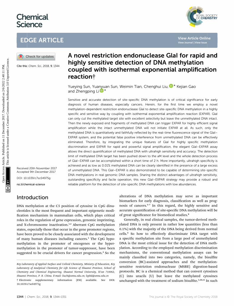
Fig. 1 schematically illustrates the work principle of the GlaI–EXPAR approach for the detection of site-specic DNA methyl-ation, which takes advantage of the high specicity of GlaI forthe digestion of methylated cytosine sites and the high sensi-tivity of EXPAR for efficient amplication and detection of theGlaI-cleaved products. In this study, a specic DNA sequence inthe septin 9 gene containing a 50-GmCGmC-30/30-mCGmCG-50
site (77373474 site) with four 5-methylcytosines (mC), isemployed as a model site-specic methylated DNA target
This journal is © The Royal Society of Chemistry 2018
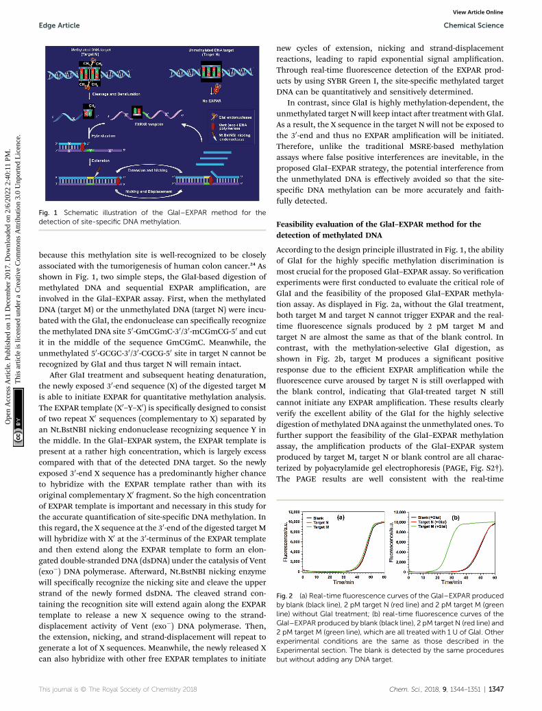
Fig. 1 Schematic illustration of the GlaI–EXPAR method for thedetection of site-specific DNA methylation.
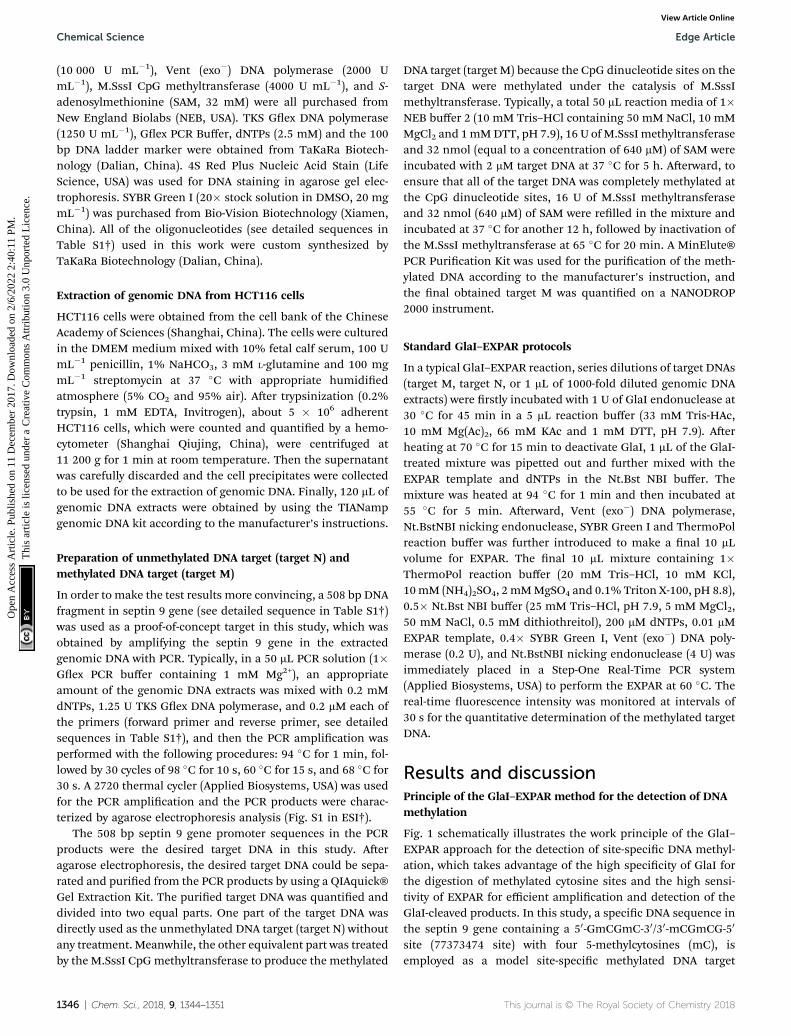
Fig. 2 (a) Real-time fluorescence curves of the GlaI–EXPAR producedby blank (black line), 2 pM target N (red line) and 2 pM target M (greenline) without GlaI treatment; (b) real-time fluorescence curves of theGlaI–EXPAR produced by blank (black line), 2 pM target N (red line) and2 pM target M (green line), which are all treated with 1 U of GlaI. Otherexperimental conditions are the same as those described in theExperimental section. The blank is detected by the same proceduresbut without adding any DNA target.
Edge Article Chemical Science
Ope
n A
cces
s A
rtic
le. P
ublis
hed
on 1
1 D
ecem
ber
2017
. Dow
nloa
ded
on 2
/6/2
022
2:40
:11
PM.
Thi
s ar
ticle
is li
cens
ed u
nder
a C
reat
ive
Com
mon
s A
ttrib
utio
n 3.
0 U
npor
ted
Lic
ence
.View Article Online
because this methylation site is well-recognized to be closelyassociated with the tumorigenesis of human colon cancer.24 Asshown in Fig. 1, two simple steps, the GlaI-based digestion ofmethylated DNA and sequential EXPAR amplication, areinvolved in the GlaI–EXPAR assay. First, when the methylatedDNA (target M) or the unmethylated DNA (target N) were incu-bated with the GlaI, the endonuclease can specically recognizethe methylated DNA site 50-GmCGmC-30/30-mCGmCG-50 and cutit in the middle of the sequence GmCGmC. Meanwhile, theunmethylated 50-GCGC-30/30-CGCG-50 site in target N cannot berecognized by GlaI and thus target N will remain intact.
Aer GlaI treatment and subsequent heating denaturation,the newly exposed 30-end sequence (X) of the digested target Mis able to initiate EXPAR for quantitative methylation analysis.The EXPAR template (X0–Y–X0) is specically designed to consistof two repeat X0 sequences (complementary to X) separated byan Nt.BstNBI nicking endonuclease recognizing sequence Y inthe middle. In the GlaI–EXPAR system, the EXPAR template ispresent at a rather high concentration, which is largely excesscompared with that of the detected DNA target. So the newlyexposed 30-end X sequence has a predominantly higher chanceto hybridize with the EXPAR template rather than with itsoriginal complementary X0 fragment. So the high concentrationof EXPAR template is important and necessary in this study forthe accurate quantication of site-specic DNA methylation. Inthis regard, the X sequence at the 30-end of the digested target Mwill hybridize with X0 at the 30-terminus of the EXPAR templateand then extend along the EXPAR template to form an elon-gated double-stranded DNA (dsDNA) under the catalysis of Vent(exo�) DNA polymerase. Aerward, Nt.BstNBI nicking enzymewill specically recognize the nicking site and cleave the upperstrand of the newly formed dsDNA. The cleaved strand con-taining the recognition site will extend again along the EXPARtemplate to release a new X sequence owing to the strand-displacement activity of Vent (exo�) DNA polymerase. Then,the extension, nicking, and strand-displacement will repeat togenerate a lot of X sequences. Meanwhile, the newly released Xcan also hybridize with other free EXPAR templates to initiate
This journal is © The Royal Society of Chemistry 2018
new cycles of extension, nicking and strand-displacementreactions, leading to rapid exponential signal amplication.Through real-time uorescence detection of the EXPAR prod-ucts by using SYBR Green I, the site-specic methylated targetDNA can be quantitatively and sensitively determined.
In contrast, since GlaI is highly methylation-dependent, theunmethylated target N will keep intact aer treatment with GlaI.As a result, the X sequence in the target N will not be exposed tothe 30-end and thus no EXPAR amplication will be initiated.Therefore, unlike the traditional MSRE-based methylationassays where false positive interferences are inevitable, in theproposed GlaI–EXPAR strategy, the potential interference fromthe unmethylated DNA is effectively avoided so that the site-specic DNA methylation can be more accurately and faith-fully detected.
Feasibility evaluation of the GlaI–EXPAR method for thedetection of methylated DNA
According to the design principle illustrated in Fig. 1, the abilityof GlaI for the highly specic methylation discrimination ismost crucial for the proposed GlaI–EXPAR assay. So vericationexperiments were rst conducted to evaluate the critical role ofGlaI and the feasibility of the proposed GlaI–EXPAR methyla-tion assay. As displayed in Fig. 2a, without the GlaI treatment,both target M and target N cannot trigger EXPAR and the real-time uorescence signals produced by 2 pM target M andtarget N are almost the same as that of the blank control. Incontrast, with the methylation-selective GlaI digestion, asshown in Fig. 2b, target M produces a signicant positiveresponse due to the efficient EXPAR amplication while theuorescence curve aroused by target N is still overlapped withthe blank control, indicating that GlaI-treated target N stillcannot initiate any EXPAR amplication. These results clearlyverify the excellent ability of the GlaI for the highly selectivedigestion of methylated DNA against the unmethylated ones. Tofurther support the feasibility of the GlaI–EXPAR methylationassay, the amplication products of the GlaI–EXPAR systemproduced by target M, target N or blank control are all charac-terized by polyacrylamide gel electrophoresis (PAGE, Fig. S2†).The PAGE results are well consistent with the real-time
Chem. Sci., 2018, 9, 1344–1351 | 1347
Chemical Science Edge Article
Ope
n A
cces
s A
rtic
le. P
ublis
hed
on 1
1 D
ecem
ber
2017
. Dow
nloa
ded
on 2
/6/2
022
2:40
:11
PM.
Thi
s ar
ticle
is li
cens
ed u
nder
a C
reat
ive
Com
mon
s A
ttrib
utio
n 3.
0 U
npor
ted
Lic
ence
.View Article Online
uorescence results. All of these results clearly indicate that theGlaI–EXPAR method is undoubtedly feasible for the accurateand highly specic detection of site-specic DNA methylation.
Analytical performance of the GlaI–EXPAR method fortarget M detection
Various experimental parameters including the amount of theinvolved enzymes and the temperature of the EXPAR, whichmay inuence the performance of the GlaI–EXPAR assay for thedetection of target M, were all investigated and optimized indetail (Fig. S3–S6 in ESI†). Accordingly, 1 U GlaI endonuclease, 4U Nt.BstNBI nicking enzyme, 0.2 U Vent (exo�) DNA poly-merase, and the EXPAR temperature of 60 �C are found to be theoptimal conditions.
Under such optimized experimental conditions, the analyt-ical performance of the proposed GlaI–EXPAR method for thedetection of target M was evaluated. Fig. 3a exhibits the well-dened real-time uorescence curves produced by differentconcentrations of target M ranging from 200 aM to 200 pM,respectively. It can be seen that with increasing concentrationsof target M, the uorescence curves arise more rapidly since thedetected EXPAR products are only dependent on the initialdosage of target M. The point of inection (POI) values, namely,the times corresponding to the maximum slope in each real-time uorescence curve, are recorded for the quantitativedetermination of target M. As shown in Fig. 3b, two good linearrelationships are obtained between the POI values and loga-rithm of the target M concentrations in the ranges of 200 aM to20 fM, and 20 fM to 200 pM, respectively. The correspondingcorrelation equations are POI ¼ 8.68 � 2.61 lg(Ctarget M/M)(correlation coefficient R ¼ 0.9811) and POI ¼ �54.41 �7.18 lg(Ctarget M/M) (R¼ 0.9986), respectively. The results shownin Fig. 3a clearly demonstrate that the uorescence responseproduced by as low as 200 aM target M (equal to an absolutequantity of 2 zmol methylated DNA molecules in a 10 mLvolume) can be clearly discriminated from the blank, indicatingan ultrahigh sensitivity of the proposed GlaI–EXPAR assay.Furthermore, compared with the most widely used BC-basedmethylation assays which typically need 16–40 h assayingtime34 due to their cumbersome procedures, the whole GlaI–
Fig. 3 (a) The real-time fluorescence curves produced by target Mwith different concentrations. From left to right, the target Mconcentrations are successively 200 pM, 20 pM, 2 pM, 200 fM, 20 fM, 2fM, 200 aM and 0 (blank); (b) the plots between POI values in the real-time fluorescence curves and logarithm (lg) of the target M concen-trations. Error bars are estimated from the standard deviation of threereplicate measurements at each data point.
1348 | Chem. Sci., 2018, 9, 1344–1351
EXPAR assay can be accomplished within 2 h including both thesimple GlaI treatment and EXPAR reaction.
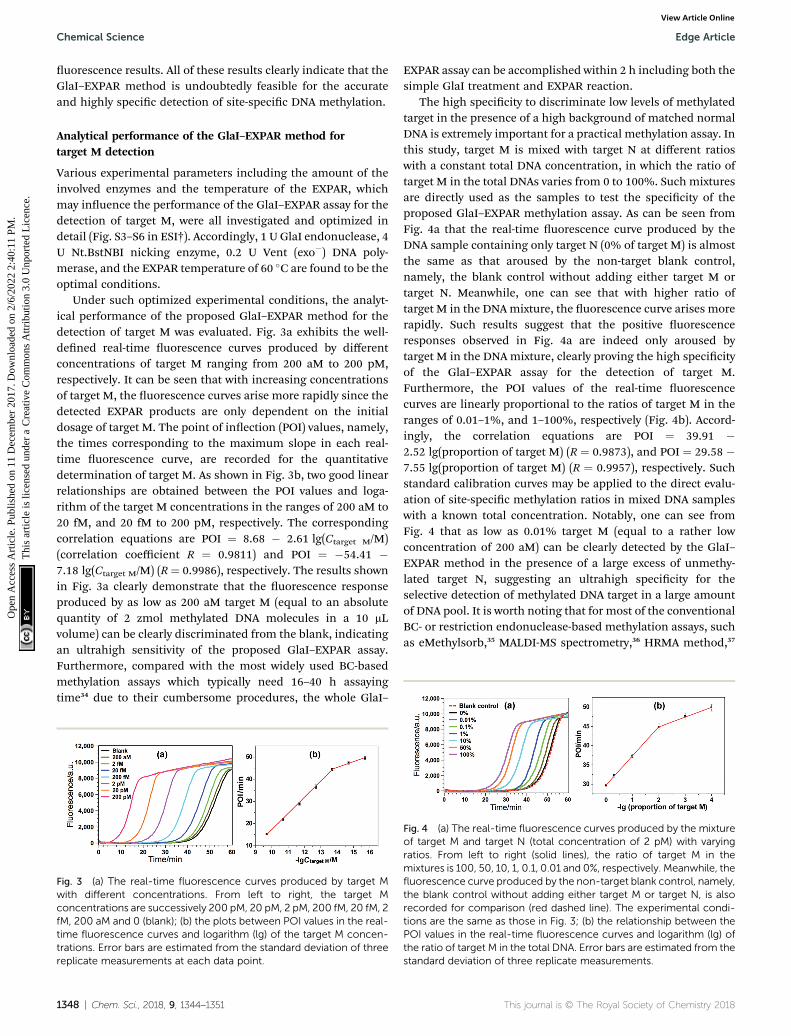
The high specicity to discriminate low levels of methylatedtarget in the presence of a high background of matched normalDNA is extremely important for a practical methylation assay. Inthis study, target M is mixed with target N at different ratioswith a constant total DNA concentration, in which the ratio oftarget M in the total DNAs varies from 0 to 100%. Such mixturesare directly used as the samples to test the specicity of theproposed GlaI–EXPAR methylation assay. As can be seen fromFig. 4a that the real-time uorescence curve produced by theDNA sample containing only target N (0% of target M) is almostthe same as that aroused by the non-target blank control,namely, the blank control without adding either target M ortarget N. Meanwhile, one can see that with higher ratio oftarget M in the DNAmixture, the uorescence curve arises morerapidly. Such results suggest that the positive uorescenceresponses observed in Fig. 4a are indeed only aroused bytarget M in the DNAmixture, clearly proving the high specicityof the GlaI–EXPAR assay for the detection of target M.Furthermore, the POI values of the real-time uorescencecurves are linearly proportional to the ratios of target M in theranges of 0.01–1%, and 1–100%, respectively (Fig. 4b). Accord-ingly, the correlation equations are POI ¼ 39.91 �2.52 lg(proportion of target M) (R ¼ 0.9873), and POI ¼ 29.58 �7.55 lg(proportion of target M) (R ¼ 0.9957), respectively. Suchstandard calibration curves may be applied to the direct evalu-ation of site-specic methylation ratios in mixed DNA sampleswith a known total concentration. Notably, one can see fromFig. 4 that as low as 0.01% target M (equal to a rather lowconcentration of 200 aM) can be clearly detected by the GlaI–EXPAR method in the presence of a large excess of unmethy-lated target N, suggesting an ultrahigh specicity for theselective detection of methylated DNA target in a large amountof DNA pool. It is worth noting that for most of the conventionalBC- or restriction endonuclease-based methylation assays, suchas eMethylsorb,35 MALDI-MS spectrometry,36 HRMA method,37
Fig. 4 (a) The real-time fluorescence curves produced by the mixtureof target M and target N (total concentration of 2 pM) with varyingratios. From left to right (solid lines), the ratio of target M in themixtures is 100, 50, 10, 1, 0.1, 0.01 and 0%, respectively. Meanwhile, thefluorescence curve produced by the non-target blank control, namely,the blank control without adding either target M or target N, is alsorecorded for comparison (red dashed line). The experimental condi-tions are the same as those in Fig. 3; (b) the relationship between thePOI values in the real-time fluorescence curves and logarithm (lg) ofthe ratio of target M in the total DNA. Error bars are estimated from thestandard deviation of three replicate measurements.
This journal is © The Royal Society of Chemistry 2018

Edge Article Chemical Science
Ope
n A
cces
s A
rtic
le. P
ublis
hed
on 1
1 D
ecem
ber
2017
. Dow
nloa
ded
on 2
/6/2
022
2:40
:11
PM.
Thi
s ar
ticle
is li
cens
ed u
nder
a C
reat
ive
Com
mon
s A
ttrib
utio
n 3.
0 U
npor
ted
Lic
ence
.View Article Online
cationic conjugated polymer-based method,38 SMART-MSP22
and HDCR,24 only 0.1% to 10% methylated target DNA can bedetected in the DNA mixtures containing excessive unmethy-lated DNA. So the specicity of the proposed GlaI–EXPAR issuperior to these traditional methylation assays. As far as weknow, only seldom reported methods such as the MS-qFRET39
and HRCA29 are capable of discerning 0.01% methylated targetDNA from their unmethylated counterparts, showing compa-rable specicity to the GlaI–EXPAR method. However,compared with the GlaI–EXPAR method, both MS-qFRET andHRCA need stringent primer/probe design, complicated proce-dures, long reaction time and show relatively low sensitivity.Therefore, due to the excellent specicity, high sensitivity andsimple operations, the proposed GlaI–EXPAR assay providesa powerful and reliable protocol for the detection of site-specicDNA methylations.
Detection of DNA methylation in real genomic DNA samples
Due to its ultrahigh sensitivity and specicity, the GlaI–EXPARmethod is further examined for the detection of site-specicmethylation in real genomic DNA samples extracted from theHCT116 colon cancer cells. The level of the methylated DNAtarget in 1 mL of 1000-fold diluted genomic DNA extracts isquantitatively evaluated according to the calibration curve asthat shown in Fig. 3b. As displayed in Fig. 5, the POI valueproduced by the genomic DNA sample falls within the linearrange of 200 aM to 20 fM, and thus the amount of target M inthe genomic DNA sample is determined to be 3.1 fM accordingto the corresponding calibration equation. When the genomicDNA sample is further spiked with 10 fM of standard target M,the time of the POI value is remarkably shortened, and theamount of target M in the spiked sample is calculated to be 12.4fM with a recovery of 93.0%. In contrast, when the equivalent
Fig. 5 Detection of target M (77373474 site in septin 9 gene) in thegenomic DNA sample extracted from HCT116 cells. The real-timefluorescence curves are produced by the genomic DNA sample (greenline), the genomic DNA sample spiked with 10 fM standard target M(blue line), the genomic DNA sample spiked with 10 fM target N (reddashed line), and the blank control (black line), respectively. Thedetermined concentration of target M in genomic DNA is calculated inthe 10 mL GlaI–EXPAR system. Error bars are estimated from threereplicate measurements.
This journal is © The Royal Society of Chemistry 2018
genomic DNA sample is spiked with 10 fM of unmethylatedtarget N, the obtained POI value is exactly identical to thatproduced by only the genomic DNA sample (red dashed line inFig. 5). These results clearly suggest that the positive responsesof the genomic DNA samples are indeed faithfully aroused bythe site-specic methylated target sequence. So this GlaI–EXPAR assay is feasible and reliable for the highly sensitive andhighly specic detection of minute amounts of site-specic DNAmethylations in real genomic DNA samples. To further provethe accuracy of the obtained results, the detected 50-GCGC-30/30-CGCG-50 site (77373474 site) of the target septin 9 genesequence in the extracted genomic DNA from HCT-116 cells issequenced (Fig. S7, ESI†). The bisulte sequencing result indi-cates that the cytosines in this site are all methylated. Therefore,the detection results obtained by the GlaI–EXPAR are consistentwith the sequencing results.
Evaluation of the generality of the GlaI–EXPAR method formethylation analysis
According to a previous study by the Tarasova group,19 threetypes of methylated sequence sites can serve as good substratesof GlaI. The list of favorable cleavage sites of GlaI includesa fully methylated site 50-GmCGmC-30/30-mCGmCG-50, a generalstructure 50-PumCGmC-30/30-PyGmCG-50 with three methylatedcytosines (Pu stands for purine nucleotides while Py stands forpyrimidine nucleotides), and one recognition sequence withtwo methylated cytosines 50-AmCGT-30/30-TGmCA-50, all ofwhich include at least one methylated CpG dinucleotide. So bycombining with the cleavage site-specic EXPAR template, theproposed GlaI–EXPAR can be applicable for the quantitativedetection of GlaI-recognizing site-specic CpG methylation.
The detected proof-of-concept target M (77373474 site) isa fully methylated 50-GmCGmC-30/30-mCGmCG-50 site. To testthe generality of the proposed method, the GlaI–EXPAR strategyis further applied to the detection of another consensus GlaI-recognizing methylation site (77373518 site, denoted as targetM0) with a 50-AmCGmC-30/30-TGmCG-50 sequence in the hyper-methylated region of the septin 9 gene, which is one of the bestGlaI substrates in the general 50-PuCGC-30/30-PyGCG-50 struc-tures.19 According to the principle of GlaI–EXPAR, for thedetection of different methylation sites, only the “X” sequenceof the EXPAR template need to be changed according to the site-specic 30-end sequence of the GlaI-cutting site, while otherexperimental conditions can stay the same. In this study, theGlaI-digested target M0 exposes a new end sequence (Z) at the 30-terminus, which can initiate EXPAR for quantitative methyla-tion analysis by using the site-specic EXPAR template (Z0–Y–Z0,see detailed sequence in Table S1†). As can be seen from thereal-time uorescence curves in Fig. 6, with increasingconcentrations of target M0 from 200 aM to 20 pM, the corre-sponding POI value is gradually shortened, and as low as 200aM target M0 can be unequivocally detected from the blankcontrol. Fig. S8† shows that the POI values are linearly propor-tional to the logarithm of the target M0 concentrations in theranges of 200 aM to 20 fM, and 20 fM to 20 pM, respectively. Thecorresponding correlation equations are POI ¼ 20.02 �
Chem. Sci., 2018, 9, 1344–1351 | 1349

Fig. 6 The real-time fluorescence curves for the detection of the77373518-site methylation in the target DNA (target M0). From left toright, the concentrations of the target M0 are successively 20 pM, 2 pM,200 fM, 20 fM, 2 fM, 200 aM and 0 (blank), respectively. Meanwhile, thefluorescence curve produced by 2 pM unmethylated target N0 is alsorecorded for comparison (red dashed line). The experimental condi-tions are all the same as those in Fig. 3 except for the methylation site-specific EXPAR template.
Chemical Science Edge Article
Ope
n A
cces
s A
rtic
le. P
ublis
hed
on 1
1 D
ecem
ber
2017
. Dow
nloa
ded
on 2
/6/2
022
2:40
:11
PM.
Thi
s ar
ticle
is li
cens
ed u
nder
a C
reat
ive
Com
mon
s A
ttrib
utio
n 3.
0 U
npor
ted
Lic
ence
.View Article Online
2.18 lg(Ctarget M0/M) (R ¼ 0.9920) and POI ¼ �56.91 �7.86 lg(Ctarget M0/M) (R ¼ 0.9956), respectively. Such results arein good consistence with those for the detection of target M(Fig. 3), suggesting that the GlaI–EXPAR can be easily extendedto the detection of different site-specic DNA methylations withsimilar high sensitivity. Notably, one can also see from Fig. 6that the uorescence response aroused by unmethylated targetN0 with a high concentration of 2 pM is almost the same withthe blank control, further verifying the ultrahigh specicity ofthe GlaI which is capable of accurately discriminating methyl-ated target DNA from a large amount of 104-fold excess ofunmethylated DNA.
Conclusions
In summary, by integrating the distinct advantages of GlaIendonuclease for highly selective recognition and cleavage ofmethylated DNA target, and EXPAR for the highly efficientamplication of GlaI-cleaved DNA fragments, we have for therst time developed a novel GlaI–EXPAR strategy which allowsfor the quantitative evaluation of site-specic DNA methylationwith ultrahigh sensitivity and specicity. Compared with theconventional MSRE-based methylation assays, since the GlaIcan only recognize and cut the methylated DNA site and thenonly the GlaI-digested DNA fragments can initiate subsequentEXPAR, the combination of GlaI with EXPAR can efficientlyavoid the potential false positive interference from unmethy-lated target DNA or non-target DNAs. Furthermore, differentfrom the current gold-standard BC-based methylation assayswhere cumbersome and time-consuming operations aregenerally involved, in the GlaI–EXPAR assay, the operations forboth the GlaI treatment and EXPAR are quite simple and rapid.Highly sensitive (with a detection limit down to the aM level)and specic (0.01% methylated target can be discerned from
1350 | Chem. Sci., 2018, 9, 1344–1351
a large DNA pool) methylation detection can be readily accom-plished within 2 h under isothermal conditions. Consideringthe high sensitivity, excellent specicity, easy probe design andsimple operation, we believe that this proposed GlaI–EXPARstrategy may provide a robust and reliable platform for theaccurate detection of site-specic DNA methylations in bothbiological and biomedical studies.
Conflicts of interest
There are no conicts to declare.
Acknowledgements
This work was supported by the National Natural ScienceFoundation of China (21335005, 21622507, and 21472120),Program for Changjiang Scholars and Innovative ResearchTeam in University (IRT_15R43), the Natural Science BasicResearch Plan of Shaanxi Province (2015KJXX-22), and theFundamental Research Funds for the Central Universities(GK201501003, GK201603039).
Notes and references
1 F. Krueger, B. Kreck, A. Franke and S. R. Andrews, Nat.Methods, 2012, 9, 145–151.
2 H. Thomassin, E. J. Oakeley and T. Grange, Methods, 1999,19, 465–475.
3 C. Rohde, Y. Zhang, T. P. Jurkowski, H. Stamerjohanns,R. Reinhardt and A. Jeltsch, Nucleic Acids Res., 2008, 36, e34.
4 I. J. V. Vlodrop, H. E. C. Niessen, S. Derks,M. M. L. L. Baldewijns, W. V. Criekinge, J. G. Herman andM. V. Engeland, Clin. Cancer Res., 2011, 17, 4225–4231.
5 C. Loon-Day, F. Model, T. Devos, R. Tetzner, J. Distler,M. Schuster, X. Song, R. Lesche, V. Liebenberg, M. Ebert,B. Molnar, R. Grutzmann, C. Pilarsky and A. Sledziewski,Clin. Chem., 2008, 54, 414–423.
6 P. W. Laird and R. Jaenisch, Annu. Rev. Genet., 1996, 30, 441–464.
7 M. R. Estecio and J. P. Issa, FEBS Lett., 2011, 585, 2078–2086.8 M. L. Gonzalgo, P. A. Jones and G. Liang, US Pat. No.7,662,563 B2, 2010.
9 F. Diehl, M. Li, D. Dressman, Y. He, D. Shen, S. Szabo,L. A. DiazJr, S. N. Goodman, K. A. David, H. Juhl,K. W. Kinzler and B. Vogelstein, Proc. Natl. Acad. Sci. U. S.A., 2005, 102, 16368–16373.
10 A. Olek, J. Oswald and J. Walter, Nucleic Acids Res., 1996, 24,5064–5066.
11 X. Wang, F. Chen, D. Zhang, Y. Zhao, J. Wei, L. Wang,S. Song, C. Fan and Y. Zhao, Chem. Sci., 2017, 8, 4764–4770.
12 K. Tanaka and A. Okamoto, Bioorg. Med. Chem. Lett., 2007,17, 1912–1915.
13 A. K. Burnett, B. H. Ramsahoye and C. Taylor, Nucleic AcidsRes., 1997, 25, 3196–3198.
14 C. Dahl and P. Guldberg, Biogerontology, 2003, 4, 233–250.15 T. O. Tollefsbol, Methods Mol. Biol., 2004, 287, 1–8.
This journal is © The Royal Society of Chemistry 2018
Edge Article Chemical Science
Ope
n A
cces
s A
rtic
le. P
ublis
hed
on 1
1 D
ecem
ber
2017
. Dow
nloa
ded
on 2
/6/2
022
2:40
:11
PM.
Thi
s ar
ticle
is li
cens
ed u
nder
a C
reat
ive
Com
mon
s A
ttrib
utio
n 3.
0 U
npor
ted
Lic
ence
.View Article Online
16 K. N. Rand and P. L. Molloy, BioTechniques, 2010, 49, xiii–xvii.
17 H. Wen, H. Wang, H. Wang, J. Yan, H. Tian and Z. Li, Anal.Methods, 2016, 8, 5372–5377.
18 V. A. Chernukhin, T. N. Najakshina, M. A. Abdurashitov,J. E. Tomilova, N. V. Mezentzeva, V. S. Dedkov,N. A. Mikhnenkova, D. A. Gonchar and D. SKh,BioTechnologia, 2006, 4, 31–35.
19 G. V. Tarasova, T. N. Nayakshina and S. K. H. Degtyarev, BMCMol. Biol., 2008, 9, 7–18.
20 I. H. N. Wong, Methods Mol. Biol., 2006, 336, 33–43.21 M. F. Fraga and M. Esteller, BioTechniques, 2002, 33, 636–
649.22 L. S. Kristensen, T. Mikeska, M. Krypuy and A. Dobrovic,
Nucleic Acids Res., 2008, 36, e42.23 M. L. Gonzalgo and P. A. Jones, Nucleic Acids Res., 1997, 25,
2529.24 K. N. Rand, G. P. Young, T. Ho and P. L. Molloy, Nucleic Acids
Res., 2013, 41, e15.25 F. Su, L. Wang, Y. Sun, C. Liu, X. Duan and Z. Li, Chem.
Commun., 2015, 51, 3371–3374.26 F. Su, L. Wang, Y. Sun, C. Liu, X. Duan and Z. Li, Chem. Sci.,
2015, 6, 1866–1872.27 K. M. Koo, E. J. Wee, S. Rauf, M. J. Shiddiky and M. Trau,
Biosens. Bioelectron., 2014, 56, 278–285.
This journal is © The Royal Society of Chemistry 2018
28 J. Nosek, A. A. Rycovska, J. Griffith and L. Tomaska, J. Biol.Chem., 2005, 280, 10840–10845.
29 A. Cao and C. Y. Zhang, Anal. Chem., 2012, 84, 6199–6205.30 Y. P. Zeng, J. Hu, Y. Long and C. Y. Zhang, Anal. Chem., 2013,
85, 6143–6150.31 H. Q. Wang, Z. Wu, Y. Zhang, L. J. Tang, R. Q. Yu and
J. H. Jiang, Anal. Chim. Acta, 2012, 710, 111–117.32 C. Zhu, Y. Wen, H. Peng, Y. Long, Y. He, Q. Huang, D. Li and
C. Fan, Anal. Bioanal. Chem., 2011, 399, 3459–3464.33 J. V. Ness, L. K. V. Ness and D. J. Galas, Proc. Natl. Acad. Sci.
U. S. A., 2003, 100, 4504–4509.34 M. Frommer, L. E. Mcdonald, D. S. Millar, C. M. Collis,
F. Watt, G. W. Grigg, P. L. Molloy and C. L. Paul, Proc.Natl. Acad. Sci. U. S. A., 1992, 89, 1827–1831.
35 K. M. Koo, A. A. Sina, L. G. Carrascosa, M. J. Shiddiky andM. Trau, Analyst, 2014, 139, 6178–6184.
36 J. Tost, P. Schatz, M. Schuster, K. Berlin and I. G. Gut, NucleicAcids Res., 2003, 31, 50e.
37 C. M. R. Lopez, B. G. Asenjo, A. J. Lloyd and M. J. Wilkinson,Anal. Chem., 2010, 82, 9100–9108.
38 F. Feng, H. Wang, L. Han and S. Wang, J. Am. Chem. Soc.,2008, 130, 11338–11343.
39 V. J. Bailey, H. Easwaran, Y. Zhang, E. Griffiths,S. A. Belinsky, J. G. Herman, S. B. Baylin, H. E. Carrawayand T. H. Wang, Genome Res., 2009, 19, 1455–1461.
Chem. Sci., 2018, 9, 1344–1351 | 1351