a new species of tigriopus (copepoda, harpacticoida, harpacticidae) from thailand with the...
TRANSCRIPT
ORIGINAL ARTICLE
A new species of Tigriopus (Copepoda, Harpacticoida,Harpacticidae) from Thailand with the descriptionof its naupliar development
Supawadee Chullasorn • Viatcheslav N. Ivanenko •
Hans-U Dahms • Pawana Kangtia • Wan-Xi Yang
Received: 11 December 2008 / Revised: 19 February 2011 / Accepted: 16 March 2011 / Published online: 5 April 2011
� Springer-Verlag and AWI 2011
Abstract Both genders of Tigriopus thailandensis sp.
nov. are described from a laboratory stock raised from
individuals collected from the seaweed Enteromorpha
clathrata in Thailand (Bangsaen Beach, Chonburi Prov-
ince). Tigriopus thailandensis sp. nov. shares with its
closest relative T. japonicus Mori, 1932 two setae on the
third exopodal segment of leg 4 while other congeners bear
3 inner setae. However, allobasis and exopod of antenna in
both genders are much more slender and elongate than in
T. japonicus. All six naupliar stages of T. thailandensis are
described from the offspring of isolated females. In com-
parison with nauplii of T. japonicus, T. thailandensis
nauplii are characterized by the following: a smaller body
size throughout the naupliar phase; first antennular segment
without seta, second antennular segment with only one
small seta plus two longer setae; third antennular segment
with additional spinules from naupliar stage II onwards;
antenna bears three small spinules on the terminal exopodal
segment; one additional seta on the anterior surface of the
antennary basis, tubular endopod of antenna with one tiny
seta midlength at naupliar stage III that increases in size;
mandibular basis with several spinules on anterior surface;
mandibular coxa with one spinulose seta that is smooth in
T. japonicus.
Keywords Zoosystematics � Species description �External morphology � Naupliar development �Larval stages � Evolution � Harpacticidae � Thailand
Abbreviations
Enp (Exp) I First segment of endopod (exopod)
1–4 ? Ae Example of antennular/antennary
armature corresponding to 1 seta on first
and 4 setae plus 1 aesthetasc on second
segment
N I and N VI Naupliar stages I and VI, respectively
Introduction
The worldwide distributed genus Tigriopus Norman, 1869,
consists of 11 valid species (Wells 2007) that are charac-
terized by their high resistance to changes in temperature,
salinity, and oxygen and inhabit supralittoral rockpools (see
McAllen and Block 1997; McAllen et al. 1998; McAllen
1998, 1999). Copepods of the genus Tigriopus became a
focal point of several in-depth studies, e.g., its ecotoxico-
logy reviewed by Raisuddin et al. (2007), its field ecology
(Dethier 1980), life history (Koga 1970), external and
internal anatomy (Ito 1973; Dahms et al. 2007), chemical
Communicated by Peter Funch.
S. Chullasorn � P. Kangtia
Faculty of Science, Ramkhamhaeng University,
Bangkok 10240, Thailand
V. N. Ivanenko
Department of Invertebrate Zoology, Biological Faculty,
Moscow State University, Moscow 119899, Russia
H.-U. Dahms (&)
Green Life Science Department, College of Natural Science,
Sangmyung University, 7 Hongij-dong, Jongno-gu,
Seoul 110-743, South Korea
e-mail: [email protected]
W.-X. Yang (&)
The Sperm Laboratory, College of Life Sciences,
Zhejiang University, Zi Jin Gang Campus,
310058 Hangzhou, Zhejiang, China
e-mail: [email protected]
123
Helgol Mar Res (2012) 66:139–151
DOI 10.1007/s10152-011-0254-3

ecology (Kelly and Snell 1998), and genetics of adaptation
(Davenport et al. 1997; Burton et al. 1999). Other areas of
research are represented by genomics in general (Machida
et al. 2002; Kim et al. 2003; Lee 2003; Lee et al. 2005; Jung
et al. 2006), medicine-related genetics (Kim et al. 2004),
and the genetics of populations (Edmands and Burton 1998;
Edmands 1999). The life cycles of Tigriopus spp. have been
studied by Guiglia (1926), Fraser (1936), and Shaw (1938).
Nauplii of T. fulvus have been described by Igarashi (1963),
and nauplii of T. japonicus Mori 1932 were described by
Dahms et al. (2007).
Nauplii can play significant ecological roles due to their
abundances and variety (Alekseev 2002). However, life-
history studies in the field and investigations into stage-
specific phenomena in the laboratory are hampered by the
lack of descriptive information as well as keys for identi-
fication (Dahms 1993; Dahms et al. 2006; Ivanenko et al.
2008). Much rearing and descriptive work has to precede
any attempt to tackle ontogeny-related problems. Although
there are some naupliar studies dealing with the postem-
bryonic development of Tigriopus (Igarashi 1963; Ito 1970;
Dahms 1990, 1993), an update is provided with the present
new species described here. Here, we provide a morpho-
logical description of adults of both genders and the nauplii
of Tigriopus thailandensis sp. nov.
Materials and methods
Adults of T. thailandensis sp. nov. were collected during
low-tide at sandy Bangsaen Beach, Chonburi Province,
Thailand (13�19’N, 100�54’E). Different developmental
stages of T. thailandensis, including ovigerous females,
were washed out from thalli of the alga Enteromorpha
clathrata. The residue containing ovigerous females was
subsequently decanted over a 50-lm mesh screen. Adults
were rinsed into smaller bowls for transport to the labo-
ratory. Cultivation was carried out in glass dishes as
described in detail by Dahms et al. (2007).
Specimens were washed in filtered seawater and fixed in
4% seawater–formaldehyde. The stages were subsequently
embedded in glycerol. Body measurements of nauplii were
taken from the frontal portion of the naupliar shield to the
caudalmost protrusion of the hindbody (length) and the
Fig. 1 Tigriopus thailandensis sp. nov. Female. a, habitus, dorsal
view; b, habitus, lateral view Fig. 2 Tigriopus thailandensis sp. nov. Female. a, urosome, dorsal
view; b, urosome, ventral view. c, terminal seta of caudal ramus
140 Helgol Mar Res (2012) 66:139–151
123
widest lateral tips of the naupliar shield (width); only speci-
mens drawn were considered for length measurements. Two
to five specimens per stage were used for the investigation of
stage-specific variability. The developmental stages used in
the present study represent the offspring of exclusively sin-
gle-female cultures. Other details and descriptive terminol-
ogy of nauplii follow Dahms et al. (2007).
Type material The type material has been deposited in
the National Museum of Natural History, Smithsonian
Institution, Washington, D.C.: the dissected female holo-
type (USNM 1115488), the dissected male allotype
(USNM 1115489), 1 female paratype (USNM 1115490),
and 1 male paratype (USNM 1115491). The type material
was collected on December 23, 2006, in Chonburi Prov-
ince, Thailand.
Descriptive part
Order Harpacticoida Sars, 1903
Suborder Oligoarthra Lang, 1944
Family Harpacticidae Dana, 1846
Genus Tigriopus Norman, 1869
Tigriopus thailandensis sp. nov.
Etymology The species name refers to the country where
this new species was found.
Distribution Tigriopus thailandensis sp. nov. was asso-
ciated with a green alga, Enteromorpha clathrata attached
to an old big plastic bag collected during low-tide at Bang
Saen sandy beach in Chonburi Province, Thailand
(13�19’N, 100�54’E).
Adult female and male
Female (Figs. 1, 2, 3, 4, 5, 6)
Total length of holotype female 0.93 mm; paratype, total
length 0.98 mm, measured from tip of rostrum to posterior
margin of caudal ramus. Body (Fig. 1a, b) orange in color,
compact, ornamented with sensillae. Cephalosome as long
as succeeding separate 4 somites of prosome combined,
with well-developed rostrum; cephalothorax and first ped-
igerous somite separate (Fig. 1a, b). Urosome (Fig. 2a, b)
slightly tapering posteriorly. Rostrum (Fig. 3a) as long as
first segment of antennule, rounded at tip, and with a pair of
sensilla at the tip. Labrum prominent, with 4 spiniform
processes and many hairs at the apical edge. Genital double
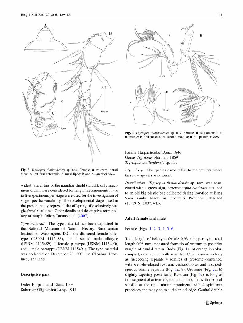
Fig. 3 Tigriopus thailandensis sp. nov. Female. a, rostrum, dorsal
view; b, left first antennule; c, maxilliped; b and c—anterior view
Fig. 4 Tigriopus thailandensis sp. nov. Female. a, left antenna; b,
mandible; c, first maxilla; d, second maxilla; b–d—posterior view
Helgol Mar Res (2012) 66:139–151 141
123

somite (Fig. 2b) with 2 oblique spinular rows on the outer
and inner corner on ventral surface.
Urosome (Fig. 2a, b) 6-segmented, comprising P5-
bearing somite and 3 abdominal somites ornamented with
short oblique rows of spinules on distal corner dorsally and
ventrally of each somite. Second urosomite furnished with
more sensillae in dorsal view. Genital double-somite with
leg 6, genital field indistinct with 2 small copulatory
apertures ventrally. Third to fifth urosomal somites orna-
mented with 1 row of minute spinules along posterior
margin. Caudal ramus as wide as long, principal terminal
caudal seta longer than length of urosome (Fig. 2c).
Antennule (Fig. 3b) 9-segmented. Second segment
largest. Fourth and terminal segment with 1 large and 1
small aesthetasc, respectively. Length of two proximal
segments as that of five apical segments combined.
Armature formula: 1-10-9-4 ? Ae.-1-4-2-2-
7 ? Ae.
Antenna (Fig. 4a): pre-antennary sclerite and coxa bare.
Endopod 2-segmented: first segment of allobasis with 1
seta on middle of anterior edge and furnished with rows of
minute spinules on the surface; second segment endopod
with 4 long setae at base, 4 short and slender setae, and 3
short spines. Exopod 3-segmented: first segment longest
with 2 setae, second segment shorter with 1 seta, third
segment with 1 lateral and 1 terminal seta.
Mandible (Fig. 4b): coxa well developed with 1 arched
spinular row at midlength. Cutting edge with 4 strong and 4
small spines, and 1 spinulose seta; basis with 1 apical seta;
exopod 3-segmented: first segment with 2 setae and few
spinules, second segment with 1 seta, third segment with 2
setae and minute spinules; endopod 1-segmented with 3
setae at middle inner edge, and 7 setae on distal end.
Maxillule (Fig. 4c): arthrite of praecoxa with 2 slender
and 1 plumose setae on surface, 4 bare and 2 pinnate spines
on inner edge. Coxa with 3 setae on inner end; basis
spinulated on outer face; 1 slender spine and 2 setae at
distal end; 2 subdistal setae, 2 of which juxtaposed; exopod
with 3 setae and some spinules near distal end; endopod
small, with 3 terminal setae.
Maxilla (Fig. 4d): syncoxa with 3 endites; proximal
endite bilobular, each lobule with 2 plumose setae on distal
Fig. 6 Tigriopus thailandensis sp. nov. Female. a, right leg 3; b, right
leg 4; c, left leg 5. Posterior view
Fig. 5 Tigriopus thailandensis sp. nov. Female. a, right leg 1; b, right
leg 2. Posterior view
142 Helgol Mar Res (2012) 66:139–151
123
end, and distal one with a spinular row transversely; each
other endite with 3 plumose setae on distal end; basis
furnished with a strong claw that is pectinate along distal
part of inner edge; 2 strong, remarkably plumose setae and
5 slender bare setae near base of claw.
Maxilliped (Fig. 3c): syncoxa and basis well developed
with outer row of spinules midlength; basis small with a
subdistal row of spinules. Endopod 2-segmented: first
segment well developed with slightly undulating inner
margin, 2 perpendicular spinule rows on outer edge and 2
short rows of slender spinules at inner border; second
segment with 2 small outer setae, ornamented with den-
ticuliform spinules at inner border. Enp II and its claw
together nearly as long as basis.
Leg 1 (Fig. 5a): praecoxa and coxa with a particular
spinular ornamentation pattern as shown in the drawing;
basis with a spiniform seta on the inner and one on the
outer distal corner; endopod much shorter than exopod,
both 3-segmented. Enp I distinctly longer than enp II and
enp III, with 1 long and strong spine-like seta at outer distal
corner; enp II as long as terminal segment without seta; enp
III with 1 big outwardly curved claw-like spinulose seta,
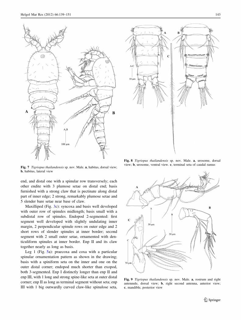
Fig. 7 Tigriopus thailandensis sp. nov. Male. a, habitus, dorsal view;
b, habitus, lateral view
Fig. 8 Tigriopus thailandensis sp. nov. Male. a, urosome, dorsal
view; b, urosome, ventral view. c, terminal seta of caudal ramus
Fig. 9 Tigriopus thailandensis sp. nov. Male. a, rostrum and right
antennule, dorsal view; b, right second antenna, anterior view;
c, mandible, posterior view
Helgol Mar Res (2012) 66:139–151 143
123
1 spine-like and 1 slender inner seta. Both exp I and exp II
elongated, exp III very small. Exp I with 1 pinnate seta on
distal outer corner; exp II with 1 pinnate seta at two-thirds
of outer margin, and 1 spinulose seta on inner distal corner;
exp III with 2 median distal claw-like setae, 1 outer and 2
inner spine-like setae.
Legs 2–3 (Figs. 5b, 6a): basis with 1 pinnate seta on
outer corner. Endopod 3-segmented; enp I and enp II each
with 1 inner plumose seta; enp III with 1 outer pinnate
spine-like seta, 3 plumose setae: 2 median distal and 1
outer. Exopod 3-segmented; exp I and exp II each with 1
outer pinnate spine, and 1 inner plumose seta; exp III with
3 outer pinnate spines, 2 plumose setae apically, and 2
inner plumose setae.
Leg 4 (Fig. 6b): as leg 2 and leg 3, except enp II without
inner seta.
Leg 5 (Fig. 6c): baseoendopod large, furnished with
rows of spinules, with 1 outer basal seta and 5 spinulose
setae of unequal length on endopodal lobe, the second
outermost longest. Exopod 1-segmented, anterior face
covered with longer-sized spinules, with 5 spinulose setae
of unequal length, the second innermost longest.
Male (Figs. 7, 8, 9, 10, 11, 12)
Total length of holotype male 0.94 mm; paratype, total
length 1.0 mm, measured from tip of rostrum to posterior
margin of caudal ramus. Male morphology as in female
except the following characters. Antennule (Fig. 9a) sub-
chirocer, 8-segmented: first segment with small spinules on
anterior inner surface, fifth one shortest, sixth one globu-
larly expanded with a big aesthetasc, seventh one forming a
claw-like outer process, and last one small. Armature for-
mula: 1-1-11-3-2-8 ? Ae.-0-9. Antenna (Fig. 9b):
anterior edge of allobasis without seta, all other armature as
in female.
Leg 2 (Fig. 11b): Basis with 1 pinnate outer seta.
Endopod 3-segmented: first segment with 1 inner seta;
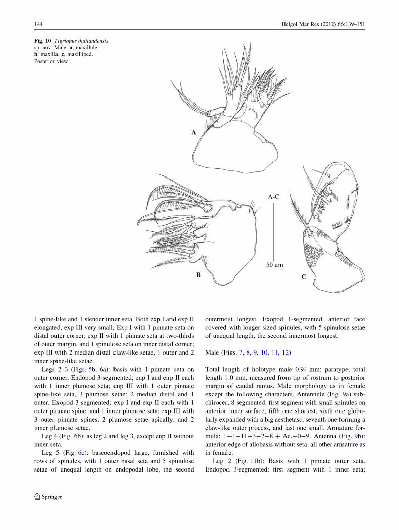
Fig. 10 Tigriopus thailandensissp. nov. Male. a, maxillule;
b, maxilla; c, maxilliped.
Posterior view
144 Helgol Mar Res (2012) 66:139–151
123
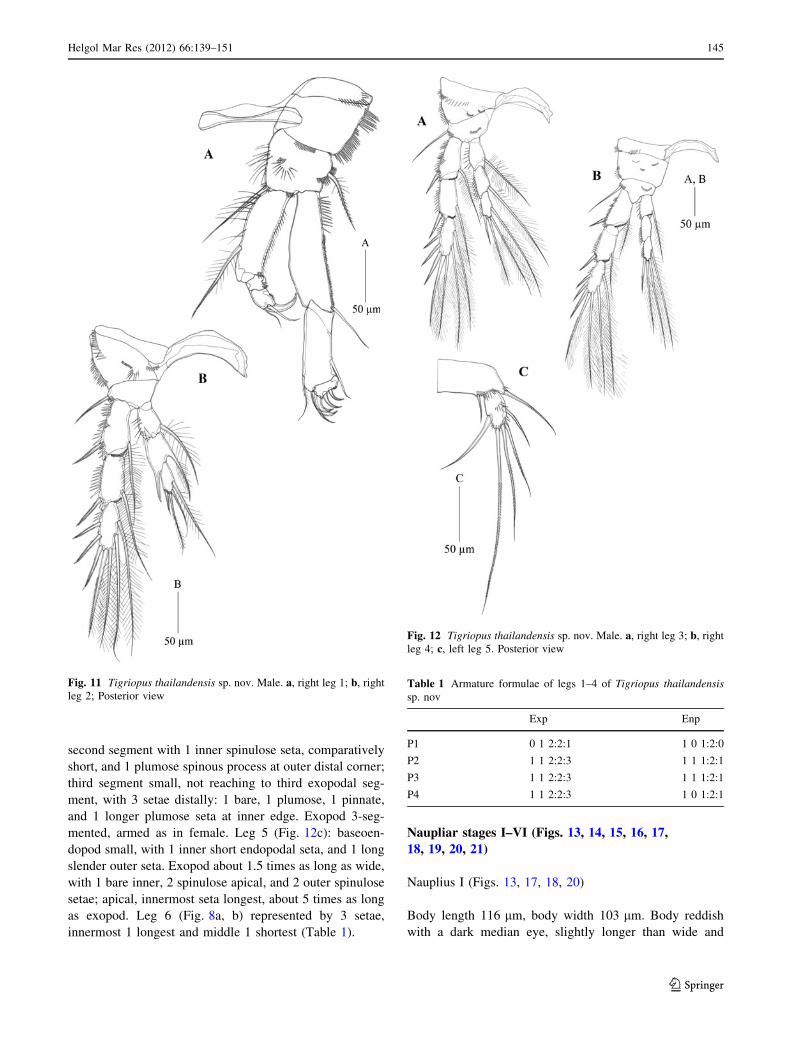
second segment with 1 inner spinulose seta, comparatively
short, and 1 plumose spinous process at outer distal corner;
third segment small, not reaching to third exopodal seg-
ment, with 3 setae distally: 1 bare, 1 plumose, 1 pinnate,
and 1 longer plumose seta at inner edge. Exopod 3-seg-
mented, armed as in female. Leg 5 (Fig. 12c): baseoen-
dopod small, with 1 inner short endopodal seta, and 1 long
slender outer seta. Exopod about 1.5 times as long as wide,
with 1 bare inner, 2 spinulose apical, and 2 outer spinulose
setae; apical, innermost seta longest, about 5 times as long
as exopod. Leg 6 (Fig. 8a, b) represented by 3 setae,
innermost 1 longest and middle 1 shortest (Table 1).
Naupliar stages I–VI (Figs. 13, 14, 15, 16, 17,
18, 19, 20, 21)
Nauplius I (Figs. 13, 17, 18, 20)
Body length 116 lm, body width 103 lm. Body reddish
with a dark median eye, slightly longer than wide and
Fig. 11 Tigriopus thailandensis sp. nov. Male. a, right leg 1; b, right
leg 2; Posterior view
Fig. 12 Tigriopus thailandensis sp. nov. Male. a, right leg 3; b, right
leg 4; c, left leg 5. Posterior view
Table 1 Armature formulae of legs 1–4 of Tigriopus thailandensissp. nov
Exp Enp
P1 0 1 2:2:1 1 0 1:2:0
P2 1 1 2:2:3 1 1 1:2:1
P3 1 1 2:2:3 1 1 1:2:1
P4 1 1 2:2:3 1 0 1:2:1
Helgol Mar Res (2012) 66:139–151 145
123
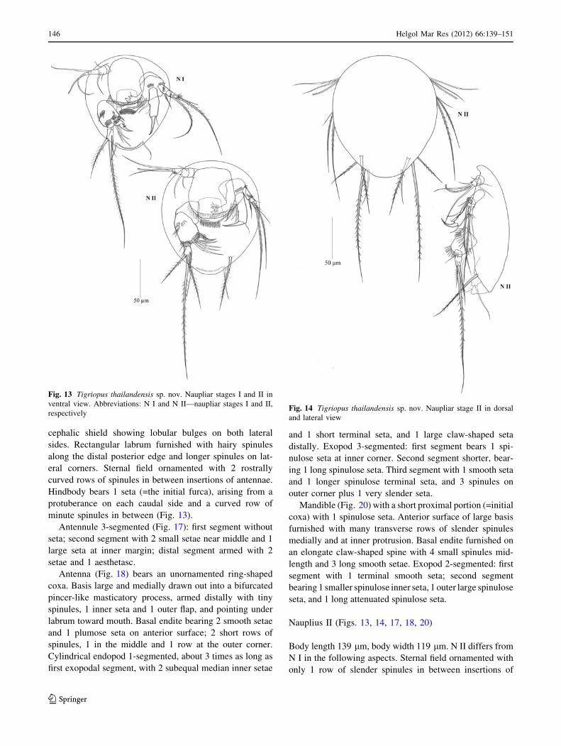
cephalic shield showing lobular bulges on both lateral
sides. Rectangular labrum furnished with hairy spinules
along the distal posterior edge and longer spinules on lat-
eral corners. Sternal field ornamented with 2 rostrally
curved rows of spinules in between insertions of antennae.
Hindbody bears 1 seta (=the initial furca), arising from a
protuberance on each caudal side and a curved row of
minute spinules in between (Fig. 13).
Antennule 3-segmented (Fig. 17): first segment without
seta; second segment with 2 small setae near middle and 1
large seta at inner margin; distal segment armed with 2
setae and 1 aesthetasc.
Antenna (Fig. 18) bears an unornamented ring-shaped
coxa. Basis large and medially drawn out into a bifurcated
pincer-like masticatory process, armed distally with tiny
spinules, 1 inner seta and 1 outer flap, and pointing under
labrum toward mouth. Basal endite bearing 2 smooth setae
and 1 plumose seta on anterior surface; 2 short rows of
spinules, 1 in the middle and 1 row at the outer corner.
Cylindrical endopod 1-segmented, about 3 times as long as
first exopodal segment, with 2 subequal median inner setae
and 1 short terminal seta, and 1 large claw-shaped seta
distally. Exopod 3-segmented: first segment bears 1 spi-
nulose seta at inner corner. Second segment shorter, bear-
ing 1 long spinulose seta. Third segment with 1 smooth seta
and 1 longer spinulose terminal seta, and 3 spinules on
outer corner plus 1 very slender seta.
Mandible (Fig. 20) with a short proximal portion (=initial
coxa) with 1 spinulose seta. Anterior surface of large basis
furnished with many transverse rows of slender spinules
medially and at inner protrusion. Basal endite furnished on
an elongate claw-shaped spine with 4 small spinules mid-
length and 3 long smooth setae. Exopod 2-segmented: first
segment with 1 terminal smooth seta; second segment
bearing 1 smaller spinulose inner seta, 1 outer large spinulose
seta, and 1 long attenuated spinulose seta.
Nauplius II (Figs. 13, 14, 17, 18, 20)
Body length 139 lm, body width 119 lm. N II differs from
N I in the following aspects. Sternal field ornamented with
only 1 row of slender spinules in between insertions of
Fig. 13 Tigriopus thailandensis sp. nov. Naupliar stages I and II in
ventral view. Abbreviations: N I and N II—naupliar stages I and II,
respectivelyFig. 14 Tigriopus thailandensis sp. nov. Naupliar stage II in dorsal
and lateral view
146 Helgol Mar Res (2012) 66:139–151
123
antennae. Hindbody with 1 additional seta on either side
(Figs. 13, 14). Antennule (Fig. 17) with 1 additional
median seta and 5 small setae at anterior inner margin on
its distal segment. Antenna (Fig. 18): basal endite bearing
1 additional seta. Tubular endopod bearing 1 small spine at
distal margin. First segment of exopod ornamented with 1
oblique row of 7 spinules. Mandibular coxal spinulose seta
became smooth seta (Fig. 20). Basal endopod bearing 1
additional seta. One additional seta at outer corner of the
first segment of exopod. Maxillule represented by a spi-
nulose seta.
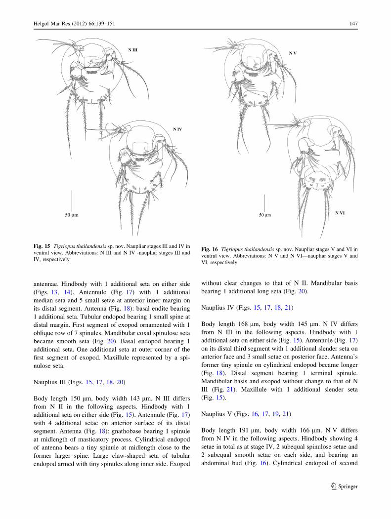
Nauplius III (Figs. 15, 17, 18, 20)
Body length 150 lm, body width 143 lm. N III differs
from N II in the following aspects. Hindbody with 1
additional seta on either side (Fig. 15). Antennule (Fig. 17)
with 4 additional setae on anterior surface of its distal
segment. Antenna (Fig. 18): gnathobase bearing 1 spinule
at midlength of masticatory process. Cylindrical endopod
of antenna bears a tiny spinule at midlength close to the
former larger spine. Large claw-shaped seta of tubular
endopod armed with tiny spinules along inner side. Exopod
without clear changes to that of N II. Mandibular basis
bearing 1 additional long seta (Fig. 20).
Nauplius IV (Figs. 15, 17, 18, 21)
Body length 168 lm, body width 145 lm. N IV differs
from N III in the following aspects. Hindbody with 1
additional seta on either side (Fig. 15). Antennule (Fig. 17)
on its distal third segment with 1 additional slender seta on
anterior face and 3 small setae on posterior face. Antenna’s
former tiny spinule on cylindrical endopod became longer
(Fig. 18). Distal segment bearing 1 terminal spinule.
Mandibular basis and exopod without change to that of N
III (Fig. 21). Maxillule with 1 additional slender seta
(Fig. 15).
Nauplius V (Figs. 16, 17, 19, 21)
Body length 191 lm, body width 166 lm. N V differs
from N IV in the following aspects. Hindbody showing 4
setae in total as at stage IV, 2 subequal spinulose setae and
2 subequal smooth setae on each side, and bearing an
abdominal bud (Fig. 16). Cylindrical endopod of second
Fig. 15 Tigriopus thailandensis sp. nov. Naupliar stages III and IV in
ventral view. Abbreviations: N III and N IV -naupliar stages III and
IV, respectively
Fig. 16 Tigriopus thailandensis sp. nov. Naupliar stages V and VI in
ventral view. Abbreviations: N V and N VI—naupliar stages V and
VI, respectively
Helgol Mar Res (2012) 66:139–151 147
123

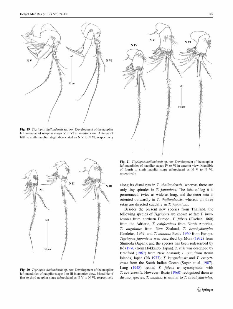
antenna bearing 4 spinules at midlength along inner margin
of its segment (Fig. 19). Claw-shaped seta of mandibular
endopod ornamented with 5 spinules at proximal inner
margin (Fig. 21).
Nauplius VI (Figs. 16, 17, 19, 21)
Body length 203 lm, body width 176 lm. N VI differs
from N V in the following aspects. The labrum without
spinules along its edge. Sternal field without row of spin-
ules in between insertions of antennae. Antenna without
basipodal endite and gnathobase; distal segment of exopod
with additional seta (Fig. 19). Mandibular coxa without
any seta, endopod expanded (Fig. 21). Inner edge with
enditic lobes; 1 seta on endopod, 1 seta on prospective
exopod. With 4 lobular precursors of postmandibular
appendages developed on both lateral sides. Anterior two
unarmed limb-buds are assumed to represent maxillule and
maxilliped precursors. First two swimming legs (legs 1–2)
indicated at the sixth naupliar stage. Lobe with 2 small
setae corresponding to leg one, and further caudally, lobe
with 2 setae corresponding to leg two (Fig. 16).
Biological remarks. Individuals of all six naupliar stages
are able to crawl and to swim-crawl on surfaces. All
developmental stages including nauplii can also swim
freely in the water column, and aggregate to swarms in
their splash-pool habitat.
Discussion
Tigriopus thailandensis sp. nov. shares with its closest
relative, T. japonicus Mori 1932, the two inner setae on the
third exopodal segment of leg 4, while other congeners
bear three inner setae. Allobasis and exopod of antenna are
much more slender and elongate in T. thailandensis than in
T. japonicus. There are nine spinule patches on the female
antenna of T. thailandensis, whereas there are only two in
T. japonicus. In both genders of T. thailandensis, the third
exopodal segment of leg 1 bears five claw-like setae, with
the outermost slender, whereas in T. japonicus it bears
three setae, with the outermost being very stout. The first
endopodal segment of leg 1 bears strong spiniform spinules
Fig. 17 Tigriopus thailandensis sp. nov. Development of the naupliar
left antennule of naupliar stages I to VI in anterior view. Antennule of
first to sixth naupliar stage abbreviated as N I to N VI, respectivelyFig. 18 Tigriopus thailandensis sp. nov. Development of the naupliar
left antennae of naupliar stages I to IV in anterior view. sp. nov
148 Helgol Mar Res (2012) 66:139–151
123
along its distal rim in T. thailandensis, whereas there are
only tiny spinules in T. japonicus. The lobe of leg 6 is
pronounced, twice as wide as long, and the outer seta is
oriented outwardly in T. thailandensis, whereas all three
setae are directed caudally in T. japonicus.
Besides the present new species from Thailand, the
following species of Tigriopus are known so far: T. brev-
icornis from northern Europe, T. fulvus (Fischer 1860)
from the Adriatic, T. californicus from North America,
T. angulatus from New Zealand, T. brachydactylus
Candeias, 1959, and T. minutus Bozic 1960 from Europe.
Tigriopus japonicus was described by Mori (1932) from
Shimoda (Japan), and the species has been redescribed by
Ito (1970) from Hokkaido (Japan). T. raki was described by
Bradford (1967) from New Zealand; T. igai from Bonin
Islands, Japan (Ito 1977); T. kerguelensis and T. crozett-
ensis from the South Indian Ocean (Soyer et al. 1987).
Lang (1948) treated T. fulvus as synonymous with
T. brevicornis. However, Bozic (1960) recognized them as
distinct species. T. minutus is similar to T. brachydactylus,
Fig. 19 Tigriopus thailandensis sp. nov. Development of the naupliar
left antennae of naupliar stages V to VI in anterior view. Antenna of
fifth to sixth naupliar stage abbreviated as N V to N VI, respectively
Fig. 20 Tigriopus thailandensis sp. nov. Development of the naupliar
left mandibles of naupliar stages I to III in anterior view. Mandible of
first to third naupliar stage abbreviated as N V to N VI, respectively
Fig. 21 Tigriopus thailandensis sp. nov. Development of the naupliar
left mandibles of naupliar stages IV to VI in anterior view. Mandible
of fourth to sixth naupliar stage abbreviated as N V to N VI,
respectively
Helgol Mar Res (2012) 66:139–151 149
123
particularly in the female setal armature of P5. The lack of
an outer seta on the exp 2 of leg 1 in T. brachydactylus
seems to be an artifact, since all other Tigriopus species
show this seta.
As in most Harpacticoida, postmandibular appendages of
naupliar stages show a great variation in both T. thailand-
ensis and T. japonicus: from their first appearance, in the
total number present at N VI, and in their shape and arma-
ture. At N VI, the first maxilla, second maxilla, maxilliped,
legs one and two may all be present, and they can then be
identified according to their position from front to rear.
Naupliar development in the Harpacticidae
Ito (1970) and Koga (1970) examined Japanese populations
of T. japonicus. The study of Ito (1970) is lacking the N IV.
In particular, Koga (1970) documents the first maxilla from
N III onwards, whereas it is expressed in Ito’s and the
present account of T. thailandensis sp. nov. as early as at
the N II stage.
According to Walker (1981), segmentation is lacking on
both the antennal and mandibular exopod in Harpacticus
sp. throughout the phase. This is in contrast to the present
observation of a four-segmented antennal and a two-seg-
mented mandibular exopod in T. thailandensis as well as in
T. japonicus from a Taiwan population (Dahms et al.
2007). These characters in T. thailandensis have also been
confirmed in another harpacticid genus Harpacticus by
Castel (1976) for H. littoralis. Clogston (1965) described
the antennal exopod of Zaus spinatus as being 2-segmented
throughout the phase, whereas it is four-segmented in
T. thailandensis and T. japonicus. In contrast to the present
report on T. thailandensis, Clogston (1965) argued that
there was no indication of the first maxilla in Z. spinatus.
Tigriopus thailandensis sp. nov. lacks armature on the
first segment of the antennule throughout the nauplius
phase, whereas one seta is present in T. japonicus
according to Dahms et al. (2007). There are three long
setae in T. japonicus on the antennule’s second segment
whereas only one small seta plus two longer setae develop
in T. thailandensis sp. nov.
There is an aesthetasc on the antennule’s third segment
in T. thailandensis sp. nov. that is not discernible on the
third segment in T. japonicus. There is one seta more on the
third segment in T. thailandensis sp. nov. than in T. japo-
nicus (that means: seven setae plus one aesthetasc at N III
versus six setae, plus one aesthetasc) until both reach
thirteen setae, plus one aesthetasc at N VI in both species.
There are no spinules on this terminal segment in T. japo-
nicus, whereas there are spinules from N II onwards in
T. thailandensis sp. nov. The second antenna of T. thai-
landensis sp. nov. bears 3 small spinules on the terminal
segment of the exopod, whereas there are no in T. japo-
nicus. In T. thailandensis sp. nov. there is one additional
seta on the anterior surface of the basis. The tubular
endopod bears a tiny seta midlength at N III that increases
in size throughout the naupliar development. The man-
dibular coxal seta bears one spinulose seta in T. thailand-
ensis sp. nov. that is smooth in T. japonicus. Several
spinules are present on the anterior surface of the basis of
T. thailandensis sp. nov., whereas there is only one row in
T. japonicus.
Acknowledgments This research is supported by the National
Research Council of Thailand (Grant 2551A1120211) and Ram-
khamhaeng University. The work of V. N. Ivanenko is supported by
the Russian Foundation for Basic Research (Grant 06-04-48918-a).
The work of W.-X. Yang is supported by the National Nature Science
Foundation of China (Grant 31072198 and 40776079).
References
Alekseev VR (2002) Copepoda. In: Fernando CH et al (eds) A guide
to tropical freshwater zooplankton—identification, ecology and
impact on fisheries. Backhuys Publishers, Leiden, pp 123–188
Bozic B (1960) Le genre Tigriopus Norman (Copepodes Harpacti-
coıdes) et ses formes europeennes; recherches morphologiques et
experimentales. Arch Zool Exp Gen 98(3):167–269
Bradford J (1967) The genus Tigriopus Norman (Copepoda, Harpac-
ticoida) in New Zealand with a description of a new species.
Trans Roy Soc NZ Zool 10(6):51–59
Burton RS, Rawson PD, Edmands S (1999) Genetic architecture of
physiological phenotypes: empirical evidence for coadapted
gene complexes. Am Zool 39:451–462
Castel J (1976) Developpement larvaire et biologie de Harpacticuslittoralis Sars, 1910 (copepode, harpacticoide) dans les etangs
saumatres de la region d’Arcachon. Cah Biol Mar 17:195–212
Clogston F (1965) Postembryonic development of species of harp-
acticoid copepods from the Pacific coast of the United States and
an application of developmental patterns to their systematics.
PhD dissertation, Univ. of Washington, Seattle, 246 pp
Dahms H-U (1990) Naupliar development of Harpacticoida (Crusta-
cea, Copepoda) and its significance for phylogenetic systematics.
Mikrof Mar 6:169–272
Dahms H-U (1993) Pictorial keys for the identification of crustacean
nauplii from the marine meiobenthos. J Crust Biol 13:609–616
Dahms H-U, Fornshell JA, Fornshell BJ (2006) Key for the
identification of crustacean nauplii. Org Divers Evol 6:47–56
Dahms H-U, Chullasorn S, Kangtia P, Ferrari FD, Hwang J-S (2007)
Naupliar development of Tigriopus japonicus Mori, 1932
(Harpacticidae, Copepoda). Zool Stud 46:746–759
Davenport J, Barnett PRO, McAllen RJ (1997) Environmental
tolerances of three species of the harpacticoid genus Tigriopus.
J Mar Biol Assoc UK 77:3–16
Dethier MN (1980) Tidepools as refuges: predation and the limits of
the harpacticoid copepod Tigriopus californicus. J Exp Mar Biol
Ecol 42:99–111
Edmands S (1999) Heterosis and outbreeding depression in interpop-
ulation crosses spanning a wide range of divergence. Evolution
53:1757–1768
Edmands S, Burton RS (1998) Variation in cytochrome-c oxidase
activity is not maternally inherited in the copepod Tigriopuscalifornicus. Heredity (Lond) 80:668–674
150 Helgol Mar Res (2012) 66:139–151
123
Fraser JH (1936) The occurrence, ecology and life history of
Tigriopus fulvus (Fischer). J Mar Biol Assoc UK 20:523–536
Guiglia D (1926) Sullo sviluppo larvale de Tigriopus fulvus (Fischer).
Boll Mus Zool Anat Comp R Univ Gen (2)6(3):11–16
Igarashi S (1963) Developmental cycle of Tigriopus japonicus Mori.
Sci Rep Tohoku Univ 29(2):59–72
Ito T (1970) The biology of a harpacticoid copepod Tigriopus japonicusMori. J Fac Sci, Hokkaido Univ, Ser VI, Zool 17:474–500
Ito T (1973) Ventral nerve cord of Tigriopus japonicus Mori
(Copepoda, Harpacticoida). Annot Zool Japon 46(1):45–48
Ito T (1977) New species of marine harpacticoid copepods of the
genera Harpacticella and Tigriopus from the Bonin Islands, with
reference to the morphology of copepodid stages. J Fac Sci
Hokkaido Univ Ser VI Zool 32:273–326
Ivanenko VN, Ferrari FD, Dahms H-U (2008) Nauplii of Tegastesfalcatus (Norman, 1868)(Harpacticoida, Tegastidae), a copepod
with an unusual naupliar mouth and mandible. J Crust Biol
28:270–280
Jung SO, Lee YM, Park TJ, Park HG, Leung KMY, Dahms H-U, Lee
W, Lee JS (2006) The complete mitochondrial genome of the
intertidal copepod Tigriopus sp. (Copepoda, Harpacticidae) from
Korea and phylogenetic considerations. J Exp Mar Biol Ecol
333:251–262
Kelly LS, Snell TW (1998) Role of surface glycoproteins in mate
guarding of the marine harpacticoid Tigriopus japonicus. Mar
Biol 130:605–612
Kim IC, Kim YJ, Song SJ, Lee J-S, Lee W (2003) The intertidal
harpacticoid copepod Tigriopus japonicus (Crustacea: Copep-
oda) b-actin gene: cloning, sequence and intraspecies variation.
DNA Seq 14:279–284
Kim IC, Kim YJ, Lee Y-M, Kim BG, Park TJ, Kim HS, Jung MM,
Williams TD, Lee W, Lee J-S (2004) cDNA cloning of
translationally controlled tumor protein/histamine releasing
factor (TCTP/HRF) from the intertidal harpacticoid copepod
Tigriopus japonicus. DNA Seq 15:159–163
Koga F (1970) On the life history of Tigriopus japonicus Mori
(Copepoda). J Oceanogr Soc Japan 26(1):11–21
Lang K (1948) Monographie der Harpacticiden I, II. Reprint Otto
Koeltz Science Publ, Konigstein 1682 p
Lee JS (2003) cDNA cloning of rhoA gene from the intertidal
harpacticoid copepod Tigriopus japonicus (Crustacea, Copep-
oda). Kor J Gen 25:403–408
Lee YM, Kim IC, Jung SO, Lee JS (2005) Analysis of 686 expressed
sequence tags (ESTs) from the intertidal harpacticoid copepod
Tigriopus japonicus (Crustacea, Copepoda). Mar Poll Bull
51:757–768
Machida RJ, Miya MU, Nishida M, Nishida S (2002) Complete
mitochondrial DNA sequence of Tigriopus japonicus (Crustacea:
Copepoda). Mar Biotechnol 4:406–417
McAllen R (1998) Life at its limits—the ecophysiology of the high
shore rockpool inhabitant Tigriopus brevicornis. PhD thesis,
University of London
McAllen R (1999) Enteromorpha intestinalis–a refuge for the
supralittoral rockpool harpacticoid copepod Tigriopus brevicor-
nis. J Mar Biol Assoc UK 79:1125–1126
McAllen R, Block W (1997) Aspects of the cryobiology of the
intertidal harpacticoid copepod Tigriopus brevicornis. Cryobi-
ology 35:309–317
McAllen R, Taylor AC, Davenport J (1998) Osmotic and body
density response in the harpacticoid copepod Tigriopus brevi-cornis in supralittoral rockpools. J Mar Biol Assoc UK
78:1143–1153
Mori T (1932) Tigriopus japonicus, a new species of neritic
Copepoda. Zool Mag Tokyo 50(5):294–295
Raisuddin S, Kwok KWH, Leung KMY, Schlenk D, Lee J-S (2007) The
copepod Tigriopus: A promising marine model organism for
ecotoxicology and environmental genomics. Aquat Tox 83:161–173
Shaw TH (1938) Some observations on the life history of a tide-pool
copepod, Tigriopus fulvus (Fischer). Bull Fan Mem Inst Biol
Zool 8:9–17
Soyer J, Thiriot-Quievreux C, Colomines JC (1987) Description de
deux especes jumelles du groupe Tigriopus angulatus (Copep-
oda, Harpacticoida) dans les archipels Crozet et Kerguelen
(Terres Australes et Antarctiques Francaises). Zool Scr
16(2):143–154
Walker X (1981) Reproductive biology and development of a marine
harpacticoid copepod reared in the laboratory. J Crust Biol
1:376–388
Wells BJ (2007) An annotated checklist and keys to the species of
Copepoda Harpacticoida (Crustacea). Zootaxa 1568:872
Helgol Mar Res (2012) 66:139–151 151
123