a new half-condensed schiff base compound: highly selective and sensitive ph-responsive fluorescent...
TRANSCRIPT

10.1021/ol201652r r 2011 American Chemical SocietyPublished on Web 08/09/2011
ORGANICLETTERS
2011Vol. 13, No. 174510–4513
A New Half-Condensed Schiff BaseCompound: Highly Selective andSensitive pH-Responsive FluorescentSensor
Uday Chand Saha,‡ Koushik Dhara,*,† Basab Chattopadhyay,§ Sushil Kumar Mandal, )
Swastik Mondal,^ Supriti Sen,‡ Monika Mukherjee,§ Sander van Smaalen,^
and Pabitra Chattopadhyay*,‡
Department of Chemistry, Burdwan University, Golapbag, West Bengal, India,Department of Chemistry, Sambhu Nath College, Labpur,Birbhum-731303, West Bengal, India, Department of Solid State Physics,Indian Association for the Cultivation of Science, Jadavpur, Kolkata,700032, India,Cytogenetics and Molecular Biology Laboratory, Department of Zoology,University of Kalyani, Kalyani-741235, India, and Laboratory of Crystallography,University of Bayreuth, D-95440 Bayreuth, Germany
[email protected]; [email protected]
Received June 20, 2011
ABSTRACT
A new probe, 3-[(3-benzyloxypyridin-2-ylimino)methyl]-2-hydroxy-5-methylbenzaldehyde (1-H) behaves as a highly selective fluorescent pHsensor in a Britton�Robinson buffer at 25 �C. The pH titrations show a 250-fold increase in fluorescence intensity within the pH range of 4.2 to8.3 with a pKa value of 6.63 which is valuable for studying many of the biological organelles.
Fluorescent probes are powerful tools in cell biology forthe nondestructive measurement of intracellular species toclarify the real-time dynamics and various biological func-tions of targeted metal cations in living cells owing to theirsimplicity and sensitivity.1 Biochemical processes frequently
involve protonation and deprotonation of biomoleculeswith concomitant changes in the pH of the environment inmany cellular events, such as cell growth,2 calcium regula-tion,3 endocytosis,4 chemotaxis,5 andother cellular processes.As minor variations of intracellular pH may induce
cellular dysfunction, development of a desirable highly
‡Burdwan University.† Sambhu Nath College.§ Indian Association for the Cultivation of Science.
)University of Kalyani.^University of Bayreuth.(1) (a)Kim,H.N.; Lee,M.H.; Kim,H. J.; Kim, J. J.; Yoon, S.Chem.
Soc. Rev. 2008, 37, 1465. (b) Que, E. L.; Domaille, D. W.; Chang, C.J. Chem. Rev. 2008, 108, 1517.
(2) Martinez-Zaguilln, R.; Gillies, R. J. Cell Physiol. Biochem. 1996,6, 169.
(3) Satoh, H.; Hayashi, H.; Katoh, H.; Terada, H.; Kobayashi, A.Am. J. Physiol. Heart Circ. Physiol. 1995, 37, H1239.
(4) Okamoto, C. T. Adv. Drug Delivery Rev. 1998, 29, 215.(5) Falke, J. J.; Bass, R. B.; Butler, S. L.; Chervitz, S. A.; Danielson,
M. A. Annu. Rev. Cell Dev. Biol. 1997, 13, 457.
Org. Lett., Vol. 13, No. 17, 2011 4511
sensitive pH fluorescent probe having excitation profiles inthe visible region is a cynosure for chemists. A limitednumber of pH-responsing fluorescent probes have beendeveloped to monitor diverse physiological and patholo-gical processes.6 Limitations of the currently available pHprobes include low sensitivity and/or excitation profiles inthe ultraviolet region. Among the fluorescent pH probesthat have been reported, only a small number are practicalfor intracellular imaging.7,8
To overcome these problems, we have designed andsynthesized a new probe, a simple half-condensed Schiff-base compound 3-[(3-benzyloxypyridin-2-ylimino)methyl]-2-hydroxy-5-methylbenzaldehyde) (1-H) which behaves asa highly selective fluorescent pH receptor in a Britton�Robinson buffer at 25 �Cand it shows a 250-fold enhancedfluorescence when pH is shifted from 4.2 to 8.3. Interest-ingly, the presence of an excess of the biologically relevant(Naþ, Kþ, Ca2þ, etc.) and other metal (Cr3þ, Mn2þ, Fe3þ,etc.) ions does not affect the intensity. To the best of ourknowledge, this type of simple half-condensed Schiff-basetype compound as fluorescent probe for sensing pH forcellular imaging in living cells is still unexplored.The synthesis of 1-H involves the addition of a solution
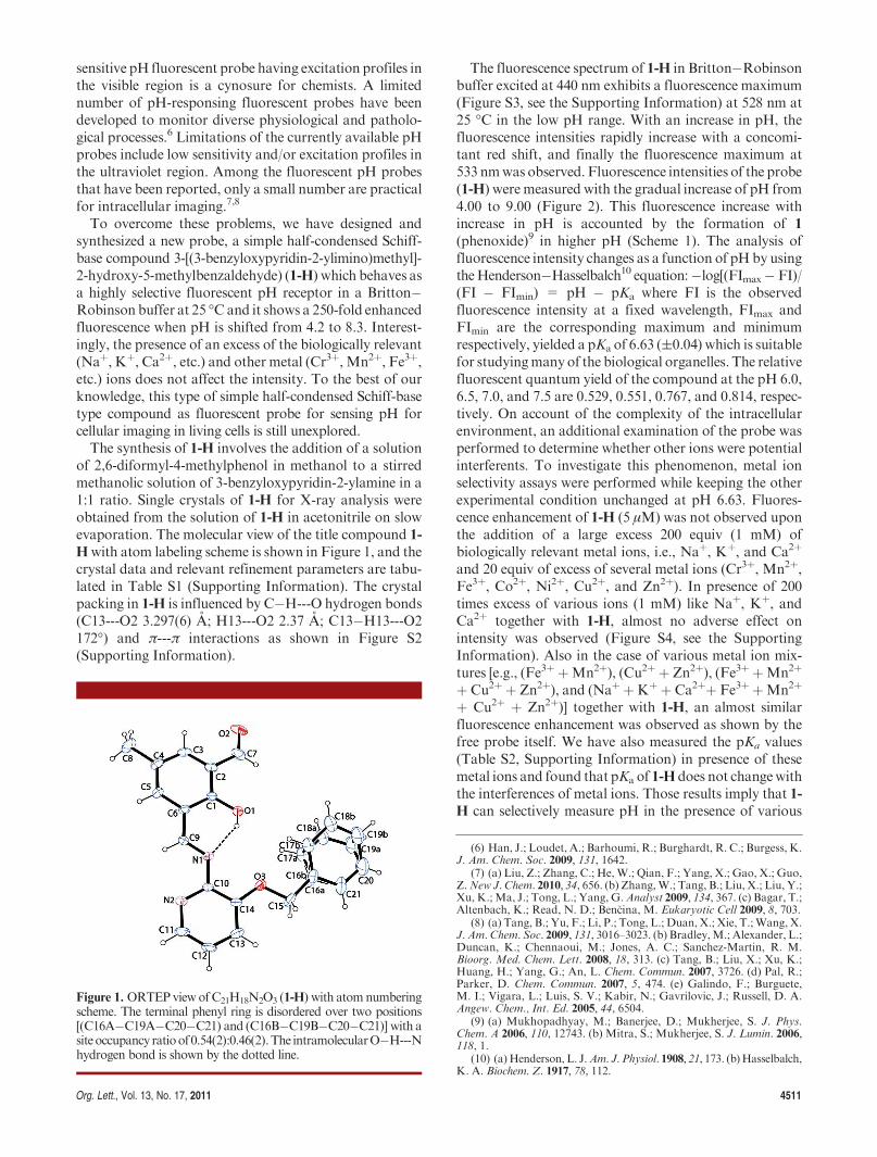
of 2,6-diformyl-4-methylphenol in methanol to a stirredmethanolic solution of 3-benzyloxypyridin-2-ylamine in a1:1 ratio. Single crystals of 1-H for X-ray analysis wereobtained from the solution of 1-H in acetonitrile on slowevaporation. The molecular view of the title compound 1-Hwith atom labeling scheme is shown in Figure 1, and thecrystal data and relevant refinement parameters are tabu-lated in Table S1 (Supporting Information). The crystalpacking in 1-H is influenced by C�H---O hydrogen bonds(C13---O2 3.297(6) A; H13---O2 2.37 A; C13�H13---O2172�) and π---π interactions as shown in Figure S2(Supporting Information).
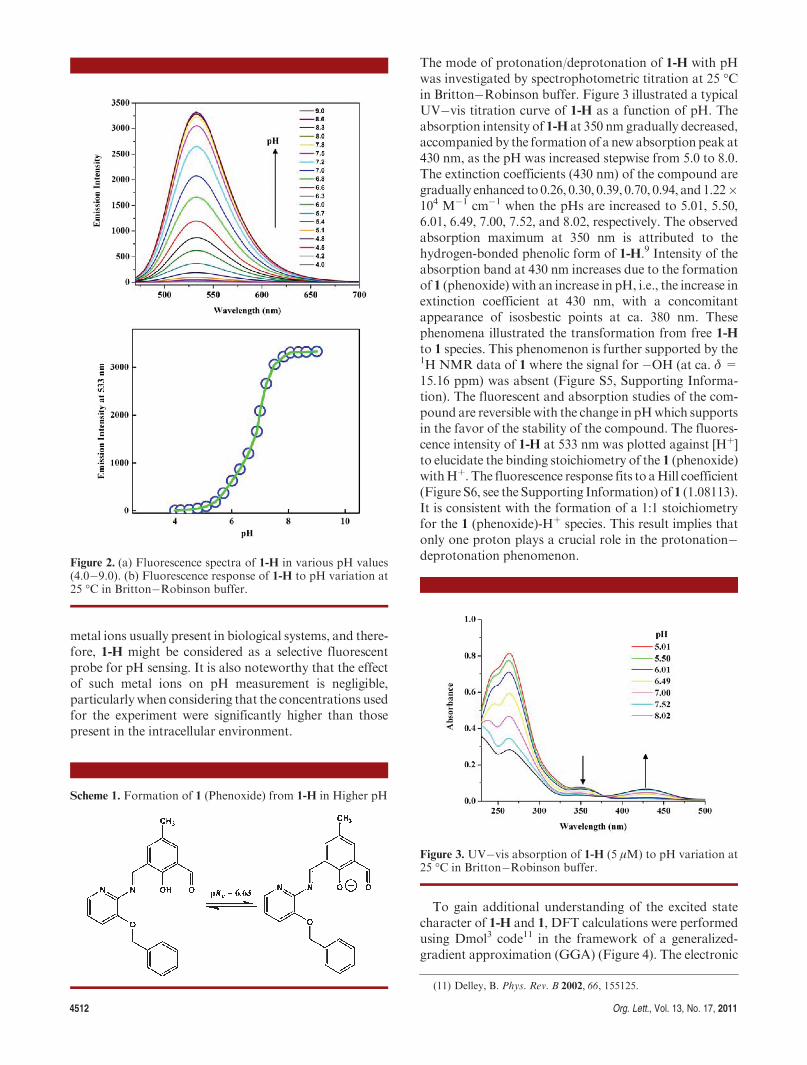
The fluorescence spectrum of 1-H in Britton�Robinsonbuffer excited at 440 nm exhibits a fluorescence maximum(Figure S3, see the Supporting Information) at 528 nm at25 �C in the low pH range. With an increase in pH, thefluorescence intensities rapidly increase with a concomi-tant red shift, and finally the fluorescence maximum at533 nmwas observed. Fluorescence intensities of the probe(1-H) weremeasured with the gradual increase of pH from4.00 to 9.00 (Figure 2). This fluorescence increase withincrease in pH is accounted by the formation of 1(phenoxide)9 in higher pH (Scheme 1). The analysis offluorescence intensity changes as a function of pHby usingtheHenderson�Hasselbalch10 equation:�log[(FImax� FI)/(FI � FImin) = pH � pKa where FI is the observedfluorescence intensity at a fixed wavelength, FImax andFImin are the corresponding maximum and minimumrespectively, yielded a pKa of 6.63 ((0.04) which is suitablefor studyingmanyof the biological organelles. The relativefluorescent quantum yield of the compound at the pH 6.0,6.5, 7.0, and 7.5 are 0.529, 0.551, 0.767, and 0.814, respec-tively. On account of the complexity of the intracellularenvironment, an additional examination of the probe wasperformed to determine whether other ions were potentialinterferents. To investigate this phenomenon, metal ionselectivity assays were performed while keeping the otherexperimental condition unchanged at pH 6.63. Fluores-cence enhancement of 1-H (5 μM) was not observed uponthe addition of a large excess 200 equiv (1 mM) ofbiologically relevant metal ions, i.e., Naþ, Kþ, and Ca2þ
and 20 equiv of excess of several metal ions (Cr3þ, Mn2þ,Fe3þ, Co2þ, Ni2þ, Cu2þ, and Zn2þ). In presence of 200times excess of various ions (1 mM) like Naþ, Kþ, andCa2þ together with 1-H, almost no adverse effect onintensity was observed (Figure S4, see the SupportingInformation). Also in the case of various metal ion mix-tures [e.g., (Fe3þ þMn2þ), (Cu2þ þ Zn2þ), (Fe3þ þMn2þ
þ Cu2þ þ Zn2þ), and (Naþ þKþ þ Ca2þþ Fe3þ þMn2þ
þ Cu2þ þ Zn2þ)] together with 1-H, an almost similarfluorescence enhancement was observed as shown by thefree probe itself. We have also measured the pKa values(Table S2, Supporting Information) in presence of thesemetal ions and found that pKa of 1-H does not changewiththe interferences of metal ions. Those results imply that 1-H can selectively measure pH in the presence of various
Figure 1. ORTEPviewofC21H18N2O3 (1-H) with atomnumberingscheme. The terminal phenyl ring is disordered over two positions[(C16A�C19A�C20�C21) and (C16B�C19B�C20�C21)] with asiteoccupancy ratioof0.54(2):0.46(2).The intramolecularO�H---Nhydrogen bond is shown by the dotted line.
(6) Han, J.; Loudet, A.; Barhoumi, R.; Burghardt, R. C.; Burgess, K.J. Am. Chem. Soc. 2009, 131, 1642.
(7) (a) Liu, Z.; Zhang, C.; He,W.; Qian, F.; Yang, X.; Gao, X.; Guo,Z.New J. Chem. 2010, 34, 656. (b) Zhang,W.; Tang, B.; Liu, X.; Liu, Y.;Xu, K.; Ma, J.; Tong, L.; Yang, G.Analyst 2009, 134, 367. (c) Bagar, T.;Altenbach, K.; Read, N. D.; Ben�cina, M. Eukaryotic Cell 2009, 8, 703.
(8) (a) Tang, B.; Yu, F.; Li, P.; Tong, L.; Duan, X.; Xie, T.;Wang, X.J. Am. Chem. Soc. 2009, 131, 3016–3023. (b) Bradley,M.; Alexander, L.;Duncan, K.; Chennaoui, M.; Jones, A. C.; Sanchez-Martin, R. M.Bioorg. Med. Chem. Lett. 2008, 18, 313. (c) Tang, B.; Liu, X.; Xu, K.;Huang, H.; Yang, G.; An, L. Chem. Commun. 2007, 3726. (d) Pal, R.;Parker, D. Chem. Commun. 2007, 5, 474. (e) Galindo, F.; Burguete,M. I.; Vigara, L.; Luis, S. V.; Kabir, N.; Gavrilovic, J.; Russell, D. A.Angew. Chem., Int. Ed. 2005, 44, 6504.
(9) (a) Mukhopadhyay, M.; Banerjee, D.; Mukherjee, S. J. Phys.Chem. A 2006, 110, 12743. (b) Mitra, S.; Mukherjee, S. J. Lumin. 2006,118, 1.
(10) (a) Henderson, L. J.Am. J. Physiol. 1908, 21, 173. (b)Hasselbalch,K. A. Biochem. Z. 1917, 78, 112.
4512 Org. Lett., Vol. 13, No. 17, 2011
metal ions usually present in biological systems, and there-fore, 1-H might be considered as a selective fluorescentprobe for pH sensing. It is also noteworthy that the effectof such metal ions on pH measurement is negligible,particularlywhen considering that the concentrations usedfor the experiment were significantly higher than thosepresent in the intracellular environment.
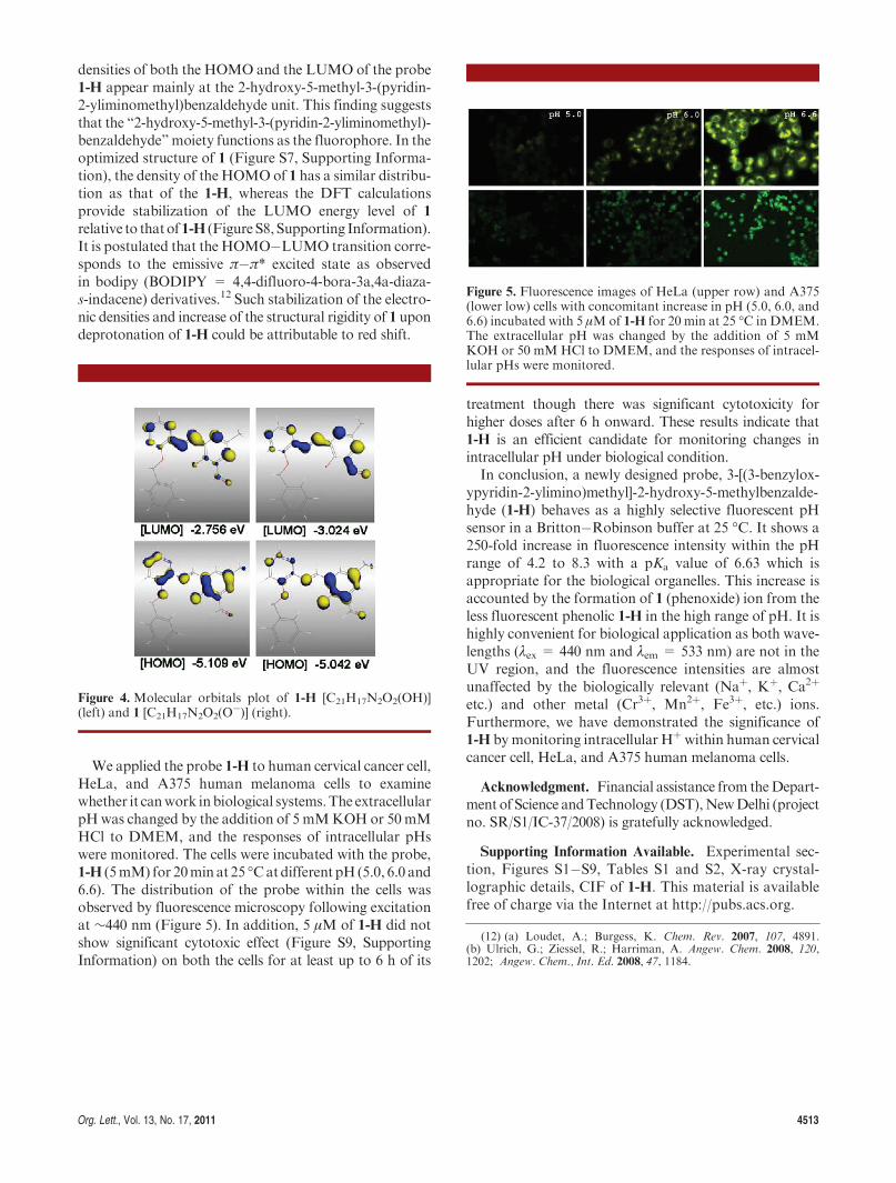
The mode of protonation/deprotonation of 1-H with pHwas investigated by spectrophotometric titration at 25 �Cin Britton�Robinson buffer. Figure 3 illustrated a typicalUV�vis titration curve of 1-H as a function of pH. Theabsorption intensity of 1-H at 350 nm gradually decreased,accompanied by the formation of a newabsorption peak at430 nm, as the pH was increased stepwise from 5.0 to 8.0.The extinction coefficients (430 nm) of the compound aregradually enhanced to 0.26, 0.30, 0.39, 0.70, 0.94, and1.22�104 M�1 cm�1 when the pHs are increased to 5.01, 5.50,6.01, 6.49, 7.00, 7.52, and 8.02, respectively. The observedabsorption maximum at 350 nm is attributed to thehydrogen-bonded phenolic form of 1-H.9 Intensity of theabsorption band at 430 nm increases due to the formationof 1 (phenoxide) with an increase in pH, i.e., the increase inextinction coefficient at 430 nm, with a concomitantappearance of isosbestic points at ca. 380 nm. Thesephenomena illustrated the transformation from free 1-Hto 1 species. This phenomenon is further supported by the1H NMR data of 1 where the signal for �OH (at ca. δ =15.16 ppm) was absent (Figure S5, Supporting Informa-tion). The fluorescent and absorption studies of the com-pound are reversible with the change in pHwhich supportsin the favor of the stability of the compound. The fluores-cence intensity of 1-H at 533 nm was plotted against [Hþ]to elucidate the binding stoichiometry of the 1 (phenoxide)withHþ. The fluorescence response fits to aHill coefficient(Figure S6, see the Supporting Information) of 1 (1.08113).It is consistent with the formation of a 1:1 stoichiometryfor the 1 (phenoxide)-Hþ species. This result implies thatonly one proton plays a crucial role in the protonation�deprotonation phenomenon.
To gain additional understanding of the excited statecharacter of 1-H and 1, DFT calculations were performedusing Dmol3 code11 in the framework of a generalized-gradient approximation (GGA) (Figure 4). The electronic
Figure 2. (a) Fluorescence spectra of 1-H in various pH values(4.0�9.0). (b) Fluorescence response of 1-H to pH variation at25 �C in Britton�Robinson buffer.
Scheme 1. Formation of 1 (Phenoxide) from 1-H in Higher pH
Figure 3. UV�vis absorption of 1-H (5 μM) to pH variation at25 �C in Britton�Robinson buffer.
(11) Delley, B. Phys. Rev. B 2002, 66, 155125.
Org. Lett., Vol. 13, No. 17, 2011 4513
densities of both the HOMO and the LUMO of the probe1-H appear mainly at the 2-hydroxy-5-methyl-3-(pyridin-2-yliminomethyl)benzaldehyde unit. This finding suggeststhat the “2-hydroxy-5-methyl-3-(pyridin-2-yliminomethyl)-benzaldehyde”moiety functions as the fluorophore. In theoptimized structure of 1 (Figure S7, Supporting Informa-tion), the density of the HOMOof 1 has a similar distribu-tion as that of the 1-H, whereas the DFT calculationsprovide stabilization of the LUMO energy level of 1relative to thatof1-H (Figure S8,Supporting Information).It is postulated that the HOMO�LUMO transition corre-sponds to the emissive π�π* excited state as observedin bodipy (BODIPY = 4,4-difluoro-4-bora-3a,4a-diaza-s-indacene) derivatives.12 Such stabilization of the electro-nic densities and increase of the structural rigidity of 1 upondeprotonation of 1-H could be attributable to red shift.
We applied the probe 1-H to human cervical cancer cell,HeLa, and A375 human melanoma cells to examinewhether it canwork inbiological systems.The extracellularpHwas changed by the addition of 5 mMKOHor 50mMHCl to DMEM, and the responses of intracellular pHswere monitored. The cells were incubated with the probe,1-H (5mM) for 20min at 25 �Catdifferent pH(5.0, 6.0 and6.6). The distribution of the probe within the cells wasobserved by fluorescence microscopy following excitationat ∼440 nm (Figure 5). In addition, 5 μM of 1-H did notshow significant cytotoxic effect (Figure S9, SupportingInformation) on both the cells for at least up to 6 h of its
treatment though there was significant cytotoxicity forhigher doses after 6 h onward. These results indicate that1-H is an efficient candidate for monitoring changes inintracellular pH under biological condition.In conclusion, a newly designed probe, 3-[(3-benzylox-
ypyridin-2-ylimino)methyl]-2-hydroxy-5-methylbenzalde-hyde (1-H) behaves as a highly selective fluorescent pHsensor in a Britton�Robinson buffer at 25 �C. It shows a250-fold increase in fluorescence intensity within the pHrange of 4.2 to 8.3 with a pKa value of 6.63 which isappropriate for the biological organelles. This increase isaccounted by the formation of 1 (phenoxide) ion from theless fluorescent phenolic 1-H in the high range of pH. It ishighly convenient for biological application as both wave-lengths (λex = 440 nm and λem = 533 nm) are not in theUV region, and the fluorescence intensities are almostunaffected by the biologically relevant (Naþ, Kþ, Ca2þ
etc.) and other metal (Cr3þ, Mn2þ, Fe3þ, etc.) ions.Furthermore, we have demonstrated the significance of1-H bymonitoring intracellular Hþwithin human cervicalcancer cell, HeLa, and A375 human melanoma cells.
Acknowledgment. Financial assistance from theDepart-ment of Science andTechnology (DST),NewDelhi (projectno. SR/S1/IC-37/2008) is gratefully acknowledged.
Supporting Information Available. Experimental sec-tion, Figures S1�S9, Tables S1 and S2, X-ray crystal-lographic details, CIF of 1-H. This material is availablefree of charge via the Internet at http://pubs.acs.org.
Figure 4. Molecular orbitals plot of 1-H [C21H17N2O2(OH)](left) and 1 [C21H17N2O2(O
�)] (right).
Figure 5. Fluorescence images of HeLa (upper row) and A375(lower low) cells with concomitant increase in pH (5.0, 6.0, and6.6) incubated with 5 μMof 1-H for 20 min at 25 �C in DMEM.The extracellular pH was changed by the addition of 5 mMKOH or 50 mMHCl to DMEM, and the responses of intracel-lular pHs were monitored.
(12) (a) Loudet, A.; Burgess, K. Chem. Rev. 2007, 107, 4891.(b) Ulrich, G.; Ziessel, R.; Harriman, A. Angew. Chem. 2008, 120,1202; Angew. Chem., Int. Ed. 2008, 47, 1184.