a new genus of marine chaetonotids (gastrotricha), with a
TRANSCRIPT

A new genus of marine chaetonotids (Gastrotricha),with a description of two new species from
Greenland and Denmark
M.A. Todaro*$, M. BalsamoO and R.M. KristensenP
*Dipartimento di Biologia Animale, Universita' di Modena e Reggio Emilia, via Campi, 213/d, I-41100 Modena, Italy.OIstituto di Scienze Morfologiche, Universita' di Urbino, Loc. Crocicchia, I-61029 Urbino, Italy.
PZoological Museum, University of Copenhagen, Universitetsparken 15, DK-2100 Copenhagen, Denmark.$Corresponding author, e-mail: [email protected]
Two new marine species of Gastrotricha: Chaetonotidae are described from West Greenland andDenmark. Some peculiar features suggest the a⁄liation of the two species to a new genus, for which thename Diuronotus is proposed. The a⁄nity of these gastrotrichs with species of Musellifer, the only genusentirely marine and hermaphroditic, and possibly the most basal one along the Chaetonotidae evolu-tionary line, is discussed from a morphological point of view. The span of the geographical distribution ofDiuronotus is discussed in the light of a record of an additional co-generic, undescribed species from theUSA. The relationships of this genus will be important in reconstructing the evolutionary pathwayswithin Gastrotricha: Chaetonotida.
INTRODUCTION
The phylum Gastrotricha includes about 690meiobenthic-sized species grouped into two orders:Macrodasyida, with 240 strap-shaped species, all but two(i.e. Marinellina £agellata Ruttner-Kolisko, 1955 andRedudasys forneris Kisielewski, 1987; see Kisielewski, 1987for discussion) of which are marine or estuarine, andChaetonotida with 450 tenpin-shaped species (exceptNeodasys) living in both marine and freshwater habitats.Among Chaetonotida, four families, Dasydytidae,Dichaeturidae, Neogosseidae and Proichthydiidae, includeonly fresh water species, two families, Neodasyidae andXenotrichulidae have only marine representatives,whereas the remaining seventh family, the Chaetonotidae,includes both marine and fresh water forms. The latterfamily is the most species-rich of the entire phylum andconsists of 12 genera, two of which (i.e. Musellifer
Hummon, 1969 and Halichaetonotus Remane, 1936),include only marine species, ¢ve other (i.e. Arenotus
Kisielewski, 1987, Fluxiderma d’Hondt, 1974, LepidochaetusKisielewski, 1991, Polymerurus Remane, 1927 and Undula
Kisielewski, 1991) are found only in fresh water eco-systems, whereas the remaining ¢ve genera (AspidiophorusVoigt, 1907, Chaetonotus Ehrenberg, 1830, HeterolepidodermaRemane, 1927, Ichthydium Ehrenberg, 1830, and perhapsLepidodermella Blake, 1933) include both marine and freshwater species.
The rare species of Musellifer bear particular relevancewithin an evolutionary framework since their primaryhabitat (i.e. the sea), coupled with the display of character-istics perceived to be plesiomorphic, at least from amorpho-functional point of view (i.e. presence of wellstructured spermatozoa and accessory reproductive struc-tures, cf. Guidi et al., 2003), make them the likely candi-dates for the most basal position in the Chaetonotidae
hypothetical evolutionary tree (however, see Hochberg &Litvaitis, 2000 for a di¡erent conclusion). Herein weprovide the description of some specimens belonging totwo undescribed species that in many respects recall thecharacteristics of the three known Musellifer species, yetthe peculiarity of other morphological traits suggeststheir a⁄liation to a newly established genus, for whichthe name Diuronotus is proposed.
MATERIALS AND METHODS
The description of the new species follows mostly theconvention of Hummon et al. (1992) whereas the locationsof some morphological characteristics along the body aregiven in percentage units (U) of total body lengthmeasured from anterior to posterior. Details aboutsampling sites, collection and specimens processing aregiven in the sections below (i.e. type locality and typematerial).
SYSTEMATICS
Order CHAETONOTIDA Remane, 1925[Rao & Clausen, 1970]
Family CHAETONOTIDAE Zelinka, 1889[sensu Kisielewski, 1991]
Subfamily CHAETONOTINAE Zelinka, 1889[sensu Kisielewski, 1991]
Diuronotus gen. nov.
Diagnosis
Chaetonotidae with weakly marked head, drawn outinto a muzzle; muzzle surrounded by a ciliary band;head plates absent; caudal furca less than 1�7 of total bodylength, showing a secondary adhesive tube on the dorso-medial side of each branch; secondary adhesive tube
J. Mar. Biol. Ass. U.K. (2005), 85, 1391^1400Printed in the United Kingdom
Journal of the Marine Biological Association of the United Kingdom (2005)
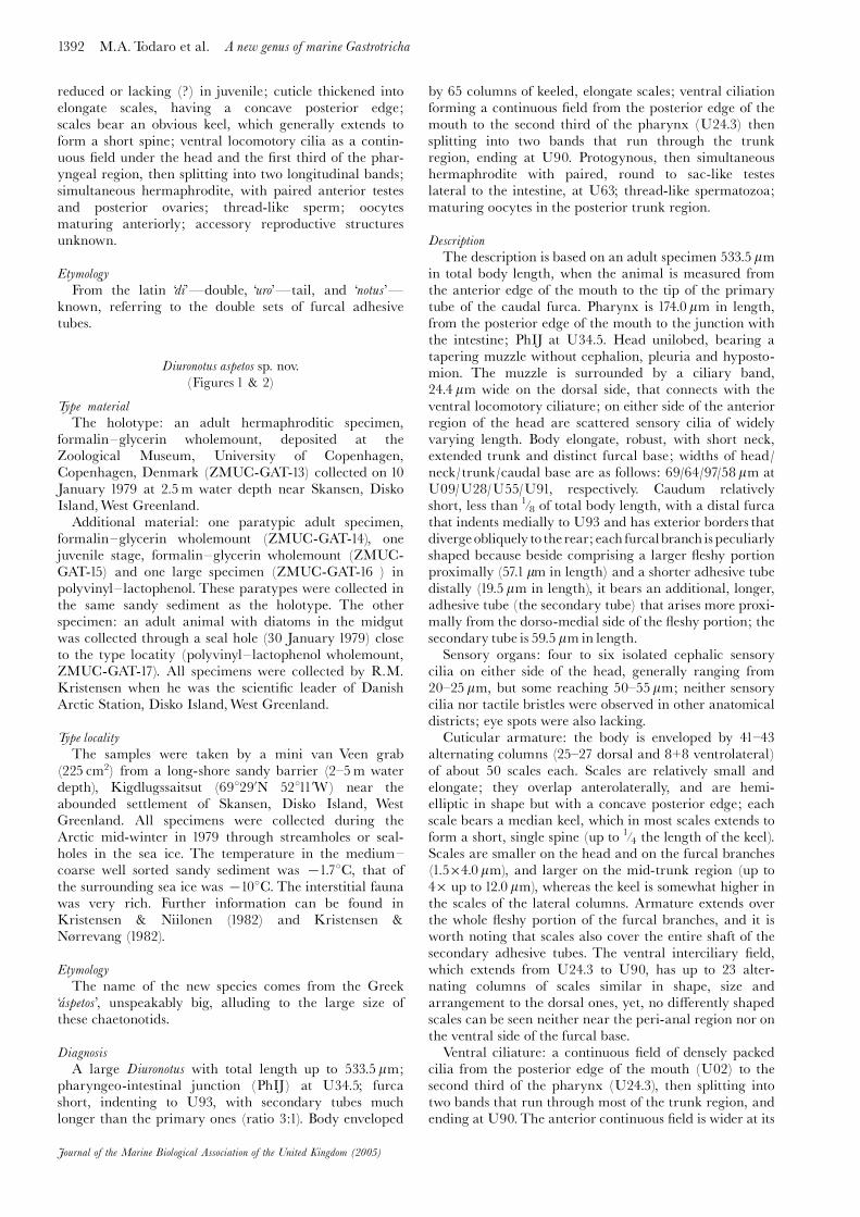
reduced or lacking (?) in juvenile; cuticle thickened intoelongate scales, having a concave posterior edge;scales bear an obvious keel, which generally extends toform a short spine; ventral locomotory cilia as a contin-uous ¢eld under the head and the ¢rst third of the phar-yngeal region, then splitting into two longitudinal bands;simultaneous hermaphrodite, with paired anterior testesand posterior ovaries; thread-like sperm; oocytesmaturing anteriorly; accessory reproductive structuresunknown.
Etymology
From the latin ‘di’�double, ‘uro’�tail, and ‘notus’�known, referring to the double sets of furcal adhesivetubes.
Diuronotus aspetos sp. nov.(Figures 1 & 2)
Type material
The holotype: an adult hermaphroditic specimen,formalin^glycerin wholemount, deposited at theZoological Museum, University of Copenhagen,Copenhagen, Denmark (ZMUC-GAT-13) collected on 10January 1979 at 2.5m water depth near Skansen, DiskoIsland,West Greenland.
Additional material: one paratypic adult specimen,formalin^glycerin wholemount (ZMUC-GAT-14), onejuvenile stage, formalin^glycerin wholemount (ZMUC-GAT-15) and one large specimen (ZMUC-GAT-16 ) inpolyvinyl^lactophenol. These paratypes were collected inthe same sandy sediment as the holotype. The otherspecimen: an adult animal with diatoms in the midgutwas collected through a seal hole (30 January 1979) closeto the type locatity (polyvinyl^lactophenol wholemount,ZMUC-GAT-17). All specimens were collected by R.M.Kristensen when he was the scienti¢c leader of DanishArctic Station, Disko Island,West Greenland.
Type locality
The samples were taken by a mini van Veen grab(225 cm2) from a long-shore sandy barrier (2^5m waterdepth), Kigdlugssaitsut (698290N 52811’W) near theabounded settlement of Skansen, Disko Island, WestGreenland. All specimens were collected during theArctic mid-winter in 1979 through streamholes or seal-holes in the sea ice. The temperature in the medium^coarse well sorted sandy sediment was 71.78C, that ofthe surrounding sea ice was 7108C. The interstitial faunawas very rich. Further information can be found inKristensen & Niilonen (1982) and Kristensen &N�rrevang (1982).
Etymology
The name of the new species comes from the Greek‘a¤ spetos’, unspeakably big, alluding to the large size ofthese chaetonotids.
Diagnosis
A large Diuronotus with total length up to 533.5 mm;pharyngeo-intestinal junction (PhIJ) at U34.5; furcashort, indenting to U93, with secondary tubes muchlonger than the primary ones (ratio 3:1). Body enveloped
by 65 columns of keeled, elongate scales; ventral ciliationforming a continuous ¢eld from the posterior edge of themouth to the second third of the pharynx (U24.3) thensplitting into two bands that run through the trunkregion, ending at U90. Protogynous, then simultaneoushermaphrodite with paired, round to sac-like testeslateral to the intestine, at U63; thread-like spermatozoa;maturing oocytes in the posterior trunk region.
Description
The description is based on an adult specimen 533.5 mmin total body length, when the animal is measured fromthe anterior edge of the mouth to the tip of the primarytube of the caudal furca. Pharynx is 174.0 mm in length,from the posterior edge of the mouth to the junction withthe intestine; PhIJ at U34.5. Head unilobed, bearing atapering muzzle without cephalion, pleuria and hyposto-mion. The muzzle is surrounded by a ciliary band,24.4 mm wide on the dorsal side, that connects with theventral locomotory ciliature; on either side of the anteriorregion of the head are scattered sensory cilia of widelyvarying length. Body elongate, robust, with short neck,extended trunk and distinct furcal base; widths of head/neck/trunk/caudal base are as follows: 69/64/97/58 mm atU09/U28/U55/U91, respectively. Caudum relativelyshort, less than 1�8 of total body length, with a distal furcathat indents medially to U93 and has exterior borders thatdivergeobliquely tothe rear; each furcalbranch ispeculiarlyshaped because beside comprising a larger £eshy portionproximally (57.1 mm in length) and a shorter adhesive tubedistally (19.5 mm in length), it bears an additional, longer,adhesive tube (the secondary tube) that arises more proxi-mally from the dorso-medial side of the £eshy portion; thesecondary tube is 59.5 mm in length.
Sensory organs: four to six isolated cephalic sensorycilia on either side of the head, generally ranging from20^25 mm, but some reaching 50^55 mm; neither sensorycilia nor tactile bristles were observed in other anatomicaldistricts; eye spots were also lacking.
Cuticular armature: the body is enveloped by 41^43alternating columns (25^27 dorsal and 8+8 ventrolateral)of about 50 scales each. Scales are relatively small andelongate; they overlap anterolaterally, and are hemi-elliptic in shape but with a concave posterior edge; eachscale bears a median keel, which in most scales extends toform a short, single spine (up to 1�4 the length of the keel).Scales are smaller on the head and on the furcal branches(1.5�4.0 mm), and larger on the mid-trunk region (up to4� up to 12.0 mm), whereas the keel is somewhat higher inthe scales of the lateral columns. Armature extends overthe whole £eshy portion of the furcal branches, and it isworth noting that scales also cover the entire shaft of thesecondary adhesive tubes. The ventral interciliary ¢eld,which extends from U24.3 to U90, has up to 23 alter-nating columns of scales similar in shape, size andarrangement to the dorsal ones, yet, no di¡erently shapedscales can be seen neither near the peri-anal region nor onthe ventral side of the furcal base.
Ventral ciliature: a continuous ¢eld of densely packedcilia from the posterior edge of the mouth (U02) to thesecond third of the pharynx (U24.3), then splitting intotwo bands that run through most of the trunk region, andending at U90.The anterior continuous ¢eld is wider at its
1392 M.A. Todaro et al. A new genus of marine Gastrotricha
Journal of the Marine Biological Association of the United Kingdom (2005)

A new genus of marine Gastrotricha M.A. Todaro et al. 1393
Journal of the Marine Biological Association of the United Kingdom (2005)
Figure 1. Diuronotus aspetos sp. nov.�drawing of the habitus�(A) internal anatomy; (B) ventral side; (C) scale of the head;(D) scale of the trunk; and (E) scale of the rear trunk. Scale bars: A, B , 100 mm; C^E, 10 mm.

1394 M.A. Todaro et al. A new genus of marine Gastrotricha
Journal of the Marine Biological Association of the United Kingdom (2005)
Figure 2. Diuronotus aspetos sp. nov.�di¡erential interference contrast optics �(A) habitus; (B) arrangement of the dorsal scales;(C) posterior end, showing the secondary adhesive tube of the left furcal branch; and (D) close-up of the secondary adhesive tubes,showing the scales along its shaft. Scale bars: A, 200 mm; B, 20 mm; C, 30 mm; D, 50 mm.

¢rst third, from U02 to U10.5, where it connects with thedorsal band, then tapers rearward before de¢nitivelysplitting.
Digestive tract: the mouth apparatus is of relativelylarge width; the external muzzle rim measures 14 mm indiameter and is characterized by a very thick wall; themouth opening measures 7.8 mm in diameter andcontinues into a 14 mm long buccal cavity, slightly enlar-ging toward the rear; 19^20 teeth-like cuticular ridgesstrengthen the mouth wall: they are arranged radiallyand can be everted, so forming a spiny basket placed infront, and surrounding the muzzle/mouth. The pharynxhas a swelling at each end, the anterior being less obvious(32 mm) than the posterior (47.5 mm), whereas its centralportion has a fairly constant width (30 mm); the intestineis broadest anteriorly (56 mm) and narrows graduallyover its length (to 8 mm); the anus opens ventrally atU90.
Reproductive tract: simultaneous hermaphrodite withpaired, round to sac-like testes, lateral to the intestine atU63. Thread-like spermatozoa. No information regardingovaries per se, however, several oocytes at di¡erent stages ofmaturation were seen in the posterior trunk region, dorso-laterally to the intestine.
Remarks
It seems that the shape of the scales undergoes some sortof change during ontogenetic growth; in fact, in the smal-lest juvenile specimen studied, 301 mm in total length, thescales, particularly on the head and on the furcal base, areshorter, rather narrow, show a deeply concave posterioredge, and bear a much higher keel, which continues intoa comparatively longer spiny process, up to the samelength as the scale. Juvenile specimen (ZMUC-GAT-15)has reduced secondary adhesive tubes. One specimen(ZMUC-GAT-17), has the intestine totally ¢lled withdiatoms. All specimens were only found during the Arcticwinter. The type locality has been visited several timessince 1979 during the summer, but the species has neverbeen found again (see Ehrhardt & Svendsen, 1994). Thismay indicate that the species is a ‘High Arctic species’,which can be found in Low Arctic (Disko Island) onlyduring the wintertime. All the other genera of gastrotrichsfound together with Diuronotus aspetos sp. nov. such asTetranchyroderma, Thaumastoderma, Paradasys, Mesodasys,Turbanella, Chaetonotus and Halichaetonotus were also foundduring the summer time.
Diuronotus rupperti sp. nov.(Figures 3^5)
Type material
The holotype: an adult hermaphroditic specimen(ZMUC-GAT-18), formalin^glycerin wholemount,deposited at the ZMUC. Collected by R.M. Kristensenon 6 April 1993.
Additional material: eight adult paratypic specimens,formalin^glycerin wholemount (ZMUC-GAT-19-26), andfour paratypic juvenile stages, formalin^glycerin whole-mount (ZMUC-GAT-27-30). Three other specimensmounted on scanning electron microscopy stubs are keptin the meiofauna collection of R.M. Kristensen (ZMUC-GAT-31-33).
Type locality
The samples were taken by a meiofauna corer (25 cm2)intertidally at low water mark in a sandy beach, (578190N11819’E) near Bansten Beach, L�s�, Denmark. Allspecimens were collected on 6 April 1993. The sedimentwas made of ¢ne, moderately sorted sand. The interstitialfauna was very rich.
Etymology
The species is dedicated to Professor Edward E.Ruppert of Clemson University, North Carolina, USA, inrecognition of his outstanding contribution to theknowledge of Gastrotricha.
Diagnosis
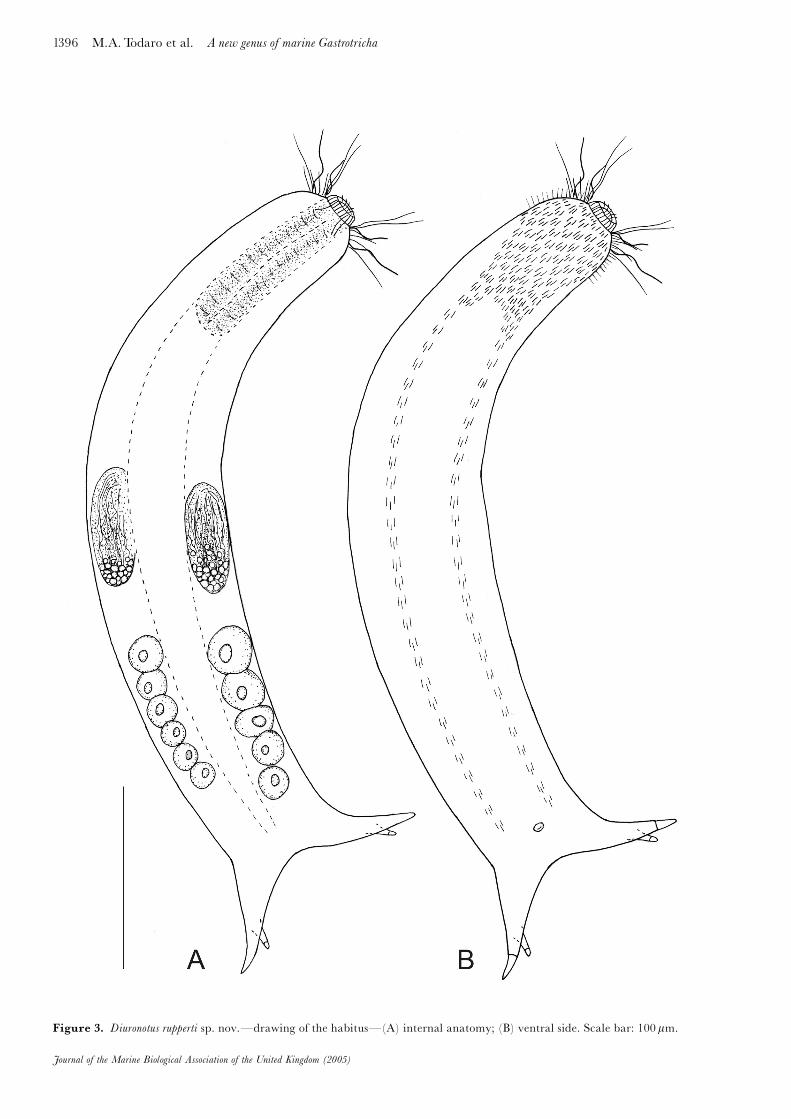
A Diuronotus with total length up to 472.5 mm; PhIJ atU22; furca short, indenting to U88, with secondary tubesshorter than the primary ones (ratio 1:2). Body envelopedby 63^65 columns of elongate, keeled scales; ventral loco-motory ciliature forming a continuous ¢eld from theposterior edge of the mouth to over the second third ofthe pharynx (U18.3), then splitting into two bands thatextend along the whole trunk, ending at U85. Protogy-nous, then simultaneous hermaphrodite with paired,round to sac-like testes, lateral to the intestine at U63;thread-like sperm; maturing oocytes in the posteriortrunk region.
Description
The description is based on an adult specimen, 472.5 mmin total body length, when the animal is measured fromthe anterior edge of the mouth to the tip of the primarytube of the caudal furca. Pharynx is 96.0 mm in length,from the posterior edge of the mouth to the junction withthe intestine; PhIJ at U22. Head unilobed, bearing atapering muzzle but lacking cephalion, pleuria and hypos-tomion. The muzzle is surrounded by a ciliary band(23.5 mm wide on the dorsal side) that connects with theventral locomotory ciliature; on either side of the anteriorregion of the head are scattered sensory cilia of widelyvarying length. Body elongate, robust, with ill-de¢nedneck, extended trunk and distinct furcal base; widths ofhead/neck/trunk/caudal base are as follows: 58/63/73/60 mm at U09/U28/U55/U91, respectively. Caudum rela-tively short, less than 1�7 of total body length, with a distalfurca that indents medially to U88 and has exteriorborders that diverge obliquely to the rear; each furcalbranch comprises an extended £eshy portion (40 mm inlength), a conical primary adhesive tube (16.8 mm inlength) and a secondary tube implanted more proximallyon the dorso-medial side of the £eshy portion; thesecondary tube is rather short, measuring only 8.5 mm inlength.
Sensory organs: four to six scattered cephalic sensorycilia on either side of the head, ranging from 15 to 25 mmin length; neither sensory cilia nor tactile bristles wereobserved in other anatomical districts; eye spots were alsolacking.
Cuticular armature: the body is enveloped by 41^43alternating columns (25^27 dorsal and 8+8 ventrolateral)of about 50 overlapping scales each. Scales are relativelysmall, generally elongate with a concave posterior edgeand bear a median keel, generally extending into a short
A new genus of marine Gastrotricha M.A. Todaro et al. 1395
Journal of the Marine Biological Association of the United Kingdom (2005)
1396 M.A. Todaro et al. A new genus of marine Gastrotricha
Journal of the Marine Biological Association of the United Kingdom (2005)
Figure 3. Diuronotus rupperti sp. nov.�drawing of the habitus�(A) internal anatomy; (B) ventral side. Scale bar: 100 mm.

A new genus of marine Gastrotricha M.A. Todaro et al. 1397
Journal of the Marine Biological Association of the United Kingdom (2005)
Figure 4. Diuronotus rupperti sp. nov.�di¡erential interference contrast optics�(A) habitus; (B) posterior end, showing theprimary tube of the furcal branches; (C) posterior end showing the secondary tubes of the furcal branches; (D) arrangement of thescales on the dorsal side; (E) scale on the dorsal side, lacking spiny process; and (F) maturing spermatids. Scale bars: A, 200 mm; B,C, 25 mm; D^F, 10 mm.

spine (up to 1�4 the length of the keel). Scales are smallerand shorter on the head and on the furcal branches(1.5�3.0 mm), and larger on the mid-trunk region (up to3� up to 10.0 mm), whereas the keel is somewhat higheron the head and the lateral scales. The armature extendsover the whole £eshy portion of the furcal branches, andalso covers most of the shaft of the secondary adhesivetubes. The ventral interciliary ¢eld, which extends fromU24.3 to U85, has up to 25 alternating columns of scalessimilar in shape, size and arrangement to the dorsal ones,yet, no di¡erently shaped scales can be seen near the peri-anal region nor under the furcal base.
Ventral ciliature: a continuous ¢eld of densely packedcilia from the posterior edge of the mouth (U02) to overthe second third of the pharynx (U18.3), then splitting intotwo bands that run through the whole trunk region,ending at U85. The anterior continuous ¢eld is wider atits ¢rst third, from U02 to U15, where it connects with
the dorsal band, then tapers rearward before de¢nitivelysplitting.
Digestive tract: the mouth apparatus is of relativelylarge width; the external muzzle rim measures 14 mm indiameter and is characterized by a very thick wall; themouth opening is 7.5 mm in diameter and continues into a12 mm long buccal cavity, slightly enlarging toward therear; 19^21 teeth-like cuticular ridges strengthen themouth wall, they are arranged radially and can beeverted, forming a spiny basket placed in front, andsurrounding the muzzle/mouth. The pharynx is of fairlyconstant width (22 mm); the intestine is broader over itsanterior third (23 mm) and narrows gradually over itslength (to 6 mm); the anus opens ventrally at U85.
Reproductive tract: simultaneous hermaphrodite withpaired, round to sac-like testes, lateral to the intestine atU47. Anterior portion of the testes ¢lled with thread-likespermatozoa maturing from round cellular elements
1398 M.A. Todaro et al. A new genus of marine Gastrotricha
Journal of the Marine Biological Association of the United Kingdom (2005)
Figure 5. Diuronotus rupperti sp. nov.�scanning electron microscope micrographs�(A) dorsolateral view of the anterior end,showing an unilobate scale with high keel extending into a crescent-shaped spiny process; (B) scales on the dorsal side; (C) ventralciliary bands and scales of the interciliary ¢eld; and (D) close-up of the posterior end showing the primary (pt) and secondary (st)tube of the left furcal branch. Scale bars: 10 mm.

(spermatocytes?) located in the posterior half of eachgonad. No information regarding ovaries per se, however,several oocytes at di¡erent stages of maturation were seenin the posterior trunk region, dorsolaterally to theintestine.
Remarks
It seems that the size of the secondary adhesive tubesundergoes some sort of change during ontogeneticgrowth; in fact, in the smallest juvenile specimen(ZMUC-GAT-30) studied, 221 mm in total length, thesetubes are totally lacking. There are no signs of these tubesin animals from 253 to 311 mm in length (ZMUC-GAT-27-29); however, when the ovaries begin to develop specimensof 372^402 mm in length (ZMUC-GAT-20-22), thesecondary adhesive tubes can be observed. Diuronotus
rupperti sp. nov. appears so similar in its traits to the unde-scribed, North American, species drawn in ¢gure 24.3g byRuppert (1988), to suggest a close relationship (sisterspecies?) between the two taxa.
Taxonomic remarks
At gross anatomy level the examined specimens can beeasily a⁄liated to the order Chaetonotida based on theabsence of anterior adhesive tubes, and to the suborderPaucitubulatina because of the absence of lateral adhesivetubes. The most striking feature of these gastrotrichs fromGreenland and Denmark are the two supernumerary(secondary) adhesive tubes present on the furcal branchesthat gives a furcate appearance to each caudal ramus.Within Chaetonotida: Paucitubulatina, the latter charac-teristic is known to occur only in members of the genusDichaetura Lauterborn, 1910, which to date counts onlytwo species, both described from freshwater habitats, i.e.D. capricornia (Metschniko¡, 1865) and D. piscator
(Murray, 1913). Observations of these small gastrotrichs,up to 150 mm in total length, are mostly old dated andtheir descriptions are largely incomplete (e.g. Murray,1913). For instance, the real structure of the secondarycaudal ramus is not yet clearly explained. It is very likely,however, that the innermost caudal branch in both thesespecies is made up by a stout spine or a cuticular hook, asreported in the most recent descriptions of D. capricornia(i.e. Martin, 1981), and thus it is not an additional adhesivetube as it appears to be in the specimens under study.
The head drawn out into a muzzle surrounded by aciliary band, along with the uniform cuticular covering(i.e. absence of scales of distinctly di¡erent shape aroundthe peri-anal area and/or on the furcal branches), aprimary hermaphroditic condition and the habitat theylive in (i.e. marine settings) approach the specimensunder study to the species of the genus Musellifer.However, Diuronotus aspetos sp. nov. and D. rupperti sp. nov.cannot be assigned to Musellifer without grossly extendingits generic boundaries. This is primarily due to thepresence of the supernumerary adhesive tubes on thefurcal branches that makes the new species unique amidstChaetonotidae, and the presence of scales along the shaftof these adhesive tubes which appears to be an autoapo-morphy of these taxa. The much smaller furca/total bodylength ratio (less than 1�7 in Diuronotus versus up to 1�3 inMusellifer), the di¡erent shape of the scales (elongate, witha strong keel ending with a spiny process in Diuronotus
versus sub-rounded to rhomboidal, with or without aweek keel ending into a tiny ¢lament or a seta inMusellifer)should further contribute to di¡erentiate the species of thetwo genera. The greatly di¡erent length of the secondaryadhesive tubes shown in specimens of the two new species(up to 59.9 mm in D. aspetos versus up to 8.5 mm inD. rupperti) should easily allow their discrimination.Furthermore, juvenile specimens of D. rupperti totally lackthe secondary adhesive tubes.
CONCLUSION
Ruppert (1988) ¢gured a chaetonotid gastrotrichbelonging to a new genus and species, of which he lateron provided ultrastructural data concerning the muscleorganization (Ruppert 1991). None of the recent paperson Gastrotricha phylogeny has taken this taxon intoaccount (e.g. Hochberg & Litvaitis, 2000; Todaro et al.,2003; Zrzavy, 2003), likely due to the fact that the speci-mens found by Ruppert were not formally described. Sincewe regard Ruppert’s species to be at least congeneric withthe present ones, it is hoped that, in a larger framework,the formal systematization of these animals may contri-bute to future debate on the reconstruction of the evolu-tionary pathways within Gastrotricha and especiallyChaetonotida. From a biogeographical point of view,Ruppert’s record is illuminating; even though the authordid not provide indications about the sampling sites of thenew genus and species, it is very likely that his specimenswere from the Atlantic coast of the USA, therefore makingthe geographical range of Diuronotus much wider than itappears from our data. Only future research can reveal apossible cosmopolitan distribution of the new genus, as it isin the case of most gastrotrich genera.
We are thankful to Miss Iben Heiner for operating the scan-ning electron microscope. We also thank the crew of ArcticStation’s research vessel ‘Porsild’ for logistics and technical assis-tance during the three expeditions to Skansen and Flakkerhuk in1979. The project bene¢ted from a grant to M.A. Todaro throughthe European Commission’s programme for Improving theHuman Research Potential and Socio-Economic KnowledgeBase (IHP); funds have been made available to provide trans-national access to researchers from members and associatedstates of the European Community to utilize the collections andother facilities of the Major Research Infrastructure CopenhagenBiosystematics Centre (COBICE). Furthermore, this paper wassupported by National Science Foundation grant no. 0334932:Assemble theTree of Life of protostome animals.
REFERENCESEhrhardt, C. & Svendsen, H.K., 1994. Marine Gastrotricha ogRotifera ved Qaamassoq�Disko. In Arktisk Biologisk Feltkursus
Qeqertasuaq/Godhavn 1994 (ed. C. Ehrhardt), pp. 53^71.Copenhagen: University of Copenhagen. [In Danish withEnglish abstract.]
Guidi, L., Marotta, R., Pierboni, L., Ferraguti, M., Todaro,M.A. & Balsamo, M., 2003. Comparative sperm ultrastruc-ture of Neodasys ciritus and Musellifer delamarei, two speciesconsidered to be basal among Chaetonotidae (Gastrotricha).Zoomorphology, 122, 135^143.
Hochberg, R. & Litvaitis, M.K., 2000. Phylogeny ofGastrotricha: a morphology-based framework of gastrotrichrelationships. Biological Bulletin. Marine Biological Laboratory,
Woods Hole, 198, 299^305.
A new genus of marine Gastrotricha M.A. Todaro et al. 1399
Journal of the Marine Biological Association of the United Kingdom (2005)

Hummon, W.D., Balsamo, M. & Todaro, M.A., 1992. Italianmarine Gastrotricha: I. Six new and one redescribed speciesof Chaetonotida. Bollettino di Zoologia, 59, 499^516.
Kisielewski, J., 1987. Two new interesting genera of Gastrotricha(Macrodasyida and Chaetonotida) from the Brazilian fresh-water psammon. Hydrobiologia, 153, 23^30.
Kristensen, R.M. & Niilonen, T., 1982. Structural studies onDiurodrilus Remane (Diurodrilidae fam. n.), with descriptionof Diurodrilus westheidei sp. n. from the Arctic interstitial meio-benthos,West Greenland. Zoologica Scripta, 11, 1^12.
Kristensen, R.M. & N�rrevang, A., 1982. Description ofPsammodrilus aedi¢cator sp. n. (Polychaeta), with notes on theArctic interstitial fauna of Disko Island, West Greenland.Zoologica Scripta, 11, 265^279.
Martin, L.V., 1981. Gastrotrichs found in Surrey. Microscopy, 34,286^300.
Murray, T., 1913. Gastrotricha. Journal of the Quekett Microscopical
Club, Series 2, 12, 211^238.
Ruppert, E.E., 1988. Gastrotricha. In Introduction to the study of
meiofauna (ed. R.P. Higgins and H. Thiel), pp. 302^311.Washington, DC: Smithsonian Institution Press.
Ruppert, E.E., 1991. Gastrotricha. In Microscopic anatomy of inver-
tebrates. Vol. 4. Aschelminthes (ed. F.W. Harrison and E.E.Ruppert), pp. 41^109. NewYork:Wiley-Liss.
Todaro, M.A., Littlewood, D.T.J., Balsamo, M., Herniou, E.A.,Cassanelli, S., Manicardi, G.,Wirz, A. & Tongiorgi, P., 2003.The interrelationships of the Gastrotricha using nuclear smallrRNA subunit sequence data, with an interpretation based onmorphology. ZoologicherAnzeiger, 242, 145^156.
Zrzavy, J., 2003. Gastrotricha and metazoan phylogeny. ZoologicaScripta, 32, 61^81.
Submitted 2 August 2005. Accepted 22 September 2005.
1400 M.A. Todaro et al. A new genus of marine Gastrotricha
Journal of the Marine Biological Association of the United Kingdom (2005)