a new discosauriscid seymouriamorph tetrapod from the ... · a new discosauriscid seymouriamorph...
TRANSCRIPT

A new discosauriscid seymouriamorph tetrapod fromthe Lower Permian of Moravia, Czech Republic
JOZEF KLEMBARA
Klembara, J. 2005. A new discosauriscid seymouriamorph tetrapod from the Lower Permian of Moravia, Czech Repub−lic. Acta Palaeontologica Polonica 50 (1): 25–48.
A new genus and species, Makowskia laticephala gen. et sp. nov., of seymouriamorph tetrapod from the Lower Permiandeposits of the Boskovice Furrow in Moravia (Czech Republic) is described in detail, and its cranial reconstruction is pre−sented. It is placed in the family Discosauriscidae (together with Discosauriscus and Ariekanerpeton) on the followingcharacter states: short preorbital region; rounded to oval orbits positioned mainly in anterior half of skull; otic notchdorsoventrally broad and anteroposteriorly deep; rounded to oval ventral scales. Makowskia is distinguished from otherDiscosauriscidae by the following characters: nasal bones equally long as broad; interorbital region broad; prefrontal−postfrontal contact lies in level of frontal mid−length (only from D. pulcherrimus); maxilla deepest at its mid−length; sub−orbital ramus of jugal short and dorsoventrally broad with long anterodorsal−posteroventral directed lacrimal−jugal su−ture; postorbital anteroposteriorly short and lacks elongated posterior process; ventral surface of basioccipital smooth;rows of small denticles placed on distinct ridges and intervening furrows radiate from place immediately laterally to artic−ular portion on ventral surface of palatal ramus of pterygoid (only from D. pulcherrimus); oblique anterior margin oftransverse flange of pterygoid directed anteromedially−posterolaterally; cultriform process of parasphenoid relativelyshort and slightly rounded; ventral surface of the posterior plate of parasphenoid heavily sculptured (only from D.pulcherrimus and Ariekanerpeton); distal ends of fourth and fifth presacral ribs distinctly anteroposteriorly broadened,and extend into the hook−like, posteriorly directed processes; shaft of ?last caudal rib anteroposteriorly broadened; poste−rior stem of interclavicle narrows anteriorly and posteriorly from broadened mid−length section.
Key words: Skeletal anatomy, Discosauriscidae, Seymouriamorpha, Permian, Boskovice Furrow, Moravia, CzechRepublic.
Jozef Klembara [[email protected]], Comenius University in Bratislava, Faculty of Natural Sciences, Mlynskádolina, 842 15 Bratislava, Slovak Republic.
Introduction
The first discoveries of stegocephalians from the BoskoviceFurrow in Moravia (Czech Republic) were made in 1872 nearthe town of Malá Lhota (Augusta 1948). Makowsky (1876)described these finds as Archegosaurus austriacus. Becausethe name Archegosaurus was preoccupied, Kuhn (1933) in−troduced the new generic name Discosauriscus for this spe−cies. A comprehensive revision of all finds of discosauriscidtetrapods since Makowsky’s first description was made byŠpinar (1952). Špinar (1952) recognized two genera, eachcontaining two species, from the region of the Boskovice Fur−row and confirmed their position within SeymouriamorphaWatson, 1917: Discosauriscus pulcherrimus (Fritsch, 1879),D. potamites (Steen, 1938), Letoverpeton austriacum (Ma−kowsky, 1876), and L. moravicum (Fritsch, 1883). Klembaraand Meszároš (1992) presented a new, chemically prepared,three−dimensional material of discosauriscids from the Bos−kovice Furrow. A subsequent revision by Klembara (1997) re−duced the number of discosauriscid taxa from the LowerPermian deposits of the Boskovice Furrow to only one genusand two species: Discosauriscus austriacus (Makowsky,1876) and D. pulcherrimus (Fritsch, 1879). The taxa Disco−
sauriscus potamites (Steen, 1938) and Letoverpeton mora−vicum (Fritsch, 1883) are junior synonyms of Discosauriscusaustriacus (see discussion in Klembara 1997). D. austriacus isvery abundant, whereas only five specimens of D. pulcher−rimus have been recorded (Klembara 1997). The present pa−per brings a description of a new genus and species, Makow−skia laticephala, from the Lower Permian deposits of theBoskovice Furrow, thus extending the number of the generaknown from this region and strata to two again. The new spe−cies is represented by a single specimen (the anterior portionof the skeleton). The three−dimensional preservation of disco−sauriscids from the Boskovice Furrow has enabled a very de−tailed knowledge of the skeletal anatomy, integument, andstructures of the sensory systems of both species of the genusDiscosauriscus (Klembara 1994a, 1996, 1997; Klembara andBartík 2000) and of Makowskia (see below). In addition, fos−silized soft parts of the body such as skin, eyes, external gills,and notochord have also been described in D. austriacus(Klembara and Meszároš 1992; Klembara 1995, 1997; Klem−bara and Bartík 2000).
According to Klembara (1997), the family Discosauris−cidae Romer, 1947 consists of two genera: Discosauriscus andAriekanerpeton (the latter genus has been recorded from the
http://app.pan.pl/acta50/app50−025.pdfActa Palaeontol. Pol. 50 (1): 25–48, 2005

Lower Permian of Tadzhikistan, Ivakhnenko 1981, 1987;Laurin 1996a; Bulanov 2003). Discosauriscus and Ariekaner−peton share character states such as short preorbital region,rounded to oval orbits positioned mainly in anterior half of theskull, otic notch dorsoventrally broad and anteroposteriorlydeep, and rounded or oval ventral scales. These characterstates are considered to be unique to the aforementioned gen−era and included into the diagnosis of the family Disco−sauriscidae. The genus Makowskia described herein fallswithin this family (see below). All the three genera differ fromthe Seymouriidae (Williston 1911) and Utegeniidae (Bulanov2003) which display the characteristic long preorbital regionof the skull and orbits located about its mid−length.
Zhang et al. (1984) described a new putative discosau−riscid, Urumqia liudaowanensis, from the Permian depositsof China. Ivakhnenko (1987) synonymized this form withUtegenia shpinari. A revision of the original material of thistetrapod is needed, but Klembara and Ruta (2004a, b) placedUtegenia shpinari (Kazakhstan) within seymouriamorphs asthe sister taxon to the discosauriscids. According to Laurin(1996b, 2000), discosauriscids appear to be a paraphyleticgroup that includes all known larval and most juvenile sey−mouriamorph specimens known (genera Utegenia, Disco−sauriscus, Ariekanerpeton and Urumquia). A new phylog−eny of Seymouriamorpha along with discussion of Laurin’s(1996b, 2000) conclusions has been recently presented byKlembara and Ruta (2004a, b). It supports the existence ofthe family Discosauriscidae with two genera Discosauriscus(Czech Republic, Germany, Poland, and France) and Arie−kanerpeton (Tadzhikistan).
The aim of this paper is to provide (i) a detailed descrip−tion of the skeletal anatomy of a new genus and species of thediscosauriscid tetrapod, Makowskia laticephala, from theLower Permian of the Boskovice Furrow in the Czech Re−public, (ii) a comparison of its skeletal anatomy with those ofother Lower Permian members of the families Discosauris−cidae and Seymouriidae and the ?Uppermost Carboniferous–Lower Permian tetrapod Utegenia shpinari (Utegeniidae);and (iii) the phylogenetic position of Makowskia withinSeymouriamorpha.
In the description below the detailed knowledge of theskeletal anatomy of Discosauriscus (Klembara 1997; Klem−bara and Bartík 2000) is used as a standard for comparisons.
Institutional abbreviations.—CM, Carnegie Museum of Nat−ural History, Pittsburgh, USA; MNG, Museum der Natur,Gotha, Germany; SNM, Slovak National Museum, Bratislava,Slovakia.
Material and methodsThe holotypic specimen (SNM Z 26506) was found pre−served in a laminated limestone, exhibiting similar condi−tions as specimens of Discosauriscus austriacus (Klembaraand Meszároš 1992), which are relatively abundant at the lo−
cality (Klembara 1997). The skeleton was completely re−moved from the rock chemically using a 10% solution of ace−tic acid (for the method, see Klembara and Meszároš 1992).Although the individual bones are three−dimensionally pre−served, several portions of the skull are slightly compresseddorsoventrally (Figs. 1A, 2A). To reconstruct the originalshape of the skull an enlarged, wax−plasticine model wasconstructed. All the bones of the skull were measured, mod−elled at seven times natural size, and then reassembled usingflat metal bars. The drawing restorations of the skull andlower jaw (Figs. 4, 5) were produced as follows: (i) mostly onthe basis of the model; (ii) when only one of the paired bonesis preserved, it is used to reproduce its mate (this was em−ployed almost completely for the maxilla, lacrimal, andjugal); (iii) missing bones are depicted in white and their out−lines were reconstructed on the basis of the anatomy of theneighbouring bones or on the anatomy of Discosauriscusaustriacus (Klembara 1997), with the outlines shown asdashed lines (this was employed for the ventrolateral tips ofthe prefrontals, posterior portions of the squamosals, andmost of the quadratojugals); (iv) where only the ornamenta−tion of the skull roof bones is damaged, these areas are re−stored in grey only (this was employed for central portion ofthe left jugal and the squamosals). The other drawings havebeen made using a WILD M8 stereomicroscope equippedwith a camera lucida.
Systematic palaeontology
Seymouriamorpha Watson, 1917Family Discosauriscidae Romer, 1947Emended and revised diagnosis (Klembara 1997; Klembaraand Bartík 2000).—Tetrapods with short preorbital region;rounded to oval orbits positioned mainly in anterior half ofskull; otic notch dorsoventrally broad and anteroposteriorlydeep; rounded to oval ventral scales.
Genus Makowskia nov.Etymology: In honor of Prof. Alexander Makowsky, professor at the for−mer German Technical University in Brno (Czech Republic). He wasthe first to describe the first specimens of discosauriscids from theBoskovice Furrow in Moravia (Czech Republic) discovered near thetown Malá Lhota in 1872.
Type and only species: Makowskia laticephala sp. nov.
Diagnosis.—As for species.
Makowskia laticephala sp. nov.Figs. 1–9.
Etymology: From Latin latus, broad and Greek kefalé, head.
Holotype: SNM Z 26506, skull and anterior portion of postcranial skele−ton, is the only known specimen, and is deposited in the collections ofthe Slovak National Museum in Bratislava (SNM).
Locality and horizon: Boskovice Furrow in Moravia (Czech Republic);Kochov−L, about 500 m NE of the village Kochov (near city Letovice)
26 ACTA PALAEONTOLOGICA POLONICA 50 (1), 2005

http://app.pan.pl/acta50/app50−025.pdf
KLEMBARA—NEW DISCOSAURISCID SEYMOURIAMORPH TETRAPOD 27
5 mm
premaxilla
maxilla
lacrimal
septomaxilla
nasal
frontal
jugal
postorbital
intertemporal
prefrontal
ectopterygoid
postfrontal
sclerotic plates
squamosal
supratemporal
tabular
tabular processoccipital flangepostparietal
parietal
squamosal
quadrato jugal
Fig. 1. Makowskia laticephala gen. et sp. nov., SNM Z 26506. Photograph of holotype skull (A) and explanatory drawing of the same (B) in dorsal view.

(Klembara and Meszároš 1992). Bačov horizon, zone 6, Lower Saxo−nian (sensu Werneburg 1989), Lower Permian.
Diagnosis.—Autapomorphies relative to discosauriscidsDiscosauriscus and Ariekanerpeton include: nasal bonesequally long as broad; interorbital region broad; prefrontal−postfrontal contact lies in level of frontal mid−length (differsonly from D. pulcherrimus); maxilla deepest at its mid−length;suborbital ramus of jugal short and dorsoventrally broad withlong anterodorsal−posteroventral directed lacrimal−jugal su−ture; postorbital anteroposteriorly short and lacks elongatedposterior process; ventral surface of basioccipital smooth;rows of small denticles placed on distinct ridges and interven−ing furrows radiate from place immediately laterally to articu−lar portion on ventral surface of palatal ramus of pterygoid(only from D. pulcherrimus); oblique anterior margin of trans−verse flange of pterygoid directed anteromedially−postero−laterally; cultriform process of parasphenoid relatively shortand slightly rounded; ventral surface of the posterior plate ofparasphenoid heavily sculptured (only from D. pulcherrimusand Ariekanerpeton); distal ends of fourth and fifth presacralribs distinctly anteroposteriorly broadened, and extend intothe hook−like, posteriorly directed processes; shaft of ?lastcaudal rib anteroposteriorly broadened; posterior stem ofinterclavicle narrows anteriorly and posteriorly frombroadened mid−length section.
Remarks.—The degree of ossification of the skeleton, thetype of ornamentation and sutures of skull roof bones ofSNM Z 26506 correspond to the similar−sized specimens ofDiscosauriscus austriacus (Klembara 1995, 1997).
Makowskia laticephala is compared with similar−sizedspecimens of Discosauriscus austriacus and D. pulcherrimus(Klembara 1997; Klembara and Bartík 2000).
Description and comparisonThe skull of Makowskia laticephala (SNM Z 26506) is rela−tively well−preserved, although several bones are missing orvariably damaged (Figs. 1, 2). The midline length of skull(Na+Fr+Pa+Pp) is 33 mm. The individual bones of the skullroof are joined via mainly simple, but in some places verysinuous sutures, and therefore paired bones may differ con−siderably in outline (e.g., supratemporal or parietal, Fig. 1).
The ornamentation of the skull roof bones consists of smalltubercles and short ridges concentrated mainly at their centres(best visible on the parietal) (Figs. 1A, 4). Radiating ridges andgrooves are present at the periphery of the parietals andfrontals, and the posterior portions of the nasals. Otherwise,the ornamentation of most bones consists of tubercles of vari−ous size and irregular short ridges and grooves. The ornamen−tation of the jugal consists of pits of various sizes and weakelevations, as seen in Discosauriscus austriacus (Klembara1997: fig. 4).
The ornamentation of several bones is badly preservedand makes the observation of the presence or course of the
sensory grooves impossible. The sensory grooves are ob−servable on the premaxilla, nasal, prefrontal, intertemporal,supratemporal, and postorbital (Figs. 1A, 4). On the right na−sal, the sensory groove is very deep, and its morphology re−sembles the confluent foraminate pits (Klembara 1994a).The distinct ornamentation of the posterior portions of thetabulars and the postparietals does not permit the grooves ofthe occipital (supratemporal) commissural canal to be identi−fied with certainty.
SkullSkull roof and sclerotic ring.—The premaxilla is a biramousstructure consisting of a ventral (maxillary) and a postero−dorsal (nasal) rami; a third, posterior (vomerine) ramus is notdistinctly developed (Figs. 1, 4). The bodies of premaxillae arejoined only in a short median suture. The nasal ramus is broad−est at its base and gradually tapers dorsalwards. Between bothnasal rami is an interpremaxillary space, the posterior part ofwhich was probably occupied by anteriormost tips of thenasals. In similar−sized Discosauriscus austriacus specimensthe presence of the interpremaxillary space is only a transi−tional, ontogenetic state that reflects the incomplete ossifica−tion of the nasal rami of the premaxillae. In larger, juvenilespecimens this space is completely closed by the posterior por−tions of the nasal rami of the premaxillae and the anteromedialtips of the nasals (Klembara 1997: fig. 29). The maxillaryramus is long and dorsoventrally narrow and has a vertical su−ture with the maxilla, whereas in D. anstriacus this suture isoblique. The maxillary ramus forms almost the entire anteriormargin of the exonarial fenestra. Each premaxilla bears sixconical, sharpely pointed teeth. The inner wall of the maxillaryramus bears a narrow horizontal lamina that forms the anteriormargin of the exochoanal fenestra. The anterior wall of theseptomaxilla abuts the dorsal surface of the horizontal laminaand the inner surface of the maxillary ramus of the premaxilla.The medial portion of the horizontal lamina extends pos−teriorly to form a short posterior ramus that meets the vomer.
The nasal is as long as it is wide (Figs. 1, 4), in contrast tothe invariably longer nasal in similar−sized Discosauriscus(Klembara 1997) and Ariekanerpeton (Ivakhnenko 1981;Laurin 1996a; Bulanov 2003). The anteromedial corner ofthe nasal produces a small process that is slightly overlappedby the dorsal ramus of the premaxilla. The anterior margin ofthe nasal borders broadly the exonarial fenestra posteriorlyand is slightly flexed ventrolaterally creating an antero−medially−posterolaterally directed ridge at the dorsal surfaceof the bone, which continues onto the prefrontal. The nasalhas a zigzag suture with the lacrimal, but an almost straightsuture with the prefrontal that is aligned with the frontal−prefrontal suture. The suture between both nasals is irregular.
The septomaxilla, best preserved on the left side of theskull (Figs. 1, 4), is a trough−shaped bone lying in the exo−narial fenestra. The septomaxilla joins the lacrimal postero−laterally, the maxilla anterolaterally, and anteriorly it abuts
28 ACTA PALAEONTOLOGICA POLONICA 50 (1), 2005

http://app.pan.pl/acta50/app50−025.pdf
KLEMBARA—NEW DISCOSAURISCID SEYMOURIAMORPH TETRAPOD 29
maxilla
tabular
tabular process occipital flange
5 mm
basioccipital
parasphenoid
basipterygoidprocess
pterygoid
vomer
stapes
surangular
angular
palatine
postsplenial
dentary
Fig. 2. Makowskia laticephala gen. et sp. nov., SNM Z 26506. Photograph of holotype skull (A) and explanatory drawing of the same (B) in ventral view.
the posterior wall of the premaxilla. As in Discosauriscus(Klembara 1997), the septomaxilla does not join the nasal.The exonarial fenestra is thus surrounded with the pre−maxilla, maxilla, septomaxilla, lacrimal, and nasal. The dor−sal margin of the septomaxilla that is adjacent to the maxillaand the lacrimal is ornamented. The ornamentation consistsof tiny pits and tubercles, and this portion of the septomaxillaparticipates in the skull roof. The septomaxillary foramen islocated in the wall of the septomaxilla, below the ornamentedportion. The ventral portion of the inner surface of the septo−maxilla adjacent to the septomaxillary foramen is roughened.A small process on the medial surface of the septomaxillaprobably joined the vomer as in D. austriacus (Klembara1997).
The frontal is an anteroposteriorly elongate, mediolaterallybroad plate (Figs. 1, 4). Its anterior half is mediolaterallybroadened, equalling that of the nasal. The suture joining thefrontals is undulating.
The prefrontal is triangular in outline and forms the antero−medial margin of the orbit (Figs. 1, 4). Anterolaterally it pro−duces a large ventral lamina that is overlapped by the lacrimal.The posterior ramus of the prefrontal is mediolaterally broadand has a broad contact with the postfrontal. This suture lies ata level of the frontal mid−length. The broad prefrontal−post−frontal suture, together with relatively broad frontal, results ina greatly widened interorbital width, a feature unique to Ma−kowskia laticephala among discosauriscids.
The postfrontal is a large, crescent−shaped plate thatforms the ventromedial margin of the orbit (Figs. 1, 4). It hasa narrow contact with the parietal and an interdigitating su−ture with the intertemporal.
The parietal is a large, anteroposteriorly elongated plate(Figs. 1, 4). A rounded pineal foramen lies at the level of theintertemporal mid−length. The suture between the parietals isstraith anterior to the pineal foramen, whereas posteriorly ithas a distinctly undulating shape, as in Discosauriscus aus−triacus (Klembara 1994b, 1997). Also as in Discosauriscus,the parietal posesses three lateral processes, though the pro−cess wedged between the frontal and postfrontal is verysmall. A posterolateral process of the right parietal is not de−veloped, eliminating a contact with the tabular but permittinga short contact between the supratemporal and postparietal.This condition probably originated by the secondary fusionof the independently ossified lateral parietal with the supra−temporal at an early growth stage. There are several speci−mens of D. austriacus in which several independent bonesexist in the region of the lateral and posterolateral processesof the parietal; these have been designated as lateral parietalsand represent the subdivisions of the parietals (Klembara1993; Klembara et al. 2002). The ventral surfaces of theposteromedial margins of the parietals are massive and to−gether they form a rounded crest that continues to the ventralsurfaces of the postparietals.
The intertemporal is a mediolaterally broad, ellipticalbone with a relatively long, lateral suture with the squamosal(Figs. 1, 4).
The supratemporal is a large, roughly square−shaped ele−ment (Figs. 1, 4). Its posterolateral corner, together with thelateral end of the tabular, is slighly flexed ventrolaterally,similar to the condition in Discosauriscus (Klembara 1997).On the ventral surface of the supratemporal a stout oticflange runs parallel to lateral margin of the bone before turn−ing medially at its posterior end. Here, it is mediolaterallybroadest and shares a digitiform suture with the crista arcuataof the tabular. The otic flange is firmly sutured to the dorso−lateral portion of the squamosal.
The postparietal is an anteroposteriorly short, latero−medially broad plate (Figs. 1, 4). The right postparietal isbroader and has a suture with the left parietal, a configurationthat is occasionally present also in Discosauriscus austriacus(Klembara 1997). The postparietals are joined by an oxbowsuture. The postparietals and tabulars are distinctly orna−mented except for a narrow, smooth, horizontal lamina ex−tending along the occipital margin of the skull table. Thelamina exhibits slight transverse striations. Close to the mid−line suture, the laminae become broader, flex steeply postero−ventrally, and unite in a median, ventrally pointed process.The posteroventrally flexed occipital flange of the postparietalis narrowest medially, broadens laterally, and joins the simi−larly oriented occipital flange of the tabular with the formerslightly overlaping the latter. There is a more or less distinctgroove between the horizontal lamina and the occipital flangesof the postparietals and the tabulars.
The tabular is a mediolaterally elongated plate and its oc−cipital flange is larger than that of the postparietal (Figs. 1, 4,5A). There are rounded crests positioned laterally and medi−ally on the external surface of the occipital flange of the
30 ACTA PALAEONTOLOGICA POLONICA 50 (1), 2005
1 mm
1 mm
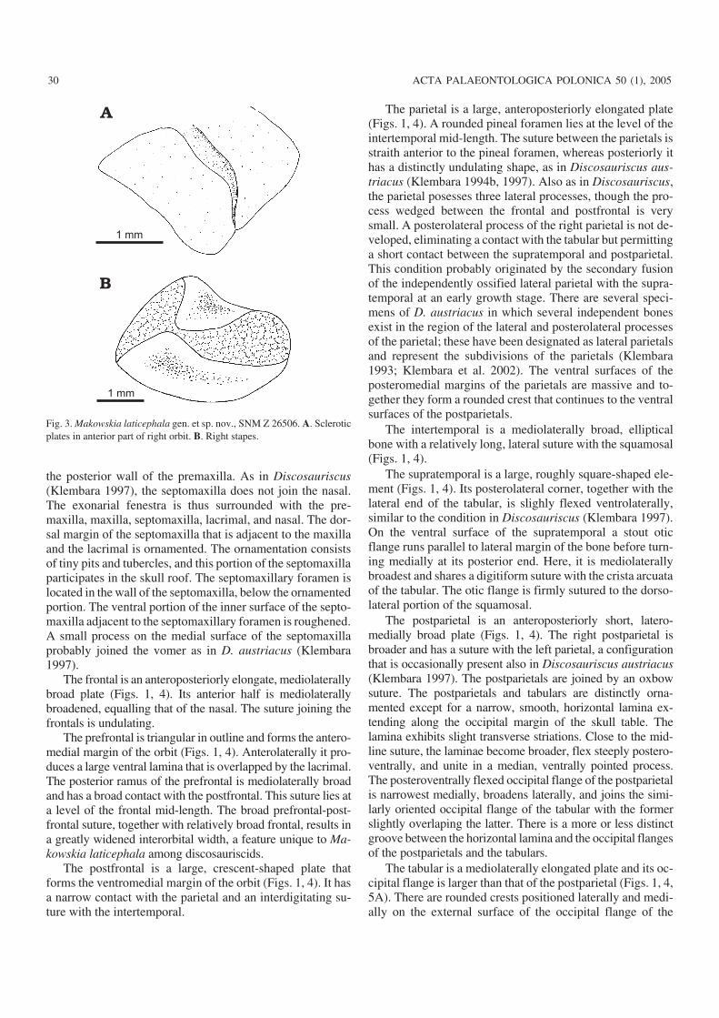
Fig. 3. Makowskia laticephala gen. et sp. nov., SNM Z 26506. A. Scleroticplates in anterior part of right orbit. B. Right stapes.

tabulars and postparietals. The crests surround an elliptical,slightly roughened depression, indicating the place of inser−tion of the cervical muscles. The right tabular process is aprolonged plate, whereas the left is a horn−like structure, andthe dorsal surfaces of both processes are smooth. The ventral
surface of both tabulars is well preserved and is basicallyidentical to that of Discosauriscus austriacus (Klembara1997). The crista arcuata is high and, its lateral surface, aswell as the surface between it and the lateral margin of thetabular, are roughened. The anterior half of the surface of the
http://app.pan.pl/acta50/app50−025.pdf
KLEMBARA—NEW DISCOSAURISCID SEYMOURIAMORPH TETRAPOD 31
5 mm
5 mm
Fig. 4. Makowskia laticephala gen. et sp. nov., SNM Z 26506. Restoration of skull in dorsal (A) and left lateral (B) views.

tabular process bears a deep sulcus, and immediately anteri−orly to it is a deep pit. Anterior to the pit and medially to thecrista arcuata there is a distinct elevation bearing a system ofgrooves, pits and, small crests at its surface.
An almost complete right maxilla and a fragment of theleft one are preserved. The maxilla is a long, narrow bone,reaching a level slightly behind the mid−length of the orbit(Figs. 1, 2, 4, 5). Not all teeth are preserved, but on the basisof their spacing the total number can be estimated to be about25. Contrary to the condition in Discosauriscus (Klembara1997) and Ariekanerpeton (Ivakhnenko 1981; Bulanov2003), the maxilla is the highest at its mid−length. At its ante−rior end the incision forming the margin of the exonarialfenestra is very shallow, whereas in Discosauriscus this por−tion of the maxilla is more deeply incised (Klembara 1997).The inner surface of the maxilla is not well−preserved, but thepresence of the broad horizontal lamina is recognizable.
Most of the right lacrimal is preserved (Figs. 1, 4). Itspreorbital part is quadrangular, whereas posteriorly it ex−tends into the suborbital or jugal process that forms theanteroventral margin of the orbit. The jugal process is rela−tively long and dorsoventrally flattened. Its posteriormostsection is broad, which corresponds to the broad articularportion of the suborbital ramus of the jugal.
The left jugal is well−preserved, forming a stout bone oftriangular shape (Figs. 1, 4). In contrast to Discosauriscus(Klembara 1977) and Ariekanerpeton (Ivakhnenko 1981;Laurin 1996a; Bulanov 2003) the suborbital ramus is rela−tively short and unusually broad dorsoventrally. A broad,anteroventral lamina of the suborbital ramus overlapped asimilarly broad, sutural scar on the jugal ramus of the lacri−mal. It is readily visible in lateral view that the suture be−tween the ornamented surfaces of the jugal and lacrimal isrelatively long, and is directed anterodorsal−posteroventral.The dorsal process of the jugal has the form of a blunt point,similar to that of the postorbital. The suture separating thetwo bones is very short. The posterior process of the jugal ispointed, and its posteroventral margin has a short suture withthe quadratojugal.
The postorbital is a triramous structure; the dorsomedialand ventrolateral rami are of about the same length, whereasthe posterior ramus is very short (Figs. 1, 4). The orbital mar−gin is slightly elevated dorsally. The inner surface of thepostorbital is smooth and as a such is continuous with thesimilar surfaces of the postfrontal and prefrontal. The ventro−lateral jugal ramus is anteroposteriorly very narrow. Thesame is true also for the dorsomedial ramus, although it isslightly broader anteroposteriorly and shorter mediolaterally.
Both squamosals are present, although not completelypreserved (Figs. 1, 4). On the right squamosal both the orna−mented area and the smooth otic flange are partially pre−served. On the left squamosal it is observable that the oticflange is much broader than in Discosauriscus (Klembara1997). The morphology of the lateral margin of the supra−temporal indicates that its suture with the squamosal reachedabout the mid−length level of the supratemporal.
Only the posterior portion of the left quadratojugal is pre−served (Figs. 1, 4, 5A). A sulcus is present on the posteriormargin of the smooth rounded flange. This indicates the pres−ence of the paraquadrate foramen in a similar position to thatin Discosauriscus (Klembara 1997). The inner surface of thisportion of the quadratojugal is roughened, indicating theplace of the articulation with the cartilaginous quadrate.
Of the sclerotic ring, only two partially articulated plates inthe anterior part and three articulated sclerotic plates posi−tioned in the posteromedial part of the right orbit are preserved(Figs. 1, 3A). Each sclerotic plate is of quadrangular shape.The plates are arranged in such a manner that each plate over−laps or fits into depression of the preceding plate. On the basisof the size of the remaining plates, it is possible to estimate theoriginal number of plates in each sclerotic ring at about 40.
Palate.—The vomer is an elongated plate and forms thewhole medial margin of the exochoanal fenestra (Figs. 2,5A). The anterior thirds of both vomers meet in a shortmidline suture. The posterior portion of the vomer forms alarge lamella that, as in D. austriacus (Klembara 1997: fig.26), is ventrally overlapped by the anterior portion of thepterygoid. The vomer has a short suture with the antero−medial process of the palatine. At about the mid−length of thevomer two tusks are present. Anterior to the tusks and alongthe exochoanal margin are several small teeth.
The right palatine is relatively well−preserved (Figs. 2,5A). It is subrectangular in outline and forms the concavemargin of the posterior border of the exochoanal fenestra.The palatine is about the same length as the ectopterygoid.The suture with the ectopterygoid is straight. The palatine isnotched posterolaterally, but the state of preservation in thisarea does not permit the unambiguous determination of thepresence or absence of the suborbital fenestra, as in Disco−sauriscus (Klembara 1997). In the skull of Makowskia lati−cephala the suborbital fenestra is restored on the basis of theconditions in Discosauriscus (Klembara 1997). At about thelevel of the mid−length of the palatine two tusks are present.
The right ectopterygoid is almost completely preserved,but is visible only in dorsal view (Figs. 1, 5A). It is asubrectangular plate wedged between the maxilla and jugallaterally and the pterygoid medially and posteromedially.The posterolateral corner of the ectopterygoid is obliquelyangled.
The pterygoid is the largest palatal bone and all its fourparts—palatal ramus, transverse process, central regionaround the basicranial (articular) fossa, and quadrate ramus—are well−preserved (Figs. 2, 5A). The palatal ramus ismediolaterally broad and its medial margin is almost straight.Immediately anterior to the basicranial articulation the me−dial margin bears a small notch. Both rami met probably attheir anterior tips. There is a noticeable difference in themediolateral width of the palatal rami. The subrectangulartransverse process is well−developed. Its anterior margin isslightly oblique and runs in anteromedial−posterolateral di−rection. In contrast, the anterior and posterior margins are
32 ACTA PALAEONTOLOGICA POLONICA 50 (1), 2005
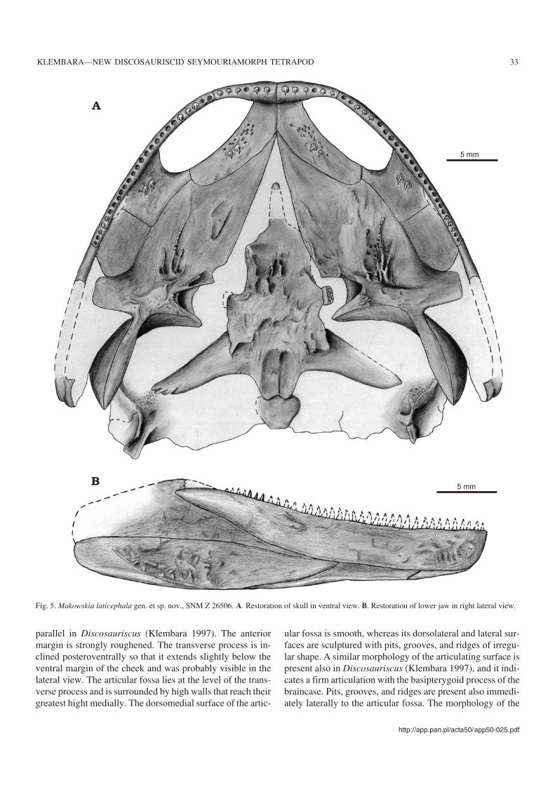
parallel in Discosauriscus (Klembara 1997). The anteriormargin is strongly roughened. The transverse process is in−clined posteroventrally so that it extends slightly below theventral margin of the cheek and was probably visible in thelateral view. The articular fossa lies at the level of the trans−verse process and is surrounded by high walls that reach theirgreatest hight medially. The dorsomedial surface of the artic−
ular fossa is smooth, whereas its dorsolateral and lateral sur−faces are sculptured with pits, grooves, and ridges of irregu−lar shape. A similar morphology of the articulating surface ispresent also in Discosauriscus (Klembara 1997), and it indi−cates a firm articulation with the basipterygoid process of thebraincase. Pits, grooves, and ridges are present also immedi−ately laterally to the articular fossa. The morphology of the
http://app.pan.pl/acta50/app50−025.pdf
KLEMBARA—NEW DISCOSAURISCID SEYMOURIAMORPH TETRAPOD 33
5 mm
5 mm
Fig. 5. Makowskia laticephala gen. et sp. nov., SNM Z 26506. A. Restoration of skull in ventral view. B. Restoration of lower jaw in right lateral view.

ventral surface of the palatal ramus is similar to that inDiscosauriscus austriacus, but dissimilar to that in D.pulcherrimus (Klembara 1997). In Makowskia laticephalaridges alternating with deep grooves diverge anteriorly fromthe central region of the pterygoid. The ridges are the highestin the central region, becoming reduced toward the peripheryof the palatal ramus. The pattern and extent of the ridges dif−fers between the right and the left pterygoids. The highestridges bear a row of small denticles, but this also differs be−tween the pterygoids. The central region of the pterygoid ismassive and bears pits and irregular grooves and ridges,which continues also onto the transverse process. Thequadrate ramus is nearly vertical, and its anterior portion, theascending lamina, is high and subquadrangular in shape. It istransversely oriented and its dorsal margin lies close to theventral surface of the skull roof. The basal portion of theascending lamina is vaulted anteriorly.
The parasphenoid is well−preserved, only the cultriformprocess is incomplete and the left posterolateral process isbroken in several places (Figs. 2, 5A). Preserved parts of thecultriform process show that it was short and roundedslightly anteriorly. The posterior parasphenoid plate is largeand forms a high, heavily sculptured, triangular wedge−likeprocess anterior to the level of the basipterygoid processes.The sculpture consists of high ridges bordering deep furrowsand pits. Immediately posteriorly to the wedge−like processthe parasphenoid plate is also sculptured, but with broad,shallow depressions divided by low rounded ridges. The par−allel lateral margins of the ventral parasphenoid plate aresharp and slightly flexed lateroventrally. Posteriorly they ex−tend onto the ventral surface of the posterolateral processesto form the ventrolateral crests or basal tubera. The postero−lateral processes of the parasphenoid plate are long and taperslightly laterally. The posteriormost portion of the para−sphenoid plate forms two processes adjacent to the midline,the posterior parts of which underlied the anterior margin ofthe ventral surface of the basioccipital. Both processes bearanteroposteriorly oriented elliptical depressions divided by anarrow and sharp ridge. The depressions probably representthe sites of muscle insertions.
Ossifications of palatoquadrate and stapes.—The quadrateis not ossified, and the epipterygoids are not represented. Insimilarly sized specimens of Discosauriscus austriacus, theepipterygoid is already partially ossified (Klembara 1997).The stapes is crescentic shaped (Figs. 2, 3B). In its shape andgrade of ossification the stapes duplicates that in similarlysized Discosauriscus specimens (Klembara 1997), except inbeing slightly more robust. The distal end of the stapes is nar−rowed, whereas the proximal end broadens and probably con−tinued as two cartilaginous processes, as in Discosauriscus(Klembara 1997). The position of stapes relative to the adja−cent cranial structures was probably identical to that in Disco−sauriscus.
Neural endocranium.—Only the basipterygoid processesof the basisphenoid are preserved (Figs. 2, 5A). They lie at
the level of the posterior portion of the anterior wedge−likeprocess of the parasphenoid. The dorsal and lateral marginsof the basipterygoid process are covered by smoothly fin−ished bone, whereas the distal margins of the processes areunfinished, indicating cartilaginous continuation.
The basioccipital is almost completely preserved, al−though it is broken along in its mid−width and the right side isslightly displaced dorsal to the left side (Figs. 2, 5A). Thebasioccipital forms a dorsoventrally thin quadrangular platewith anterolaterally rounded corners. In Makowskia lati−cephala the basioccipital is broad anteriorly and its lateralmargins converge posteromedially. Its anterior margin isslightly anteriorly convex, whereas the posterior margin isstraight and about half of the length of the anterior margin.The ventral surface of the basioccipital is smooth and lacksthe posteromedially converging ridges typical of that ofDiscosauriscus (Klembara 1997). The shape of the basi−occipital of Makowskia is very similar to that in Utegenia,however, it is slightly broader in the former species.
The exoccipital is not preserved.
Lower jaw.—Only the incomplete right lower jaw is pre−served (Figs. 2, 5B). The individual bones are more or lessarticulated and best exposed in lateral and ventral views,whereas those of the medial surface are poorly preserved.The relative size of the preserved bones, as well as their inter−vening sutures, are very similar to those in Discosauriscusaustriacus (Klembara 1997). The lower jaw is deepest in theregion of the anterior end of the surangular.
The dentary is a very long, narrow plate that is broadestanteriorly and narrows gradually to a pointed end posteriorly.Ventrally it joins the splenial and postsplenial, whereas pos−teriorly it overlaps laterally the anterior portion of the angu−lar. There is a large, anteroposteriorly elongated fenestra be−tween the dentary and splenial on the medial surface of thejaw, as in Discosauriscus austriacus (Klembara 1997). Onlysome of the posteriormost teeth are preserved.
The surangular is a large plate, but its posterodorsal por−tion is missing and its anterodorsal portion is only partiallypreserved. The anterior margin of the surangular extends al−most perpendicularly and indicates a complicated suture withthe posterior coronoid. The posterior end of the dentary liesslightly below this surangular portion, indicating that thesurangular−posterior coronoid suture should be visible in lat−eral view, as in Discosauriscus austriacus (Klembara 1997).The coronoids are not preserved.
The splenial is dorsoventrally narrow in lateral view andits ventral surface, together with those of the postsplenial andangular, form a continuous trough−like channel along theventral margin of the jaw. As in D. austriacus (Klembara1997), the border between the external ornamented and ven−tral smooth surfaces of all these three bones is demarcated bya sharp and distinct edge. The anteriormost end of the sple−nial forms the ventral portion of the symphysis. The posteriorportion of the splenial overlaps laterally the posterior end ofthe postsplenial.
34 ACTA PALAEONTOLOGICA POLONICA 50 (1), 2005

In lateral view the postsplenial is dorsoventrally narrow,but dorsally higher than the splenial. Externaly the anteriorend of the postsplenial inserts between the splenial and den−tary, but the suture with the latter is much longer. The poste−rior end of the postsplenial underlies the anterior portion ofthe angular.
The angular is a long and massive plate, whose anteriorend narrows and inserts between the postsplenial and denta−ry. The external surface of the angular is ornamented, withthe ventral margin of ornamentation forming a distinct, ven−trally convex edge.
Only small fragments of the prearticular are preservedand the articular was cartilaginous.
Dentition.—The teeth are preserved on both premaxillae,maxillae, and vomers and the right palatine; small denticlesare preserved on the vomers and pterygoids (Figs. 1, 2, 4B,5). Several posteriormost teeth are present on the dentary andare cylindrical with slightly posteriorly recurved, sharplypointed crowns. The bases of one anterior maxillary and oneof the largest premaxillary teeth bear slightly elongated, finestriations indicating the initial stage of infolding of the den−tine. None of the other preserved teeth bears the traces ofbasal striations. There are six teeth on each premaxilla (sev−eral of them are broken). Most of the maxillary teeth are notpreserved, but the number of tooth positions indicates acount of about 25 teeth. The number of visible palatal tusks isas follows: two on each vomer and two on the right palatine.The ectopterygoid is not exposed in ventral view. Smallpointed denticles are present on the ventral surface of thevomer anterior to the tusks, whereas further posteriorly thedenticles are aligned in a row lying laterally to the tusks andmarginal to the internal margin of the exochoanal fenestra.Similar small denticles are present also on the ridges on theventral surface of the palatal ramus of the pterygoid. Thedenticles are aligned in the rows on the crests of ridges. Onlya few denticles were present on the right pterygoid, as indi−cated by several small tooth positions on the surface of theridges. Such variation in the number of the denticles on theright and left sides are observable also in Discosauriscus(Klembara 1997).
Reconstructed skull.—The skull width exceeds the skulllength by about 27%, and the short preorbital length is onlyabout 23% of the skull length (Figs. 4, 5). The nasals areequally long as broad, which is unique within the familyDiscosauriscidae. The lateral portions of the nasal and theprefrontal are slightly flexed ventrally and aligned with theangle of the adjacent portion of the cheek. As a consequence,this portion of the cheek is morphologically divided from theadjacent dorsal surface of the skull roof by an edge directedanteromedially−posterolaterally. The prefrontal−postfrontalsuture is broad. This, together with the relatively broadfrontals, accentuates the interorbital width. The inter−temporal, supratemporal, and tabular bones are medio−laterally very broad. This is true also for the parietal and thusfor the whole parietal table. In dorsal view the skull has a
semielliptical shape with the long axis along the midline. Theorbits lie in the posterior portion of the anterior half of theskull. They are of oval shaped and face dorsolaterally. Thepineal foramen is relatively large and lies at the level slightlyposterior to the posterior orbital margins. The jaw joint lies atthe level of the supratemporal−tabular suture. The cheekmakes an angle of about 60° with the parietal table. The oticnotch is anteroposteriorly deep and dorsoventrally broad.The suborbital part of the cheek is very high, contrary toother discosauriscids (Klembara 1997; Klembara and Ruta2004a). The posterolateral part of the transverse process ofthe pterygoid is flexed posteroventrally and most probablyextended below the ventral cheek margin at the level of thejugal−quadratojugal suture. The posterolateral corner of thesupratemporal and the lateralmost portion of the tabular areslightly flexed ventrally, similarly to those in Discosauriscus(Klembara 1997) and Seymouria sanjuanensis (Laurin1995). The parietal table−cheek articulation is firm, as in allother seymouriamorphs. The palate is not closed, a conditioncorresponding to the similar−sized ontogenetic stages of D.austriacus (Klembara 1997). The lower jaw was deepest atthe level of the posterior coronoid−surangular suture.
Postcranial skeletonA partial anterior vertebral column with ribs, endoskeletal anddermal pectoral elements and both humeri are preserved inone block (Fig. 6). In addition, the following bones are alsopresent in a more or less disarticulated state: several presacralvertebrae, a short series of caudal vertebrae, right ischium, fe−mur, fibula, left tibia, and one phalanx (Figs. 6–9).
Vertebrae.—A few vertebrae are relatively well−preserved,and their individual structures are easily recognizable. Onewell−preserved neural arch lies close to the fourth rib (Fig.6A) and comparison with the vertebral column of Disco−sauriscus austriacus (Klembara and Bartík 2000) indicatesthat it is probably the 4th presacral neural arch. Its anatomy issimilar to that in D. austriacus. Several other preserved ver−tebrae have slightly swollen neural arches halves, whichwere joined in the median plane via cartilage in life (Fig. 7A).In similarly large specimens of D. austriacus, vertebrae withsuch morphology are present in the posterior half of thepresacral vertebral column.
One isolated trunk pleurocentrum and several caudalpleurocentra are present (Fig. 7B1, B2, C1). The isolatedpleurocentrum forms a complete ring. A distinct parapo−physis is present in the anteroventral portion of its lateralwall. In similar−sized specimens of D. austriacus the parapo−physes are preserved on first caudal pleurocentra, whichform completely ossified rings (Klembara and Bartík 2000:fig. 2). Immediately behind this pleurocentrum is a com−pletely ossified intercentrum (Fig. 7B). It is a bilaterally ex−panded plate with slightly dorsally curved lateral margins.The neural arches of the caudal vertebrae are flexed posteri−
http://app.pan.pl/acta50/app50−025.pdf
KLEMBARA—NEW DISCOSAURISCID SEYMOURIAMORPH TETRAPOD 35

36 ACTA PALAEONTOLOGICA POLONICA 50 (1), 2005
torus interclavicle
3rd rib
4th neural arch
5th rib
scapula
insertion oflatissimus dorsimuscle
ectepicondyle
humerus
scapula
cleithrum
humerus
clavicle
2nd rib
claviclescapula
interclavicle
cleithrum
coracoid
ribs
neural arch
clavicle
torus
scales
cleithrum
scapula
deltopectoralcrest
humerus
entepicondylar foramen
entepicondyle
4th rib
5 mm
Fig. 6. Makowskia laticephala gen. et sp. nov., SNM Z 26506. A. Anterior portion of postcranial skeleton in dorsal view. B. The same skeleton in ventral view.

orly (Fig. 7C1) and have a similar shape as those in D.austriacus. Several haemal arches are preserved (Fig. 7C).One is almost completely preserved, and it lies posterior tothe last caudal rib in the sequence of vertebrae (Fig. 7C2). Itshaemal canal is large, and its distal end is unfinished,indicating a cartilaginous continuation.
Ribs.—On the basis of conditions in Discosauriscusaustriacus (Klembara and Bartík 2000: fig. 13) it may be in−ferred that the series including the second to fifth ribs is pre−served in Makowskia laticephala (Fig. 6). The second rib is aslender oval rod in cross−section. Its unfinished distal end isnot broadened. Its proximal end, although partially damaged,is anteroposteriorly expanded and indicates the presence ofboth a capitulum and tuberculum. The third rib is relativelymassive with a slightly flattened shaft. Its distal end is unfin−ished and slightly broadened. At its proximal end a distinctcapitulum and tuberculum are present. The fourth and fifthribs are dorsoventrally flattened, and their proximal and dis−tal ends are unfinished. However, their distal ends are dis−tinctly anteroposteriorly broadened, and they extend into thehook−like, posteriorly directed processes. A similar morphol−ogy of the corresponding ribs is present also in Seymouriabaylorensis (White 1939: fig. 14), although the ribs have rel−atively short shafts. Presently, it is not possible to determine,whether the length of the shaft changes during ontogeny inMakowskia. In Discosauriscus, Ariekanerpeton, and Ute−genia the distal portions of the corresponding ribs arebroadened equally anteriorly and posteriorly.
Several caudal ribs are also present, and they correspondto the fourth to sixth caudal ribs of Discosauriscus austriacus(Klembara and Bartík 2000) (Fig. 7C2). The first two are longand approximately circular in cross−section. Their proximalends bear distinct capitular and tubercular portions. The thirdrib is probably the last caudal rib, as it is smallest of the series,and the original sequence of vertebrae is preserved. In addi−tion, the vertebrae with haemal arches first appear posteriorto the last rib. In contrast to the conditions of D. austriacus,the whole shaft of this rib is flattened and anteroposteriorlybroad. Its proximal end is broadened and the capitular and tu−bercular portions are only partially divided. The distalportions of all three ribs are unfinished.
Pectoral girdle and forelimb.—The interclavicle consistsof a large anterior plate and a long posterior stem (Figs. 6B,8A). In contrast to Discosauriscus, the anterior plate is trans−versely very broad, and its anterior margin bears digitiformprocesses. The external (ventral) surface of the plate, not cov−ered by clavicles, is triangular in outline and slighly orna−mented. The ornamentation consists of low ridges and shal−low grooves riadiating from the slightly elevated central partof this triangular portion. Except for the posteriormost por−tion, the posterior stem is well−preserved. The stem is longand its anteriormost section is bilaterally narrow. Furtherposteriorly the stem gradually broadens before again narrow−ing. This shape of the posterior stem is unique within Sey−mouriamorpha.
Both clavicles are present and complete except for lack−ing the dorsal tips of their ascending processes (Fig. 6). Theventral plate of the clavicle is elliptical in outline. Its anteriormargin bears small pointed processes, which are recordedalso in Discosauriscus austriacus (Klembara and Bartík2000: fig. 19b). The lateral portion of the ventral plate is mas−sive, and on its external side, at the transition to the ascendingprocess, there is a blunt torus. The ventral portion of the as−cending process is massive and anteroposteriorly broad. Theascending process gradually narrows distally and the anteriormargin of its external surface forms a sharp edge.
Both cleithra are preserved (Fig. 6). The cleithrum is longand narrow and is hollowed along its entire length. Whereasno other features are recognizable, its general shape is verysimilar to that in Discosauriscus austriacus (Klembara andBartík 2000).
The scapula is crescent−shaped, consisting of a large platewith a massive supraglenoid buttress that is triangular inshape (Fig. 6). With the exception of the anterior margin, allother margins are unfinished. The supraglenoid fossa is deep,and the supraglenoid foramen remains open ventrally. A sim−ilar degree of ossification of the scapula is seen in compara−bly sized individuals of Discosauriscus austriacus (Klem−bara and Bartík 2000). On the internal surface of the left scap−ula is a well−developed subscapular fossa.
Both coracoids are present, but are exposed only in exter−nal view, revealing a mostly scaled−covered surface (Fig.6B). The coracoid is a subquadrangular plate with slightlyrounded corners. The posterior margin of the left coracoid isof unfinished bone.
A complete right humerus and a proximal portion of theleft are preserved (Fig. 6). The morphology of the humerus isin general very similar to that in Discosauriscus austriacus(Klembara and Bartík 2000). Both ends of the humerus areunfinished and most of the structures were cartilaginous. Theproximal articular surface extends onto the massively devel−oped, L−shaped deltopectoral crest. The shaft is relativelybroad, entepicondyle is very short, and the entepicondylar fo−ramen is not enclosed distally. In most of the similarly sizedspecimens of D. austriacus this foramen is already closed(Klembara and Bartík 2000). However, this may also reflectthe individual variability in the rate of ossification of thepostcranial bones, which occurs in D. austriacus. The ectepi−condyle is distinctly developed, exhibiting a massive crest atits distal end and a well−developed process on its proximalend for the insertion of the latissimus dorsi muscle. The distalprocess is joined with the proximal articular surface of thehumerus via a narrow strip of bone. The process for the inser−tion of the subcoracoscapularis muscle is robust, whereas thesupinator process is not developed.
Pelvic girdle and hind limb.—Of the pelvis, only the ante−rior half of the right ischium is present (Fig. 9A). Its morphol−ogy is similar to that in Discosauriscus austriacus (Klembaraand Bartík 2000) except in being much broader medio−laterally.
http://app.pan.pl/acta50/app50−025.pdf
KLEMBARA—NEW DISCOSAURISCID SEYMOURIAMORPH TETRAPOD 37

Most of the right femur is preserved (Fig. 7C1). Its cres−centic−shaped proximal articular surface is of unfinishedbone, as is the margin of the well−developed internal tro−chanter. The intertrochanteric fossa is deep.
The right fibula is compressed (Fig. 7C1), but its generalshape is similar to that in Discosauriscus austriacus (Klem−bara and Bartík 2000: fig. 26).
The left tibia is well−preserved (Fig. 9B). Its proximal endis more robust than the distal end, and both end in unfinishedbone. The proximal articulating surface is mediolaterally ex−panded, whereas the distal articulating surface is oval in out−line. The lateral surface of the tibia is concave and bears twoprolonged crests separated by a depression between them.These features of the lateral wall of the tibia are not present inDiscosauriscus austriacus (Klembara and Bartík 2000).
IntegumentSeveral rows and isolated scales are well−preserved (Fig.6B). They are preserved adhering to the external surface ofsome elements of the pectoral girdle, particularly the inter−clavicle and the coracoids. The scales are oval in outline. Thediameter of the largest scales varies from 1 to about 1.2 mm.
In comparisons with the scales of Discosauriscus austriacus(Klembara and Bartík 2000), those of Makowskia latice−phala are more massive. The external surfaces of the scalesare ornamented by concentric elevations separated bygrooves of varying depth and width. In addition to the con−centric grooves, there are also radially diverging grooves, sothat the surface of the scale is divided into small, sub−quadrangular fields. On the internal surface of the scale, therelatively high concentric ridges are observable. Betweenthem, the fine concentric grooves are present. Fine groovesrun also radially. As a whole, a similar morphology of the ex−ternal and internal surfaces is present also in D. austriacus(Klembara and Bartík 2000: fig. 16c, d).
DiscussionComparisons with discosauriscids and other seymouri−amorphs.—The comparisons within the seymouriamorphsbelow include all ?Upper Carboniferous–Lower Permian taxaknown to date: Utegenia shpinari Kuznetsov and Ivakhnenko,1981 (Kuznetsov and Ivakhnenko 1981; Ivakhnenko 1987;Ivakhnenko et al. 1997; Laurin 1996b; Bulanov 2003; Klem−bara and Ruta 2004a, b), Seymouria baylorensis Broili, 1904
38 ACTA PALAEONTOLOGICA POLONICA 50 (1), 2005
2 mm intercentrumpleurocentrum
2 mmparapophysis
5 mm
femur
fibula
neural arch
haemal arch
pleurocentrum
haemal arch
last caudal ribcaudal rib
5 mm
rib
Fig. 7. Makowskia laticephala gen. et sp. nov., SNM Z 26506. A. Neural arch from the posterior half of the presacral vertebral column in dorsal view.B. Pleurocentrum and intercentrum from anterior section of caudal vertebral column in posterolateral (B1) and right lateral (B2) views. C. Femur, fibulaand anterior section of caudal vertebral column in left lateral view (C1), selected postcranial elements of the same section of vertebral column in right lat−eral view (C2).

(White 1939; Laurin 1996a), S. sanjuanensis Vaughn, 1966(Berman et al. 1987, 2000; Berman and Martens 1993; Laurin1995, 1996c), Ariekanerpeton sigalovi (Tatarinov, 1968) (Iva−khnenko 1981, 1987; Ivakhnenko et al. 1997; Laurin 1996a;Bulanov 2003), Discosauriscus austriacus (Makowsky,1876) and D. pulcherrimus (Fritsch, 1879) (Klembara 1997;Klembara and Bartík 2000), and Makowskia laticephala gen.et sp. nov. The comparisons do not include the Upper Permianforms from Russia, the kotlassiids and leptorophids, attributedby some authors to Seymouriamorpha (e.g., Ivakhnenko1987; Bulanov 2003).
Makowskia laticephala gen. et sp. nov. is included in thefamily Discosauriscidae on basis of the following skeletaland integumentary characters:
(1) Short preorbital region. Makowskia is a typical short−snouted discosauriscid. The skulls of Discosauriscus austria−cus and Ariekanerpeton are also short−snouted, being about aslong as there are broad; the skull of D. pulcherrimus is slightlylonger as broad. In Makowskia the skull is even broader.
(2) Orbits are positioned mainly in the posterior portionof the anterior half of the skull.
(3) Otic notch is dorsoventrally broad and antero−posteriorly deep. This results in the skull being high in thepostorbital region.
(4) Rounded to oval ventral scales.Those skull proportions that are displayed in all three
discosauriscid genera are in contrast with the conditions inthe members of two other seymouriamorph genera—Ute−genia and Seymouria in which:
(1) The preorbital region is long and also the whole skullis long.
(2) The orbits lie in the mid−length of the skull.(3) The otic notch is very shallow in Utegenia and its mor−
phology is similar to that in embolomeres (Klembara and Ruta2004a). In Seymouria, the otic notch is anteroposteriorly deep,but dorsoventrally narrow. In Utegenia and Seymouria thepostorbital region of the skull is thus relatively dorsoventrallylower.
(4) In Seymouria the ventral scales are absent. In Utegeniathe ventral scales are elongated and arranged in the chevron−like pattern. Such an arrangement is present in embolomeres(e.g., Proterogyrinus scheelei, Holmes 1984).
Makowskia laticephala differs from both species of thegenus Discosauriscus (Klembara 1997; Klembara and Bartík2000) and Ariekanerpeton sigalovi (Ivakhnenko 1981, 1987;Ivakhnenko et al. 1997; Laurin 1996a; Bulanov 2003) in thefollowing characters:
(1) Nasal equally long as broad. In known size range ofMakowskia the nasal is longer than broad in Discosauriscusand Ariekanerpeton. The broad nasal, together with the wideinterorbital region, result in a very broad preorbital region inMakowskia.
(2) Broad interorbital region. The width of the frontal andthat of the prefrontal−postfrontal suture are together muchbroader than those in Discosauriscus and Ariekanerpeton.
(3) Prefrontal−postfrontal contact lies in level of frontalmid−length. This difference is only relative to the conditionin Discosauriscus pulcherrimus in which the prefrontal−post−
http://app.pan.pl/acta50/app50−025.pdf
KLEMBARA—NEW DISCOSAURISCID SEYMOURIAMORPH TETRAPOD 39
5 mm
Fig. 8. Interclavicles in ventral views of Makowskia laticephala gen. et sp. nov., SNM Z 26506 (A) and Discosauriscus austriacus (specimen KO 65) (B).

frontal contact lies at the level of the anterior third of thefrontal.
(4) Maxilla is deepest at mid−length. The maxilla in Disco−sauriscus and Ariekanerpeton is deepest in its anterior portion.
(5) A relatively short, dorsoventrally broad suborbitalramus of jugal. In Makowskia the jugal is stout and togetherwith the stout suborbital ramus of the lacrimal produce a rela−tively high suborbital wall, and their long contact extends inan anterodorsal−posteroventral direction. A high suborbitalwall is not present in similarly sized specimens of Disco−sauriscus and Ariekanerpeton. In Discosauriscus and Arie−kanerpeton the jugal−lacrimal suture is relatively short be−tween the tips of both bones.
(6) Postorbital anteroposteriorly short and without anelongated, pointed posterior process. The postorbital in Ma−kowskia is relatively short compared to those in Discosauris−cus and Ariekanerpeton in which the postorbital forms a dis−tinct, posteriorly directed pointed process.
(7) Ventral surface of basioccipital smooth. In Disco−sauriscus and Ariekanerpeton the ventral surface of the basi−occipital carries strong oblique ridges running anterolaterallyto posteromedially, and delimiting a weakly concave depres−sion.
(8) Rows of small denticles placed on distinct ridges andintervening furrows radiate from place immediately laterallyto the articular portion on the ventral surface of the palatalramus of the pterygoid. This difference is relative only toDiscosauriscus pulcherrimus in which rows of small den−ticles radiate from the midline of the posterior half of the ven−tral surface of the palatal ramus of the pterygoid, and theridges are absent.
(9) Anterior margin of transverse flange of pterygoid ex−tends obliquely anteromedially−posterolaterally. In Disco−sauriscus and Ariekanerpeton the anterior and posterior mar−gins of the transverse flange are in parallel to one another.
(10) Relatively short and slightly rounded cultriform pro−
cess of parasphenoid. In Discosauriscus and Ariekanerpetonthe cultriform process is relatively longer and pointed.
(11) Ventral surface of posterior plate of parasphenoidheavily sculptured. This difference is relative only to Disco−sauriscus pulcherrimus and Ariekanerpeton. In D. pulcherri−mus the ventral surface of the parasphenoid plate is smoothand bears small denticles arranged in rows or scattered. InAriekanerpeton, the ventral surface of the parasphenoid plateis smooth.
(12) Distal ends of fourth and fifth presacral ribs distinctlyanteroposteriorly broadened, and extend into the hook−like,posteriorly directed processes. In Discosauriscus and Arie−kanerpeton the distal portions of the corresponding ribs arebroadened equally anteriorly and posteriorly.
(13) Shaft of ?last caudal rib flattened and antero−posteriorly broad. In Discosauriscus the lst caudal rib is nar−row. The caudal ribs are not preserved in Ariekanerpeton.
(14) Posterior stem of interclavicle narrows anteriorlyand posteriorly from broadened mid−length section. InDiscosauriscus and Ariekanerpeton the anterior half of theposterior stem is broader than its posterior half (Fig. 8). Thebroader, mid−length section is delimited by shallow lateralconstrictions.
Makowskia laticephala differs from both species of thegenus Discosauriscus, Ariekanerpeton sigalovi and otherseymouriamorphs—Utegenia shpinari (Kuznetsov and Iva−khnenko 1981; Ivakhnenko 1987; Ivakhnenko et al. 1997;Laurin 1996b; Bulanov 2003; Klembara and Ruta 2004a, b),and both species of Seymouria (White 1939; Berman et al.1987, 2000; Berman and Martens 1993; Laurin 1995, 1996c)in the following characters:
(1) Nasal equally long as broad. In Utegenia and Sey−mouria the nasal is always longer than broad.
(2) Broad interorbital region. The width of the frontal andthat of the prefrontal−postfrontal suture are together muchbroader than those in Utegenia and Seymouria. The pre−frontal−postfrontal suture is broad in similar−sized Utegeniaand larger Discosauriscus austriacus (Klembara 1997) andSeymouria sanjuanensis (Berman and Martens 1993), how−ever, the frontal is relatively narrow in all these forms. Theprefrontal−postfrontal suture is very short in the smallestknown S. sanjuanensis specimen (MNG 7859, skull lengthabout 20 mm) from Germany mentioned by Berman andMartens (1993) (personal observation). The length of the su−ture in this specimen correspons to that in similar−sized spec−imens of Discosauriscus, Ariekanerpeton, and Utegenia.
(3) Maxilla is deepest at mid−length. The maxilla in Ute−genia and Seymouria is deepest in its anterior portion.
(4) A relatively short, dorsoventrally broad suborbitalramus of jugal. A high suborbital wall is not present in simi−larly sized specimens of Utegenia and the juvenile specimenof S. sanjuanensis from Germany (Berman and Martens1993). In Utegenia the jugal−lacrimal suture is relativelyshort between the tips of both bones. In larger S. baylorensisand S. sanjuanensis specimens this suture is relatively long,but extends in an anteroventral−posterodorsal direction.
40 ACTA PALAEONTOLOGICA POLONICA 50 (1), 2005
5 mm
Fig. 9. Makowskia laticephala gen. et sp. nov., SNM Z 26506. Rightischium in ventral view (A), left tibia in anterior view (B).

http://app.pan.pl/acta50/app50−025.pdf
KLEMBARA—NEW DISCOSAURISCID SEYMOURIAMORPH TETRAPOD 41
OUTGROUPAcanthostegaIchthyostegaCrassigyrinusGreererpetonWhatcheeriaEoherpetonProterogyrinusArcheriaPholiderpetonGephyrostegusLimnoscelisUtegeniaAriekanerpetonD.austriacusD.pulcherrimusMakowskiaS. baylorensisS. sanjuanensisDiadectesTseajaiaEocaptorhinusPetrolacosaurusMicrobrachisSaxonerpetonDendrerpetonBalanerpetonEcolsoniaEryopsBaphetesMegalocephalusLethiscusSauropleura
OUTGROUPAcanthostegaIchthyostegaCrassigyrinusWhatcheeriaEoherpetonProterogyrinusArcheriaPholiderpetonGephyrostegusUtegeniaMakowskiaAriekanerpetonD. austriacusD. pulcherrimusS. baylorensisS. sanjuanensisDiadectesTseajaiaLimnoscelisEocaptorhinusPetrolacosaurusGreererpetonDendrerpetonBalanerpetonEcolsoniaEryopsBaphetesMegalocephalusLethiscusSauropleuraMicrobrachisSaxonerpeton
Dis
co
sa
uriscid
ae
OUTGROUPAcanthostegaIchthyostegaCrassigyrinusWhatcheeriaEoherpetonProterogyrinusArcheriaPholiderpetonGephyrostegusUtegeniaMakowskiaD. pulcherrimusAriekanerpetonD. austriacusS. baylorensisS. sanjuanensisDiadectesTseajaiaLimnoscelisEocaptorhinusPetrolacosaurusGreererpetonDendrerpetonBalanerpetonEcolsoniaEryopsBaphetesMegalocephalusLethiscusSauropleuraMicrobrachisSaxonerpeton
6351
9875
53
97
59
71
90
88
60
93
98
81
85
60
53
99
88
Seym
ouriam
orp
ha
76
Fig. 10. Phylogenetic analysis. A, B. Two of six most parsimonious trees recovered by PAUP* 40b10 from a heuristic search of 33 taxa and 150 characters.C. Bootstrap percentages on a 50% majority−rule consensus tree.

(5) Postorbital anteroposteriorly short and without anelongated, pointed posterior process. The postorbital is rela−tively short compared to those in Utegenia and juvenileSeymouria sanjuanensis in which the postorbital forms a dis−tinct, posteriorly directed pointed process.
(6) Anterior margin of transverse flange of pterygoid ex−tends obliquely anteromedially−posterolaterally. In Utegeniaand Seymouria sanjuanensis the anterior and posterior mar−gins of the transverse flange are in parallel to one another. InS. baylorensis (in the sense of White 1939) the margins areparallel one to another. However, according to Laurin(1996b), the lateral half of the anterior margin in S. baylo−rensis is redirected anterolaterally−posteromedially so as toform a wedge−like suture with the ectopterygoid.
(7) Relatively short and slightly rounded cultriform pro−cess of parasphenoid. In Utegenia and Seymouria the cultri−form process is relatively longer and pointed.
(8) Shaft of ?last caudal rib flattened and anteropos−teriorly broad. In Seymouria the last caudal rib is narrow andpointed. The caudal region of the vertebral column is notwell−preserved in Utegenia but several caudal ribs preservedare not broadened.
(9) Posterior stem of interclavicle narrows anteriorly andposteriorly from broadened mid−length section. In Utegeniathe stem is very broad anteriorly, then narrows posteriorly, butwith its posterior end slightly broadened. In Seymouriabaylorensis the stem is broadest anterorly, gradually narrowsposteriorly before slightly broadening at its posterior end(White 1939). In S. sanjuanensis, however, the posterior endof the stem (completely preserved in CM 28597) is not broad−ened, but is of the same width as the stem in the level of itsmid−length. Outlines of the stems of Utegenia and Seymouriabaylorensis are similar except for the stem of Utegenia beingrelatively short, with a length subequal to the anterior plate.
Phylogenetic analysis.—Phylogenetic analysis of ?UpperCarboniferous–Lower Permian seymouriamorphs has beenrecently made by Klembara and Ruta (2004b). As well asseymouriamorphs, their analysis includes also several otherearly tetrapods, 31 taxa in total and one hypothetical, all−zerooutgroup. The taxa were coded for 150 morphologicalcharacters.
Here I included Makowskia laticephala into the data ma−trix of Klembara and Ruta (2004b), and retained their 150characters; as for the discussion of topology of other taxathan those belonging into Seymouriamorpha, see Klembaraand Ruta (2004b). I have made several changes in the defini−tions of the characters here; these are as follows: (i) defini−tion of character 6 is emended; (ii) new states are added tocharacters 3, 47, and 56 (Appendix 1). Besides this, I haverecoded several characters of seymouriamorph taxa (Appen−dix 2). The data matrix was processed with PAUP* (v.40b.10; Swofford 2001) on Pentium PC and the obtainedtrees printed using TreeView (Page 1996). Characters are un−ordered and of equal weight. The matrix was analysed usingthe heuristic search algorithm and ACCTRAN optimization.
The result of analysis are six most parsimonious trees of 525steps (CI = 0.4019; RI = 0.6948; RC = 0.2793). In all sixtrees, Makowskia forms a sister taxon to Ariekanerpeton,Discosauriscus austriacus, and D. pulcherrimus. In three ofthem D. austriacus and D. pulcherrimus are sister taxa toAriekanerpeton (Fig. 10A), and in three other trees, Arie−kanerpeton and D. austriacus are sister taxa to D. pulcher−rimus (Fig. 10B). The strict consensus tree of six trees showsa trichotomy of Ariekanerpeton, D. austriacus, and D. pul−cherrimus. To resolve this trichotomy, additional materialespecially of the postcranial skeleton of Ariekanerpeton andD. pulcherrimus is required.
Contrary to the cladogram of Klembara and Ruta (2004b),Makowskia, Ariekanerpeton, Discosauriscus austriacus, andD. pulcherrimus form a clade, which is the sister taxon to bothspecies of Seymouria, and Utegenia is basal to both Sey−mouria species. A bootstrap analysis shows a strong supportfor separation of Discosauriscidae and Seymouriidae, and thenode linking Utegenia with Seymouriidae plus Discosaurisci−dae is also strongly supported (Fig. 10C).
AcknowledgmentsComments from Drs. David S. Berman (Carnegie Museum of NaturalHistory, Pittsburgh), Ralf Werneburg (Naturhistorisches Museum,Schleusingen), Andrew R. Milner (Birkbeck College, London), Stuart S.Sumida (California State University, San Bernardino), and one anony−mous referee considerably improved the quality of this paper. Figs. 3, 4,and 8A were drawn by Mr. Andrej Čerňanský (Comenius University inBratislava), and the photographs of Figs. 1A and 2A were made by Dr.Ján Kodada (Comenius University in Bratislava). Thanks are also ex−tended to Mrs. Zuzana Šulavíková (Comenius University in Bratislava)for the chemical preparation of the holotype of Makowskia and her assis−tance in the construction of wax−plasticine model of its skull. This projectwas supported in part by the Scientific Grant Agency, Ministry of Educa−tion of the Slovak Republic and the Slovak Academy of Sciences, GrantNo. 1/0018/03.
ReferencesAmalitzky, V.P. [Amalickij, V.P.] 1921. Seymouriidae, North Dvina Exca−
vations of Prof. V.P. Amalitzky [in Russian]. Petrogradskaâ AkademiâNauk 2: 1–14.
Augusta, J. 1948. Our present knowledge of the Stegocephali in the LowerPermian of Moravia [in Czech]. Přírodovědní sborník ostravskéhokraje (Opava) 9: 82–101.
Berman, D.S., Henrici, A.C., Sumida, S.S., and Martens, T. 2000. Redes−cription of Seymouria sanjuanensis (Seymouriamorpha) from the LowerPermian of Germany based on complete, mature specimens with a discus−sion of paleoecology of the Bromacker locality assemblage. Journal ofVertebrate Paleontology 20: 253–268.
Berman, D.S. and Martens, T. 1993. First occurrence of Seymouria (Am−phibia: Batrachosauria) in the Lower Permian Rotliegend of CentralGermany. Annals of Carnegie Museum 62: 63–79.
Berman, D.S., Reisz, R.R., and Eberth, D.A. 1987. Seymouria sanjuanensis(Amphibia, Batrachosauria) from the Lower Permian Cutler Formationof north−central New Mexico and the occurrence of sexual dimorphism
42 ACTA PALAEONTOLOGICA POLONICA 50 (1), 2005

in that genus questioned. Canadian Journal of Earth Sciences 24:1769–1784.
Broili, F. 1904. Permische Stegocephalen und Reptilien aus Texas. Palae−ontographica 51: 1–120.
Bulanov, V.V. 2003. Evolution and systematics of seymouriamorph para−reptiles. Paleontological Journal 37 (Supplement 1): 1–105.
Fritsch, A. 1879. Neue Übersicht der in der Gaskohle und den Kalksteinen derPermformation in Böhmen vorgefundenen Tierreste. Sitzungsberichte derköniglichen böhmischen Gesellschaft der Wissenschaften, Prague 1879:184–195.
Fritsch, A. 1883. Fauna der Gaskohle und der Kalksteine der Perm−formation Böhmens. Band I. 182 pp. Selbstverlag, Prague.
Holmes, R.B. 1984. The Carboniferous amphibian Proterogyrinus scheeleiRomer, and the early evolution of tetrapods. Philosophical Transac−tions of the Royal Society of London B 306: 431–527.
Ivakhnenko, M.F. [Ivahnenko, M.F.] 1981. Discosauriscidae from thePermian of Tadzhikistan [in Russian]. Paleontologičeskij žurnal 1:114–128.
Ivakhnenko, M.F. [Ivahnenko, M.F.] 1987. Permskie parareptilii SSSR. 160pp. Nauka, Moskva.
Ivakhnenko, M.F. [Ivahnenko, M.F.], Golubev, V.K., Gubin, Yu.M. [Gubin,Û.M.], Kalandadze, N.N. [Kalandadze, N.N.], Novikov, I.V., Sennikov,A.G., and Rautian, A.S. 1997. Permian and Triassic tetrapods of EasternEurope [in Russian]. Trudy Paleontologičeskogo Instituta Akademii Nauk268: 1–216.
Klembara, J. 1993. The subdivisions and fusions of the exoskeletal skullbones of Discosauriscus austriacus (Makowsky 1876) and their possi−ble homologues in rhipidistians. Paläontologische Zeitschrift 67:145–168.
Klembara, J. 1994a. Electroreceptors in the Lower Permian tetrapod Disco−sauriscus austriacus (Makowsky 1876). Palaeontology 37: 609–626.
Klembara, J. 1994b. The sutural pattern of skull−roof bones in Lower Perm−ian Discosauriscus austriacus from Moravia. Lethaia 27: 85–95.
Klembara, J. 1995. The external gills and ornamentation of skull roof bonesof the Lower Permian tetrapod Discosauriscus (Kuhn 1933) with re−marks to its ontogeny. Paläontologische Zeitschrift 69: 265–281.
Klembara, J. 1996. The lateral line system of Discosauriscus austriacus(Makowsky 1876) and the homologization of skull roof bones betweentetrapods and fishes. Palaeontographica A 240: 1–27.
Klembara, J. 1997. The cranial anatomy of Discosauriscus Kuhn, a sey−mouriamorph tetrapod from the Lower Permian of the Boskovice Fur−row (Czech Republic). Philosophical Transactions of the Royal Societyof London B 352: 257–302.
Klembara, J. and Bartík, I. 2000. The postcranial skeleton of DiscosauriscusKuhn, a seymouriamorph tetrapod from the Lower Permian of theBoskovice Furrow (Czech Republic). Transactions of the Royal Societyof Edinburgh, Earth Sciences 90 (for 1999): 287–316.
Klembara, J. and Meszároš, Š. 1992. New finds of Discosauriscus austriacus(Makowsky 1876) from the Lower Permian of the Boskovice Furrow(Czecho−Slovakia). Geologica Carpathica 43: 305–312.
Klembara, J. and Ruta, M. 2004a. The seymouriamorph tetrapod Utegeniashpinari from the ?Upper Carboniferous–Lower Permian of Kazakh−stan. Part. I: Cranial anatomy and ontogeny. Transactions of the RoyalSociety of Edinburgh, Earth Sciences 94 (for 2003): 45–74.
Klembara, J. and Ruta, M. 2004b. The seymouriamorph tetrapod Utegeniashpinari from the ?Upper Carboniferous–Lower Permian of Kazakh−stan. Part II: Postcranial anatomy and relationships. Transactions of theRoyal Society of Edinburgh: Earth Sciences 94 (for 2003): 75–93.
Klembara, J., Tomášik, A., and Kathe, W. 2002. Subdivisions, fusions and
extended sutural areas of dermal skull bones in Discosauriscus Kuhn(Seymouriamorpha). Neues Jahrbuch für Geologie und Paläontologie,Abhandlungen 223: 317–349.
Kuhn, O. 1933. Labyrinthodontia. In: W. Quenstedt (ed.), Fossilium Cata−logus, I. Animalia. Pars 84, 1–114. W. Jung, Den Haag.
Kuznetsov, V.V. [Kuznecov, V.V.] and Ivakhnenko, M.F. [Ivahnenko, M.F.]1981. Discosauriscids from the Upper Palaeozoic of South Kazakhstan [inRussian]. Paleontologičeskij žurnal 3: 102–110.
Laurin, M. 1995. Comparative cranial anatomy of Seymouria sanjuanensis(Tetrapoda: Batrachosauria) from the Lower Permian of Utah and NewMexico. PaleoBios 16: 1–8.
Laurin, M. 1996a. A reevaluation of Ariekanerpeton, a Lower Permianseymouriamorph (Tetrapoda: Seymouriamorpha) from Tadzhikistan.Journal of Vertebrate Paleontology 16: 653–665.
Laurin, M. 1996b. A reappraisal of Utegenia, a Permo−Carboniferousseymouriamorph (Tetrapoda: Batrachosauria) from Kazakhstan. Jour−nal of Vertebrate Paleontology 16: 374–383.
Laurin, M. 1996c. A redescription of the cranial anatomy of Seymouriabaylorensis, the best known seymouriamorph (Tetrapoda: Batracho−sauria). PaleoBios 17: 1–16.
Laurin, M. 2000. Seymouriamorpha. In: H. Heatwole and R.L. Carroll(eds.), Amphibian Biology, Volume 4, Palaeontology, 1064–1080. Sur−rey Beatty and Sons, Chipping Norton.
Makowsky, A. 1876. Über einen neuen Labyrinthodonten ‘Archegosaurusaustriacus nov. spec.’. Sitzungsberichte der keiserischen Akademie derWissenschaft 73: 155–166.
Page, R.D.M. 1996. TreeView: An application to display phylogenetic treeson personal computers. Computer Applications in the Biosciences 12:357–358.
Romer, A.S. 1947. Review of the Labyrinthodontia. Bulletin of the Museumof Comparative Zoology, Harvard College 99: 1–368.
Steen, M.C. 1938 On the fossil Amphibia from the Gas Coal of Nýřany andother deposits in Czechoslovakia. Proceedings of the Zoological Soci−ety of London B 108: 205–283.
Špinar, Z.V. 1952. Revision of some Moravian Discosauriscidae (Labyrintho−dontia) [in Czech]. Rozpravy Ústředního ústavu geologického 15: 1–115.
Swofford, D. L. 2001. PAUP*: Phylogenetic analysis using parsimony(*and other methods). Version 4.0b10. Sinauer Associates, Sunderland,Massachusetts.
Tatarinov, L.P. 1968. Upper Permian and Mesozoic amphibians and reptiles ofUSSR [in Russian]. In: N.N. Kalandadze, V.G. Očev, L.P. Tatarinov, P.K.Čudinov, M.A. Šiškin (eds.), Verhnepermskie i mezozoijskie zemno−vodnye i presmykaûŝiesâ CCCP, 73–92. Nauka, Moskva.
Vaughn, P.P. 1966. Seymouria from the Lower Permian of southeasternUtah, and possible sexual dimorphism in that genus. Journal of Paleon−tology 40: 603–612.
Watson, D.M.S. 1917. A sketch classification of the pre−Jurassic tetrapodvertebrates. Proceedings of the Zoological Society of London 1917:167–186.
Werneburg, R. 1989. Labyrinthodontier (Amphibia) aus dem Oberkarbonund Unterperm Mitteleuropas – Systematik, Phylogenie und Strati−graphie. Freiberger Forschungshefte, Reihe C 436: 7–57.
White, T.E. 1939. Osteology of Seymouria baylorensis Broili. Bulletin ofthe Museum of Comparative Zoology, Harvard College 85: 325–409.
Williston, S.W. 1911. A new family of reptiles of the Permian of New Mex−ico. American Journal of Sciences 31: 378–398.
Zhang, F., Li, Y., and Wan, X. 1984. A new occurence of Permianseymouriamorphs in Xinjiang, China [in Chinese, English abstract].Vertebrata PalAsiatica 22: 294–304.
http://app.pan.pl/acta50/app50−025.pdf
KLEMBARA—NEW DISCOSAURISCID SEYMOURIAMORPH TETRAPOD 43

Appendix 1
Data matrix
OUTGROUP0000000000 0000000000 0000000000 0000000000 0000000000 0000000000
0000000000 0000000000 0000000000 0000000000 0000000000 0000000000
0000000000 0000000000 0000000000
Acanthostega gunnari2000000000 00000000?? 00???00010 0?00000000 11001000?0 00000?0000
0000010000 0000000000 ?000000000 0000000000 0000000111 0000001100
0000000000 0000000000 00000?0000
Archeria crassidisca2000010000 10001010?? 2100101010 1?10100000 11101301?1 1111331???
???????01? 00?0?00000 00?2100001 0?01011111 1100111111 1220000111
1112200000 0010112101 0111?10001
Ariekanerpeton sigalovi0011010011 1122300101 2100101101 0111100011 2210111201 1122321001
1001110111 1111111110 0111211111 0112001111 1??0111111 ?1?011??11
11420101?0 21?1012100 0??1?????1
Balanerpeton woodi1000001021 1000000??? 1000000000 0111100000 22102200?0 1001310011
1011101110 0000100000 000?000001 ?100001010 0010111111 000001??11
1040100000 20001?2101 01020??000
Baphetes kirkbyi2100000000 0000010100 1000011000 0111000000 03100100?? 1101000011
1011101110 0000000000 ?000?01?00 ?????????? ?????????? ??????????
????0?0??? ???0002100 0?????????
Crassigyrinus scoticus1000000001 1000400101 0000000010 1?00000002 01101100?0 1100130000
1000100010 0000000000 ?00??00?01 ?000000011 0000000111 ?001000111
?01?10?00? 0000011000 00????0??1
Dendrerpeton acadianum1000001000 1000000100 1000000100 0211000000 12102200?0 1120300011
1011101110 0000000000 ?00?000001 ?100????10 ???01?1??? ?1000?????
??4010000? 20101?2100 110?0????0
Diadectes absitus2000010000 1000000100 001?101000 1211100010 1110014201 1012331102
1011101110 0001200000 00?1201001 020221??11 1001?11??1 000111????
0142010111 1120112100 0111011111
Discosauriscus austriacus0011010010 1122300101 2101101101 1111100011 2210012201 1122321001
1001110110 1111111110 0111211111 0112011111 1000111111 111011??11
1142010110 2121012101 0??110?000
D. pulcherrimus0001010020 1122300101 2101101101 1111101011 2210013201 1122351001
1001110110 1111111110 0111211111 0112011111 1000111111 ????11??11
1142010110 ???1012101 0??11?????
Ecolsonia cutlerensis10000011?? 1000000111 401?000000 0101000000 32002200?0 100133001?
?01??01??0 0000100000 000?001001 ??02011010 1010111111 010001??11
11?030000? 220011211? 010??????0
44 ACTA PALAEONTOLOGICA POLONICA 50 (1), 2005

Eocaptorhinus laticeps10000101?? 0020???11? 111??21?01 0111000010 00?1114211 1110031100
11011????0 1?0??00000 ?010??10?? 121211??11 1001?11??1 01?011????
0142010111 2200?????1 1111010111
Eoherpeton watsoni2000000000 1000401??? 1000100010 1?01000012 1110???0?? 11???31???
?0?0?0?0?0 0000000000 ???2?000?? ??01001111 ??00101111 1100????11
1??110000? 0010011101 0????????1
Eryops megacephalus2000001000 1000000111 3000000010 0?00000000 13102200?1 1001330011
1011101110 0000100000 0000001001 1?02011010 1010111111 010001????
1040100000 2210112110 01020?0001
Gephyrostegus bohemicus20000101?? 1?20001??? ?000100000 1111?00011 11101?01?? ??????0111
0011101110 00010????? ??0?201011 0002011111 1?00011001 100101??11
104100010? 0010012101 0111?10001
Greererpeton burkemorani1000100000 00000100?? 10??010000 0100000000 00001200?0 1110310001
1000100010 0000000000 0000000000 ?000001010 0000010011 000100010?
1020100000 2000011100 0101000000
Ichthyostega stensioei0000000000 00000000?? 00???00010 0?00000000 00000170?0 00003?0100
1100110010 0000100000 ?002100000 ??00001001 0000000111 0000000100
0040100000 0000010000 0100?????1
Lethiscus stocki0000110020 0000??0??? 101????00? 00?1100000 0000?1???? ??????????
?????????? ?????????? ?????????? ?0???????? ?????????? ??????????
??22?01??? ?????????? ?????10100
Limnoscelis paludis2000010000 0020000110 311?121001 0101000010 0010116210 1100351101
1101110110 0001200000 ?0?1?01011 021?????11 1?01??1??? ?001??????
??42010111 2120112100 0111?11111
Makowskia laticephala0021010010 1122300101 2101101101 1111101111 2211012201 1122321001
1001110110 0011111111 0111411111 ?11??1??1? ???01????? ??????????
?1?2010??? ???101?10? 0???1?????
Megalocephalus pachycephalus2100000000 0000010100 101?0?0000 0111000000 03100100?? 1101000011
1011101110 0000000000 ?000?????? ??00001111 0100111111 0010000100
00???????? ?????????? ??????????
Microbrachis pelikani0000010010 0020000??? 101?111?00 0001000010 00001110?0 1020131111
1111111111 1100200000 ?000201011 0?13001011 1000111111 010001??11
1012?00110 3000???101 10?3010001
Petrolacosaurus kansensis20000101?? 01210001?? 111?12100? 0111100010 00?011421? 1100101112
1111110110 1001200000 ?010201001 021111??11 1000111??1 000011????
0142010111 2000112101 1111010111
Pholiderpeton scutigerum2000011??? 10001010?? ??00?10010 1?100000?2 11101301?0 11??331101
1????00010 0000000000 ?002100001 ?001011111 1100111111 1220000011
10?220000? ???011??0? 01??010001
http://app.pan.pl/acta50/app50−025.pdf
KLEMBARA—NEW DISCOSAURISCID SEYMOURIAMORPH TETRAPOD 45

Proterogyrinus scheelei1000010001 10001010?? 1000101000 1?01010002 11101300?1 1111301???
?00110001? 00?0000000 0002100001 ?001?01?1? 0??0?111?1 12200???1?
1014100000 0010111101 0111?00001
Sauropleura pectinata/scalaris0000100010 10000000?? 111?100000 0001000000 00001150?0 1000331101
1100110010 0000?00000 ?00?000001 ?00310??11 0001?01??1 000001????
?142?01000 20000??110 01?2?10100
Saxonerpeton geinitzi20000101?? 0020000??? 111?1?1?01 0001000010 000011?0?0 1020131111
11111????0 0?0??00000 ?00?201011 0?13?????? ?????????? ??????????
??42??0110 20001??101 1102?10001
Seymouria baylorensis1000010010 1122300111 3101100101 1211100111 3210010201 1112331011
1011111110 00011?0010 ?1?1201111 1212011111 1000111111 ?11011??11
1142010111 0121012100 0?01110000
S. sanjuanensis1000010010 11223001?? 3101100101 1111100011 3210010201 1112331011
1011111110 000?111010 01?1201111 121??????? ?0?01????? ??????????
??42010111 ???1012100 0101???000
Tseajaia campi2000010000 0000000??? 211?001000 1011000010 121012421? 1?02331102
1011101110 0001200000 00??301101 02?2???111 1001?11?11 0001?1???1
1142010111 1120112100 0101011111
Utegenia shpinari1000010000 1012200101 2100101001 1111010012 1110111201 1122231001
1000110011 1111210000 01112?0111 001300??1? 1000111111 1???11??11
11320?0??0 21???????? ???1?????1
Whatcheeria deltae2000000000 1120??1??? 10000??0?0 1?00001000 0?10??0??? 1??133?0?0
?0?0?0?0?? ?????????? ????200101 ??000????? 0??00??111 0000?01011
1013100000 0000001100 01???????1
Appendix 2
Character list
For sources of each taxon, see Klembara and Ruta (2004b).
1. Position of mid−point of maximum anteroposterior orbit diameter:closer to tip of snout than to posterior end of skull (0); in the middleof skull length (1); closer to posterior end of skull than to tip ofsnout (2).
2. Absence (0) or presence (1) of antorbital vacuities.3. Skull longer than broad (0) as broad as long (1) or broader than long
(2).4. Preorbital region of skull less than twice as wide as long (0) or at
least twice as wide as long (1).5. Absence (0) or presence (1) of condition: nasals much shorter than
frontals (less than one−third as long as frontals).6. Total width of premaxillae more (0) or less (1) than two−thirds of
the width of the skull table.7. Absence (0) or presence (1) of distinct, triangular or digitiform, dor−
sal alary processes of premaxillae, widely separated by interveningnasals.
8. Presence (0) or absence (1) of prefrontal−postfrontal suture.9. Position of prefrontal−postfrontal suture relative to the mid−length
of the frontal: posterior (0); at the same level (1); anterior (2).10. Broad (0) or point−like (1) prefrontal−postfrontal contact.11. Postorbital medial margin straight for most of its length or very
gently curved (1), irregularly sinuous (0).12. Postorbital not broader than long (0) or broader than long (1).13. Postorbital without distinct ventrolateral (jugal) ramus (0), with in−
cipient ramus (1), with well−developed and elongate ramus (2).14. Postorbital without distinct dorsomedial ramus for postfrontal (0),
with incipient ramus (1), with elongate ramus (2).15. Absence of outgrowth from posterolateral ventral surface of tabular
(0), presence of outgrowth in the form of spike−like unornamentedcomponent (1), elongate and recurved blade (2), rectangular, plate−like process (3), presence of conical extension of unornamentedportion of posterolateral corner of tabular (4).
46 ACTA PALAEONTOLOGICA POLONICA 50 (1), 2005

16. Absence (0) or presence (1) of tabular buttons.17. Absence (0) or presence (1) of prolonged posterolateral ornamented
surface of tabular.18. Septomaxilla absent (0) or present (1).19. Septomaxilla forming part of skull roof (0) or not (1).20. Septomaxilla touching nasal (0) or not (1).21. Nostrils slit−like (0), round (1), elliptical and wide (2), elliptical and
elongate (3), keyhole−shaped and longer than premaxilla (4).22. Maximum diameter of nostril less wide than (0) or almost as wide
as premaxilla (1).23. Intertemporal present (0) or absent (1).24. Intertemporal smaller than supratemporal (0) or larger than/compa−
rable in size with supratemporal (1).25. Parietal−tabular contact absent (0) or present (1).26. In lateral view, quadratojugal underlies jugal (0), jugal−quadrato−
jugal suture is oriented approximately dorsoventrally (1), jugal un−derlies quadratojugal (2).
27. Jugal not interposed between maxilla and quadratojugal (0) or inter−posed (1).
28. Absence (0) or presence (1) of flange−like, ventrolateral sheet ofbone formed by tabular and supratemporal.
29. Prefrontal−jugal suture absent (0) or present (1) (lacrimal not enter−ing orbit).
30. Dorsalmost part of quadratojugal above (0) or below (1) highestpoint of maxilla.
31. Absence (0) or presence (1) of posteromedial extensions of un−ornamented portions of postparietals projecting posteroventrally(presence of a rearward process).
32. Suborbital ramus of lacrimal present and long (0) (more than onethird of ventral half of orbit rim), present and short (1) (less than onethird of ventral half of orbit rim), absent (2).
33. Postorbital region of jugal longer (0) or shorter (1) than suborbitalregion.
34. Depth of suborbital region of jugal more (0) or less (1) than half ofthe length of anteroposterior orbit diameter.
35. Length of postorbital region of jugal more (0) or less (1) than onethird of the length of the postorbital cheek region.
36. Postorbital process of jugal rectangular, with bluntly terminatedposterior end (1) or not (0).
37. Broad (0) or narrow (1) postorbital−jugal contact (length of sutureless than three times the distance between dorsalmost and ventral−most point of orbital edge of postorbital).
38. Posterior apex of postorbital lying posterior to (0) or anterior to/atsame level as rearmost part of postfrontal (1).
39. Together, parietals not (0) broader than long or broader (1).40. Lateral margins of skull table irregular (0), straight or gently con−
cave (1), convex at level of supratemporals (2).41. Squamosal embayment absent (0), shallow (squamosal straight)
(1), deep and dorsoventrally broad (squamosal semicircular) (2),deep and dorsoventrally narrow (3).
42. Unornamented otic flange of squamosal absent (0), narrow (1),broad ventrally (2), narrow ventrally and broad dorsally (3).
43. Squamosal−tabular suture present (0) or absent (1).44. Maxilla highest point in anterior third of its length (0) or in the mid−
dle (1).45. Palatal vacuities absent (0), present narrow (1), present broad (2).46. Shape of cultriform process biconvex (0), narrow triangular (1),
parallel−sided (2), with proximal constriction followed by swelling(3).
47. Ventral surface of pterygoid palatal ramus covered with shagreen(0), with radiating, densely spaced low ridges with denticle rows(1), with radiating sharp ridges with denticles (2), with denselyspaced denticle rows radiating from posterior mid−length of palatalramus (3), with tooth row(s) along medial margin (4), with multiple
rows running mostly parallel to mesial and lateral margins of palatalramus (5), smooth (6).
48. Transverse pterygoid flange absent (0), present as an incipientdownturning [torus transiliens] (1), present as a distinct ridge (2).
49. Absence (0) or presence (1) of teeth on transverse pterygoid flange.50. Pterygoids not visible in lateral aspect below ventral margin of
jugal and quadratojugal (0) or visible (1).51. Absence (0) or presence (1) of posterior plate of parasphenoid.52. Absence (0) or presence (1) of ventrolateral crests of parasphenoid.53. Median posterior process of parasphenoid absent (0), present and
unpaired (1), present and paired (2).54. Parasphenoid posterolateral processes absent (0), present and short
(1), present, elongate and wing−like (2).55. Parasphenoid cultriform process with shagreen (0), with patch of
denticles (1), with radiating ridges and denticle rows (2), smooth(3).
56. Posterior plate of parasphenoid behind cultriform process withshagreen (0), with denticulated field (1), sculptured with or withoutdenticles (2), with light striations (3), smooth (4), smooth withdenticles arranged in rows or scattered (5).
57. Ventral, exposed surface of vomers narrow, elongate and strip−like(1) or not (0).
58. Presence (0) or absence (1) of fangs on vomer.59. Absence (0) or presence (1) of shagreen on vomer.60. Presence (0) or absence (1) of subcentral tooth row (teeth compara−
ble in size to marginal series, often aligned with fangs) on vomer, orpresence of row along medial margin (2).
61. Presence (0) or absence (1) of denticle row (line of small teeth be−tween tooth row and lateral margin of the bone) on vomer.
62. Presence (0) or absence (1) of fangs on palatine.63. Absence (0) or presence (1) of shagreen on palatine.64. Presence (0) or absence (1) of tooth row on palatine.65. Presence (0) or absence (1) of denticle row on palatine.66. Presence (0) or absence (1) of fangs on ectopterygoid.67. Absence (0) or presence (1) of shagreen on ectopterygoid.68. Presence (0) or absence (1) of tooth row on ectopterygoid.69. Presence (0) or absence (1) of denticle row on ectopterygoid.70. Denticle rows on vomer (continuation of rows on pterygoid) absent
(0) or present (1).71. Denticle rows on palatine (continuation of rows on pterygoid) ab−
sent (0) or present (1).72. Denticle rows on ectopterygoid (continuation of rows on pterygoid)
absent (0) or present (1).73. Row(s) of denticles lining the exochoanal margin absent (0) or pres−
ent (1).74. Ectopterygoid partly separated from cheek (1) or not (0).75. Ectopterygoid longer than (0), as long as (1), shorter than (2) pala−
tine.76. Anterior, triangular, wedge−like, more or less distinct process im−
mediately anterior to basipterygoid processes absent (0) or present(1).
77. Lateral margins of posterior plate of parasphenoid not sharp (0) orsharp (1).
78. Lateral margins of posterior plate of parasphenoid raised all around(1) or not (0).
79. Lateral margins of posterior plate of parasphenoid confluent withanterior wedge−like process (1) or not (0).
80. Jagged margins of posterior plate of parasphenoid behind basi−pterygoid processes present (1) or absent (0).
81. Posterolateral angles of posterior plate of parasphenoid extendinglaterally on medioventral part of posterolateral processes (0) or not(1) (forming acute angles with horizontal plane).
82. Absence (0) or presence (1) of otic tubes.83. Absence (0) or presence (1) of suborbital fenestra.
http://app.pan.pl/acta50/app50−025.pdf
KLEMBARA—NEW DISCOSAURISCID SEYMOURIAMORPH TETRAPOD 47
84. Posttemporal fossae large (0) (dorsal surface of fossa occupyingmore than half of maximum width of tabular ornamented surface),small (1), absent (2).
85. Interclavicular stem absent (0), elongate and subtriangular withblunt posterior end (1), elongate and rod−like but without expan−sion along its shaft (2), elongate and rod−like and with broadenedposterior portion (3), elongate and rod−like and with middle ex−pansion along its shaft (4).
86. Anterior margin of ventral plate of clavicle without (0) or with (1)fimbriate indentations.
87. Clavicle ventral plate with anterior expansion (0) or antero−posteriorly narrowed (1).
88. Scapular and coracoid not separate (0) or separate (1).89. Cleithrum with (0) or without dorsal expansion (1).90. Cleithrum with (0) or without (1) branchial lamina.91. Atlantal rib present (0) or absent (1).92. Chevron−like rectangular gastralia present (0), round gastralia
present (1), absence of gastralia (2).93. Angular reaches rear end of jaw (1) or not (0) in lateral aspect.94. Adductor (“surangular”) crest absent (0), formed only by sur−
angular (1), formed by third coronoid and surangular (2), formedby dentary (3).
95. Three (0) or one (1) coronoids.96. Posterior coronoid not exposed in lateral view (0) or exposed (1).97. Contact between anterior coronoid and mesial lamina of splenial
absent (0) or present (1).98. Contact between middle coronoid and mesial lamina of splenial
absent (0) or present (1).99. Mesial lamina of angular absent (0) or present (1).
100. Rearmost part of mesial lamina of splenial closer to adductor fossa(1) than to anterior margin of lower jaw (0).
101. Posterior coronoid without (0) or with (1) posterodorsal process.102. Suture between prearticular and surangular absent (0) or present
(1).103. Suture between prearticular and splenial present (0) or absent (1).104. Postsplenial present (0) or absent (1).105. Mesial lamina of postsplenial absent (0) or present (1).106. Denticles on prearticular absent (1) or present (0).107. Adductor fossa faces dorsally (0) or mesially (1).108. Fangs on anterior coronoid present (0) or absent (1).109. Fangs on middle coronoid present (0) or absent (1).110. Fangs on posterior coronoid present (0) or absent (1).111. Mesially projecting flange on dorsal edge of prearticular absent
(0) or present (1).112. Relatively small posterior Meckelian fenestra between prearti−
cular and angular absent (0), present and small (1), present andlarge (2) (depth comparable with size of adductor fossa; occasion−ally shifted slightly anteriorly to involve postsplenial).
113. Relatively small anterior Meckelian fenestra between splenial,postsplenial and prearticular absent (0), present and small (1),present and large (2) (depth comparable with size of adductorfossa; occasionally shifted slightly anteriorly to involve post−splenial).
114. Single, large elongate Meckelian fenestra leaving narrow mesialexposure of splenial (diadectid pattern) absent (0) or present (1).
115. Anterior elongate fenestra between ventromedial anterior laminaof dentary and splenial absent (0) or present (1).
116. Parasymphysial plate present (0) or absent (1).117. Tooth row of parasymphysial plate absent (0) or present (1).
118. Fang pair of parasymphysial plate absent (0) or present (1).119. Denticle field on anterior coronoid absent (0) or present (1).120. Denticle field on middle coronoid absent (0) or present (1).121. Denticle field on posterior coronoid absent (0) or present (1).122. Anterior fang pair on dentary present (0) or absent (1).123. Number of presacral vertebrae: 30 (0), 30–38 (1), more than 38
(2), 30–28 (3), fewer than 28 (4).124. Pleurocentra paired (0), fused ventrally but forming incomplete
rings dorsally (1), forming complete rings (2), fused dorsally butnot ventrally (3), with dorsal extremities in contact but not fused(4).
125. Intercentra low (0), crescent (1), disc−like (2), with dorsal extremi−ties in contact but not fused (3).
126. Swollen neural arches absent (0) or present (1).127. Absence (0) or presence (1) of laterally compressed, rectangular to
fan−shaped neural arches.128. Absence (0) or presence (1) of condition: dorsalmost extremity of
ossified part of neural spine aligned vertically with posterior levelof pleurocentrum of the same vertebral segment.
129. Absence (0) or presence (1) of condition: height of ossified portionof neural spine plus arch reduced (less than the distance betweenpre− and postzygapophysis).
130.One (0) or two (1) sacral ribs.131. Dorsal process of iliac blade high (0), low rounded (1), absent (2),
with dorsal notch dividing the blade into small anterior and poste−rior processes (3).
132. Posterodorsal process of iliac blade elongate and slender (0), stoutand abbreviated posteriorly (1), stout and broad posterodorsally(2).
133. Transverse line of ilium absent (0), present (1), presence of iliacshelf (2).
134. Absence (0) or presence (1) of condition: deltopectoral crestL−shaped.
135. Deltopectoral crest in the middle of humerus length (0), more orless halfway between caput humeri and radial condyle, or moreproximal (1).
136. Ectepicondyle offset relative to latissimus dorsi process (0) oraligned with the latter (1).
137. Distal extremity of ectepicondyle aligned with ulnar facet (0), be−tween ulnar and radial facets (1), aligned with radial facet (2).
138. Presence (0) or absence (1) of ectepicondylar foramen.139. Presence (0) or absence (1) of entepicondylar foramen.140. Distance from most proximal point of articular surface of caput
humeri to radial condyle smaller (0) or greater than twice thewidth of caput humeri (1) in extensor view.
141. Entepicondyle about half as long as humeral shaft (0) or less (1).142. Olecranon process absent (0) or present (1).143. Tarsus without (0) or with (1) L−shaped proximal element.144. Manus with more than five (0), five (1), four (2), three (3) digits.145. Stapedial foramen present (0) or absent (1).146. Atlantal pleurocentrum paired (0) or fused (1) in mature individu−
als.147. Absence (0) or presence (1) of anteriorly directed, midventral pro−
cess of axial intercentrum−atlantal pleurocentrum complex.148. Axial neural arch and pleurocentrum unfused (0) or fused (1).149. Axial intercentrum and atlantal pleurocentrum unfused (0) or ar−
ticulated/fused (1).150. Longest mid−trunk ribs shorter than (0) or at least as long as (1)
four mid−trunk vertebrae.
48 ACTA PALAEONTOLOGICA POLONICA 50 (1), 2005