a new bioavailable fenretinide formulation with
TRANSCRIPT

Orienti et al. Cell Death and Disease (2019) 10:529
https://doi.org/10.1038/s41419-019-1775-y Cell Death & Disease
ART ICLE Open Ac ce s s
A new bioavailable fenretinide formulationwith antiproliferative, antimetabolic, andcytotoxic effects on solid tumorsIsabella Orienti1, Federica Francescangeli2, Maria Laura De Angelis2, Katia Fecchi3, Lucilla Bongiorno-Borbone4,Michele Signore 5, Angelo Peschiaroli4, Alessandra Boe6, Alessandro Bruselles2, Angelita Costantino2,7,Adriana Eramo2, Valentina Salvati2, Giovanni Sette2, Paola Contavalli2, Lello Zolla8, Toshihiko Oki9,10, Toshio Kitamura9,10,Massimo Spada11, Alessandro Giuliani12, Marta Baiocchi2, Filippo La Torre13, Gerry Melino 4, Marco Tartaglia14,Ruggero De Maria15 and Ann Zeuner 2
AbstractFenretinide is a synthetic retinoid characterized by anticancer activity in preclinical models and favorable toxicologicalprofile, but also by a low bioavailability that hindered its clinical efficacy in former clinical trials. We developed a newformulation of fenretinide complexed with 2-hydroxypropyl-beta-cyclodextrin (nanofenretinide) characterized by anincreased bioavailability and therapeutic efficacy. Nanofenretinide was active in cell lines derived from multiple solidtumors, in primary spheroid cultures and in xenografts of lung and colorectal cancer, where it inhibited tumor growthindependently from the mutational status of tumor cells. A global profiling of pathways activated by nanofenretinidewas performed by reverse-phase proteomic arrays and lipid analysis, revealing widespread repression of the mTORpathway, activation of apoptotic, autophagic and DNA damage signals and massive production of dihydroceramide, abioactive lipid with pleiotropic effects on several biological processes. In cells that survived nanofenretinide treatmentthere was a decrease of factors involved in cell cycle progression and an increase in the levels of p16 andphosphorylated p38 MAPK with consequent block in G0 and early G1. The capacity of nanofenretinide to inducecancer cell death and quiescence, together with its elevated bioavailability and broad antitumor activity indicate itspotential use in cancer treatment and chemoprevention.
Over the last decades, the use of targeted agents andimmunotherapeutics has increasingly flanked classiccytotoxics, leading to a significant survival improvement
for selected subsets of cancer patients. Nevertheless, newtherapies are often associated to high economic burden,toxic side effects and absence of predictive biomarkers ofefficacy. Therapy failure in cancer patients is tightly linkedto the presence of tumor-initiating cells responsible fordrug resistance and cancer relapse1,2. Therefore, effectivetherapeutic strategies should ideally be able not only tokill cancer cells but also to prevent the reactivation ofremaining tumorigenic cells. Fenretinide (N-4-hydro-xyphenyl-retinamide) is a synthetic derivative of all-transretinoic acid characterized by high cytotoxic efficacyagainst cancer cells in vitro and previously investigated asa potential anticancer and chemopreventive drug3–11.
© The Author(s) 2019OpenAccessThis article is licensedunder aCreativeCommonsAttribution 4.0 International License,whichpermits use, sharing, adaptation, distribution and reproductionin any medium or format, as long as you give appropriate credit to the original author(s) and the source, provide a link to the Creative Commons license, and indicate if
changesweremade. The images or other third partymaterial in this article are included in the article’s Creative Commons license, unless indicated otherwise in a credit line to thematerial. Ifmaterial is not included in the article’s Creative Commons license and your intended use is not permitted by statutory regulation or exceeds the permitted use, you will need to obtainpermission directly from the copyright holder. To view a copy of this license, visit http://creativecommons.org/licenses/by/4.0/.
Correspondence: Ruggero De Maria ([email protected]) orAnn Zeuner ([email protected])1Department of Pharmacy and Biotechnology, University of Bologna via SanDonato 19/2, 40127 Bologna, Italy2Department of Oncology and Molecular Medicine, Istituto Superiore di Sanità,Viale Regina Elena 299, 00161 Rome, ItalyFull list of author information is available at the end of the article.These authors contributed equally: Isabella Orienti, Federica Francescangeli,Maria Laura De AngelisThese authors jointly supervised this work: Katia Fecchi, Lucilla Bongiorno-BorboneEdited by M. Agostini
Official journal of the Cell Death Differentiation Association
1234
5678
90():,;
1234
5678
90():,;
1234567890():,;
1234
5678
90():,;

Previous clinical phase I-III evaluations of fenretinidehave shown minimal systemic toxicity and good toler-ability7,12,13, fostering additional studies aimed atexploiting the selective anticancer effect of this com-pound. However, clinical trials aimed at evaluating theactivity of fenretinide in cancer patients yielded frustrat-ing results as therapeutic plasma levels could not beattained due to the poor aqueous solubility and con-sequent low bioavailability of the drug3,8,13. In fact plasmaconcentrations of fenretinide remained below the mini-mum threshold for the onset of the antitumor activity alsoafter multiple and protracted administrations3,13. For-mulations aimed at increasing fenretinide bioavailabilitywere previously prepared by drug incorporation into lipidmatrices or oil-in-water emulsions. They showed animproved performance compared to previous studies butfaced some problems related to the increase of theadministration dose and the correspondent increase inplasma concentration14–17. We have prepared a newfenretinide formulation characterized by improved aqu-eous solubility through drug salification and complexationwith 2-hydroxypropyl β-cyclodextrin, a solubilizing exci-pient endowed with favorable biodistribution and reducedtoxicity18. The new fenretinide formulation (referredthereafter as nanofenretinide, NanoFEN) showed highanticancer efficacy in vitro and in vivo against cell linesfrom multiple tumors, primary spheroid cultures of lungand colon cancer cells and tumor xenografts in theabsence of macroscopic toxic effects. A global analysis ofNanoFEN-activated events revealed a widespread inhibi-tion of the mTOR pathway, cell death with mixedautophagic/apoptotic features and cell cycle block withinduction of quiescence. Such events occurred in parallelwith a massive accumulation of bioactive dihydrocer-amide lipids, pleiotropic inhibitors of cell cycle andmetabolism. Altogether, these results indicate thatNanoFEN activates a multifactorial program in cancercells composed by signals of apoptosis, autophagy andproliferative/metabolic inhibition, resulting in a wide-spread and durable antitumor effect. Although additionalstudies will be required to establish the efficacy andabsence of toxicity of NanoFEN in human subjects, ourresults indicate this compound as a candidate for futureclinical studies.
ResultsGeneration of a new bioavailable fenretinide formulationThe extremely poor aqueous solubility of fenretinide
traditionally represents a strong drawback limiting thebioavailability of this molecule and its use in anticancertherapy. A possible solution for increasing fenretinide
bioavailability is its complexation with cyclodextrins, inorder to improve the drug aqueous solubility and providechemical stability. Compared to other solubilizing exci-pients marketed in pharmaceutical products, cyclodex-trins are endowed with favorable biodistribution andreduced toxicity also after repeated administrations18.However, the very low solubility of fenretinide in waterprevents its spontaneous inclusion into the hydrophobiccavity of the cyclodextrin, compromising the effectivenessof the complexation process. We overcame this obstacleby rising the aqueous solubility of fenretinide through anunprecedented procedure of fenretinide salification thatallowed drug complexation to take place in the presenceof hydroxypropyl β-cyclodextrin (Fig. 1a). The formationof the fenretinide-cyclodextrin complex is demonstratedby the chemical shift changes in the nuclear magneticresonance (1H NMR) spectrum corresponding to protonsH8, H3, H5, H2, H6 of the complexed fenretinide (Fig. 1b,right panel) with respect to the fenretinide salt (Fig. 1b,left panel). Figure 1c reports the aqueous solubility ofNanoFEN, showing it is >1000 times higher than purefenretinide (PureFEN). Importantly, in order to provide aneffective antitumor activity, the NanoFEN complex shouldbe stable in aqueous body fluids and ideally release itsactive molecule only after accumulation in the tumor site,behaving as an hydrophobic absorbing phase andfavouring accumulation by the enhanced permeability andretention effect (EPR effect)19. Figure 1d shows that therelease of fenretinide from the complex takes place only inthe presence of an adsorbing hydrophobic phase while itis suppressed in a plain aqueous environment, thus sup-porting a productive use of this formulation in the anti-tumor therapy. Pharmacokinetics and bioavailability ofNanoFEN in mice were assessed by comparing intrave-nous administration of NanoFEN versus fenretinide con-tained in the oral gelatin capsules provided by theNational Cancer Institute and previously used in themajority of clinical trials, which contain fenretinide dis-persed in corn oil and polysorbate-80 (referred thereafteras NCI fenretinide). We compared intravenous adminis-tration of NanoFEN versus oral administration of NCIfenretinide (Fig. 1e, left), oral administration of NanoFENversus oral administration of NCI fenretinide (Fig. 1e,center) and intravenous administration of NanoFEN ver-sus intravenous administration of PureFEN (Fig. 1e, right).The latter was dissolved in 10% ethanol and saline toobtain drug solubilization, although this procedure couldnot be applicable to human use due to the presence offenretinide precipitates. In all three cases, NanoFENprovided significantly higher drug exposure, being AreaUnder the Curve (AUC) values obtained after NanoFEN
Orienti et al. Cell Death and Disease (2019) 10:529 Page 2 of 19
Official journal of the Cell Death Differentiation Association

administration always higher than those obtained withother systems either by oral or intravenous route (Fig. 1eand Table 1). Previous formulations of fenretinide used inclinical trials were characterized by a favorable tox-icological profile, with side effects generally limited todiminished dark adaptation and dermatologic disorderseven at high dosages7,12,13,20,21. In order to determinewhether NanoFEN may produce macroscopic side effectsin mice, we evaluated body weight, hepatic and
hematologic parameters in mice treated intravenouslywith NanoFEN. Body weight, alanine transaminase (ALT),aspartate transaminase (AST), bilirubin and hematologicvalues at the end of the treatment were comparable tocontrols, providing preliminary evidences on the absenceof systemic toxicity, hepatic damage and myelosuppres-sion (Supplementary Fig. 1a–c). These results show thatthe new physico-chemical properties of NanoFEN providea substantial increase in fenretinide plasma levels without
Fig. 1 Generation and analysis of a new bioavailable fenretinide formulation. a Tridimensional representation of fenretinide (yellow)complexed with 2-hydroxypropyl-β-cyclodextrin (blue). b 1H NMR analysis of fenretinide salt (left panel) and fenretinide complex with cyclodextrin(right panel). Chemical shift changes corresponding to protons H8, H3, H5, H2, H6 of the complexed fenretinide (right panel). c Solubility of PureFENand NanoFEN in water at 25 °C. d Release of fenretinide from NanoFEN in a plain aqueous environment (black squares, NanoFEN+Vehicle) or in thepresence of an absorbing phase (red diamonds, NanoFEN+Absorbing phase). Values represent mean ± SD of three independent experiments. **P <0.01 from two-tailed t-test. e Plasma pharmacokinetic profiles of fenretinide after single administration of 5 mg/kg from different formulations: leftpanel, NanoFEN i.v. (black circles) compared to NCI p.o. (grey circles); central panel, NanoFEN p.o. (black circles) compared to NCI p.o. (grey circles);right panel, NanoFEN i.v. (black circles) compared to PureFEN i.v. (grey circles). Tables show the Cmax and AUC values of the relative graphs
Orienti et al. Cell Death and Disease (2019) 10:529 Page 3 of 19
Official journal of the Cell Death Differentiation Association

macroscopic side effects, although in-depth toxicologicalanalyses will be required to rule out potential organ-specific toxicities.
NanoFEN-induced killing of cancer cells from solid tumorsMulticellular spheroid cultures derived from primary
tumors have been shown to constitute a reliable systemfor drug testing as they provide a closer model of cancerheterogeneity as compared to commercial cell lines22–25.Primary spheroid cultures were derived from both lungand colorectal cancer (CRC) patients, biobanked at earlypassages after isolation, characterized for common geneticmutations and validated for their ability to reproduce thehistology of the original tumor in mouse xenografts25,26.We tested NanoFEN on twenty-one spheroid lines of CRCand six spheroid lines of lung cancer, the latter beingrepresentative of all the major histological tumor types(adenocarcinoma, squamous cell carcinoma, large cellneuroendocrine carcinoma). NanoFEN was able to inducedeath of colon and lung spheroids, with the latter showinga sensitivity related to tumor histotype (Fig. 2a and Sup-plementary Fig. 2a, b), although an analysis on a largernumber of samples would be required to confirm thiscorrelation. Notably, the sensitivity of colorectal spheroidsto NanoFEN was independent of the presence of commonmutations incompatible with anti-EGFR agents, suggest-ing that patients not eligible for targeted therapies couldin principle benefit from NanoFEN treatment. NanoFENwas also tested on three ATCC CRC cell lines and nineATCC lung cancer cell lines, where it induced cell deathwith higher IC50 as compared to primary spheroid lines(Fig. 2b and Supplementary Fig. 2a, b). We reasoned thatthe presence of 10% serum in the culture media of ATCClines may account for their lower sensitivity to NanoFENas compared to spheroid lines (which are grown in serum-free media). Thus, we treated ATCC cells with NanoFENin the presence of 1% serum and noticed that the IC50
was affected in CRC cell lines but not in lung cancer celllines (Supplementary Fig. 2a, b). Thus, the differentialsensitivity of ATCC lines and of spheroid lines likelydepends on a combination of culture conditions and of yetunidentified factors. Finally, we tested the effect ofNanoFEN on primary cultures of melanoma, breast can-cer, glioblastoma and sarcoma, and on ATCC lines ofhematologic cancer cells. NanoFEN displayed a broadspectrum cytotoxic effect on all the cells tested and par-ticularly on primary cultures, which displayed IC50 valuesranging from 0.2 to 4.9 µM (Supplementary Fig. 2c–g).These results suggest that NanoFEN interferes withmechanisms shared by multiple tumors to inhibit cellviability.
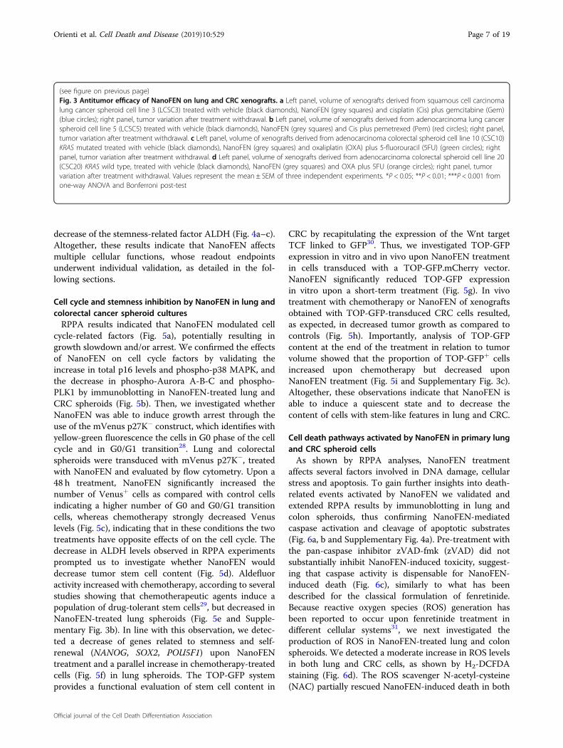
In vivo antitumor efficacy of NanoFEN on lung and CRCxenograftsWe assessed the antitumor effects of NanoFEN com-
pared to conventional chemotherapeutic agents on lungand CRC xenografts derived from four primary spheroidlines by monitoring tumor growth during treatment andthe variation of tumor size after treatment withdrawal.NanoFEN and chemotherapy treatment significantlyinhibited tumor progression in lung squamous carcinomaxenografts, lung adenocarcinoma xenografts (Fig. 3a, bleft) and CRC xenografts, the latter with or withoutmutated KRAS (Fig. 3c, d left). After treatment interrup-tion, xenografts were monitored for two additional weeksin order to monitor the rate of tumor regrowth. Whilexenografts treated with chemotherapy showed acceleratedgrowth rates after treatment cessation, NanoFEN-treatedxenografts grew significantly slower, suggesting thatNanoFEN does not induce an enrichment of tumorigeniccells (Fig. 3a–d right). Finally, we investigated the feasi-bility of a prolonged treatment with NanoFEN in mice.Mice bearing lung cancer xenografts were treated withNanoFEN for 70 days, during which we observed an
Table 1 Main pharmacokinetic parameters of Fenretinide in CD1 female mice after administration of Nanofenretinideby oral or intravenous route in comparison with the NCI capsules and pure Fenretinide administered at the same dose(5mg/Kg fenretinide)
Pk parameters NanoFEN i.v. PureFEN i.v. NanoFEN p.o. NCI p.o. Units
AUC 0-last 13563.5 6420.32 9378.9 3201 h*ng/ml
AUC inf 13656.8 6574.35 9382.5 3209.7 h*ng/ml
Ke 0.094 0.088 0.106 0.089 1/h
HL 7.41 7.87 6.54 7.79 h
Tmax – – 4 2 h
Vd 3.9 6.52 – – L/kg
Cmax 6932.6 4134.77 730.06 298.13 ng/ml
Cl 0.366 0.574 – – L/hr/kg
Orienti et al. Cell Death and Disease (2019) 10:529 Page 4 of 19
Official journal of the Cell Death Differentiation Association

extended arrest of tumor growth without any animaldeath or evident signs of toxicity (Supplementary Fig. 3a).Such observations, if confirmed in humans, could candi-date NanoFEN as a compound suitable for long-termtreatments such as those aimed at preventing tumorrecurrence.
Targeted phospho-proteomics on NanoFEN-treatedprimary cancer cellsWe aimed to generate a comprehensive picture of
NanoFEN-induced changes in pathway activation byReverse-Phase Protein Array (RPPA), a high-throughputtechnology based on the detection of proteins along withtheir post-traslational protein modifications, e.g., cleavageand phosphorylation27. To this end, we performed RPPAusing a selection of 72 antibodies (Supplementary Table 1)
on lung cancer spheroids left untreated or treated withNanoFEN at the IC50 dose. RPPA analysis revealed aNanoFEN-dependent modulation of multiple pathwaysinvolved in cell proliferation and biosynthesis, cell cycleinhibition, cell death and stemness. In detail, treatmentwith NanoFEN was associated with down-modulation ofmultiple components of the mTOR pathway, i.e.,decreased levels of phospho-mTOR, phospho-eIF4G andphospho-4EBP1 or proliferation pathway, i.e., decreasedlevels of phospho-ERK 1/2; induction of cell death andDNA damage response, i.e., increased amounts of cleavedCaspase 7, cleaved PARP and phospho-H2A.Xγ and adecrease of cell cycle-related factors, i.e., phospho-AuroraA/B/C, phospho-CDC2, phospho-PLK1, phospho-CDC25c, phospho-Histone H3 as well as increased levelsof p16 and of phospho-p38 MAPK, accompanied by a
Fig. 2 Effects of NanoFEN on lung and colorectal cancer cells. a Cell viability of CRC spheroid cells (CSCs) (left) and lung cancer spheroid cells(LCSCs) (right) treated with NanoFEN at the indicated concentrations. b Cell viability of CRC commercial cell lines (left) and lung cancer commercialcell lines (right) treated with NanoFEN at the indicated concentrations. Values represent mean ± SD of three independent experiments
Orienti et al. Cell Death and Disease (2019) 10:529 Page 5 of 19
Official journal of the Cell Death Differentiation Association

Fig. 3 (See legend on next page.)
Orienti et al. Cell Death and Disease (2019) 10:529 Page 6 of 19
Official journal of the Cell Death Differentiation Association
decrease of the stemness-related factor ALDH (Fig. 4a–c).Altogether, these results indicate that NanoFEN affectsmultiple cellular functions, whose readout endpointsunderwent individual validation, as detailed in the fol-lowing sections.
Cell cycle and stemness inhibition by NanoFEN in lung andcolorectal cancer spheroid culturesRPPA results indicated that NanoFEN modulated cell
cycle-related factors (Fig. 5a), potentially resulting ingrowth slowdown and/or arrest. We confirmed the effectsof NanoFEN on cell cycle factors by validating theincrease in total p16 levels and phospho-p38 MAPK, andthe decrease in phospho-Aurora A-B-C and phospho-PLK1 by immunoblotting in NanoFEN-treated lung andCRC spheroids (Fig. 5b). Then, we investigated whetherNanoFEN was able to induce growth arrest through theuse of the mVenus p27K− construct, which identifies withyellow-green fluorescence the cells in G0 phase of the cellcycle and in G0/G1 transition28. Lung and colorectalspheroids were transduced with mVenus p27K−, treatedwith NanoFEN and evaluated by flow cytometry. Upon a48 h treatment, NanoFEN significantly increased thenumber of Venus+ cells as compared with control cellsindicating a higher number of G0 and G0/G1 transitioncells, whereas chemotherapy strongly decreased Venuslevels (Fig. 5c), indicating that in these conditions the twotreatments have opposite effects of on the cell cycle. Thedecrease in ALDH levels observed in RPPA experimentsprompted us to investigate whether NanoFEN woulddecrease tumor stem cell content (Fig. 5d). Aldefluoractivity increased with chemotherapy, according to severalstudies showing that chemotherapeutic agents induce apopulation of drug-tolerant stem cells29, but decreased inNanoFEN-treated lung spheroids (Fig. 5e and Supple-mentary Fig. 3b). In line with this observation, we detec-ted a decrease of genes related to stemness and self-renewal (NANOG, SOX2, POU5F1) upon NanoFENtreatment and a parallel increase in chemotherapy-treatedcells (Fig. 5f) in lung spheroids. The TOP-GFP systemprovides a functional evaluation of stem cell content in
CRC by recapitulating the expression of the Wnt targetTCF linked to GFP30. Thus, we investigated TOP-GFPexpression in vitro and in vivo upon NanoFEN treatmentin cells transduced with a TOP-GFP.mCherry vector.NanoFEN significantly reduced TOP-GFP expressionin vitro upon a short-term treatment (Fig. 5g). In vivotreatment with chemotherapy or NanoFEN of xenograftsobtained with TOP-GFP-transduced CRC cells resulted,as expected, in decreased tumor growth as compared tocontrols (Fig. 5h). Importantly, analysis of TOP-GFPcontent at the end of the treatment in relation to tumorvolume showed that the proportion of TOP-GFP+ cellsincreased upon chemotherapy but decreased uponNanoFEN treatment (Fig. 5i and Supplementary Fig. 3c).Altogether, these observations indicate that NanoFEN isable to induce a quiescent state and to decrease thecontent of cells with stem-like features in lung and CRC.
Cell death pathways activated by NanoFEN in primary lungand CRC spheroid cellsAs shown by RPPA analyses, NanoFEN treatment
affects several factors involved in DNA damage, cellularstress and apoptosis. To gain further insights into death-related events activated by NanoFEN we validated andextended RPPA results by immunoblotting in lung andcolon spheroids, thus confirming NanoFEN-mediatedcaspase activation and cleavage of apoptotic substrates(Fig. 6a, b and Supplementary Fig. 4a). Pre-treatment withthe pan-caspase inhibitor zVAD-fmk (zVAD) did notsubstantially inhibit NanoFEN-induced toxicity, suggest-ing that caspase activity is dispensable for NanoFEN-induced death (Fig. 6c), similarly to what has beendescribed for the classical formulation of fenretinide.Because reactive oxygen species (ROS) generation hasbeen reported to occur upon fenretinide treatment indifferent cellular systems31, we next investigated theproduction of ROS in NanoFEN-treated lung and colonspheroids. We detected a moderate increase in ROS levelsin both lung and CRC cells, as shown by H2-DCFDAstaining (Fig. 6d). The ROS scavenger N-acetyl-cysteine(NAC) partially rescued NanoFEN-induced death in both
(see figure on previous page)Fig. 3 Antitumor efficacy of NanoFEN on lung and CRC xenografts. a Left panel, volume of xenografts derived from squamous cell carcinomalung cancer spheroid cell line 3 (LCSC3) treated with vehicle (black diamonds), NanoFEN (grey squares) and cisplatin (Cis) plus gemcitabine (Gem)(blue circles); right panel, tumor variation after treatment withdrawal. b Left panel, volume of xenografts derived from adenocarcinoma lung cancerspheroid cell line 5 (LCSC5) treated with vehicle (black diamonds), NanoFEN (grey squares) and Cis plus pemetrexed (Pem) (red circles); right panel,tumor variation after treatment withdrawal. c Left panel, volume of xenografts derived from adenocarcinoma colorectal spheroid cell line 10 (CSC10)KRAS mutated treated with vehicle (black diamonds), NanoFEN (grey squares) and oxaliplatin (OXA) plus 5-fluorouracil (5FU) (green circles); rightpanel, tumor variation after treatment withdrawal. d Left panel, volume of xenografts derived from adenocarcinoma colorectal spheroid cell line 20(CSC20) KRAS wild type, treated with vehicle (black diamonds), NanoFEN (grey squares) and OXA plus 5FU (orange circles); right panel, tumorvariation after treatment withdrawal. Values represent the mean ± SEM of three independent experiments. *P < 0.05; **P < 0.01; ***P < 0.001 fromone-way ANOVA and Bonferroni post-test
Orienti et al. Cell Death and Disease (2019) 10:529 Page 7 of 19
Official journal of the Cell Death Differentiation Association

lung and colon CSCs and reduced ROS levels (Fig. 6e andSupplementary Fig. 4b) suggesting that ROS generationplays a role in NanoFEN cytotoxicity, in line to whatreported in leukemic stem cells32. Since the repression ofthe mTOR pathway that emerged from RPPA analyses isknown to promote autophagy, we investigated the pre-sence of autophagy-related markers in lung and CRCspheroids treated in vitro with NanoFEN. Interestingly,we found in the majority (3 out of 4) of the spheroid linestested an increased expression of both LC3 and itsphosphatidylethanolamine conjugate (LC3-II), which is
recruited to autophagosomal membranes33 (Fig. 6f andSupplementary Fig. 4c). We also observed that theautophagy inhibitor 3-MA was able to counteractNanoFEN-induced death (Fig. 6g), suggesting thatautophagy contributes to NanoFEN-induced death inboth lung and CRC cells.
Modulation of cellular metabolism by NanoFENFenretinide treatment of cancer cells has been pre-
viously associated with the production of lipid secondmessengers including ceramide, gangliosides (both
Fig. 4 RPPA analysis of pathways activated by NanoFEN in primary lung cancer spheroid cells (LCSCs). a Hierarchical clustering of RPPAresults obtained on six LCSCs derived from different histotypes and one commercial lung cancer cell line (H292) treated with the respective dose ofNanoFEN for 48 h: LCSC1 4.3 μM; LCSC2 3.8 μM and 1.9 μM; LCSC3 0.1μM and 50 nM; LCSC4 0.9 μM; LCSC5 4.2 μM; LCSC6 4.9 μM; H292 29 μM. Thecolor-coded values (high= yellow, average= black, cyan= low) in the heatmap correspond to normalized intensities of averaged sample replicates(n= 3), standardized over the sample set analyzed (z score). A list of RPPA antibodies used here is reported in Supplementary Table 1. b Graphicalrepresentation of the activated (colored) and repressed (grey) pathways emerged from RPPA analysis in LCSCs treated as described in (a).Phosphorylation sites are outlined in green when they result in protein activation. c Table highlights the endpoints upregulated in control (whitepanel) and treated (yellow panel) lung cancer cells as described in (a)
Orienti et al. Cell Death and Disease (2019) 10:529 Page 8 of 19
Official journal of the Cell Death Differentiation Association

involved in apoptosis induction) and dihydrocer-amide34,35. Moreover, RPPA results showed a globalinhibition of the mTOR pathway (Fig. 7a) that was sub-sequently confirmed by immunoblotting (Fig. 7b andSupplementary Fig. 5a), thus linking NanoFEN mechan-ism of action to metabolism, biosynthesis and lipid
signaling networks. We assessed the effect of NanoFENon cellular lipids by liquid chromatography-mass spec-trometry (LC-MS) in lung and CRC spheroid cultures,revealing a massive accumulation of d18 dihydroceramidein treated cells (Fig. 7c and Supplementary Fig. 5b) ascompared to other ceramides (Supplementary Fig. 5c–g).
Fig. 5 (See legend on next page.)
Orienti et al. Cell Death and Disease (2019) 10:529 Page 9 of 19
Official journal of the Cell Death Differentiation Association

Dihydroceramide production was previously shown toelicit multiple biological effects in cancer cells such as G0/G1 cell cycle arrest, impairment of mitochondrial functionand autophagy induction36–38, corresponding to the bio-logical effects observed in NanoFEN-treated cells. Theseobservations suggest that NanoFEN (as was previouslyshown for fenretinide) targets dihydroceramide desatur-ase (DES), thus inducing dihydroceramide accumulation.Accordingly, we observed a strong increase of sphinga-nine (SA, the lipid mediator upstream of DES) bothin vitro in NanoFEN-treated lung and CRC cells (Fig. 7d)and in vivo in lung cancer xenografts (Supplementary Fig.6a). In order to determine the functional relevance ofdihydroceramide production in NanoFEN-treated cells weused the serine palmitoyltransferase inhibitor myriocin,which blocks de novo sphingolipid biosynthesis39. Pre-treatment of spheroid lines with myriocin partially pro-tected cells from NanoFEN-induced death, indicating anactive role of the sphingolipid pathway in mediatingNanoFEN effects (Fig. 7e). In line with an important roleof dihydroceramide in mediating NanoFEN effects, SAand the DES inhibitor GT11 induced a dose-dependentviability decrease in lung cancer spheroids (Supplemen-tary Fig. 6b). By contrast, the glucosylceramide synthaseinhibitor 1-phenyl-2-palmitoylamino-3-morpholino-1-propanol (PPMP) was unable to block NanoFEN-induceddeath, indicating that the cytotoxic effects of NanoFENare determined by the accumulation of dihydroceramiderather than by glucosylceramide/ceramide formation (Fig. 7f).Finally, we ought to determine whether the sphingolipidpathway would cooperate with repression of the mTORpathway in mediating NanoFEN-induced toxicity. To thisaim, we mimicked the effect of NanoFEN by treatingspheroids with both sphinganine and GT11 at lowdoses. Additional treatment with the mTOR pathway
inhibitor rapamycin significantly potentiated the toxiceffects of the SA/GT11 combination, indicating a coop-erative effect of the two pathways in inducing cancer celldeath (Fig. 7g and Supplementary Fig. 6c). Western blotanalysis of key factors implicated in mTOR signalingshowed that the combined treatment with SA/GT11/rapamycin effectively inhibited the mTOR pathway andrecapitulated NanoFEN-related events (Fig. 7h).
DiscussionIn accordance to their multifaceted role in cell pro-
liferation, differentiation and death, retinoids have longbeen studied for their chemopreventive and chemother-apeutic effects in cancer patients, although theirmechanism of action has not been completely elucidated.One of the most promising retinoid derivatives is fenre-tinide, which has been extensively investigated in phase I-III clinical trials, revealing long-term safety and toler-ability but modest anticancer effects3,4,7–15,21,40–43. Thescarce efficacy of fenretinide in previous clinical studies isrelated to its low bioavailability, as even high-dose sche-dules and multiple administrations did not provideplasma concentrations sustained over time at the levelsrequired for a therapeutic activity3,8,10–13. We report herethat a nanoencapsulated form of fenretinide (previouslymodified through a salification procedure in order toincrease complexation effectiveness) has an enhancedsolubility and reaches significantly higher plasma levelsthan formulations previously used in clinical trials. Themechanism of action of NanoFEN was analyzed in pri-mary spheroid cultures of lung and CRC, thus providing acomprehensive picture of protein and lipid pathwaysactivated in cancer cells. The results emerging fromproteomic and lipidomic analyses revealed a complex setof molecular events activated by NanoFEN, resulting in a
(see figure on previous page)Fig. 5 Effects of NanoFEN on cell cycle and stem-like features in vitro and in vivo on lung cancer spheroid cells (LCSCs) and CRC spheroidcells (CSCs). a Schematic representation of cell cycle regulators activated (black) or repressed (grey) arised from RPPA analysis of LCSCs treated withNanoFEN as described in Fig. 4a. b Immunoblot analysis of cell cycle regulators phospho-Aurora A-B-C, phospho-PLK1, p38, phospho-p38 and p16 onLCSC6 (left panel) and CSC7 (right panel) untreated and treated with NanoFEN at the IC50 concentration (4.9 μM and 0.2 μM respectively) for 48 h.c Flow cytometry analysis of mVenus p27K−-transduced LCSC6 (left panel) and CSC7 (right panel) untreated and treated with NanoFEN at the IC50dose (4,9 μM and 0,2 μM respectively) for 48 h, Gem 25 μM and OXA 10 μM. Values represent mean ± SD of three independent experiments. **P <0.01; ***P < 0.001 from two-tailed t-test. d Schematic representation of stem-like factors (repressed in grey) emerged from RPPA analysis of lung CSCstreated as described in Fig. 4a. e Cytofluorimetric analysis of Aldefluor activity on LCSC6 treated with NanoFEN at IC50 dose and Gemcytabine 25 μMfor 48 h. Values represent mean ± SD of three independent experiments. **P < 0.01; ***P < 0.001 from two-tailed t-test. f qRT-PCR analysis of indicatedgenes on whole tumor lysates derived from LCSC6 xenografts, untreated or treated with NanoFEN 30mg/Kg/week and Cis 3 mg/Kg/week and Gem60mg/Kg/week (Cis+ Gem). Values represent mean ± SD of three independent experiments. *P < 0.05; **P < 0.01 from two-tailed t-test. g Flowcytometry analysis of TOP-GFP.mc-transduced CSC7, untreated and treated with NanoFEN at the IC50 dose 0.2 μM and OXA 10 μM for 48 h. Valuesrepresent mean ± SD of three independent experiments. **P < 0.01 from two-tailed t-test. h Volume of xenografts derived from CSC10 transducedwith TOP-GFP.mc untreated (black downward triangles) or treated with vehicle or treated NanoFEN 30mg/Kg/week (green upward triangles) andOXA 5mg/Kg/week plus 5FU 12.5 mg/Kg/week (red squares). Values represent mean ± SEM three independent experiments. *P < 0.05; **P < 0.01from one-way ANOVA and Bonferroni post-test. i Total stem cell content in tumor xenografts obtained as in (h), relative to the experiment shown inSupplementary Fig. 3c. Values were calculated as tumor weight × percentage of TOP-GFP.mc positive cells/100. Values represent mean ± SD of threeindependent experiments. *P < 0.05; ***P < 0.001 from two-tailed t-test
Orienti et al. Cell Death and Disease (2019) 10:529 Page 10 of 19
Official journal of the Cell Death Differentiation Association

broad landscape of functional effects ranging from celldeath (with features of both autophagy and apoptosis) tocell cycle arrest and to a generalized metabolic repressionmediated by inhibition of the mTOR pathway and massive
production of dihydroceramide. The effect of NanoFEN ininducing growth arrest/quiescence is particularly inter-esting in view of a potential use of this compound inchemopreventive settings. In fact, maintaining tumor cells
Fig. 6 (See legend on next page.)
Orienti et al. Cell Death and Disease (2019) 10:529 Page 11 of 19
Official journal of the Cell Death Differentiation Association

(particularly those disseminated in distant organs) in aprolonged and stable quiescent state is progressivelyemerging as a potential therapeutic strategy aimed atpreventing tumor relapse44. In line with this hypothesis, acombination of all-trans retinoic acid and 5-azacitidinehas been reported to induce a quiescent state in bonemarrow-disseminated cancer cells, supporting a role ofretinoids in promoting a long-term metastasis-free con-dition45–47 and is currently being investigated in a clinicaltrial (NCT03572387). If confirmed in additional pre-clinical and clinical studies, the low toxicity of NanoFENwould allow prolonged treatment schedules aimed atavoiding the reactivation of disseminated tumor cells andsubsequent tumor relapse, particularly in cancers subjectto late recurrences such as those of breast and prostate.Moreover, it would be interesting to investigate the effectof NanoFEN in retinoid-sensitive tumors such as somehematological tumors as well as neuroblastoma, where theincreased bioavailability of NanoFEN may allow moreeffective treatment schedules as compared to previousclinical trials. In summary, the results reported in thisstudy indicate NanoFEN as a bioavailable fenretinideformulation with broad-range inhibitory effects on cancercells that may find a future use in several clinical settings.
Materials and methodsCells, tissues, mice, antibodies, and reagentsColon and lung cancer specimens were obtained from
patients undergoing surgical resection upon informedconsent and approval by the Sapienza-PoliclinicoUmberto I Ethical Committee (RIF.CE: 410717/10/2016). Spheroid cultures were established and validated aspreviously described48,49. All the commercial cancer cellline were purchased from the American Type CultureCollection (ATCC, Manassas, VA, USA) or Sigma-Aldrich(St. Louis, MO, USA) and cultured following manu-facturer’s instructions. Fenretinide, N-(4-Hydroxyphenyl)retinamide (code 65646-68-6) was purchased from Olon
(Milan, Italy). 2-hydroxypropyl-β-cyclodextrin (averageMw ~1380), potassium hydroxide, 1-octanol, D2O,DMSO-d6 were purchased from Sigma-Aldrich. Anti-bodies used for RPPA and western blot were listed inSupplementary Table 1. Oxaliplatin and 5-fluorouracilwere from Teva Italia (Milano, Italy) gemcitabine andcisplatin were from Selleckchem (Houston, TX, USA).Aldefluor assay was from STEMCELL Technologies(Vancouver, Canada). Myriocin was purchased fromENZO Life Science (New York, NY, USA), N-[(1R,2S)-2-hydroxy-1-hydroxymethyl-2-(2-tridecyl-1-cyclopropenyl)ethyl]octanamide (GT11) and Sphinganine from AvantiPolar Lipids (Alabaster, AL, USA), Rapamycin from Sell-eckchem, z-Val-Ala-DL-Asp-fluoromethylketone (zVAD)from BACHEM (Torrance, CA, USA), DL-threo-1-Phe-nyl-2-hexadecanoylamino-3-morpholino-propanol-HCL(PPMP) from Matreya (State College, PA, USA), N-Acetyl-L-Cysteine (NAC) was purchased from Sigma-Aldrich, 5-chloromethylfluoresceindiacetate (CMFDA)was purchased from Molecular Probes (Eugene, OR,USA). Anti-LC3 and anti-β-Actin antibodies were pur-chased from Sigma-Aldrich; phospho-Aurora A (Thr288)/Aurora B (Thr232)/Aurora C (Thr198) (D13A11), phos-pho-PLK1, phospho-S6 ribosomal protein (Ser240/244)and total S6 ribosomal protein (5G10), caspase3, caspase7,caspase9, PARP, fodrin cleaved, phospho-p38 MAP kinase(Thr180/Tyr182), mTOR, phospho-mTOR (Ser2448), 4E-BP1(53H11), phospho-4E-BP1(Ser65) antibodies werefrom Cell Signaling Technology (Danvers, MA, USA).Anti-CDKN2A (p16) was from Biolegend (London, Uni-ted Kingdom). Secondary anti-mouse and anti-rabbitantibodies coupled to horseradish peroxidase were fromBio-Rad (Hercules, CA, USA).
Preparation of fenretinide 2-hydroxypropyl-β-cyclodextrincomplex (NanoFEN)The first step of NanoFEN preparation involved fenre-
tinide salification to raise its aqueous solubility to the
(see figure on previous page)Fig. 6 Analysis of cell death pathways activated by NanoFEN in lung cancer spheroid cells (LCSCs) and CRC spheroid cells (CSCs). aSchematic representation of RPPA endpoints involved in DNA damage, cell stress and apoptosis activated (black) in LCSCs treated with NanoFEN asdescribed in Fig. 4a. Phosphorylation sites are outlined in green when they result in protein activation. b Immunoblot analysis of cell death-relatedproteins Caspase-9, 3, and 7, PARP and α-fodrin on LCSC6 (left panel) and CSC7 (right panel) untreated and treated with NanoFEN at IC50 dose (4.9and 0.2 μM, respectively) for 48 h. c Cell viability of LCSC6 (left panel) and colon CSC7 (right panel) pretreated with zVAD 40 μΜ and then withNanoFEN at IC50 dose (4.9 and 0.2 μM, respectively) for 48 h. Values represent mean ± SD of three technical replicates of a representative experimentof two biological replicates. *P < 0.05 from two-tailed t-test. d Production of reactive oxygen species (ROS) was measured by flow cytometer in LCSC6(left panel) and CSC7 (right panel) untreated or treated with NanoFEN (4.9 and 0.2 μM, respectively) for 24 h and then with 5 μM of CM-H2DCFDA.Values represent mean ± SD of three independent experiments. *P < 0.05 from two-tailed t-test. e Cell viability of LCSC6 (left panel) and CSC7 (rightpanel) pretreated with N-acetyl-cysteine (NAC) 2 mM for 1 h and after with NanoFEN for 48 h (4.9 and 0.2 μM respectively). Values represent mean ±SD of three technical replicates of a representative experiment. *P < 0.05; **P < 0.01 from two-tailed t-test. f Immunoblot analysis of autophagy-related marker LC3 I-II on LCSC6 (left panel) and CSC7 (right panel) untreated and treated with NanoFEN at IC50 dose (4.9 and 0.2 μM, respectively) for24 h. g Cell viability of LCSC6 (left panel) and CSC7 (right panel) treated with 3-Methyladenine (3-MA) 5 mM plus NanoFEN (4.9 and 0.2 μM,respectively) for 24 h. Values represent mean ± SD of a three technical replicates of a representative experiment. *P < 0.05; **P < 0.01; ***P < 0.001from two-tailed t-test
Orienti et al. Cell Death and Disease (2019) 10:529 Page 12 of 19
Official journal of the Cell Death Differentiation Association

extent required for complexation with cyclodextrins.Salification was carried out by dissolving fenretinide(2 mol in 20mL ethanol containing a stoichiometric
amount of potassium hydroxide. The solvent wasremoved in a rotary evaporator to obtain a solid redresidue of fenretinide potassium salt (4-HPRK). The salt
Fig. 7 (See legend on next page.)
Orienti et al. Cell Death and Disease (2019) 10:529 Page 13 of 19
Official journal of the Cell Death Differentiation Association

was physically mixed with 2-hydroxypropyl-β-cyclodextrin at 1:10 w:wsalt:cyclodetrin ratio and grin-ded to homogeneity. The physical mixture obtained wasdispersed in 30mL water, stirred at room temperature 6 hin the dark and subsequently left at 4 °C overnight. Afterthis period the suspension was stirred again for 2 h atroom temperature, diluted with 20mL water and finallyfiltered through a 0.2 µm filter to eliminate any excess ofundissolved material. The filtered solution was lyophilizeduntil the solid complex was obtained as an orange-redhomogeneous powder. The complex was analyzed by 1H-NMR to detect the presence of chemical shift changeswith respect to the free drug as indicators of fenretinideinclusion into the cyclodextrin hydrophobic cavity50. Forthe analysis the complex was dissolved in D2O and thefenretinide salt in DMSO-d6. The 1H-NMR spectra wererecorded by an Inova 600MHz High Resolution NMRSpectrometer. The amount of fenretinide incorporatedinto the complex was spectrophotometrically determinedin solutions obtained by dissolving known amounts of thesolid complex in a water:ethanol (1:1, v-v) mixture withthe aim to dissociate the drug from the complex andmaintain its solubilization in the aqueous phase in thepresence of ethanol. The hydro-alcoholic solutions wereanalyzed for their fenretinide content at the maximumwavelength of the fenretinide absorption (360 nm). Theaqueous solubility of fenretinide complexed with 2-hydroxypropyl-β-cyclodextrin was measured in compar-ison with the pure drug. Excess amounts of the complexor pure drug were placed in water, stirred 12 h at 25 °C inthe dark to saturation of the aqueous phase and filteredthrough 0.2 µm filters. After filtration the saturatedsolutions were spectrophotometrically analysed at 360 nmfor their fenretinide content.
Release studiesRelease studies of fenretinide from the complex were
carried out by placing saturated aqueous solutions of thecomplex in a dialysis membrane bag (Mw cutoff 5KD),
allowing diffusion only to the free drug and not to thecomplex. The membrane separated the saturated aqueoussolution of the complex from a receiving compartmentcontaining an aqueous buffer (PBS pH 7.4) and 1-octanol(10:1v:v ratio) used to extract the drug diffused throughthe dialysis membrane and to simulate the presence ofhydrophobic absorbing phases such as biomembranes51.The release of the drug from the complex was evaluatedby a spectrophotometric analysis of the complex solutionin the releasing compartment at the maximum absorptionwavelength of fenretinide (360 nm) at appropriate timepoints.
Pharmacokinetics studiesThe experiments were performed in CD1 female mice,
7 weeks of age, (body weight 25 ± 2 g) obtained fromEnvigo RMS SrL (Udine, Italy). Mice were maintainedunder specific-pathogen-free conditions with constanttemperature and humidity, according to the institutionalguidelines of the Istituto Superiore di Sanità Animal CareCommittee. Animal experimentation was conducted inconformance with the following laws, regulations, andpolicies governing the care and use of laboratory animals:Italian Governing Law (D. l. 26/2014; Authorization n.19/2008-A issued March 6, 2008 by the Ministry of Health).The pharmacokinetics of NanoFEN, administered by oralor intravenous route, was investigated in mice receivingthe formulation at doses corresponding to 5mg/kg offenretinide. The intravenous administration was carriedout by injection in the tail vein of an intravenous bolus ofNanoFEN dissolved in water or PureFEN suspended in amixture of ethanol 10 % in saline as a comparison. For theoral administration the mice were gavaged the withNanoFEN dissolved in water or with the reconstitutedcontent of the NCI capules, i.e., fenretinide (100 mg) incorn oil (704 mg) and Polysorbate 80 (60 mg) as a com-parison. After the intravenous or oral treatments, a seriesof blood samples were taken at 15 and 30min and at 1, 2,4, 8, 10, 24 and 48 h. Blood was collected in heparinized
(see figure on previous page)Fig. 7 NanoFEN inhibits cellular metabolism in lung cancer spheroid cells (LCSCs) and CRC spheroid cells (CSCs). a Schematic representationof RPPA results of proliferative and biosynthetic pathways repressed (grey) in LCSCs treated with NanoFEN. b Immunoblot analysis of proteinsinvolved in metabolic process on LCSC6 (left panel) and CSC7 (right panel) untreated and treated with NanoFEN at IC50 dose (4.9 and 0.2 μM,respectively) for 48 h. c Graphs represent NanoFEN effects on lipid metabolism by liquid chromatography-mass spectrometry (LC-MS) on LCSC6 (leftpanel) and CSC7 (right panel). Values represents the fold over control of treated cells with NanoFEN (4.9 and 0.2 μM, respectively) for 24 h. dSphinganine peak intensity in LCSC6 (left panel) and CSC7 (right panel) treated with NanoFEN (4.9 and 0.2 μM, respectively) for 24 h. e Cell viability ofLCSC6 (left panel) and CSC7 (right panel) treated with Myriocin 1 μM plus NanoFEN (4.9 and 0.2 μM, respectively) for 48 h. Values represent mean ±SD of three independent experiments. *P < 0.05, from two-tailed t-test. f Cell viability of LCSC6 (left panel) and CSC7 (right panel) treated withglucosylceramide synthase inhibitor PPMP 10 μM plus NanoFEN (4.9 and 0.2 μM, respectively) for 48 h. Values represent mean ± SD of threeindependent experiments. *P < 0.05, from two-tailed t-test. g Cell viability of LCSC6 (left panel) and CSC7 (right panel) treated with dihydroceramidedesaturase inhibitor (GT11) 0.5 μM plus sphinganine (SA) 2 μM plus mTOR pathway inhibitor Rapamycin 10 nM for 48 h. Values represent mean ± SDof three independent experiments. *P < 0.05; ***P < 0.001, from two-tailed t-test. h Immunoblot analysis of mTOR pathway components mTOR,phospho-mTOR, S6RP and phospho-S6RP on LCSC6 (left panel) and CSC7 (right panel) treated as in (g)
Orienti et al. Cell Death and Disease (2019) 10:529 Page 14 of 19
Official journal of the Cell Death Differentiation Association

tubes from the retro-orbital plexus of the mice underisoflurane anesthesia. To obtain plasma, blood sampleswere centrifuged at 4000 rpm for 10 min at 4 °C. Thesamples were stored at −20 °C until analysis. The totalconcentration of fenretinide in plasma of mice wasdetermined according to a method previously described52.Briefly, aliquot of 200 μl of each plasma sample was addedto 400 μl of Acetonitrile (CH3CN) containing 125 μg/mLButylated hydroxytoluene (BHT), and the mixture wasvortexed and centrifuged to pellet the precipitated pro-teins. The recovered supernatants were analysed on aliquid chromatograph (Perkin-Elmer, Norwall, CT) fittedwith a C18 (50 mm× 2.0 mm, 5 μm) reverse-phase col-umn and a C18 precolumn (Perkin-Elmer, Milan, Italy).The mobile phase consisted of CH3CN:H2O:CH3COOH(75:2:2, v:v:v) delivered at a flow rate of 2 mL/min.Detection was carried out with a Perkin-Elmer LC95absorbance detector at 362 nm. N-(4-ethoxyphenyl)-reti-namide (EPR) was used as internal standard. For thequantitative evaluation, reference standard curves wereset up in control mouse plasma with different knownamounts of NanoFEN or PureFEN. The measured fenre-tinide concentration-vs-time data were elaborated bynon-compartmental analysis (WinNonLin software v3) toobtain the main pharmacokinetic parameters ofNanoFEN.
Animal proceduresAll animal procedures were performed according to the
Italian national animal experimentation guidelines (D.L.116/92) upon approval of the experimental protocol bythe Italian Ministry of Health’s Animal ExperimentationCommittee (DM n. 292/2015 PR 23/4/2015). 6-week-oldfemale NOD.Cg-Prkdcscid Il2rgtm1Wjl/SzJ (NSG) micewere purchased from The Jackson Laboratory (Bar Har-bor, Maine, USA) for colon and lung cancer xenografts.Tumors were measured twice weekly by an externaldigital caliper, and volumes were calculated using thefollowing formula: π/6 x d2 x D, where d and D representshorter and longer tumor measurements, respectively.Drug treatments started when tumor volume was 50-100mm3: mice were randomized in control and treatmentgroup and treated with NanoFEN 10mg/Kg/threeweekly/intraperitoneally (IP), chemotherapeutic agent combina-tions as cisplatin 3 mg/kg/biweekly/IP+pemetrexed200mg/kg/biweekly/IP or cisplatin 3 mg/kg/biweekly/IP+gemcitabine 60 mg/kg/biweekly/IP or 5-fluorouracil12,5 mg/Kg+oxaliplatin 5 mg/Kg/biweekly/IP. Controlanimals were treated with vehicle only. Tumors weremeasured at the indicated timepoints and mice weremonitored for signs of drug-induced toxicity and weighedregularly. At the end of experiments, tumors were col-lected and dissociated to obtain cell suspension for sub-sequent studies or monitored to evaluate tumor growth
rate after treatment interruption. Relative tumor growth isindicated as ratio of tumor volume at the indicated daysafter drug suspension versus volume at the last day oftreatment. Animals were euthanized according to thenational Animal Welfare Guidelines.
Lentiviral infectionPrimary spheroid cultures of colon and lung cancer cells
were stably transduced with mVenus-p27K−28 or TOP-GFP.mCherry (purchased from Addgene, Cambridge,MA, USA) using ProFection® Mammalian TransfectionSystem from Promega (Madison, Wisconsin, USA) fol-lowing manufacturer’s instructions.
Metabolite extractionSamples were extracted following the protocol by
D’Alessandro et al53. Samples were resuspended in0.15 mL of ice cold ultra-pure water (18 MΩ) to lyse cells,then the tubes were plunged into a water bath at 37 °C for0.5 min. Samples were mixed with 0.6 mL of −20 °Cmethanol and then with 0.45 mL chloroform. Subse-quently, 0.15 mL of ice cold ultra-pure water were addedto each tube and then transferred to −20 °C for 2–8 h. Anequivalent volume of acetonitrile was added to the tubeand transferred to 4 °C for 20min. Samples were thuscentrifuged at 10,000×g for 10 min at 4 °C. Finally, sam-ples were dried in a rotational vacuum concentrator (RVC2-18—Christ Gmbh; Osterode am Harz, Germany) andre-suspended in 200 µl of water, 5% formic acid andtransfer to glass auto-sampler vials for LC/MS analysis.
Rapid resolution reversed-phase HPLCAn Ultimate 3000 Rapid Resolution HPLC system (LC
Packings, DIONEX, Sunnyvale, USA) was used to performmetabolite separation. The system featured a binary pumpand vacuum degasser, well-plate autosampler with a six-port micro-switching valve, a thermostated columncompartment. Samples were loaded onto a Reprosil C18column (2.0 mm × 150mm, 2.5 µm - Dr Maisch, Ger-many) for metabolite separation. For lipids multi-stepgradient program was used. It started with 8% solvent A(ddH20, 20 mmol L-1 ammonium formiate; pH 5) to 6%solvent A for 3 min than to 2% solvent A for 35min andfinally to 100% solvent B (methanol) in 30 min. At the endof gradient, the column was reconditioned with 8% sol-vent A for 5 min.
Mass spectrometry analysis through microTOF-QMass spectrometry analysis was carried out on an
electrospray hybrid quadrupole time of Flight massspectrometer MicroTOF-Q (Bruker-Daltonik, Bremen,Germany) equipped with an ESI-ion source. MS analysiswas carried out capillary voltage 2800 V, nebulizer 45psiand dry gas of 9 l/min, scan mode 100–1500m/z. Data
Orienti et al. Cell Death and Disease (2019) 10:529 Page 15 of 19
Official journal of the Cell Death Differentiation Association

were acquired with a stored mass range of 100–2000m/z.Tandem mass spectrometry (MS/MS) is used for lipidspecies structural characterization. Automatic isolationand fragmentation (AutoMSn mode) was performed onthe four most intense ions simultaneously throughout thewhole scanning period (30 min per run). Calibration ofthe mass analyzer is essential in order to maintain an highlevel of mass accuracy. Instrument calibration was per-formed externally every day with a sodium formatesolution consisting of 10 mM sodium hydroxide in 50 %isopropanol, water, 0.1 % formic acid. Automated internalmass scale calibration was performed through directautomated injection of the calibration solution at thebeginning and at the end of each run by a 6-port divert-valve.
Real time PCRRNA from vehicle and treated xenografts was extracted
with TRIzol (Thermo Fisher Scientific, Waltham, Massa-chusetts, USA) following manufacturer’s instructions.1 µg of RNA were reverse transcribed with M-MLVreverse transcriptase (Thermo Fisher Scientific) and 50 ngof cDNA were used as template in the PCR reactions.Specific probes used for NANOG (Hs04399610_g1), SOX2 (Hs04260357_g1) and POU5F1 (Hs00999634_gH) wereall from Thermo Fisher Scientific. Normalization wasperformed using β-actin (Hs00194899_m1) as reference.Values were expressed in terms of 2−ΔΔCt where ΔΔCt=ΔCt sample−ΔCt calibrator. ΔCt is the difference inthreshold cycles between the specific RNA target andreference gene amplicons given by StepOne Plus Real-Time PCR software by negative correlation with aninternal reference dye (ROX).
Reverse-phase protein arrayFor RPPA experiments, primary lung cancer spheroid
cells (LCSCs) and commercial lung cancer cell lines wereleft untreated or treated with NanoFEN (IC50 con-centration) as follows: LCSC1 (4.3 µM), LCSC 2 (1.9 and3.8 µM) LCSC 3 (50 nM and 1 µM) LCSC 4 (0.9 µM)LCSC 5 (4.2 µM) LCSC 6 (4.9 µM) H292 (29 µM). Fol-lowing 48 h treatment of the six LCSCs and H292 com-mercial cell line, cell pellets were promptly lysed in abuffer containing T-PER reagent (Thermo Fisher Scien-tific), 300 mM NaCl (J.T.Baker; Avantor PerformanceMaterials, Center Valley, PA), protease and phosphataseinhibitors cocktails (Sigma-Aldrich) and stored at −80 °C.In order to prepare protein samples for RPPA printing,protein lysates were allowed to thaw on ice, their totalprotein concentration was measured using the Bradfordreagent method (Thermo Fisher Scientific) and thevolume corresponding to 50 μg of total protein was usedto further dilute samples to a final concentration of0.5 mg/mL, as follows: 50% 2X Tris-Glycine SDS Sample
Buffer (Life Technologies Corporation, Carlsbad, CA), upto 47.5% sample volume and T-PER reagent (ThermoFisher Scientific) and 2.5% Tris (2-carboxyethyl)phos-phine hydrochloride (TCEP) reagent (Thermo FisherScientific). RPPA lysates were boiled for 3 min and storedat −80 °C until further processing. Prior to printing ontonitrocellulose-coated slides (GRACE Bio-Labs, Bend, OR)using a robotic arrayer (Aushon Biosystems, Billerica,MA, USA), RPPA lysates were allowed to thaw up toroom temperature and boiled for an additional 3 min. Allsamples were printed in technical triplicate as neat and 1:4dilution pairs. Reference standard lysates, i.e., HeLa+Pervanadate (Becton, Dickinson and Company, FranklinLakes, NJ, USA), A431+EGF (Becton, Dickinson andCompany), Jurkat+Etoposide (Cell Signaling Technology)and Jurkat+Calyculin A (Cell Signaling Technology),were printed in 10-point decreasing mixtures of treated tountreated samples as positive and quality controls ofantibody staining. Each reference standard curve wasprinted in technical triplicates at a final concentration of0.5 mg/mL. A selected subset of the printed microarrayslides were stained with Sypro Ruby Protein Blot Stain(Thermo Fisher Scientific) to estimate sample total pro-tein concentration and the remaining slides were storedunder desiccated conditions at -20 °C. Immediately beforeantibody staining, printed slides were treated with 1XReblot Mild Solution (Chemicon, Burlington, MA, USA)for 15 min, washed 2 × 5min with PBS and incubated for2 h in blocking solution containing 2% I-Block (AppliedBiosystems, Foster, CA, USA) and 0.1% Tween-20 in PBS.Immunostaining was carried out using a tyramide-biotinsignal amplification kit (DAKO, Santa Clara, CA, USA).Primary antibody binding was detected using a biotiny-lated goat anti-rabbit IgG H+L (diluted at 1:7500; VectorLaboratories, Burlingame, CA, USA) or rabbit anti-mouseIg (diluted at 1:10, DAKO) followed by streptavidin-conjugated IRDye®-680LT fluorophore (LI-COR Bios-ciences, Lincoln, Nebraska, USA. Primary antibodiesundergo pre- and post-RPPA validation for single bandspecificity by western-blot using complex cellular lysates.Negative control slides, incubated only with secondaryantibody were included in each staining run. All SyproRuby, i.e., per-spot total protein content,as well asimmunostained slides were scanned using a Tecan PowerScanner™ (Tecan Group Ltd, Männedorf, Switzerland) at5 μm resolution. Acquired images were analyzedwith MicroVigene v5.2, http://www.vigenetech.com/MicroVigene.htm (VigeneTech Inc, Carlisle, MA, USA),for spot detection, local and negative control backgroundsubtraction, replicate averaging and total protein nor-malization. RPPA data analysis was performed by meansof ‘R’ v3.5.0 https://www.R-project.org/ (R Foundation forStatistical Computing) and ‘RStudio’ v1.1.414 https://www.rstudio.com/ (RStudio), using the following installed
Orienti et al. Cell Death and Disease (2019) 10:529 Page 16 of 19
Official journal of the Cell Death Differentiation Association

packages:base, plyr, tidyverse, FactoMineR, factoextra,RColorBrewer, Bioconductor and shiny. Custom ‘R’scripts for slide quality control, internal standardizationand two-way hierarchical clustering (correlation distanceand complete method) are available upon request. Adetailed list of antibodies used for RPPA is available inSupplementary Table 1.
Flow cytometryFor flow cytometry analysis, primary lung and CRC
spheroid cultures transduced with the mVenus p27K−
and treated with NanoFEN, oxaliplatin or gemcitabinewere dissociated at a single cell level mechanically or withTrypLE Express (Thermo Fisher Scientific) respectively.Cells were resuspended at a concentration of 500.000cells/mL of PBS/0.4% BSA/0.5M EDTA. The percentageof Venus positive cells was analyzed with a FACSCantoflow cytometer (Becton Dickinson) equipped with a DIVAsoftware. 10 μg/mL 7-aminoactinomycin D (Sigma-Aldrich) was used for dead cells exclusion. The sameprocedure was applied for primary CRC spheroids trans-duced with the TOP-GFP.mCherry construct treated withNanoFEN, oxaliplatin and 5-fluorouracil for in vitro andex vivo experiments.
Measurement of intracellular ROS levelsThe intracellular generation of ROS was measured
using the oxidation-sensitive fluorescent dye 5-chloromethylfluorescein diacetate (CM-H2DCFDA), withthe conversion of CM-H2DCFDA to dichlorofluorescein(DCF) assessed as previously described54. An equalnumber of dissociated spheroid cells (2.5 × 10⁵) wereseeded in 6-well cell culture plates. Briefly, each cell linewas incubated with NanoFEN at IC50 concentration andpre-treated for 1 h with the antioxidant N-Acetyl-Cysteine(NAC). At different times, the cells were washed twicewith pre-warmed Phosphate Saline Buffer (PBS) and thenincubated in the same buffer containing 5 μM CM-H2DCFDA with DMSO alone at 37 °C. The fluorescenceintensity of DCF was measured at 527 nm emissionwavelength after excitation at 492 nm at 1 h intervals forup to 48 h using a Flow Cytometer Accuri (BectonDickinson). An increase in fluorescence intensity asarbitrary units indicated the net generation ofintracellular ROS.
Western blottingImmunoblotting was performed as previously descri-
bed55. Briefly, whole cell extracts were obtained by incu-bating the cells in lysis buffer supplemented with proteaseand phosphatase inhibitor cocktail. Lysates concentrationwas determined and equal amounts of proteins wereloaded on a 4–12% precast gel (Thermo Fisher Scientific)and transferred to nitrocellulose membranes. Blots were
blocked with TBST 5% non-fat dry milk (Bio-RadLaboratories, Hercules, CA, USA) and incubated over-night at 4 °C with primary antibodies (described in theAntibodies and Reagents section) then incubated for45min with secondary HRP-conjugated antibodies dis-solved in TBST/1% BSA. Chemiluminescent signals weredetected with Amersham ECL prime or select westernblotting detection reagent (GE Healthcare Life Sciences,Barrington, IL, USA). Images were taken and analyzedwith Bio-Rad ChemiDoc Imagers (Bio-Rad Laboratories).
Viability assayCell viability upon treatment with NanoFEN, zVAD,
NAC, 3-MA, Myriocin, PPMP, GT11, SA and Rapamycinwas determined by CellTiter-Glo luminescent cell viabi-lity assay (Promega, Madison, WI, USA) according to themanufacturer’s directions. Briefly, lung and colorectalspheroids were dissociated mechanically or with TrypLEExpress (Thermo Fisher Scientific) respectively and see-ded 3 × 10³ per well in 96-well plates (three replicates perexperimental point) in serum-free medium, and incubatedin a humidified atmosphere at 37 °C, 5% CO2. Cells weretreated with the respective compounds and analysed afterthe appropriate time period as described in detail in Fig-ure Legends. Luminescence was detected with a DTX880multimode microplate reader (Beckman Coulter, Brea,CA, USA).
Statistical analysisAnalyses were performed using GraphPad Prism ver-
sion 4.0 for Windows (GraphPad Software) with non-paired double-tailed t-test (after verifying normal dis-tribution of the population with Shapiro–Wilk test) orwith one-way ANOVA where appropriate. Results arepresented as the mean ± standard deviation (SD) orstandard error of the mean (SEM) where appropriate.Statistical significance is expressed as *P < 0.05; **P < 0.01and ***P < 0.001.
AcknowledgementsWe thank Paola Di Matteo and Stefano Guida for excellent technical assistance,Antonio Di Virgilio and Daniele Macchia for their essential contribution forin vivo experiments. This work was supported by the following grants: AIRC IG2017 Ref: 20744, AIRC IG 2014 Ref: 15749, ERA-NET TRANSCAN Ref TACTIC JTC2014 to A.Z., by Awards 2015 Sapienza University of Rome C26H15ZKWL grantto F.L.T. and by AIRC 5 × 1000 Molecular Clinical Oncology Extension ProgramRef 9979 grant to R.D.M., G.M. and M.T.
Author details1Department of Pharmacy and Biotechnology, University of Bologna via SanDonato 19/2, 40127 Bologna, Italy. 2Department of Oncology and MolecularMedicine, Istituto Superiore di Sanità, Viale Regina Elena 299, 00161 Rome, Italy.3Center for Gender-Specific Medicine, Istituto Superiore di Sanità, Viale ReginaElena 299, 00161 Rome, Italy. 4Department of Experimental Medicine andSurgery, University of Rome Tor Vergata, Via Montpellier 1, 00133 Rome, Italy.5RPPA Unit, Proteomics, Core Facilities, Istituto Superiore di Sanità, Viale ReginaElena 299, 00161 Rome, Italy. 6Core Facilities, Istituto Superiore di Sanità, Rome,Italy. 7Department of Biomedical and Biotechnological Sciences BIOMETEC,University of Catania, via Santa Sofia 97, 95123 Catania, Italy. 8DAFNE
Orienti et al. Cell Death and Disease (2019) 10:529 Page 17 of 19
Official journal of the Cell Death Differentiation Association

Department, University Tuscia, Via S. Camillo de Lellis, 01100 Viterbo, Italy.9Division of Cellular Therapy, The Institute of Medical Science, The University ofTokyo, Minato-ku, Tokyo 108-8639, Japan. 10Division of Stem Cell Signaling, TheInstitute of Medical Science, The University of Tokyo, 4-6-1 Shirokanedai,Minato-ku, Tokyo 108-8639, Japan. 11Center of Animal research and Welfare,Istituto Superiore di Sanità, Rome, Italy. 12Environment and Health Department,Istituto Superiore di Sanita’, Rome, Italy. 13Surgical Sciences and EmergencyDepartment, Division of Emergency & Trauma Surgery, EmergencyDepartment, Policlinico Umberto I/Sapienza University, Viale del Policlinico155, 00161 Rome, Italy. 14Genetics and Rare Diseases Research Division,Ospedale Pediatrico Bambino Gesù, Viale di San Paolo 15, 00146 Rome, Italy.15Institute of General Pathology, Catholic University of the Sacred Heart, LargoFrancesco Vito 1, 00168 Rome, Italy
Author contributionsI.O. performed the generation and chemical validation of the fenretinide-cyclodextrin complex, F.F., L.B., M.L.D.A., A.C., A.E., G.S., P.C., A.P., M.B. performedexperiments, K.F., V.S. and M.Sp. performed in vivo experiments, M.Si.performed Reverse Phase Protein microArray, A.Bo., performed FACS analyses,A.Br., M.T., performed genetic analyses of spheroid cultures. L.Z. performedlipidomic analysis, T.O., T.K. generated the mVenus p27K− vector, F.L.T.provided clinical samples, A.G. performed statistical analyses, G.M. providedessential supervision. R.D.M. and A.Z. devised the experiments and supervisedthe project. A.Z. wrote the manuscript.
Conflict of interestThe authors declare that they have no conflict of interest.
Publisher’s noteSpringer Nature remains neutral with regard to jurisdictional claims inpublished maps and institutional affiliations.
Supplementary Information accompanies this paper at (https://doi.org/10.1038/s41419-019-1775-y).
Received: 17 June 2019 Accepted: 26 June 2019
References1. Kreso, A. & Dick, J. E. Evolution of the cancer stem cell model. Cell Stem Cell 14,
275–291 (2014).2. Zeuner, A. The secret life of quiescent cancer stem cells. Mol. Cell Oncol. 2,
e968067 (2015).3. Garaventa, A. et al. Phase I trial and pharmacokinetics of fenretinide in children
with neuroblastoma. Clin. Cancer Res. 9, 2032–2039 (2003).4. Maurer, B. J. et al. Phase I trial of fenretinide delivered orally in a novel
organized lipid complex in patients with relapsed/refractory neuroblastoma: areport from the New Approaches to Neuroblastoma Therapy (NANT) con-sortium. Pediatr. Blood Cancer 60, 1801–1808 (2013).
5. Moore, M. M. et al. A phase II study of fenretinide in patients with hormonerefractory prostate cancer: a trial of the Cancer Therapeutics Research Group.Cancer Chemother. Pharmacol. 66, 845–850 (2010).
6. Schneider, B. J. et al. Phase II trial of fenretinide (NSC 374551) in patients withrecurrent small cell lung cancer. Investig. New Drugs 27, 571–578 (2009).
7. Veronesi, U. et al. Fifteen-year results of a randomized phase III trial of fen-retinide to prevent second breast cancer. Ann. Oncol. 17, 1065–1071 (2006).
8. Villablanca, J. G. et al. Phase II study of oral capsular 4-hydroxyphenylretinamide (4-HPR/fenretinide) in pediatric patients withrefractory or recurrent neuroblastoma: a report from the Children’s OncologyGroup. Clin. Cancer Res. 17, 6858–6866 (2011).
9. Reynolds, C. P., et al. High plasma levels of fenretinide (4-HPR) were associatedwith improved outcome in a phase II study of recurrent ovarian cancer: astudy by the California Cancer Consortium. J Clin Oncol. 25 (2007).
10. Puduvalli, V. K. et al. Phase II study of fenretinide (NSC 374551) in adults withrecurrent malignant gliomas: a North American Brain Tumor Consortiumstudy. J. Clin. Oncol. 22, 4282–4289 (2004).
11. Vaishampayan, U. et al. Phase II trial of fenretinide in advanced renal carci-noma. Investig. New Drugs 23, 179–185 (2005).
12. Villablanca, J. G. et al. Phase I trial of oral fenretinide in children with high-risksolid tumors: a report from the Children’s Oncology Group (CCG 09709). J. Clin.Oncol. 24, 3423–3430 (2006).
13. Jasti, B. R., et al. Phase I clinical trial of fenretinide (NSC374551) in advancedsolid tumors. Proc. Am. Soc. Clin. Oncol. 20 (2001).
14. Maurer, B. J. et al. Improved oral delivery of N-(4-hydroxyphenyl)retinamidewith a novel LYM-X-SORB organized lipid complex. Clin. Cancer Res. 13,3079–3086 (2007).
15. Kummar, S. et al. Phase I trial of fenretinide lym-x-sorb oral powder inadults with solid tumors and lymphomas. Anticancer Res. 31, 961–966(2011).
16. Mohrbacher, A. M. et al. Phase I study of fenretinide delivered intravenously inpatients with relapsed or refractory hematologic malignancies: a CaliforniaCancer Consortium Trial. Clin. Cancer Res. 23, 4550–4555 (2017).
17. Cooper, J. P., Reynolds, C. P., Cho, H. & Kang, M. H. Clinical development offenretinide as an antineoplastic drug: pharmacology perspectives. Exp. Biol.Med 242, 1178–1184 (2017).
18. Brewster, M. E. & Loftsson, T. Cyclodextrins as pharmaceutical solubilizers. Adv.Drug Deliv. Rev. 59, 645–666 (2007).
19. Islam, W. et al. Augmentation of the enhanced permeability and retentioneffect with nitric oxide-generating agents improves the therapeutic effects ofnanomedicines. Mol. cancer Ther. 17, 2643–2653 (2018).
20. Camerini, T. et al. Safety of the synthetic retinoid fenretinide: long-term resultsfrom a controlled clinical trial for the prevention of contralateral breast cancer.J. Clin. Oncol. 19, 1664–1670 (2001).
21. Cobleigh, M. A. et al. Phase I/II trial of tamoxifen with or without fenretinide, ananalog of vitamin A, in women with metastatic breast cancer. J. Clin. Oncol. 11,474–477 (1993).
22. Dieter, S. M. et al. Distinct types of tumor-initiating cells form human coloncancer tumors and metastases. Cell Stem Cell 9, 357–365 (2011).
23. Francescangeli, F. et al. Proliferation state and polo-like kinase1 dependence oftumorigenic colon cancer cells. Stem Cells 30, 1819–1830 (2012).
24. Vermeulen, L. et al. Single-cell cloning of colon cancer stem cells reveals amulti-lineage differentiation capacity. Proc. Natl. Acad. Sci. USA 105,13427–13432 (2008).
25. De Angelis, M. L. et al. Cancer stem cell-based models of colorectal cancerreveal molecular determinants of therapy resistance. Stem Cells Transl. Med 5,511–523 (2016).
26. Sette, G. et al. Tyr1068-phosphorylated epidermal growth factor receptor(EGFR) predicts cancer stem cell targeting by erlotinib in preclinical models ofwild-type EGFR lung cancer. Cell Death Dis. 6, e1850 (2015).
27. Espina, V., Wulfkuhle, J., Calvert, V. S., Liotta, L. A. & Petricoin, E. F. 3rd Reversephase protein microarrays for theranostics and patient-tailored therapy.Methods Mol. Biol. 441, 113–128 (2008).
28. Oki, T. et al. A novel cell-cycle-indicator, mVenus-p27K-, identifies quiescentcells and visualizes G0-G1 transition. Sci. Rep. 4, 4012 (2014).
29. Martins-Neves, S. R., Cleton-Jansen, A. M. & Gomes, C. M. F. Therapy-inducedenrichment of cancer stem-like cells in solid human tumors: Where do westand? Pharmacol. Res. 137, 193–204 (2018).
30. Vermeulen, L. et al. Wnt activity defines colon cancer stem cells and isregulated by the microenvironment. Nat. cell Biol. 12, 468–476 (2010).
31. Oridate, N. et al. Involvement of reactive oxygen species in N-(4-hydro-xyphenyl)retinamide-induced apoptosis in cervical carcinoma cells. J. Natl.Cancer Inst. 89, 1191–1198 (1997).
32. Zhang, H. et al. Preferential eradication of acute myelogenous leukemia stemcells by fenretinide. Proc. Natl. Acad. Sci. USA 110, 5606–5611 (2013).
33. Tanida, I., Ueno, T. & Kominami, E. LC3 and Autophagy. Methods Mol. Biol. 445,77–88 (2008).
34. Lovat, P. E. et al. Gangliosides link the acidic sphingomyelinase-mediatedinduction of ceramide to 12-lipoxygenase-dependent apoptosis of neu-roblastoma in response to fenretinide. J. Natl. Cancer Inst. 96, 1288–1299(2004).
35. Rahmaniyan, M., Curley, R. W. Jr., Obeid, L. M., Hannun, Y. A. & Kraveka, J. M.Identification of dihydroceramide desaturase as a direct in vitro target forfenretinide. J. Biol. Chem. 286, 24754–24764 (2011).
36. Kraveka, J. M. et al. Involvement of dihydroceramide desaturase in cell cycleprogression in human neuroblastoma cells. J. Biol. Chem. 282, 16718–16728(2007).
Orienti et al. Cell Death and Disease (2019) 10:529 Page 18 of 19
Official journal of the Cell Death Differentiation Association

37. Siddique, M. M. et al. Ablation of dihydroceramide desaturase 1, a ther-apeutic target for the treatment of metabolic diseases, simultaneouslystimulates anabolic and catabolic signaling. Mol. Cell. Biol. 33, 2353–2369(2013).
38. Gagliostro, V. et al. Dihydroceramide delays cell cycle G1/S transition viaactivation of ER stress and induction of autophagy. Int. J. Biochem. cell Biol. 44,2135–2143 (2012).
39. Lee, Y. S. et al. Myriocin, a serine palmitoyltransferase inhibitor, suppressestumor growth in a murine melanoma model by inhibiting de novo sphin-golipid synthesis. Cancer Biol. Ther. 13, 92–100 (2012).
40. Decensi, A. et al. Randomized double-blind 2 x 2 trial of low-dose tamoxifenand fenretinide for breast cancer prevention in high-risk premenopausalwomen. J. Clin. Oncol. 27, 3749–3756 (2009).
41. Decensi, A. et al. Randomized trial of fenretinide in superficial bladder cancerusing DNA flow cytometry as an intermediate end point. Cancer Epidemiol.,Biomark. Prev. 9, 1071–1078 (2000).
42. Johansson, H. et al. Effect of fenretinide and low-dose tamoxifen on insulinsensitivity in premenopausal women at high risk for breast cancer. Cancer Res.68, 9512–9518 (2008).
43. Pienta, K. J., Esper, P. S., Zwas, F., Krzeminski, R. & Flaherty, L. E.Phase II chemoprevention trial of oral fenretinide in patients at riskfor adenocarcinoma of the prostate. Am. J. Clin. Oncol. 20 , 36–39(1997).
44. Aguirre-Ghiso, J. A. & Sosa, M. S. Emerging topics on disseminated cancer celldormancy and the paradigm of metastasis. Annu. Rev. Cancer Biol. 2, 377–393(2018).
45. Adam, A. P. et al. Computational identification of a p38SAPK-regulated tran-scription factor network required for tumor cell quiescence. Cancer Res. 69,5664–5672 (2009).
46. Bragado, P. et al. TGF-beta2 dictates disseminated tumour cell fate in targetorgans through TGF-beta-RIII and p38alpha/beta signalling. Nat. cell Biol. 15,1351–1361 (2013).
47. Kim, R. S. et al. Dormancy signatures and metastasis in estrogen receptorpositive and negative breast cancer. PloS One 7, e35569 (2012).
48. De Angelis, M. L. et al. Colorectal cancer spheroid biobanks: multi-levelapproaches to drug sensitivity studies. Cell Biol. Toxicol. 34, 459–469 (2018).
49. Eramo, A. et al. Identification and expansion of the tumorigenic lung cancerstem cell population. Cell Death Differ. 15, 504–514 (2008).
50. Munoz Botella, S. et al. Analytical applications of retinoid-cyclodextrin inclusioncomplexes. 1. Characterization of a retinal-beta-cyclodextrin complex. J. Pharm.Biomed. Anal. 14, 909–915 (1996).
51. Esteves, F., Moutinho, C. & Matos, C. Correlation between octanol/water andliposome/water distribution coefficients and drug absorption of a set ofpharmacologically active compounds. J. Liposome Res. 23, 83–93 (2013).
52. Carosio, R. et al. Enhanced anti-neuroblastoma activity of a fenretinide com-plexed form after intravenous administration. J. Pharm. Pharmacol. 64,228–236 (2012).
53. D’Alessandro, A., D’Amici, G. M., Timperio, A. M., Merendino, N. & Zolla, L.Docosohaexanoic acid-supplemented PACA44 cell lines and over-activation ofKrebs cycle: an integrated proteomic, metabolomic and interactomic over-view. J. Proteom. 74, 2138–2158 (2011).
54. Bilski, P., Belanger, A. G. & Chignell, C. F. Photosensitized oxidation of 2′,7′-dichlorofluorescin: singlet oxygen does not contribute to the formation offluorescent oxidation product 2′,7′-dichlorofluorescein. Free Radic. Biol. Med.33, 938–946 (2002).
55. Francescangeli, F. et al. Dynamic regulation of the cancer stem cell com-partment by Cripto-1 in colorectal cancer. Cell Death Differ. 22, 1700–1713(2015).
Orienti et al. Cell Death and Disease (2019) 10:529 Page 19 of 19
Official journal of the Cell Death Differentiation Association