a mutation downstream from the signal peptidase cleavage site
TRANSCRIPT
Proc. NatL Acad. Sci. USAVol. 78, No. 3, pp. 1717-1721, March 1981Cell Biology
A mutation downstream from the signal peptidase cleavage siteaffects cleavage but not membrane insertion of phage coat protein
(filamentous phage/phage assembly)
MARJORIE RUSSEL AND PETER MODELThe Rockefeller University, New York, New York 10021
Communicated by Norton D. Zinder, November 24, 1980
ABSTRACT Morphogenesis of filamentous phage includessynthesis of the phage major coat protein in precursor form, itsinsertion into the host cell plasma membrane, its cleavage to themature form of the protein, and its assembly there into virions.The M13 mutant am8HlR6 encodes a coat protein in which leu-cine replaces glutamic acid as residue 2 of the mature protein[Boeke, J. D., Russel, M. & Model, P. (1980)J. Mol. BioL 144,103-1161. The coat protein' precursor produced by this variant isa poor substrate for the Escherichia coli signal peptidase both invivo and in vitro. This pre-coat protein, which is eventually pro-cessed and, assembled into viable phage particles, is associatedwith the membrane fraction of the infected cell. We conclude thatthe domain recognized by the signal peptidase extends beyond thesignal peptide itself. Furthermore, membrane association and sig-nal peptide cleavage can be separated temporally under conditionsthat permit membrane insertion, cleavage, and phage assembly.
Under normal conditions the closely related filamentous bac-teriophages fl and M13 grow on their Escherichia coli hostwithout causing cell death (1, 2). Phage morphogenesis involvessynthesis of large amounts of major coat protein in precursorform (3, 4), insertion of that precursor protein into the cyto-plasmic membrane (5), and concomitant cleavage of a 23-amino-acid peptide from the NH2 terminus of the precursor to formthe mature protein (6, 7). These steps appear to mimic the bio-synthesis of E. coli membrane proteins, and the phage coatprotein has many of the characteristics of a typical host mem-brane protein (8-10). Subsequently phage particles are formedvia a poorly understood morphogenetic pathway, which re-quires the participation of other phage gene products (11).Phage particles are extruded from the host (as they are formed)without causing lysis. The major coat protein of the filamentousphages is, on a molar basis, the predominant protein synthe-sized in infected cells. This system has been-used in order togain a better understanding of the biosynthesis and route ofcompartmentalization of prokaryotic membrane proteins.On the basis of experiments in vitro, Chang et al. (6, 7) have
concluded that formation of the mature filamentous phage coatprotein involves cotranslational insertion of the coat proteinprecursor into membrane vesicles rapidly followed by cleavageof the precursor to the mature form. On the basis of rather sim-ilar experiments in vitro (12, 13) and subsequent work in vivo(14-16), Wickner and coworkers have concluded that pre-coatprotein is synthesized on free ribosomes, that the precursor isdischarged into the cytoplasmic milieu, and that it subsequentlyfinds its way to the membrane, with proteolytic cleavage againa consequence of membrane insertion.
In consequence of these experimental findings, the twogroups have postulated different functions for the precursor
portion of the molecule. Chang et al. (6, 7) propose that theprecursor amino acid sequence functions as a "signal" in thescheme proposed by Blobel and Dobberstein (17), and that thefunction of the signal is to facilitate the attachment of the na-scent chain to the cell membrane (presumably via a receptor)and to differentiate by this means proteins destined for othercell compartments from those that will remain in the cytoplasm.Wickner (18) has proposed that the function of the precursorpart of the molecule is to increase the solubility of the proteinand to permit protein folding into the membrane without re-quirement for a receptor-mediated translocation step.
Earlier work (14, 15) together with experiments to be de-scribed below shows that newly synthesized coat protein inwild-type-infected cells associates with the cell membrane sorapidly and is cleaved so swiftly that experimental analysis isvery difficult. However, bacteria infected with any of severalmutant phage process coat protein slowly (14, 15). What thesemutants have in common is that phage morphogenesis and ex-trusion is blocked, while synthesis of the coat protein continuesat its normal rate. Cells do not survive infection with such mu-tant phage, but are rapidly killed, with symptoms that suggestpermeabilization of the cell envelope and leakage of cell con-tents (19). Ultrastructural studies show gross membrane hy-perplasia (20) and a number of other changes in cell structure(21). The phospholipid composition of membranes isolated fromsuch mutant-infected cells is abnormal, showing a pronouncedelevation of cardiolipin and a depletion of phosphoserine andphosphoethanolamine (22). Spheroplasts formed from such cellsare unusually fragile and cannot be isolated under usual con-ditions. That these changes are due to accumulation of coatprotein is shown by infection with gene VIII amber mutantphage, which do not make coat protein and do not exhibit thesemembrane changes (22, 23).
Mutations in all but one of the phage genes, growth at hightemperature, or growth under a variety of suboptimal condi-tions leads to the early death of the host cell (ref. 1; unpublisheddata). Thus experimental results obtained under such circum-stances may not be relevant to events that occur in uninfectedor wild-type-infected E. coli.We recently described some properties of a mutant phage
(am8HlR6), isolated by Pratt (24), whose pre-coat protein isprocessed much more slowly than is wild-tvpe pre-coat protein(25). Cells infected with this mutant produce phage at nearlynormal rates for at least 1 hr after infection. Thus this mutantmay be a better analog for studying processing and (export or)membrane insertion of wild-type filamentous phage coat pro-
Abbreviations: CCCP, carbonyl cyanide m-chlorophenylhydrazone; Vp,fl gene V protein; Lp, Escherichia coli lipoprotein; Cp, coat protein;preCp, pre-coat protein.
The publication costs of this article were defrayed in part by page chargepayment. This article must therefore be hereby marked "advertise-ment" in accordancewith 18 U. S. C. §1734 solely to indicate this fact.
1717
1718 Cell Biology: Russel and Model
tein and, by extension, other transported proteins of the un-infected cell.
MATERIALS AND METHODSChemicals. ["4C]Phenylalanine (460.mCi/mmol; 1 Ci = 3.7
X 1010 becquerels) was purchased from Schwarz/Mann.[3S]Methionine (1-1.2 X 10³ Ci/mmol) was from New EnglandNuclear. Crystallized egg white lysozyme was from ReheisChemical Co. (Chicago, IL), and deoxyribonuclease I (DPFF)was from Worthington. Carbonyl cyanide m-chlorophenylhy-drazone (CCCP) was from Sigma.Growth Conditions and Phage Strains. E. coli K38 were
grown at 370C in DO salts (26) containing 0.2% glucose, vitaminB1 at 5 ,ug/ml, and each of 18 amino acids (no phenylalanineor methionine) at 2 mM in all experiments. am8HlR6 is a non-amber revertant of the gene VIII amber mutant of M13, am8Hl(24), with leucine at position 2 of the mature coat protein (25),and was kindly provided by David Pratt. Wild-type fl is fromour collection. Conditions for measuring phage production, forlabeling, and for NaDodSO4/urea acrylamide gel electropho-resis have been described (25). In vitro translation of fl RFDNA was by the method of Zubay (27). Subsequent analysis ofthe products was as described (25).
RESULTS
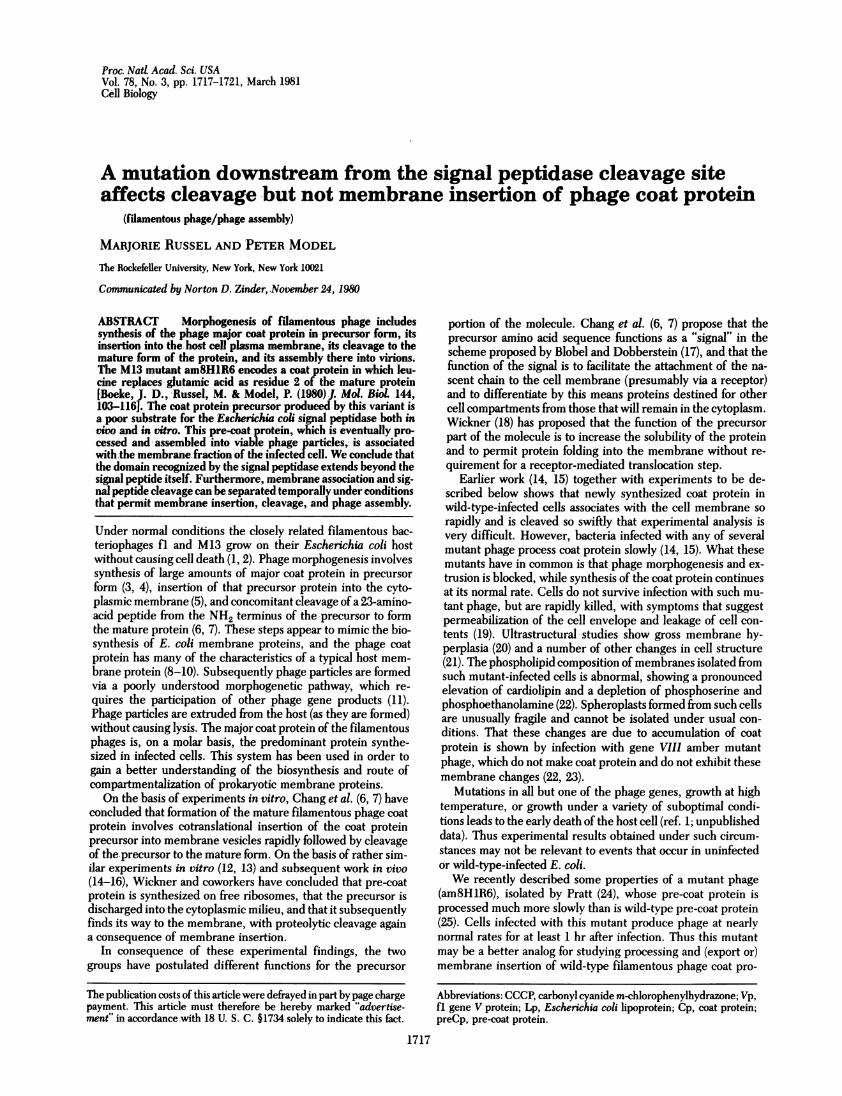
Processing of Pre-Coat Protein in En coli Infected with fl.In agreement with previous work (14, 15), we find that cellsinfected with wild-type fl phage cleave pre-coat protein veryrapidly, either while it is still a nascent chain or very shortly afterits synthesis is completed. Fig. 1 shows the results of the fastestpulse experiment we have been able to carry out. The timebetween the addition of [3S]methionine and the addition of the
I1cI .....i.X."..
w.s
m.'a'Z's'In...!!Xww..i}.*' . is _l s"I And _ _ __
¢SEg§l .buRg -
' ______
DreCup ._
_,Lp-oreCp
Cp
a. b c d e f g h k
FIG. 1. Rapid processing of wild-type pre-coat protein. Bacteriawere infected with wild-type fl at a multiplicity of 50. At 30 min afterinfection, 25 A.l of culture was added to 20 tLCi of [3S]methionine, and500 A1 of 5% trichloroacetic acid was added as rapidly as possible. Theelapsed time between addition of isotope and trichloroacetic acid was3 sec for one pulse and 6 sec for a second pulse. The subsequent pulseswere obtained by sampling 25 IL from a 300-pul aliquot of the infectedculture to which 100 uCi of [ S]methionine had been added. Incor-poration reached amaximum by 20 sec. The samples were precipitatedwith trichloroacetic acid, dried over NaOH under reduced pressure,resuspended in sample buffer, and analyzed on a NaDodSO4/urea/acrylamide gel. Lanes: a, 3 sec; b, 6 sec; c, 15 sec; d, 35 sec; e, 52 sec;f, 72 sec; g, 100 sec; h, 2 min; i, 4 min; j, 12 min; k, pre-coat protein; 1,
uninfected control. Lane a is a 6-day exposure. Lanes b-i are a 2.5-dayexposure. Vp, fl gene V protein; Lp, E. coli lipoprotein; preCp, pre-coatprotein; Cp, coat protein.
bp e-- m- -
Lpl-preCp= d..
,pr _ r_ ___
a b c d e f g h
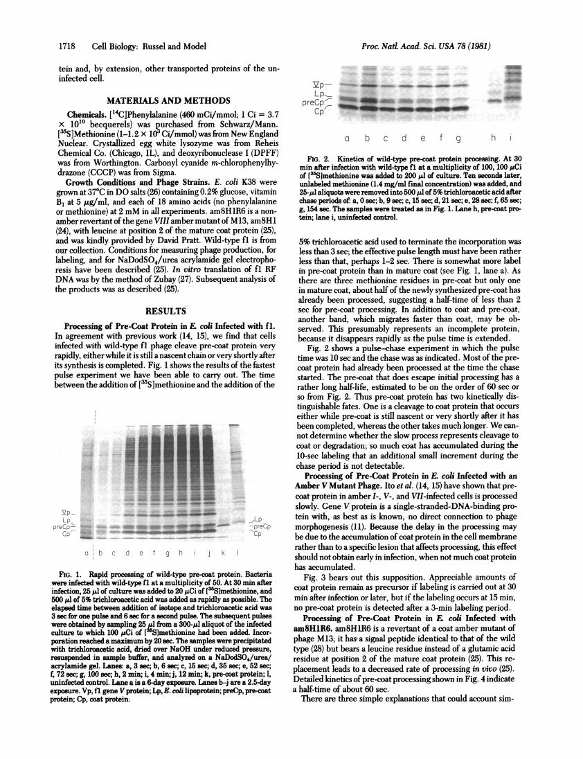
FIG. 2. Kinetics of wild-type pre-coat protein processing. At 30min after infection with wild-type fl at a multiplicity of 100, 100 ;LCiof [3Slmethionine was added to 200 Al of culture. Ten seconds later,unlabeled methionine (1.4 mg/ml final concentration) was added, and25-,M' aliquotswere removed into 500 ,ul of 5% trichloroacetic acid afterchase periods of: a, 0 sec; b, 9 sec; c, 15 sec; d, 21 sec; e, 28 sec; f, 65 sec;g, 154 sec. The samples were treated as in Fig. 1. Lane h, pre-coat pro-tein; lane i, uninfected control.
5% trichloroacetic acid used to terminate the incorporation wasless than 3 sec; the effective pulse length must have been ratherless than that, perhaps 1-2 sec. There is somewhat more labelin pre-coat protein than in mature coat (see Fig. 1, lane a). Asthere are three methionine residues in pre-coat but only onein mature coat, about half of the newly synthesized pre-coat hasalready been processed, suggesting a half-time of less than 2see for pre-coat processing. In addition to coat and pre-coat,another band, which migrates faster than coat, may be ob-served. This presumably represents an incomplete protein,because it disappears rapidly as the pulse time is extended.
Fig. 2 shows a pulse-chase experiment in which the pulsetime was 10 sec and the chase was as indicated. Most of the pre-coat protein had already been processed at the time the chasestarted. The pre-coat that does escape initial processing has arather long half-life, estimated to be on the order of 60 sec orso from Fig. 2. Thus pre-coat protein has two kinetically dis-tinguishable fates. One is a cleavage to coat protein that occurseither while pre-coat is still nascent or very shortly after it hasbeen completed, whereas the other takes much longer. We can-not determine whether the slow process represents cleavage tocoat or degradation; so much coat has accumulated during the10-sec labeling that an additional small increment during thechase period is not detectable.
Processing of Pre-Coat Protein in E. coli Infected with anAmber V Mutant Phage. Ito et al. (14, 15) have shown that pre-coat protein in amber I-, V-, and VII-infected cells is processedslowly. Gene V protein is a single-stranded-DNA-binding pro-tein with, as best as is known, no direct connection to phagemorphogenesis (11). Because the delay in the processing maybe due to the accumulation of coat protein in the cell membranerather than to a specific lesion that affects processing, this effectshould not obtain early in infection, when not much coat proteinhas accumulated.
Fig. 3 bears out this supposition. Appreciable amounts ofcoat protein remain as precursor if labeling is carried out at 30min after infection or later, but if the labeling occurs at 15 min,no pre-coat protein is detected after a 3-min labeling period.
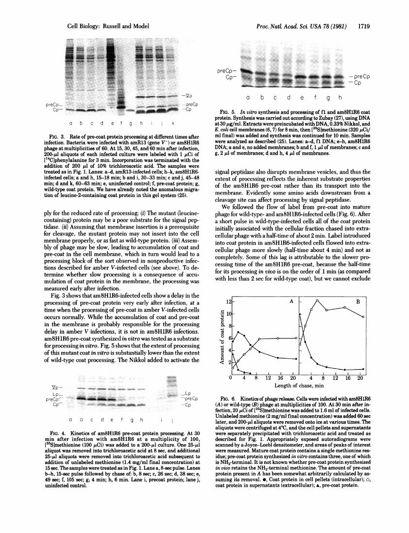
Processing of Pre-Coat Protein in E. coli Infected witham8HlR6. am8HlR6 is a revertant of a coat amber mutant ofphage M13; it has a signal peptide identical to that of the wildtype (28) but bears a leucine residue instead of a glutamic acidresidue at position 2 of the mature coat protein (25). This re-placement leads to a decreased rate of processing in vivo (25).Detailed kinetics of pre-coat processing shown in Fig. 4 indicatea half-time of about 60 sec.There are three simple explanations that could account sim-
Proc. Nad Acad. Sci. USA 78 (1981)
Proc. Nati Acad. Sci. USA 78 (1981) 1719
S-w sas-!om...... ....
.. ......
-momm -lfa VP
preCp-.. ... -preCpprCp_ l-prCp
a b c d e f g h k
FIG. 3. Rate of pre-coat protein processing at different times afterinfection. Bacteria were infected with amR13 (gene V-) or am8HlR6phage at multiplicities of 60. At 15, 30, 45, and 60 min after infection,200-gl aliquots of each infected culture were labeled with 1 uCi of[14C]phenylalanine for 3 min. Incorporation was terminated with theaddition of 200 /.l of 10% trichloroacetic acid. The samples weretreated as in Fig. 1. Lanes: a-d, amR13-infected cells; h-k, am8HlR6-infected cells; a and h, 15-18 min; b and i, 30-33 min; c and j, 45-48min; d and k, 60-63 min; e, uninfected control; f, pre-coat protein; g,
wild-type coat protein. We have already noted the anomalous migra-tion of leucine-2-containing coat protein in this gel system (25).
ply for the reduced rate of processing: (i) The mutant (leucine-containing) protein may be a poor substrate for the signal pep-
tidase. (ii) Assuming that membrane insertion is a prerequisitefor cleavage, the mutant protein may not insert into the cellmembrane properly, or as fast as wild-type protein. (iii) Assem-bly of phage may be slow, leading to accumulation of coat andpre-coat in the cell membrane, which in turn would lead to a
processing block of the sort observed in nonproductive infec-tions described for amber V-infected cells (see above). To de-termine whether slow processing is a consequence of accu-
mulation of coat protein in the membrane, the processing wasmeasured early after infection.
Fig. 3 shows that am8HlR6-infected cells show a delay in theprocessing of pre-coat protein very early after infection, at a
time when the processing of pre-coat in amber V-infected cellsoccurs normally. While the accumulation of coat and pre-coatin the membrane is probably responsible for the processingdelay in amber V infections, it is not in am8HlR6 infections.am8HlR6 pre-coat synthesized in vitro was tested as a substratefor processing in vitro. Fig. 5 shows that the extent of processingof this mutant coat in vitro is substantially lower than the extentof wild-type coat processing. The Nikkol added to activate the
Vp-
Lp-preCp-
Cp-
..... _Lp-preCp-Cp
a b c de f g h
FIG. 4. Kinetics of am8HlR6 pre-coat protein processing. At 30min after infection with am8HlR6 at a multiplicity of 100,[355]methionine (100 uCi) was added to a 200-,ul culture. One 25-t.laliquot was removed into trichloroacetic acid at 8 sec, and additional25-1.I aliquots were removed into trichloroacetic acid subsequent toaddition of unlabeled methionine (1.4 mg/ml final concentration) at15 sec. The samples were treated as in Fig. 1. Lane a, 8-sec pulse. Lanesb-h, 15-sec pulse followed by chase of: b, 8 sec; c, 26 sec; d, 38 sec; e,49 sec; f, 105 sec; g, 4 min; h, 6 min. Lane i, precoat protein; lane j,uninfected control.
preCp- 1%, m- rep
O F 41 dw _iOf -peCp* 4AW -OCp
a b c d e f g h
FIG. 5. In vitro synthesis and processing of fl and am8HlR6 coatprotein. Synthesis was carried out according to Zubay (27), using DNAat 30 ,ug/ml. Extracts were preincubated with DNA, 0.33% Nikkol, andE. coli cell membranes (6, 7) for 8 min, then [35S]methionine (320 ,uCi/ml final) was added and synthesis was continued for 10 min. Sampleswere analyzed as described (25). Lanes: a-d, fl DNA; e-h, am8HlR6DNA; a and e, no added membranes; b and f, 1 1.l of membranes; c andg, 2 gl of membranes; d and h, 4 .lI of membranes.
signal peptidase also disrupts membrane vesicles, and thus theextent of processing reflects the inherent substrate propertiesof the am8HlR6 pre-coat rather than its transport into themembrane. Evidently some amino acids downstream from acleavage site can affect processing by signal peptidase.We followed the flow of label from pre-coat into mature
phage for wild-type- and am8HlR6-infected cells (Fig. 6). Aftera short pulse in wild-type-infected cells all of the coat proteininitially associated with the cellular fraction chased into extra-cellular phage with a half-time of about 2 min. Label introducedinto coat protein in am8HlR6-infected cells flowed into extra-cellular phage more slowly (half-time about 4 min) and not ascompletely. Some of this lag is attributable to the slower pro-cessing time of the am8HlR6 pre-coat, because the half-timefor its processing in vivo is on the order of 1 min (as comparedwith less than 2 sec for wild-type coat), but we cannot exclude
o1 :02 A8 2 1B048 1 6 2
0
0
0
Length of chase, mmn
FIG. 6. Kinetics of phage release. Cells were infected with am8HiR6(A) or wild-type (B) phage at multiplicities of 100. At 30 mmn after in-fection, 20 ,uCi of [355]methionine was added to 1.6 ml of infected cells.Unlabeled methionine (2mg/mi final concentration) was added 60 seclater, and 200-,ul aliquots were removed onto ice at various times. Thealiquots were centrifuged at 400, and the cell pellets and supernatantswere separately precipitated with trichloroacetic acid and treated asdescribed for Fig. 1. Appropriately exposed autoradiograms werescanned by a Joyce-Loebl densitometer, and areas of peaks of interestwere measured. Mature coat protein contains a single methionine res-idue; pre-coat protein synthesized in vitro contains three, one of whichis NH2-terminal. It is not known whether pre-coat protein synthesizedin vivo retains the NH2-terminal methionine. The amount of pre-coatprotein present in A has been somewhat arbitrarily calculated by as-suming its removal. *, Coat protein in cell pellets (intracellular); a,coat protein in supernatants (extracellular); *, pre-coat protein.
Cell Biology: Russell and Model
I
1720 Cell Biology: Russel and Model
preCp- qW0UfCp-
d e f g h j k m
0 20 40 60 80 100 120 140Time after infection, min
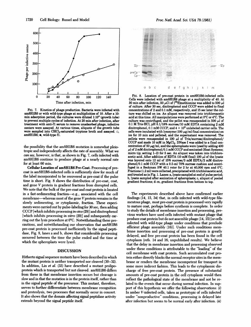
FIG. 7. Kinetics of phage production. Bacteria were infected witham8H1R6 or with wild-type phage at multiplicities of 10. After a 10-min adsorption period, the cultures were diluted 1:104 (growth tube)to prevent multiple cycles of infection. At 20 min after infection, aftertreatment with anti-fl serum to remove unadsorbed phage, infectivecenters were assayed. At various times, aliquots of the growth tubewere sampled into CHCl3-saturated tryptone broth and assayed. o,am8H1R6; *, wild-type fl.
the possibility that the am8H1R6 mutation is somewhat pleio-tropic and independently affects the rate of assembly. What wecan say, however, is that, as shown in Fig. 7, cells infected witham8HlR6 continue to produce phage at a nearly normal ratefor at least 60 min.
Cellular Location of am8HlR6 Pre-Coat. Processing of pre-coat in am8HlR6-infected cells is sufficiently slow for much ofthe label incorporated to be recovered as pre-coat if the pulsetime is short. Fig. 8 shows the distribution of pre-coat, coat,and gene V protein in gradient fractions from disrupted cells.We note that the bulk of the pre-coat and coat protein is locatedin a fast-sedimenting fraction-e.g., associated with the cellmembrane-whereas most of the gene V protein remains in theslowly sedimenting, or cytoplasmic, fraction. These experi-ments were carried out by pulse-labeling for 30 sec, then addingCCCP [which inhibits processing in vivo (29)] and dinitrophenol[which inhibits processing in vitro (29)] and subsequently car-
ring out the lysis procedure at 0°C. Notwithstanding these pre-
cautions, and notwithstanding our observation that am8HlR6pre-coat protein is processed inefficiently by the signal pepti-dase, Fig. 8, lanes a and b, shows that considerable processingoccurred between the time the pulse ended and the time atwhich the spheroplasts were lysed.
DISCUSSIONHitherto signal sequence mutants have been described in whichthe mutant protein is neither transported nor cleaved (30-32).In addition, Lin et al. (33) have described a mutant prolipo-protein which is transported but not cleaved. am8HlR6 differsfrom these in that membrane insertion occurs but cleavage isslow and in that the mutation is in the protein itself, rather thanin the signal peptide of the precursor. This mutant, therefore,serves to further differentiate between membrane recognitionand proteolysis, two processes that normally occur in concert.It also shows that the domain affecting signal peptidase activityextends beyond the signal peptide itself.
FIG. 8. Location of pre-coat protein in am8HlR6-infected cells.Cells were infected with am8HlR6 phage at a multiplicity of 40. At30 min after infection, 50 ,uCi of [3Slmethionine was added to 500 Alof culture. After 30 sec, dinitrophenol and CCCP were added to finalconcentrations of 2 and 0.1 mM, respectively, and 15 sec later the cul-ture was chilled on ice. An aliquot was removed into trichloroaceticacid at this time. All manipulations were performed at 000 or 4VC. Theculture was centrifuged, and the pellet was resuspended in 500 Al of0.1 M Tris-HCl, pH 8.1/18% sucrose/10 mM EDTA containing 2 mMdinitrophenol, 0.1 mM CCCP, and 4 x 10⁹ unlabeled carrier cells. Thecells were incubated with lysozyme (100 ,g/ml final concentration) onice for 10 min and pelleted, and the supernatant was removed. Thepellets were resuspended in 100 Al of Tris/sucrose/dinitrophenol/CCCP and made 10 mM in MgCl2. DNase I was added to a final con-centration of 50 ,ug/ml, and the spheroplasts were lysed by adding 400,ul of 2 mM dinitrophenol/0.1 mM CCCP and sonicated (Heat Systems;micro tip; setting 1-2) for 5 sec. An aliquot was taken into trichloro-acetic acid. After addition of EDTA (10mM final) 100 yl of the lysatewas layered onto 12 ml of 15% sucrose/3 mM EDTA/2 mM dinitro-phenol/0.1 mM CCCP with a 0.5-ml 70% sucrose cushion and centri-fuged in a Beckman SW 40.1 rotor for 2 hr at 40,000 rpm and 00C.Fractions (1.2 ml) were collected, precipitated with trichloroacetic acid,and treated as in Fig. 1. Lanes: a, lysate sampled at end of pulse period;b, lysate sampled after sonication of spheroplasts; c, aliquot of pooledgradient fractions; d-m, gradient fractions from bottom to top.
The experiments described above have confirmed earlierfindings (14, 15, 34) that, in cells infected with wild-type fila-mentous phage, most pre-coat protein is processed very rapidlyto mature coat, perhaps before synthesis is complete. In orderto study the details of membrane insertion and processing, pre-vious workers have used cells infected with mutant phage thatproduce coat protein but do not assemble phage (14, 15) [or cellsinfected with wild-type phage under conditions that precludeefficient phage assembly (16)]. Under such conditions mem-brane insertion and processing of pre-coat protein is greatlydelayed, and free pre-coat protein has been found in the cellcytoplasm (refs. 14 and 16; unpublished results). We believethat the delay in membrane insertion and processing observedunder these conditions is attributable to the "loading" of thecell membrane with coat protein. Such accumulated coat pro-tein either directly blocks the normal receptor sites in the mem-brane or renders the membrane incompetent for transport insome more indirect fashion. This leads to the cytoplasmic dis-charge of free pre-coat protein. The presence of substantialamounts of pre-coat protein in the cell cytoplasm would thenreflect the pathological state of the membrane and not be re-
lated to the events that occur during normal infection. In sup-port of this hypothesis we offer the following observations: (i)In amber V-infected cells, taken as prototypic of cells infectedunder "nonproductive" conditions, processing is delayed lateafter infection but seems to be normal early after infection. (ii)
600F
500 -
400_
300_
200k_
CD)
0)
0)0Q
0)cO~
100_a b c
Proc. Nad Acad. Sci. USA 78 (1981)
0
1 1 1 1

Proc. NatL Acad. Sci. USA 78 (1981) 1721
Processing of the pre-coat protein of am8HlR6 is delayed bothearly and late after infection. The proximal cause of this pro-cessing delay appears to be a defective signal processing site,because in vitro cleavage of the mutant pre-coat protein is slow,and because assembly and export of viable phage in cells in-fected with this mutant seem to be normal. (iii) Pre-coat proteinproduced in cells infected with am8HlR6 is membrane asso-ciated; it is not found free in the cytoplasm. (iv) Pre-coat proteinin cells infected with wild-type M13 that are treated with CCCPto inhibit processing is membrane associated, not free in thecytoplasm (13, 35).
Even in wild-type infections a small fraction of pre-coat pro-tein may fail to associate properly with the membrane. Thiswould account for the minor, kinetically distinguishable formof pre-coat seen in Figs. 1 and 2, which has a long half-life.We reported earlier (6, 7) that efficient sequestration and
proteolytic cleavage of fl pre-coat protein into membrane ves-icles occurs only when the vesicles are present during synthesisof the pre-coat protein, and not when they are added after trans-lation is finished. This result was also obtained by Mandel andWickner (12). On this basis, and reasoning by analogy with eu-karyotic systems, we postulated that the "signal hypothesis" (17,36) is applicable to the fl coat protein system.
Mandel and Wickner (12) made the additional finding thatproteolysis by a detergent extract of membranes can also occurposttranslationally, but only if a detergent is present duringtranslation. They suggested that the detergent prevents thefolding of the completed protein and leaves the signal peptideexposed. This latter result supports our hypothesis that in vivothe interaction between pre-coat protein and the membraneoccurs while the pre-coat is still a nascent chain.
Our current outline of the process of membrane insertion/proteolysis of filamentous phage coat protein follows: Synthesisof fl pre-coat occurs with concomitant binding of the NH2-ter-minal amino acids (signal peptide) to the plasma membrane ofthe infected cell, which accounts for the rapidity with whichwild-type filamentous coat and pre-coat become membraneassociated. It also accounts for the rapid membrane associationof the pre-coat protein of mutants such as am8HlR6 and for theappearance in wild-type-infected cells of structures that, in theelectron microscope, closely resemble eukaryotic rough en-doplasmic reticulum (20). Under "nonproductive" conditions,the membrane is overloaded and becomes incompetent to sup-port this association. The resulting pre-coat protein will tendto end up in the cytoplasm. Proteolytic cleavage of pre-coat iscontingent upon and follows successful insertion of the coatprotein into the membrane but is not required for the mem-brane association per se. From our failure to find pre-coat pro-tein in mature am8HlR6 phage, and on a priori grounds, pro-teolytic cleavage is probably a prerequisite for phage assembly.
There are probably at least three discrete steps required forthe successful integration of coat protein: the initial recognitionbetween the signal peptide and the membrane (or membranereceptor), actual insertion, and cleavage. Date et al. (35) reportthat, in cells treated with CCCP, pre-coat is membrane asso-ciated but not accessible to the signal peptidase. Furthermore,while an intact signal peptide is necessary for transport (30-32),it is not always sufficient (37), which again suggests a postrec-ognition step.We have no direct evidence bearing on the orientation of
am8HlR6 pre-coat protein in the membrane of the infectedcell. However, we observe cleavage to mature coat protein in
dinitrophenol. In contradistinction to pre-coat protein formedin the presence of CCCP, therefore, am8HlR6 pre-coat canbecome accessible to the signal peptidase and must thereforebe in a different state.
We thank N. D. Zinder for advice and for critically reading the man-
uscript, J. D. Boeke for helpful suggestions, and D. Pratt for providingam8HlR6. This work was supported in part by grants from the NationalInstitutes of Health and the National Science Foundation.
1. Marvin, D. A. & Hohn, B. (1969) Bacteriot Rev. 33, 172-209.2. Hoffman-Berling, H., Durwald, H. & Beulke, I. (1963) Z. Na-
turforsch. 18b 893-898.3. Pieczenik G., Model P. & Robertson, H. D. (1974)J. Mol Biol 90,
191-214.4. Sugimoto, K., Sugisaki, H., Okamoto, T. & Takanami, M. (1977)
J. Mol Biol 111, 487-507.5. Webster, R. E. & Cashman, J. S. (1978) in The Single-Stranded
Phages eds. Denhardt, D. T., Dressler, D. & Ray, D. S. (ColdSpring Harbor Laboratory, Cold Spring Harbor, New York, pp.
557-569.6. Chang, C. N., Blobel, G. & Model, P. (1978) Proc. Natl Acad. Sci.
USA 75, 361-365.7. Chang, C. N., Model, P. & Blobel, G. (1979) Proc. Natl Acad. Sci.
USA 76, 1251-1255.8. Makino, S., Woolford, J. L., Tanford, C. & Webster, R. E. (1976)
J. Biol Chem. 250, 4327-4332.9. Wickner, W (1975) Proc. Natl Acad. Sci. USA 72, 4749-4753.
10. Wickner, W (1976) Proc. Natl Acad. Sci. USA 73, 1159-1163.11. Horiuchi, K., Vovis, G. F. & Model, P. (1978) in The Single-
Stranded DNA Phages, eds. Denhardt, D. T., Dressler, D. & Ray,D. (Cold Spring Harbor Laboratory, Cold Spring Harbor, NewYork), pp. 113-137.
12. Mandel, G. & Wickner, W. (1979) Proc. Natl Acad. Sci. USA 76,236-240.
13. Wickner, W., Mandel, G., Zwizinski, C., Bates, M. & Killick, T.(1978) Proc. Natl Acad. Sci. USA 75, 1754-1758.
14. Ito, K., Mandel, G. & Wickner, W. (1979) Proc. Natl Acad. Sci.USA 76, 1199-1203.
15. Ito, K., Date, T. & Wickner, W. (1980) J. Biol Chem. 255,2123-2130.
16. Date, T. & Wickner, W. T. (1980)J. Virol, in press.
17. Blobel, G. & Dobberstein, B. (1975)J. Cell Biol 67, 835-851.18. Wickner, W. (1979) Annu. Rev. Biochem. 48, 23-45.19. Hohn, B., von Schutz, H. & Marvin, D. A. (1971)J. Mol Biol 56,
155-165.20. Schwartz, F. M. & Zinder, N. D. (1967) Virology 34, 352-355.21. Onishi, Y. & Kuwano, M. (1971)J. Virol 7, 673-678.22. Woolford, J. L., Jr., Cashman, J. S. & Webster, R. E. (1974) Vi-
rology 58, 544-560.23. Webster, R. E. & Rementer, M. (1980)J. Mol Biol. 139,393-405.24. Pratt, D., Tzagoloff, H. & Beaudoin, J. (1969) Virology 39, 42-53.25. Boeke, J. D., Russel, M. & Model, P. (1980) J. Mol Biol 144,
103-116.26. Vogel, H. J. & Bonner, D. M. (1956)J. Biol Chem. 218, 97-106.27. Zubay, G. (1973) Annu. Rev. Genet. 7, 267-287.28. Boeke, J. D. & Model, P. (1979) Virology 96, 299-301.29. Date, T., Zwizinski, C., Ludmerer, S. & Wickner, W. (1980) Proc.
Natl Acad. Sci. USA 77, 827-831.30. Emr, S. D., Schwartz, M. & Silhavy, T. (1978) Proc. Natl Acad.
Sci. USA 75, 5802-5806.31. Bedouelle, H., Bassford, P. J., Jr., Fowler, A. V., Zabin, I., Beck-
with, J. & Hofnung, M. (1980) Nature (London) 285, 78-85.32. Bassford, P. & Beckwith, J. (1979) Nature (London) 277, 538-541.33. Lin, J. J. C., Kanazawa, H., Ozols, J. & Wu, H. C. (1978) Proc.
NatL Acad. Sci. USA 75, 4891-4895.34. Smilowitz, H., Carson, J. & Robbins, P. W. (1972) 1. Supramot
Struct. 1, 8-18.35. Date, T., Goodman, J. M. & Wickner, W. (1980) Proc. Natl Acad.
Sci. USA 77, 4669-4673.36. Emr, S. D., Hall, M. N. & Silhavy, T. J. (1980)J. Cell Biology 86,
701-711.am8HlR6-infected cells during the preparation of membraneseven when the preparations are held in a mixture of CCCP and
Cell Biology: Russel and Model
37. Moreno, F., Fowler, A., Hall, M., Silhavy, T. J., Zabin, I. &Schwartz, M. (1980) Nature (London) 286, 356-359.