a mobile secretory vesicle cluster involved in mass transport from the golgi to the plant cell
TRANSCRIPT

A Mobile Secretory Vesicle Cluster Involved in Mass Transportfrom the Golgi to the Plant Cell Exterior W OA
Kiminori Toyooka,a Yumi Goto,a Satoru Asatsuma,a,b Masato Koizumi,a Toshiaki Mitsui,c and Ken Matsuokaa,b,1
a RIKEN Plant Science Center, Tsurumi-ku, Yokohama 230-0045, Japanb Laboratory of Plant Nutrition, Faculty of Agriculture, Kyushu University, Higashi-ku, Fukuoka 812-8581, Japanc Laboratories of Plant and Microbial Genome Control, Graduate School of Science and Technology, Niigata University, Ikarashi,
Nishi-ku, Niigata 950-2181, Japan
Secretory proteins and extracellular glycans are transported to the extracellular space during cell growth. These materials
are carried in secretory vesicles generated at the trans-Golgi network (TGN). Analysis of the mammalian post-Golgi
secretory pathway demonstrated the movement of separated secretory vesicles in the cell. Using secretory carrier
membrane protein 2 (SCAMP2) as a marker for secretory vesicles and tobacco (Nicotiana tabacum) BY-2 cell as a model
cell, we characterized the transport machinery in plant cells. A combination of analyses, including electron microscopy of
quick-frozen cells and four-dimensional analysis of cells expressing fluorescent-tagged SCAMP2, enabled the identification
of a clustered structure of secretory vesicles generated from TGN that moves in the cell and eventually fuses with plasma
membrane. This structure was termed the secretory vesicle cluster (SVC). The SVC was also found in Arabidopsis thaliana
and rice (Oryza sativa) cells and moved to the cell plate in dividing tobacco cells. Thus, the SVC is a motile structure involved
in mass transport from the Golgi to the plasma membrane and cell plate in plant cells.
INTRODUCTION
The division and expansion of cells requires trafficking of lipids,
proteins, and polysaccharides to the plasma membrane (PM)
and extracellular space. Thesemolecules are synthesized and/or
modified in the Golgi apparatus and sorted into secretory ves-
icles at the trans-Golgi network (TGN) for transport to the PM.
The Golgi apparatus in plants differs from that in mammals in
several aspects, including spatial organization, dynamic prop-
erties, and functional activity. Numerous Golgi apparatus are
dispersed throughout the cytoplasm in plant cells and move
along actin cytoskeletal elements (Nebenfuhr et al., 1999). One
function of the plant Golgi apparatus is as the factory for
noncellulose extracellular glycans, including hemicellulose and
pectin (Cosgrove, 2005). Rapidly dividing plant cells need to
synthesize large quantities of these glycans and contain several
hundred to thousands of Golgi stacks (Nebenfuhr et al., 1999).
The unique features of the plant Golgi apparatus also reflect a
difference in the secretory machinery between plants and mam-
mals. Mammalian and yeast cells share a similar transport system
for secretory vesicles (Bednarek and Falbel, 2002), and the sorting
of proteins at the late secretory pathway is influenced by early,
late, and recycling endosomes. In plant cells, the prevacuolar
compartments (PVCs), multivesicular bodies (MVBs), partially
coated reticulum, tubuvesicular endosomes, and vacuoles are
involved in secretion and are collectively termed the post-Golgi
compartments (Surpin and Raikhel, 2004; Lam et al., 2007b).
However, it is not yet clear whether these are the sole elements in
the late secretory pathway of plants and how they interact during
the secretory process.
Electron microscopy observations of high-pressure frozen
cells have indicated that isolated TGN-like structures or post-
Golgi vesicle clusters are found in a number of plant cells
(Samuels et al., 2002; Saint-Jore-Dupas et al., 2004, Staehelin
and Kang, 2008). In recent years, various trafficking components
in plant cells have also been genetically characterized (Rojo and
Denecke, 2008); however, functional analysis of these structures
and trafficking components has been limited by the lack of
suitable molecular markers in this compartment.
Secretory vesicles contain divergent classes of molecules: not
only the soluble protein cargo, but also membrane proteins that
are involved in the trafficking process itself. One group of
proteins that has been identified as a component of secretory
vesicles are the secretory carrier membrane proteins (SCAMPs)
(Brand et al., 1991; Takamori et al., 2006). The conserved
structure of SCAMPs consists of a cytoplasmic N-terminal
domain with repeated asparagin-proline-phenylalanine (NPF)
sequences, four transmembrane regions, and a cytoplasmic
tail (Fernandez-Chacon and Sudhof., 2000). Genome-wide anal-
ysis has shown that plants also possess a SCAMP gene family
(Fernandez-Chacon and Sudhof, 2000), and analysis of rice
(Oryza sativa) SCAMP1 revealed an involvement endocytosis
(Lam et al., 2007a, 2007b).
In this article, we investigated the tobacco (Nicotiana tabacum)
SCAMP2 protein based on its putative function in the late
secretory pathway. Fluorescent tagging and immunoelectron
1Address correspondence to [email protected] author responsible for distribution of materials integral to thefindings presented in this article in accordance with the policy describedin the Instructions for Authors (www.plantcell.org) is: Ken Matsuoka([email protected]).WOnline version contains Web-only data.OAOpen Access articles can be viewed online without a subscription.www.plantcell.org/cgi/doi/10.1105/tpc.108.058933
The Plant Cell, Vol. 21: 1212–1229, April 2009, www.plantcell.org ã 2009 American Society of Plant Biologists
Dow
nloaded from https://academ
ic.oup.com/plcell/article/21/4/1212/6095306 by guest on 25 O
ctober 2021

microscopy (immuno-EM) analyses of tobacco BY-2 cells re-
vealed that SCAMP2 is localized in the PM, the TGN, and a
mobile structure that we term the secretory vesicle cluster (SVC).
Analysis of the SVC using total internal reflection fluorescence
microscopy, four-dimensional confocal laser scanning micros-
copy, and SCAMP2 as a marker demonstrated that it matures
from a tubulovesicular structure that might be identical to or is
derived from the TGN. Moreover, the SVC was observed to fuse
with the PM in nondividing cells and to be targeted to the cell
plate in dividing cells. Based on these results, we propose that
the SVC is a mobile compartment containing SCAMP2 that is
involved in mass transport from the Golgi apparatus to the cell
exterior in plants.
RESULTS
Subcellular Localization of Tobacco SCAMP2
Toobtain a deeper understanding of endomembrane organelles in
plant cells, we have been characterizing membrane proteins
potentially involved in vesicular trafficking as suggested by tran-
scriptomic analysis of tobacco BY-2 cells (Matsuoka et al., 2004).
Polyclonal antibodies developed against candidate proteins were
used to compare their distribution in BY-2 cells with that of
organelle markers (Figure 1; Matsuoka et al., 1997). One of these
proteins, a tobaccoSCAMPhomolog (referred toasSCAMP2; see
below), displayed a unique distribution pattern on sucrose gradi-
ents for subcellular fractionation (Figure 1). Because its fraction-
ation pattern corresponded to localization to the PM and Golgi
apparatus, SCAMP2 was chosen for further analysis.
The anti-SCAMP2 antibody recognized two closely associated
bands of ;29 kD and a band of 27 kD that were present in
identical membrane fractions of tobacco BY-2 cells at varied
intensity ratios. Similar multiple bands were observed with an-
tibodies against mammalian SCAMP2 (Liu et al., 2002) and
SCAMP2-yellow fluorescent protein (YFP) in BY-2 cells (de-
scribed below), suggesting that the signals observed in Figure
1 were specific for the tobacco SCAMP homolog(s) and an
accurate representation of its distribution. Little signal was found
in the higher molecular weight regions in the same immunoblot,
suggesting that the antibody raised against tobacco SCAMP2 is
specific for the protein. The sequence of the encoding full-length
cDNA and deduced SCAMP2 protein was then determined.
Based on amino acid sequence comparison with other species,
the tobacco SCAMP homolog was classified as a member of the
SCAMP2 family and thus is hereafter referred to as SCAMP2 (see
Supplemental Figure 1 online). Interestingly, the C-terminal Tyr
motif was only found in the plant homologs (see Supplemental
Figure 1 online).
Staining of tobacco BY-2 cells with anti-SCAMP2 antibody
followed by confocal laser scanningmicroscopy (CLSM) analysis
Figure 1. Distribution of SCAMP2 within Endomembrane Organelles.
Microsomes were prepared from BY-2 cells in the presence of Mg2+ and subjected to isopycnic sucrose density gradient ultracentrifugation. The
resulting gradients were fractionated from the bottom into 24 fractions. The concentration of sucrose in the gradient is shown at the top. The distribution
of marker proteins was analyzed by immunoblotting with specific antibodies, against PM intrinsic protein for PM, Sec61 for the ER, and V-PPase for the
vacuolar membrane. The enzyme activity of inosine disphosphatase (IDPase) as a marker for the Golgi apparatus was measured. The distribution of
SCAMP2 was analyzed by immunoblotting with anti-SCAMP2 antibody.
SCAMP2 and the Secretory Vesicle Cluster 1213
Dow
nloaded from https://academ
ic.oup.com/plcell/article/21/4/1212/6095306 by guest on 25 O
ctober 2021

revealed strong fluorescence at intracellular dot structures (Fig-
ure 2A). The PM and cell plates were also stained with this
antibody, although the signals at the PM were sometimes ob-
scure. Localization in the PM and at intracellular dots was also
observed for fusion proteins of SCAMP2 with monomeric red
fluorescent protein (mRFP) and with YFP in tobacco BY-2 cells
(Figure 2B). Colocalization of signals in the same intracellular
dots and PM was also observed when both fusion constructs
were coexpressed in the same BY-2 cell (Figure 2B) and when
BY-2 cells expressing SCAMP2-YFP were stained with anti-
SCAMP2 antibody (Figure 2C). In addition, analysis of SCAMP2-
YFP subcellular localization by cell fractionation showed a similar
distribution in the gradient and a similar multiple band pattern as
the endogenous SCAMP2, corresponding to the PM and Golgi
fractions (Figure 2D). These results confirmed that the SCAMP2-
YFP fusion protein correctly represents localization of endoge-
nous SCAMP2.
To test if the SCAMP2-containing dot structure could be an
endosomal organelle, the localization of SCAMP2-YFP was
compared with that of the endosomal marker dye FM4-64 by
confocal microscopy (Figure 3A). Cells were stained with 33 mM
FM4-64 on ice and washed with cold medium, after which they
were returned to room temperature to resume intracellular traf-
ficking. At early stages (5 to 10 min) after FM4-64 incubation, we
observed dotted structures of FM4-64 signals in a background
weak fluorescence signal showing similar pattern to cytosol
stained by 5- (and 6-) chloromethyl SNARF-1 (Yuasa et al.,
2005). The cytosolic pattern was clearly distinct from the pattern
of the endoplasmic reticulum (ER) (see Supplemental Figure 2A
online). Lower concentration of FM4-64 (17 mM) showed an
essentially identical pattern (see Supplemental Figure 2B online).
The dotted structures emitting strong red FM4-64 fluorescence
rarely colocalizedwith YFP signals inmajor population of the cells
(9%6 5.9%colocalization, n = 7 cells; Figure 3A, top), whereas a
minor population of cells showed more frequent colocalization
(63%6 11% colocalization, n = 4 cells). After 30 to 40 min, there
was a uniform and high colocalization between the YFP-positive
SCAMP2-YFP dots and FM4-64 positive dots (88% 6 6.2%, n =
10; Figure 3A,middle). At later stages (90 to 120min), FM4-64was
transported to vacuolarmembranes, whereasSCAMP2-YFPwas
not detected at the tonoplast (Figure 3A, bottom). Cells stained at
room temperature with FM4-64 and AM4-65 (a fixable analog of
FM4-64; Lam et al., 2008) showed strong staining of the PM,
followed by appearance of FM4-64 (Figure 3B) and AM4-65
(Figure 3C) positive dots at 5 to 10min after incubation thatmainly
colocalized with SCAMP2-YFP. These results suggest that the
styrl dye–stainedPMwas rapidly internalized into theplant cells at
room temperature and colocalized with SCAMP2-positive dots.
Following internalization, FM4-64 fluorescence is transported
to the Golgi apparatus in addition to endosomes and vacuolar
membranes (Bolte et al., 2004; Dettmer et al., 2006). The pos-
sibility that the SCAMP2-positive dots represented the Golgi
apparatus was therefore investigated. We have previously re-
ported that prolyl hydroxylase (PH) cycles between the ER and
the Golgi apparatus and is predominantly found in the cis-
located cisternae of the Golgi (Yuasa et al., 2005). This feature
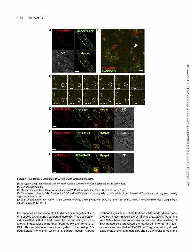
was used to mark the cis-Golgi by coexpressing a PH-mRFP
fusion protein together with SCAMP2-YFP (Figure 4A). Many, but
not all, of the YFP-positive dots were localized in close proximity
to the mRFP signals (Figure 4B) and were seen moving together
in the cell when traced using a confocal time-lapse scanning
program (Figure 4C). Based on these results, we predicted that a
significant proportion of SCAMP2would localize to the TGN or to
organelles located near the trans-cisternae of the Golgi appara-
tus. When internalization of FM4-64 was observed in BY-2 cells
expressing a fusion protein of PH with green fluorescent protein
(GFP), FM4-64 signals were detected on the sides of the Golgi
apparatus after 30 to 40 min of incubation (see Supplemental
Figure 3A online). These observations strongly suggest that the
SCAMP2-positive dots showing the FM4-64 signal in Figure 3
represent the trans-Golgi cisternae or the TGN.
Figure 2. Subcellular Localization of SCAMP2 by CLSM.
(A) Immunolocalization of SCAMP2 in BY-2 cells using an anti-SCAMP2
antibody.
(B) Localization of SCAMP2-mRFP and SCAMP2-YFP fusion proteins in
BY-2 cells.
(C) Immunofluorescence labeling of BY-2 cells expressing SCAMP2-YFP
using anti-SCAMP2 antibody. Bars = 10 mm in (A) to (C).
(D) The distribution of SCAMP2-YFP analyzed by subcellular fractiona-
tion. Proteins in microsomal membranes prepared from SCAMP2-YFP
expressing BY-2 cells and separated by isopycnic sucrose density
gradient ultracentrifugation were separated by SDS-PAGE and fluores-
cence of SCAMP2-YFP in the polyacrylamide gel was detected using a
fluoroimager.
1214 The Plant Cell
Dow
nloaded from https://academ
ic.oup.com/plcell/article/21/4/1212/6095306 by guest on 25 O
ctober 2021

We next investigated whether SCAMP2 also localizes to the
post-Golgi component in the plant secretory pathway. In Arabi-
dopsis thaliana, the SNARE proteins SYP22 and SYP41 localize
to the membranes of the MVB/PVC/vacuoles and trans-Golgi/
TGN, respectively (Uemura et al., 2004; Foresti et al., 2006).
Fusion proteins containing YFP together with tobacco SYP22 or
tobacco SYP41 were expressed in BY-2 cells and showed
fluorescence patterns conceptually identical to that seen in
Arabidopsis (see Supplemental Figures 3B and 3C online). Co-
expression of the SNARE protein fusions with SCAMP2-mRFP
revealed a colocalization between YFP-SYP41 and SCAMP2-
mRFP positive dots (Figure 4D). Most, but not all, of the
SCAMP2-mRFP positive dots showed YFP-SYP41 positive sig-
nal (91%6 3% colocalization, n = 5 cells), although some of the
YFP-SYP41 positive structures did not contain SCAMP2-mRFP,
as the percentage of green dots showing red fluorescence
was a little lower in the same cells (80% 6 18% colocalization,
n = 5 cells). This observation suggests that most of the SCAMP2
and SYP41 localized in the same compartment, but some
population of each of them localized in compartments where
the other is absent.
In contrast with SYP41, YFP-SYP22 fluorescence did not
colocalize with SCAMP2-mRFP signals (Figure 4E), although
YFP-SYP22 fluorescence frequently colocalized with FM4-64
signals (see Supplemental Figure 3C online). These results
indicated that SCAMP2 does not accumulate in the MVB/PVC
post-Golgi compartment.
It has been reported that the Ypt3/Rab11 subfamily of Rab
GTPases is localized in TGN and subsequent secretory com-
partments (Chow et al., 2008). We isolated the tobacco Rab11D
cDNA and expressed the protein as a fusion with mRFP in the
BY-2 cells. mRFP-Rab11D fluorescence almost completely
colocalized with GFP-Pra3, which is reported to localize in
TGN (Inaba et al., 2002) (see Supplemental Figure 3D online).
Coexpression of mRFP-Rab11D and SCAMP2-YFP indicated
that the fluorescence dots of both proteins colocalized in the
same compartment about half the time (47% 6 5.9% colocal-
ization, n = 4 cells), but some dots were independent (Figure 4F).
These results are consistent with localization of SCAMP2 in the
TGN and organelles involved in the secretory pathway.
To confirm the involvement of SCAMP2 in the secretory path-
way, SCAMP2-YFP and PH-mRFP localization was studied in the
presence of Brefeldin A (BFA). BFA inhibits transport vesicle
formation at the Golgi apparatus and other membranes in the
secretory pathway.After 2 h in thepresenceof a lowconcentration
of BFA (5 mg/mL), SCAMP2-YFP had moved to the PM and PH-
mRFP was redistributed from the Golgi to the ER (Figure 5A). We
also analyzed the effect of wortmannin, which at 10 to 100 mM
inhibits the biosynthesis of both phosphatidylinositol 3- and
4- phosphates as well as phospholipids in tobacco BY-2 cells
(Matsuoka et al., 1995a). It was reported recently that wortmannin
at 16.5 mM induces a morphological change of rice SCAMP1
positive compartments in tobacco BY-2 cells from a punctate
structure to a ring shape (Lam et al., 2007a). By contrast, even in
the presence of the same concentration of wortmannin, most
SCAMP2-YFP structures remained punctate, with <1% assuming
a ring shape structure in transformed BY-2 cells (see Supplemen-
tal Figure 4 online). These data confirmed that SCAMP2-positive
dots are not the cis-Golgi, PVC/MVB, and rice SCAMP1 positive
endosomal organelles. Furthermore, these observations suggest
that the tobacco SCAMP2 and rice SCAMP1 trafficking pathways
in tobacco BY-2 cells are not identical.
We next investigated whether SCAMP2 recycles between the
trans-Golgi/TGN and the PM. Cells expressing both SCAMP2-
YFP and PH-mRFP were first treated with 5 mg/mL BFA for 2 h to
remove the signal from intracellular dot-shaped compartments
(Figure 5A). Cells were then washed with fresh medium without
BFA and transferred tomedium containing cycloheximide (CHX),
a potent inhibitor of protein synthesis. After 1 h, cells contained
intracellular dots emitting both YFP and mRFP fluorescence
(Figure 5B), and the percentage of YFP signal showing internal
localization increased from 3.7 to 32.9% (Figure 5D). The relative
percentage of internal YFP signal after washing out BFA in both
Figure 3. Subcellular Localization of SCAMP2 with Styryl Fluorescent
Dyes.
Distribution of endocytotic marker dyes FM4-64 and AM4-65 in BY-2
cells expressing SCAMP2-YFP.
(A) Cells were incubated with medium containing FM4-64 on ice for 10
min and then washed with coldmedium. Samples were then incubated at
room temperature for 5 to 10 min, 30 to 40 min, and 90 to 120 min before
collecting fluorescent images.
(B) Cells were incubated with medium containing FM4-64 for 2 min at
room temperature, washed with medium, and then incubated at room
temperature. Samples were observed 5 to 10 min after washing.
(C) Distribution of AM4-65 in BY-2 cells expressing SCAMP2-YFP. The
cells were incubated with medium containing AM4-65 for 2 min at room
temperature, washed with medium, and then incubated at room tem-
perature. The samples were observed 5 to 10 min after washing. Bars =
20 mm.
SCAMP2 and the Secretory Vesicle Cluster 1215
Dow
nloaded from https://academ
ic.oup.com/plcell/article/21/4/1212/6095306 by guest on 25 O
ctober 2021
the presence and absence of CHX did not differ significantly to
that of cells without any treatment (Figure 5D). This observation
indicates that SCAMP2 had moved to the trans-Golgi/TGN or
another intracellular compartment from the PM after removal of
BFA. This redistribution was investigated further using 2,3-
butanedione monoxime, which is a general myosin ATPase
inhibitor (Higaki et al., 2006) that can inhibit endocytosis medi-
ated by the actin-myosin system (Samaj et al., 2004). Treatment
with 2,3-butanedione monoxime for an hour after washing of
BFA-treated cells prevented the increase in internal YFP fluo-
rescence and resulted in SCAMP2-YFP signal remaining almost
exclusively at the PM (Figures 5C and 5D), whereas some of the
Figure 4. Subcellular Localization of SCAMP2 with Organelle Markers.
(A) to (C) cis-Golgi was marked with PH-mRFP, and SCAMP2-YFP was expressed in the same cells.
(A) Lower magnification.
(B) Higher magnification. The arrowhead shows a YFP dot independent from PH-mRFP. Bar = 2 mm.
(C) Time-lapse pictures of (B). Most of the YFP and mRFP dots are moving side by side (white circle). Several YFP dots are touching and running
together (yellow circle).
(D) to (F) Localization of YFP-SYP41 with SCAMP2-mRFP (D), YFP-SYP22 with SCAMP2-mRFP (E), and SCAMP2-YFP with mRFP-Rab11D (F). Bars =
20 mm in (A) and (D) to (F).
1216 The Plant Cell
Dow
nloaded from https://academ
ic.oup.com/plcell/article/21/4/1212/6095306 by guest on 25 O
ctober 2021

PH-mRFP positive dots reappeared in the cell. These results
suggested that SCAMP2 likely returns to the trans-Golgi/TGN or
intracellular dot structures from the PM in a myosin-dependent
manner.
Characterization of SCAMP2-Containing Compartments
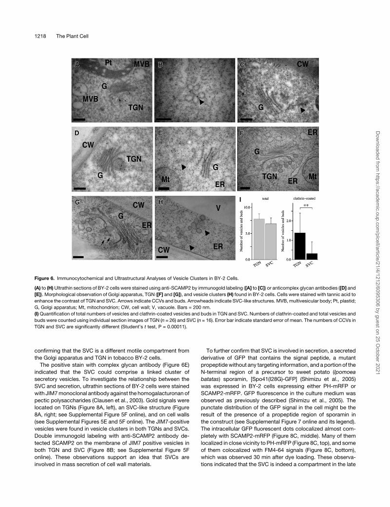
The nature of the SCAMP2-containing structures was studied in
further detail using immuno-EM. Ultrathin sections of BY-2 cells
were prepared by high-pressure freezing/freeze substitution
(HPF/FS) and stained using an anti-SCAMP2 antibody and
immunogold labeling. Gold particles were present at the PM,
the TGN, and in clusters of vesicles (Figures 6A to 6C; see
Supplemental Figures 5A to 5D online). The clusters generally
contained 5 to 12 vesicles of ;50 to 100 nm in diameter.
Consistent with the fluorescence colocalization experiments,
gold particles were not detected in theMVBs and small vacuoles
(Figure 6A). These results indicated that the SCAMP2 positive
fluorescence dots correspond to the TGN and to a vesicle cluster
not associated with the Golgi apparatus. These vesicle clusters
were termed the SVCs, as vesicles in this structure contained
secretory markers, such as secretory GFP and pectin (described
below), and the morphology of this structure was distinct from
TGN (described below). The ultrastructure of the Golgi appara-
tus, TGN, and SVC were next compared by staining with tannic
acid, which enhances the contrast of membrane structures rich
in glycoprotein as well as coat proteins of transport vesicles. The
observed tannic acid-enhanced ultrastructure of the TGN and
SVCwas consistent with the observation that SVC and TGNwere
stained with an antibody that recognized complex glycans
(Figures 6D to 6H). Some clusters of vesicles existed on one
side of the Golgi stacks where the TGN was localized (Figure 6F;
see Supplemental Figure 6A online), and vesicles in the TGN
sometimes had structures of clathrin-coated vesicles (CCVs) and
clathrin-coated buds (Figure 6G). By contrast, few clathrin coats
were detected on SVCs, especially those located close to PM
(Figures 6H and 6I). Thin-section images of both showed that
SVCs and TGNs contained similar numbers of vesicles (6.9 and
7.8, respectively; Figure 6I, left). By contrast, the average num-
bers of CCVs in each SVC was significantly lower than that in
each TGN (0.31 and 1.4, respectively; Figure 6I, right). The TGN
tended to have thick tubules in addition to vesicles with luminal
space (Figure 6G), whereas SVCs, particularly those located
close to the PM, did not display thick tubules and only occa-
sionally contained a thin tubule in addition to the vesicles (Figure
6B and H). These observations indicated that the TGN and SVCs
are related, but are distinct compartments in the cell.
To confirm that SVCs exist as structures separate from the
Golgi stacks and the TGN, BY-2 cells expressing both SCAMP2-
YFP and PH-mRFP were fixed and z-series images collected
(Figure 7A). Several SCAMP2-YFP dots were detected in the
cells, and some of these were independent from PH-mRFP dots
(Figure 7A, arrowheads). Four-dimensional (4D) analysis of SVC
movement using 4D-CLSM showed that many of the SCAMP2-
YFP dots were located at the side of the PH-mRFP and occa-
sionally moved separately from the PH-mRFP positive Golgi
apparatus (Figure 7B; see Supplemental Movie 1 online). To
provide further evidence that SVCs exist as structures separate
from TGN, BY-2 cells expressing both SCAMP2-YFP andmRFP-
Rab11 were fixed and z-series images also collected (Figure 7C).
Some of the SCAMP2-YFP dots were independent from mRFP-
Rab11 dots (Figure 7C, arrowheads). These observations indi-
cated that SVCs can move separately from the Golgi apparatus
and TGN in the cell. Moreover, ultrastructural observation of
continuous 80-nm serial sections of BY-2 cells demonstrated
that SVCs were present at a distance from the Golgi stacks and
the TGN (Figure 7C; seeSupplemental Figures 6B and 6Conline),
Figure 5. Subcellular Localization of SCAMP2 in the Presence of Inhib-
itors.
(A) Effects of BFA. BY-2 cells expressing PH-mRFP and SCAMP2-YFP
were treated with 5 mg/mL of BFA for 120 to 140 min.
(B) Recovery from BFA treatment. After BY-2 cells expressing SCAMP2-
YFP and PH-mRFP had been treated with 5 mg/mL of BFA for 2 h, they
were washed and transferred to fresh growth medium containing 50 mM
CHX or DMSO alone as a negative control for 1 h.
(C) Recovery from BFA treatment in the presence of a myosin inhibitor,
2,3-butanedione monoxime (BDM). BY-2 cells expressing SCAMP2-YFP
were treated with BFA for 2 h and then washed and transferred to fresh
medium containing 20 mM 2,3-butanedione monoxime and incubated
for 1 h. Bars = 20 mm in (A) to (C).
(D) Quantification of SCAMP2-YFP signals before and after various
treatments. Percentage of intracellular YFP signals was shown. Error
bars indicate SD.
SCAMP2 and the Secretory Vesicle Cluster 1217
Dow
nloaded from https://academ
ic.oup.com/plcell/article/21/4/1212/6095306 by guest on 25 O
ctober 2021
confirming that the SVC is a different motile compartment from
the Golgi apparatus and TGN in tobacco BY-2 cells.
The positive stain with complex glycan antibody (Figure 6E)
indicated that the SVC could comprise a linked cluster of
secretory vesicles. To investigate the relationship between the
SVC and secretion, ultrathin sections of BY-2 cells were stained
with JIM7monoclonal antibody against the homogalacturonan of
pectic polysaccharides (Clausen et al., 2003). Gold signals were
located on TGNs (Figure 8A, left), an SVC-like structure (Figure
8A, right; see Supplemental Figure 5F online), and on cell walls
(see Supplemental Figures 5E and 5F online). The JIM7-positive
vesicles were found in vesicle clusters in both TGNs and SVCs.
Double immunogold labeling with anti-SCAMP2 antibody de-
tected SCAMP2 on the membrane of JIM7 positive vesicles in
both TGN and SVC (Figure 8B; see Supplemental Figure 5F
online). These observations support an idea that SVCs are
involved in mass secretion of cell wall materials.
To further confirm that SVC is involved in secretion, a secreted
derivative of GFP that contains the signal peptide, a mutant
propeptidewithout any targeting information, and a portion of the
N-terminal region of a precursor to sweet potato (Ipomoea
batatas) sporamin, [Spo41(I28G)-GFP] (Shimizu et al., 2005)
was expressed in BY-2 cells expressing either PH-mRFP or
SCAMP2-mRFP. GFP fluorescence in the culture medium was
observed as previously described (Shimizu et al., 2005). The
punctate distribution of the GFP signal in the cell might be the
result of the presence of a propeptide region of sporamin in
the construct (see Supplemental Figure 7 online and its legend).
The intracellular GFP fluorescent dots colocalized almost com-
pletely with SCAMP2-mRFP (Figure 8C, middle). Many of them
localized in close vicinity to PH-mRFP (Figure 8C, top), and some
of them colocalized with FM4-64 signals (Figure 8C, bottom),
which was observed 30 min after dye loading. These observa-
tions indicated that the SVC is indeed a compartment in the late
Figure 6. Immunocytochemical and Ultrastructural Analyses of Vesicle Clusters in BY-2 Cells.
(A) to (H)Ultrathin sections of BY-2 cells were stained using anti-SCAMP2 by immunogold labeling ([A] to [C]) or anticomplex glycan antibodies ([D] and
[E]). Morphological observation of Golgi apparatus, TGN ([F] and [G]), and vesicle clusters (H) found in BY-2 cells. Cells were stained with tannic acid to
enhance the contrast of TGN and SVC. Arrows indicate CCVs and buds. Arrowheads indicate SVC-like structures. MVB, multivesicular body; Pt, plastid;
G, Golgi apparatus; Mt, mitochondrion; CW, cell wall; V, vacuole. Bars = 200 nm.
(I)Quantification of total numbers of vesicles and clathrin-coated vesicles and buds in TGN and SVC. Numbers of clathrin-coated and total vesicles and
buds were counted using individual section images of TGN (n = 26) and SVC (n = 16). Error bar indicate standard error of mean. The numbers of CCVs in
TGN and SVC are significantly different (Student’s t test, P = 0.00011).
1218 The Plant Cell
Dow
nloaded from https://academ
ic.oup.com/plcell/article/21/4/1212/6095306 by guest on 25 O
ctober 2021

Figure 7. SVCs Are Separated from the Golgi Apparatus.
(A) A z axis scanning series of fixed BY-2 cells expressing PH-mRFP and SCAMP2-YFP. Arrowheads indicate SVCs.
(B) The transparency projection of three-dimensional reconstructions of a BY-2 cell expressing PH-mRFP and SCAMP2-YFP using 4D-CLSM at four
time points. The panels show time-lapse pictures; arrows show SVCs. Bar = 5 mm.Movement can be seen in Supplemental Movie 1 online. Bars = 5 mm
in (A) and (B).
(C) A z axis scanning series of fixed BY-2 cells expressing mRFP-Rab11D and SCAMP2-YFP. Arrowheads indicate SVCs. Bar = 5 mm.
(D) Electron micrographs of 80-nm serial sections of SVCs in tobacco BY-2 cells. Arrowheads indicate SVCs. Bar = 200 nm.
SCAMP2 and the Secretory Vesicle Cluster 1219
Dow
nloaded from https://academ
ic.oup.com/plcell/article/21/4/1212/6095306 by guest on 25 O
ctober 2021

secretory pathway. To get another insight into the role of SVCs in
secretion, we observed transformed BY-2 cells expressing both
SCAMP2-YFP and PH-mRFP, using 4D-CLSM. Some SCAMP2-
YFP dots quickly moved to the PM from the PH-mRFP–tagged
Golgi apparatus (Figure 9A; see Supplemental Movie 2 online).
Morphological evidence of a role for SVCs in secretion was
also obtained as some SVCs were observed in contact with the
PM (Figure 9B) and vesicles in these SVCs were fusing with the
PM (Figure 9C). In some cases, several vesicles were connected
with each other to a region of the PM by electron-dense strings
(Figure 9C). Taken together, these results support a conclusion
that the SVC comprises a linked cluster of secretory vesicles.
To address whether the SVC is present in different cell types
and plant species, the ultrastructure of tobacco root tip cells, rice
cultured cells, and epidermal cells of expanding Arabidopsis
cotyledons were also examined. All of these cell types showed
the presence of SVCs (Figure 10). Additional analysis of contin-
uous 80-nm serial sections of rice cultured cells confirmed that
SVCs can exist at a distance from the Golgi stacks and TGN in
these cells also (see Supplemental Figure 8 online). Moreover,
SCAMP2 positive dots were observed in tobacco root cells by
immunofluorescence staining using anti-SCAMP2 antibody (Fig-
ures 10E and 10F).
SVCs Are Targeted to the Cell Plate in Dividing Cells
Because secretion is important for construction of the cell plate
in dividing cells, we next examined SCAMP2 localization during
mitosis in BY-2 cells. During mitosis, SCAMP2 signals accumu-
lated on newly synthesized cell plates and expanded during the
progression of the cell cycle together with expansion of each cell
plate (Figure 11; see Supplemental Figure 9 online). In mitotic
phase cells, SCAMP2 was detected by immunostaining and as a
YFP-tagged fusion and predominantly accumulated at the cell
plate (Figures 10E, 10F, 11A, and 11B). Detection of tubulin with
antitubulin antibody and of actin with fluorescent phalloidin in
cells expressing SCAMP2-mRFP revealed that SCAMP2 local-
ized between the opposing halves of the phragmoplast in divid-
ing cells (see Supplemental Figures 9A and 9B online). The
SCAMP2-positive cell plates at late telophase were stained by
aniline blue, which confirmed that the site where SCAMP2
accumulated in mitotic cells was the cell plate containing callose
(see Supplemental Figure 9C online). Interestingly, almost all of
the SCAMP2-YFP signals were accumulated on cell plate in
mitotic cells and were nearly devoid from the PM and the TGN
(Figure 11B). This was clearly evident in sequential images taken
during cell division of BY-2 cells expressing SCAMP2-mRFP
(Figure 11C).
We next analyzedwhether SVCs were targeted to the cell plate.
When movement of SCAMP2-YFP was observed using total in-
ternal reflection fluorescence microscopy, SCAMP2-YFP–tagged
dots fused rapidly and continuously with the edge ormiddle of the
cell plate (Figure 11D; see Supplemental Movies 3 and 4 online).
Tannic acid staining of ultrathin sections of dividing BY-2 cells
showed that the SVCs were present in the region where the cell
plate is generated (Figure 12A). This association with the cell plate
was further confirmed by staining with anti-1,3-b-glucan and anti-
SCAMP2 antibodies, which showed that SCAMP2-positive SVCs
without 1,3-b-glucan were present on the cell plate membrane
(Figure 12B). The number of SCAMP2 gold particles per 10 mm of
membranewere 2.46 0.74 (n= 4) on the PMat the side of the cells
and 6.0 6 2.0 (n = 4) on the cell plate, respectively. This quanti-
fication confirmed the previous fluorescence observation that
SCAMP2 is largely absent from the PM in mitotic cells and
suggested that most of the SCAMP2 present at the cell plates
was redirected from the PM.
FM4-64 has been reported to also accumulate at the cell plate
(Dettmer et al., 2006), and when FM4-64 was applied to BY-2
Figure 8. SVCs Contain Secretory Materials.
(A) An ultrathin section of a BY-2 cell was labeled with JIM7 antibody.
Bars = 200 nm.
(B) The ultrathin section was labeled with JIM7 antibody (12 nm gold) and
anti-SCAMP2 antibody (18 nm gold). Arrowheads indicate large gold
particles indicating the presence of SCAMP2. CW, cell wall; V, vacuole;
Mt, mitochondrion; G, Golgi. Bars = 200 nm.
(C) The secreted GFP fusion, Spo41(I28G)-GFP, was transiently ex-
pressed in BY-2 cells expressing SCAMP2-mRFP (middle) or PH-mRFP
(top). Spo41(I28G)-GFP expressing BY-2 cells were incubated with FM4-
64 for 30 min (bottom). Bar = 20 mm.
1220 The Plant Cell
Dow
nloaded from https://academ
ic.oup.com/plcell/article/21/4/1212/6095306 by guest on 25 O
ctober 2021

cells expressing SCAMP2-YFP, the corresponding fluorescence
accumulated to the cell plate in a similar manner to SCAMP2-
YFP (see Supplemental Figure 10A online). This accumulation of
the SCAMP2-YFP signal and the transport of SVCs to the cell
plate were not inhibited by a low concentration of BFA (see
Supplemental Figure 11 online), as in the case of SVCs targeted
to the PM (Figure 5). Using the confocal time-lapse system, we
observed that SCAMP2-YFP and FM4-64 positive SVCs were
fused to the edge of the cell plate (see Supplemental Figure 10B
and Supplemental Movie 5 online). These results further suggest
that SCAMP2 derived from the PM is transported to the TGN by
endocytosis and then targeted to the cell plate by SVCs.
DISCUSSION
The Secretory Vesicle Cluster
In this study, we analyzed the subcellular localization and trans-
port of SCAMP2 and found a previously undescribed vesicle
cluster structure, the SVC. The SVC was identified in tobacco
cultured BY-2 cells as well as tobacco root tip cells, rice sus-
pension cells, and epidermal cells of the expanding Arabidopsis
cotyledon (Figure 10).Morphology of the SVCwas clearly distinct
from the MVB, as it consisted of vesicles of 50 to 100 nm in
diameter in all species analyzed. Our observations that the SVC
Figure 9. SVCs Are Components in the Late Secretory Pathway.
(A) Three-dimensional reconstruction of a BY-2 cell expressing PH-mRFP and SCAMP2-YFP using 4D-CLSM at six time points (right panels). The left
panel shows a lowmagnification image at the 18 s time point with the area of the inset indicating the image shown in the right panels. White curved lines
show the position of cell wall. The movement of a YFP dot to the PM is indicated by arrowheads. Movement can be seen in Supplemental Movie 2
online. Bar = 5 mm.
(B) Electron micrographs showing the fusion of an SVC with the PM.
(C) Serial sections showing the fusion of an SVC with the PM. The bottom panels show a higher magnification of the top panels. The arrowhead
indicates the fusion point, and arrows show strings connected between vesicles and the PM. CW, cell wall; V, vacuole; Mt, mitochondrion; Pt, plastid.
Bars = 200 nm in (B) and (C).
SCAMP2 and the Secretory Vesicle Cluster 1221
Dow
nloaded from https://academ
ic.oup.com/plcell/article/21/4/1212/6095306 by guest on 25 O
ctober 2021

derived from the TGN (Figures 6 and 7) contained soluble
secretory proteins and cell wall components (Figure 8) generally
lacked clathrin-coated structures (Figure 6I) and fused with the
PM (Figures 9B and 9C) all indicated that the SVC is a motile
structure at a later step of the secretory pathway. A proposed
model for the generation of SVCs and SCAMP2 traffic is shown in
Figure 13. First, buds and vesicles containing SCAMP2 are
generated from the edges of trans-Golgi cisternae or at the TGN.
During or after separation of this structure from the Golgi appa-
ratus, the separated structure changes its shape through the
budding of CCVs. The remaining structure, the SVC that we have
identified, consists of a cluster of secretory vesicles. The SVC
then moves to the PM in nondividing cells or to the cell plate in
dividing cells. Finally, secretory vesicles in the SVC fuse with the
PM or the developing cell plate. Due to the large number of
vesicle fusion and tubule formation events taking place at the cell
plate in dividing cells (Samuels et al., 1995), it was impossible to
clearly demonstrate whether fusion of the SVC to the expanding
cell plate involves a change in the clustered shape or dissociation
of vesicles from the SVC prior to cell plate fusion. In any case,
SCAMP2on the PMwas shown to be recycled to the trans-Golgi/
TGN (Figure 5) by a route different from the MVB/PVC pathway.
In EM images, the SVC membranes show greater electron
density than other membranes. This difference is especially pro-
nouncedwhen sections are stained with tannic acid (Figures 6F to
6H, 7C, 9B, and 9C; see Supplemental Figures 6 and 8 online). As
tannic acid stains glycoproteins as well as vesicle coats, it is
possible that themembrane of the SVChas a thin protein coat.We
frequently observed electron-dense strings between vesicles
within SVCs and between SVCs and the PM (Figure 9C). The
images of SVC vesicles tethered to the PM in this manner are
similar to those of coat protein I–coated vesicles tethered to the
Golgi stacks (Orci et al., 1998). In the case of the Golgi apparatus,
such electron-dense linear structures are known to be formed by
vesicle-tethering proteinswith coiled-coil structures, suchasp115
and Golgins. The string between the SVC and PM might indicate
the presence of such a tethering protein. In Arabidopsis, there are
several possible coiled-coil proteins of unknown function (see
http://www.coiled-coil.org/Arabidopsis/). Therefore, it will be in-
teresting to analyze whether such coiled-coil proteins are involved
in tethering of SVCs to the PM.
In this study, we used HPF/FS for EM analysis. This is a
powerful method for studying membrane structure as cells are
frozen in milliseconds, thus avoiding many artifacts of fixation
and incubation. The ultrastructure of the TGN and partially
coated reticulum in several plant species has been described
using this method (Tanchak et al., 1984; Hillmer et al., 1988;
Mollenhauer et al., 1991; Segui-Simarro and Staehelin, 2006).
Similar analysis has been performed to monitor intracellular
events during wood formation (Samuels et al., 2002). When we
reexamined the images of the TGN in those reports, we observed
two classes of vesicular structures at the TGN: vesicles similar to
those found in SVCs and CCVs. This was also observed in
tobacco cell TGNs (Figure 6G). However, such structures were
scarce in SVCs located close to or fusing with the PM (Figure 6I).
CCVs in the plant TGN contain sorting receptors for vacuolar
targeting (Kirsch et al., 1994). The cytosolic tails of these recep-
tors contain motifs for interaction with adaptor proteins in CCVs
(McNiven and Thompson, 2006). Thus, the TGN represents the
sorting site for vacuolar delivery and secretion. Together with our
observations that secretory GFP and pectin were present in the
TGN and SVCs (Figure 8C), and that mature SVCs did not have
vesicles with clathrin coat–like morphology, this suggests that
the mature SVC formed by budding of CCVs from the TGN
(Figure 13). However, we cannot rule out a possibility that some
of the SVC-like images that we observed in simple sectioning
were the side-cut sections of the TGN, and this possibility thus
affects the quantification of the numbers of clathrin-coated buds
and vesicles. Recently, Staehelin and Kang (2008) proposed two
structurally different TGNs, namely, early TGN and late TGN,
based on electron tomographic images of Arabidopsismeristem
cells. The late TGNs are somehow separate structures from
the Golgi apparatus but contain numbers of CCV. Therefore,
Figure 10. Morphological Observation of Several Plant Cells.
(A) to (D) Tobacco BY-2 cultured cells (A), tobacco seedling root cell (B),
rice cultured cells (C), and epidermal cell of growing Arabidopsis coty-
ledon (D). Arrowheads show SVC. Pt, plastid; G, Golgi apparatus; Mt,
mitochondrion; CW, cell wall; V, vacuole. Bars = 200 nm.
(E) Immunofluorescence staining of the mitotic zone of a tobacco root
using anti-SCAMP2 antibody.
(F)Higher magnification of the root cells showing SCAMP2 localization to
the developing cell plate. Bars = 10 mm in (E) and (F).
1222 The Plant Cell
Dow
nloaded from https://academ
ic.oup.com/plcell/article/21/4/1212/6095306 by guest on 25 O
ctober 2021

immature SVCmay correspond to late TGN and SVC is the post-
late TGN organelle generated by budding of CCV from late TGN.
During pine (Pinus contorta) wood formation, cell wall mate-
rials are found in TGN-like vesicle clusters (Samuels et al., 2002).
As many such structures are separate from the Golgi apparatus
(Figure 3E in Samuels et al., 2002), it is possible that these
represent the maturing SVCs. Pine belongs to different taxo-
nomic division from all the plants analyzed in this work; therefore,
the formation of SVC in plant cells might be evolutionally con-
served in a wide variety of seed-forming plants.
Figure 11. Movement of SVCs to the Cell Plate.
Localization of SCAMP2 in BY-2 cells at M phase.
(A) Immunofluorescence staining using anti-SCAMP2 antibody by CLSM.
(B) Fluorescence of SCAMP2-YFP was detected by CLSM.
(C) Time-lapse images of BY-2 cells expressing SCAMP2-mRFP for 50 min using video fluorescent microscopy.
(D) Time-lapse images of cell plate formation in a BY-2 cell expressing SCAMP2-YFP at 1-s intervals using total internal reflection fluorescence
microscopy. Top and bottom panels show two different cell divisions. Arrowheads indicate the position of SVCs fusing with cell plates. Movement can
be seen in Supplemental Movies 3 and 4 online.
Bars = 20 mm in (A) to (C) and 5 mm in (D).
SCAMP2 and the Secretory Vesicle Cluster 1223
Dow
nloaded from https://academ
ic.oup.com/plcell/article/21/4/1212/6095306 by guest on 25 O
ctober 2021

In Arabidopsis meristematic cells, the TGN is usually associ-
ated with the Golgi apparatus (Segui-Simarro and Staehelin,
2006). This difference in the location of TGN and the presence or
absence of solitary TGN and SVC in Arabidopsis meristematic
cells (Segui-Simarro and Staehelin, 2006) with the other cells
used in this study and pine fiber cells can be explained by
differences in cell size. Similar to many cells in crop plants, the
length of cylinder-like BY-2 cells ranges from 30 to 50 mmwith a
diameter of 30 to 50 mm, whereas rectangular tobacco root tip
cells are ;40 mm long and 30 mm wide. Rice cultured cells are
spherical but somewhat irregular and have a diameter of 30 to 50
mm, Arabidopsis epidermal cells have an irregular shape and are
;20 mmwide and 50 mm long, whereas pine bark cells are 15 to
20 mm wide and >100 mm long. By contrast, shoot apical
meristematic cells of Arabidopsis are roughly cubical with 4- to
5-mm sides (Segui-Simarro and Staehelin, 2006). Unlike these
differences in cell sizes, the Golgi apparatus and secretory
vesicles in these species are similar in size: ;500 nm wide in
Golgi apparatus and 50 to 100 nmdiameter in secretory vesicles,
respectively. In addition, Golgi apparatus are scattered through-
out the cells in all Arabidopsis, pine, rice, and tobacco cells.
Thus, it can be calculated that it is;10 times the distance from
the Golgi apparatus to the PM in tobacco cells and other cells
used in this work than in Arabidopsismeristematic cells. Shorter
distances in Arabidopsismeristematic cells might limit our ability
to detect any SVCs in this cell type as SVCs are transient mobile
units during the final step of secretion.
Mass transport of proteins to vacuoles is mediated by large
vesicles in plant cells (Hara-Nishimura et al., 1998; Toyooka
et al., 2000). Although such large vesicles might be useful for the
transport of large amounts of soluble and uniform contents to a
final destination, such machinery is not adequate to transport
large quantities of membrane proteins and lipids. By contrast,
small vesicles with relatively high surface-to-volume ratios are
likely to be better carriers for membrane constituents. However,
transporting one vesicle with one motor protein, the mechanism
found in axonal transport in neurons (Scholey, 2002), would not
be an efficient system to transport massive amounts of mem-
brane constituents. Moving arrays of tethered vesicles would be
a more efficient approach to transport such vesicles efficiently.
Thus, we speculate that plants developed such a transport
mechanism with SVCs to deliver large quantities of lipid and
membrane proteins to the PM over a long distance in relatively
large plant cells.
In mammalian cells, SCAMP is involved in regulation of the
insulin-responsive glucose transporter (Laurie et al., 1993) and
(Na+, K+)/H+ exchanger (Lin et al., 2005). SCAMP2 also plays an
important role in the regulation of the subcellular distribution of
serotonin transporters in neurons (Muller et al., 2006). Plant SVCs
containing SCAMP2 might therefore also take part in the trans-
port and regulation of transporters in plants. Recently, Jaillais
et al. (2006) reported that PIN2, which is an auxin transporter, is
located in a novel endomembranous compartment and auxin
influx carrier AUX1 is also transported to the PM through a novel
pathway distinct from PIN1 (Kleine-Vehn et al., 2006). Reichardt
et al. (2007) reported that cytokinesis requires de novo secretory
transport but not endocytosis using syntaxin KNOLLE as a
marker. Likewise, Chow et al. (2008) reported that Rab-A2/-A3
localized to a novel post-Golgi membrane compartment con-
tributes to the cell plate formation in Arabidopsis root tip cell.
Thus, it will be interesting to investigate whether these proteins
are localized in the SVC.
Cytokinesis and SVCs
The PM has to expand rapidly during cytokinesis in plant cells.
During this period, Golgi-derived vesicles accumulate between
separated chromosomes along with the cytoskeleton in a struc-
ture known as the phragmoplast. Vesicles then fuse to generate
connected tubules that form into meshes and plates, collectively
called the cell plate (Samuels et al., 1995; Segui-Simarro et al.,
2004). Although some of the Golgi stacks move to the cell plate
and its vicinity during cell division (Nebenfuhr et al., 2000), not all
the Golgi do. It therefore remained unclear how Golgi-derived
vesicles could be targeted efficiently to the cell plate until our
discovery of SVCs in this work.
It has been reported that FM4-64 accumulates at the cell plate
(Bolte et al., 2004; Dhonukshe et al., 2006). Here, we found that
SCAMP2-YFP at interphase was localized in SVCs together with
FM4-64 (see Supplemental Figure 8A online). Movement of the
SCAMP2-YFP signal from SVCs to the cell plate was prominent
at mitosis (see Supplemental Figure 10B online). As discussed
above, clusters of vesiclesmight have an advantage for themass
transport of membranes. Thus, SVC-mediated transport to the
cell plate might be an efficient transport mechanism for the
generation of newPMduring cytokinesis. Similar to the observed
SCAMP2 localization to the cell plate, it was reported recently
that ectopically expressed rice SCAMP1 is targeted to the cell
plate in tobacco BY-2 cells (Lam et al., 2008). Analysis of the
Figure 12. Immunocytochemical and Ultrastructual Analysis of Cell
Plates.
(A) Electron micrographs of the cell plate region of dividing BY-2 cells.
(B) Immunogold staining of cell plate region with anti-SCAMP2 and anti-
b-1,3-glucan antibodies. The 12- and 18-nm gold particles indicate the
position of b-1,3-glucan and SCAMP2, respectively. CW, cell wall; G,
Golgi apparatus; Mt, mitochondrion; CP, cell plate.
Bars = 500 nm (A) and 200 nm in (B).
1224 The Plant Cell
Dow
nloaded from https://academ
ic.oup.com/plcell/article/21/4/1212/6095306 by guest on 25 O
ctober 2021

trafficking of rice SCAMP1 in tobacco BY-2 cells indicated that
this protein is transported back to clathrin-coated tubulovesi-
cluar structures from the PM, which is likely the TGN (Lam et al.,
2007a). Although it is not clear how rice SCAMP1 is directed to
the PM from the TGN, it is possible that some of the SVCs
observed by EM could be heterogeneous andmight also contain
rice SCAMP1. This would be consistent with the observed varied
labeling efficiency of SVCs using the anti-SCAMP2 antibody.
Future characterization of endogenous SCAMP1 in tobacco BY-2
cells will clarify this possibility.
Analysis of the trafficking of membrane proteins, including the
KORRIGAN protein (Zuo et al., 2000), revealed the importance of
Tyr-containing motifs in cytosolic domains (Robert et al., 2005).
We identified a Tyr-containing motif present in the cytosolic
domain of SCAMP2 (see Supplemental Figure 1 online). This
motif was conserved in plant SCAMP2 amino acid sequences
and may contribute to the targeting of SCAMP2 to the cell plate.
SCAMPs and the Trafficking in Plant Cells
Subcellular fractionation analysis using a sucrose gradient indi-
cated that SCAMP2was broadly distributed from the PM toGolgi
fractions (Figures 1 and 2D). Interestingly, both endogenous and
YFP-tagged SCAMP2 showed multiple bands on SDS-poly-
acrylamide gels. Relative intensities of these bands differed
depending on the fraction in both cases, suggesting that SCAMP2
is posttranslationally modified and that this modification may
contribute the localization of this protein. It was reported previ-
ously that some mammalian SCAMPs are phosphorylated and
that phosphorylation affected the localization of SCAMP3 in
CHO cells (Wu and Castle, 1998). In addition, phosphorylation of
the Tyr motif for endocytosis affected the localization of TrkA in
mammalian cells (de Pablo et al., 2008). As plant SCAMPs have a
Tyrmotif in theirC-terminal tail (seeSupplemental Figure 1online)
and as phosphorylation of proteins tended to cause a mobility
shift on SDS-polyacrylamide gels, it will be interesting to deter-
mine whether this modification is the result of the formation of
multiple forms of SCAMP2 and a difference in the distribution of
different forms in the cell.
Themovement of FM4-64 from the PM to the TGN (Bolte et al.,
2004; Figure 3; see Supplemental Figure 2 online) suggests that
membrane was likely transported from the PM to TGN. It has
been reported that the TGN might act as an early endosomal
compartment (Dettmer et al., 2006) and that endocytosed PM
and cell wall materials are used for cell plate formation via
endosomes (Dettmer et al., 2006). However, our results indicated
that SCAMP2 is not localized in an endosomal compartment
(Figure 3A, taken at 5 to 10 min). We observed that prolonged
incubation with FM4-64 allowed targeting of this dye to the
TGN, where SCAMP2-YFP was also localized (Figure 3A). The
discrepancy of our observation with these previous reports is
probably due to a difference in temperature of incubation and
Figure 13. A Model of the Localization of SCAMP2 and the Secretory Pathway in BY-2 Cells.
SCAMP2 (blue) is localized to the trans-Golgi, TGN, PM, and SVC. The SVC separated from trans-Golgi migrates to the PM or to the cell plate
membrane (see Discussion). Inset: proposed maturation pathway of the SVC.
SCAMP2 and the Secretory Vesicle Cluster 1225
Dow
nloaded from https://academ
ic.oup.com/plcell/article/21/4/1212/6095306 by guest on 25 O
ctober 2021

time taken to detect the internalization of FM4-64. Most studies
using FM4-64 to detect endosomes used;30 min of incubation
at room temperature, a duration that allowed the dye to reach the
TGN in our studies. Therefore, the TGN in tobacco BY-2 cells
might not be a counterpart of the mammalian early endosome,
but may function as a recycling or sorting organelle as secretory
proteins pass through this compartment and SCAMP2 is local-
ized here (Figure 4; see Supplemental Figure 3 online).
Analysis of the trafficking of rice SCAMP1 in tobacco BY-2
cells (Lam et al., 2007a) indicated that this protein localizes in a
tubulovesicular endosome, which is also marked with FM4-64
and Ara7. The absence of SCAMP2 in such structures and the
absence of colocalization of the early FM4-64 compartment with
SCAMP2-YFP indicated that SCAMP1 and SCAMP2 are local-
ized in distinct compartments in the early endocyotic pathway.
This is consistent with the observation in mammalian cells that
SCAMP1 and SCAMP2 use distinct trafficking pathways, al-
though some compartments overlap (Castle and Castle, 2005).
Taken together, our data suggest the presence of at least two
endocytosis routes in tobacco cells that merge in the TGN
(Figure 13), one that is mediated through the FM4-64, rice
SCAMP1 and Ara7-marked endosomes, and the other mediated
by the SCAMP2 pathway.
METHODS
Cell Culture, Transformation, and Treatment of Cells
Culture and transformation of the tobacco (Nicotiana tabacum) cell line
BY-2 was performed as described (Matsuoka and Nakamura, 1991). Log
phase cells (3 or 4 d after culture) were used throughout the study unless
otherwise stated. Stock solutions of BFA (Sigma-Aldrich), CHX (Wako
PureChemical industries), andwortmannin (Signa-Aldrich) in DMSOwere
prepared at 5 mg/mL, 50 mM, and 10 mM, respectively. BFA (5 mg/mL;
17.8mMfinal concentration) was added to culture medium and incubated
with the cells for 2 h. In some case, after the cells had been treated with 5
mg/mL BFA for 2 h, cells were washed and transferred to fresh growth
medium containing 50 mM CHX for 1 h. Control treatments were
performed with equal amounts of DMSO. Alternatively, cells were incu-
bated with 20 mM 2,3-butanedione monoxime in culture medium for 1 h.
In some cases, wortmannin (16.5 mM final concentration) was added to
culture medium and incubated with the cells for 1 h.
Cloning of SCAMP2 cDNA, Construction of Plasmids, and
Transformation into BY-2 Tobacco Cells
A tobacco full-length enriched cDNA library was constructed from an
mRNA fraction prepared from log-phase growing BY-2 cells into
pGCAPsp2 vector as described (Kato et al., 2005). Full-length cDNAs
for SCAMP2, SYP41, and SYP22 were isolated from the library using
partial cDNA information reported previously (Matsuoka et al., 2004; Galis
et al., 2006). To create the SCAMP2-mRFP, mRFP-Rab11D, and PH-
mRFP fusion constructs, SCAMP2 (DDBJ accession number AB295617),
Rab11D (DDBJ accession number AB470307), and PH (Yuasa et al.,
2005; DDBJ accession number AB119250) cDNA were amplified by PCR
from the BY-2 EST plasmids containing SCAMP2, Rab11D, or PH (Yuasa
et al., 2005). See Supplemental Table 1 online for a listing of PCR primers.
PCR products were then cloned into the BamHI and KpnI sites of
pMAT330, which contains a synthetic mRFP coding sequence with plant
codon usage (Toyooka et al., 2006). The fused cDNAs were placed
downstream of the cauliflower mosaic virus 35S promoter in the binary
vector pMAT037 and used for the Agrobacterium tumefaciens–mediated
transformation of BY-2 cells (Matsuoka et al., 1995b). The SCAMP2-YFP,
YFP-SYP41, and YFP-SYP22 were constructed using Gateway Technol-
ogy (Invitrogen). Briefly, PCR products were cloned into the pENTR/
D-TOPO vector using the pENTR Directional TOPO Cloning Kit (Invitro-
gen). The entry clone and Gateway binary vector, pH35YG2 (N-terminal
YFP fusion) and pH35GY (C-terminal YFP fusion) (Kubo et al., 2005), were
incubated with the LR Clonase Enzyme Mix (Invitrogen). Spo41(I28G)-
GFP was prepared as described (Shimizu et al., 2005).
Antibodies
The anti-SCAMP2 antibody was raised in rabbits against the recombinant
SCAMP2 protein (N-terminal 125 amino acids) prepared with the pET-
Directional TOPO Expression Kit (Invitrogen) according to the manufac-
turer’s instructions. The recombinant protein purified by Ni-Agarose was
injected into a rabbit and then the serum was purified using an immobi-
lized antigen column. The columnwasmade using HiTrap NHS-activated
Sepharose column (GEHealthcare). In some cases, IgGwas purified from
the serum using the Melon Gel IgG Spin Purification Kit (Pierce). The
antibody against complex glycan was provided by I. Hara-Nishimura
(Kyoto University, Japan). The JIM7 rat monoclonal antibody was
obtained from PlantProbes. A monoclonal antibody against 1,3-b-glucan
was obtained from Biosupplies Australia. The anti-PM intrinsic protein
was a gift from M. Maeshima (Nagoya University, Japan) and anti-PM-
ATPase was a gift from T. Sugiyama (RIKEN Plant Science Center,
Japan). Anti-plant Sec61 antibody was as described (Yuasa et al., 2005).
Anti-vacuolar membrane H+-pyrophosphatase (V-PPase) antibody was
raised in rabbits against the mixture of synthetic peptides (CLVGKVER-
NIPEDDPRNP and CGDIAGMGSDLFGSYAES, corresponding to the
sequences in the fifth loop of tobacco H+-PPase, CAA58700) conjugated
with KLA, and used after affinity purification using the antigen peptides.
This antibody specifically recognized an ;80-kD poloypeptide in the
tonoplast. Alexa Fluor 488/568 secondary antibodies and FM 4-64 were
from Invitrogen. AM 4-65 was from Biotium. The secondary antibody-
conjugated alkaline phosphatase used for immunoblot analysis was
obtained from Bio-Rad.
Fractionation, SDS-PAGE, and Detection of Proteins
Organelles in the microsomal fraction were separated by isopycnic
ultracentrifugation, and the inosine disphosphatase activity of the frac-
tions wasmeasured as described (Matsuoka et al., 1995b). Proteins were
separated by SDS-PAGE and analyzed by immunoblotting as described
(Yuasa et al., 2005) using antibodies to plant Sec61 (1:1000), V-PPase
(1:5000), anti-PM intrinsic protein (1:1000) and SCAMP2 (1:500).
SCAMP2-YFP signals separated by SDS-PAGE were recorded using a
Typhoon 9400 fluoroimager (GE Healthcare Bioscience) using a 488-nm
excitation laser at a setting of 550 V and a 520BP40 filter.
Fluorescence Microscopy
To visualize endosomal organelles, cells were stained with 17 or 33 mM
FM4-64 or 12mMAM4-65 dye inmedium at cold or room temperature for
10 min and then washed with medium at the same temperature. Cells
were then observed at 268C. To analyze the localization of SCAMP2, BY-2
cells were washed and fixed with formaldehyde as described (Toyooka
et al., 2006). Ten-day-old tobacco seedlings were fixed with formalde-
hyde in PBS for an hour and digested for 2 h at 308C in 0.1%Pectolyase in
water and then permeabilized for an hour in 10% DMSO and 1% Triton
X-100 in PBS. Fixed cells were soaked with primary antibodies to
SCAMP2 (diluted 1:100) in PBS for 1 h at room temperature, washed
with PBS, and incubated with a secondary antibody diluted 1:500 in PBS.
1226 The Plant Cell
Dow
nloaded from https://academ
ic.oup.com/plcell/article/21/4/1212/6095306 by guest on 25 O
ctober 2021
The cells were mounted in PBS on glass slides. Living cells stably
expressing YFP or mRFP fusion proteins were mounted in culture
medium. These cells were observed using a CLSM system (LSM510
META, Axioplan2 Imaging; Carl Zeiss) with a Plan-Apochromat lens (633
1.4 oil differential interference contrast; optical slices of 1 mm). We used a
25-mW argon laser (power, 5%) with 488-nm excitation and a 505- to
530-nm band-pass filter for YFP, GFP, and Alexa Fluor 488. For FM4-64
and AM4-65, we used a 650-nm long-pass filter and 488-nm excitation. A
560-nm long-pass filter and 1- mW He-Ne laser at 80% power with 543-
nm excitation was used for Alexa Fluor 568 and mRFP. Crosstalk was
prevented using a multitrack configuration with line sequential scanning.
Composite figures were prepared using Zeiss LSM Image Browser
software. Intensity of YFP fluorescence and colocalization frequency
was measured using Image J software (http://rsbweb.nih.gov/ij/).
To monitor movements of cis-Golgi and SVC, BY-2 cells expressing
PH-mRFP and SCAMP2-YFP were observed using a 4D-CLSM
(LSM5LIVE, Axiovert200; Carl Zeiss) with a Plan-Apochromat lens (as
above). Cells were mounted in glass-bottomed dishes and scanned at a
distance of 1.5mm for 10 slices using 488- and 532-nm lasers. Time-lapse
movies for cell division of BY-2 cells expressing SCAMP2-mRFP were
obtained using a fluorescence microscope (Olympus; Toyooka et al.,
2006). To visualize fusion of SVCwith cell plate, time-lapse images of BY-2
cells expressing SCAMP2-YFP were observed using a total internal
reflection fluorescence microscope (AM-TIRF; Leica) using an HCX PL
APO 1003 1.46 oil lens, YFP filter, and a DFC350FX monochrome digital
camera. Cells were mounted on glass-bottomed dishes and scanned
with a penetration depth of 310 nm (oblique illumination) using a 100-mW
multi-argon laser. Supplemental movies were prepared using Adobe
Photoshop CS2 and Apple QuickTime Pro.
HPF/FS and Tannic Acid Staining
Growing or synchronized BY-2 cells (Samuels et al., 1995) and rice (Oryza
sativa) culture cells (Mitsui et al., 1996) were placed on a flat specimen
carrier and frozen in a high-pressure freezer (EM-PACT; Leica). For
morphological observations with tannic acid staining, the frozen samples
were fixedwith anhydrous acetone containing 2%osmic acid (OsO4) for 3
to 4 d at2808C. For immunocytochemistry, the frozen sampleswere fixed
with anhydrous acetone containing 1% glutaraldehyde (GA) and 1%
OsO4 or 2% GA for 3 to 4 d at 2808C. The tubes containing the frozen
samples were warmed at 38C/h to a temperature of 2208C, at 18C/h to a
temperature of 48C and kept for 2 h at 48C using an automatic freeze-
substitution system (EM-AFS; Leica). Fixed samples were stained with
1% tannic acid (Mallinckrodt) in acetone for 1 h at room temperature for
morphological observation. The samples were washed with 100% ace-
tone or methanol and then embedded in epoxy resin or LRWhite resin for
morphological observations or immunological observations, respec-
tively. For morphological observation of tobacco roots and Arabidopsis
thaliana epidermal cells, seeds were planted in 0.8% (w/v) agar plates for
2 weeks and then moved to 2% sucrose on papers for 24 h to inhibit the
formation of ice-crystal formation during HPF/FS. The roots and cotyle-
donswere cut into 1-mm rectangles and frozen in a high-pressure freezer.
EM and Immuno-EM
Treatment of ultrathin sections fixed by GA and OsO4 on nickel 600 mesh
grids was performed as described by Follet-Gueye et al. (2003). The grids
were treatedwith 0.5MNaIO4 for 30min, washed twicewithwater (10min
each), then with 0.1 N HCl (10 min), washed with water, and then treated
with 0.1 M glycine containing 0.1% Triton X-100 for 15min. After washing
with TBS, the grids were blocked with 10%BSA in TBS for 30min at room
temperature. The sections were labeled with antibodies against SCAMP2
(1:100), complex glycan (1:25), or 1,3-b-glucan (1:200) in TBS. After being
washed with TBS, sections were indirectly labeled with 12- or 18-nm
colloidal gold particles coupled to goat anti-rabbit IgG or anti-mouse IgG
(Jackson ImmunoResearch). The ultrathin sections fixed by GA were
labeled with anti-JIM7 antibody (1:100) and anti-rat IgG antibody 12 nm
gold. Gold-labeled sections were washed with TBS and then rinsed in
water. For morphological observations, ultrathin sections were mounted
on 400 mesh Cu grids. The grids were stained with 4% aqueous uranyl
acetate for 10min and examinedwith a transmission electronmicroscope
(JEOL JEM-1011) at 80 kV. Images were acquired using a Gatan DualView
CCD camera and Gatan Digital Micrograph software or films. Number of
gold particles on PM and cell plate were counted from sections prepared
from four different blocks and measured by Image J software.
Accession Numbers
Sequence data from this article can be found in the GenBank/EMBL/
DDBJ data libraries under the following accession numbers: Nt SCAMP2,
DDBJ accession number AB295617; Nt SYP41, DDBJ accession number
AB295618; Nt SYP22, DDBJ accession number AB295619; and Nt
Rab11D, DDBJ accession number AB470307 .
Supplemental Data
The following materials are available in the online version of this article.
Supplemental Figure 1. Alignment of SCAMP2 Amino Acid Se-
quences.
Supplemental Figure 2. Subcellular Localization of FM4-64.
Supplemental Figure 3. Subcellular Localization of FM4-64 with
Organelle Markers.
Supplemental Figure 4. Subcellular Localization of SCAMP2 in the
Presence of Wortmannin.
Supplemental Figure 5. Immunogold Labeling Using Anti-SCAMP2
Antibody.
Supplemental Figure 6. Electron Micrographs of Ultrathin Serial
Sections of BY-2 Cells for the Comparison of TGN and SVC.
Supplemental Figure 7. Localization of SPO41(I28G)-GFP, Sporamin
Signal Peptide-GFP, and SPO41(I28G)-GFP-KDEL in Tobacco BY-2
Cells.
Supplemental Figure 8. Electron Micrographs of Ultrathin Serial
Sections of Rice Culture Cells for the Comparison of TGN and SVC.
Supplemental Figure 9. Distribution of SCAMP2 and Cytoskeletones
in Dividing Cells.
Supplemental Figure 10. Both SCAMP2 and FM4-64 Accumulate in
the Cell Plate.
Supplemental Figure 11. Accumulation of SCAMP2-YFP and Trans-
port of SVCs to the Cell Plate in the Presence of BFA.
Supplemental Table 1. PCR Primers.
Supplemental Movie 1. Video of Figure 7B.
Supplemental Movie 2. Video of Figure 9A.
Supplemental Movie 3. Video of Figure 11C.
Supplemental Movie 4. Video of Figure 11D.
Supplemental Movie 5. Video of Supplemental Figure 8B.
Supplemental Movie Legend.
ACKNOWLEDGMENTS
We thank Y. Suzuki, T. Narisawa, and I. Galis in RIKEN Plant Science
Center for the screening of full-length cDNA clones used in this study, I.
SCAMP2 and the Secretory Vesicle Cluster 1227
Dow
nloaded from https://academ
ic.oup.com/plcell/article/21/4/1212/6095306 by guest on 25 O
ctober 2021

Hara-Nishimura at Kyoto University for the complex glycan antibody, T.
Demura and M. Kubo at the RIKEN Plant Science Center for the
Gateway YFP fusion vectors, T. Inaba at Iwate University for the gift of
GFP-PsPra3 plasmid, and S. Hamamoto at University of California,
Berkeley, for the gift of tannic acid. We also thank M. Shimizu and S.
Takata at the RIKEN Plant Science Center for the construction of
plasmids, M. Araki and S. Oyama at the RIKEN Plant Science Center for
DNA sequencing, and Derek B. Goto at Hokkaido University for im-
proving the manuscript. This work was supported in part by grants from
the Japan Society for the Promotion of Science (17770056 to K.T.) and
from Grants-in-Aid for Scientific Research in Priority Areas from MEXT
(17078009 to K.M.).
Received February 21, 2008; revised March 8, 2009; accepted March 30,
2009; published April 17, 2009.
REFERENCES
Bednarek, S.Y., and Falbel, T.G. (2002). Membrane trafficking during
plant cytokinesis. Traffic 3: 621–629.
Bolte, S., Talbot, C., Boutte, Y., Catrice, O., Read, N.D., and Satiat-
Jeunemaitre, B. (2004). FM-dyes as experimental probes for dis-
secting vesicle trafficking in living plant cells. J. Microsc. 214:
159–173.
Brand, S.H., Laurie, S.M., Mixon, M.B., and Castle, J.D. (1991).
Secretory carrier membrane proteins 31–35 define a common protein
composition among secretory carrier membranes. J. Biol. Chem. 266:
18949–18957.
Castle, A., and Castle, D. (2005). Ubiquitously expressed secretory
carrier membrane proteins (SCAMPs) 1-4 mark different pathways
and exhibit limited constitutive trafficking to and from the cell surface.
J. Cell Sci. 118: 3769–3780.
Chow, C.M., Neto, H., Foucart, C., and Moore, I. (2008). Rab-A2 and
Rab-A3 GTPases define a trans-Golgi endosomal membrane domain
in Arabidopsis that contributes substantially to the cell plate. Plant Cell
20: 101–123.
Clausen, M.H., Willats, W.G., and Knox, J.P. (2003). Synthetic methyl
hexagalacturonate hapten inhibitors of anti-homogalacturonan mono-
clonal antibodies LM7, JIM5 and JIM7. Carbohydr. Res. 338: 1797–
1800.
Cosgrove, D.J. (2005). Growth of the plant cell wall. Nat. Rev. Mol. Cell
Biol. 6: 850–861.
de Pablo, Y., Perez-Garcia, M.J., Georgieva, M.V., Sanchis, D.,
Lindqvist, N., Soler, R.M., Comella, J.X., and Llovera, M. (2008).
Tyr-701 is a new regulatory site for neurotrophin receptor TrkA
trafficking and function. J. Neurochem. 104: 124–139.
Dettmer, J., Hong-Hermesdorf, A., Stierhof, Y.D., and Schumacher,
K. (2006). Vacuolar H+-ATPase activity is required for endocytic and
secretory trafficking in Arabidopsis. Plant Cell 18: 715–730.
Dhonukshe, P., Baluska, F., Schlicht, M., Hlavacka, A., Samaj, J.,
Friml, J., and Gadella, T.W., Jr. (2006). Endocytosis of cell surface
material mediates cell plate formation during plant cytokinesis. Dev.
Cell 10: 137–150.
Fernandez-Chacon, R., and Sudhof, T.C. (2000). Novel SCAMPs
lacking NPF repeats: ubiquitous and synaptic vesicle-specific forms
implicate SCAMPs in multiple membrane-trafficking functions. J.
Neurosci. 20: 7941–7950.
Follet-Gueye, M.L., Pagny, S., Faye, L., Gomord, V., and Driouich, A.
(2003). An improved chemical fixation method suitable for immuno-
gold localization of green fluorescent protein in the Golgi apparatus of
tobacco Bright Yellow (BY-2) cells. J. Histochem. Cytochem. 51:
931–940.
Foresti, O., daSilva, L.L., and Denecke, J. (2006). Overexpression of
the Arabidopsis syntaxin PEP12/SYP21 inhibits transport from the
prevacuolar compartment to the lytic vacuole in vivo. Plant Cell 18:
2275–2293.
Galis, I., Simek, P., Narisawa, T., Sasaki, M., Horiguchi, T., Fukuda,
H., and Matsuoka, K. (2006). A novel R2R3 MYB transcription factor
NtMYBJS1 is a methyl jasmonate-dependent regulator of phenyl-
propanoid-conjugate biosynthesis in tobacco. Plant J. 46: 573–592.
Hara-Nishimura, I.I., Shimada, T., Hatano, K., Takeuchi, Y., and
Nishimura, M. (1998). Transport of storage proteins to protein stor-
age vacuoles is mediated by large precursor-accumulating vesicles.
Plant Cell 10: 825–836.
Higaki, T., Kutsuna, N., Okubo, E., Sano, T., and Hasezawa, S.
(2006). Actin microfilaments regulate vacuolar structures and dynam-
ics: Dual observation of actin microfilaments and vacuolar membrane
in living tobacco BY-2 Cells. Plant Cell Physiol. 47: 839–852.
Hillmer, S., Freundt, H., and Robinson, D.G. (1988). The partially
coated riticulum and its relationship to the Golgi apparatus in higher
plant cells. Eur. J. Cell Biol. 47: 206–212.
Inaba, T., Nagano, Y., Nagasaki, T., and Sasaki, Y. (2002). Distinct
localization of two closely related Ypt3/Rab11 proteins on the traf-
ficking pathway in higher plants. J. Biol. Chem. 277: 9183–9188.
Jaillais, Y., Fobis-Loisy, I., Miege, C., Rollin, C., and Gaude, T. (2006).
AtSNX1 defines an endosome for auxin-carrier trafficking in Arabi-
dopsis. Nature 443: 106–109.
Kato, S., Ohtoko, K., Ohtake, H., and Kimura, T. (2005). Vector-
capping: A simple method for preparing a high-quality full-length
cDNA library. DNA Res. 12: 53–62.
Kirsch, T., Paris, N., Butler, J.M., Beevers, L., and Rogers, J.C.
(1994). Purification and initial characterization of a potential plant
vacuolar targeting receptor. Proc. Natl. Acad. Sci. USA 91: 3403–
3407.
Kleine-Vehn, J., Dhonukshe, P., Swarup, R., Bennett, M., and Friml,
J. (2006). Subcellular trafficking of the Arabidopsis auxin influx carrier
AUX1 uses a novel pathway distinct from PIN1. Plant Cell 18: 3171–
3181.
Kubo, M., Udagawa, M., Nishikubo, N., Horiguchi, G., Yamaguchi,
M., Ito, J., Mimura, T., Fukuda, H., and Demura, T. (2005). Tran-
scription switches for protoxylem and metaxylem vessel formation.
Genes Dev. 19: 1855–1860.
Lam, S.K., Cai, Y., Hillmer, S., Robinson, D.G., and Jiang, L. (2008).
SCAMPs highlight the developing cell plate during cytokinesis in
tobacco BY-2 cells. Plant Physiol. 147: 1637–1645.
Lam, S.K., Siu, C.L., Hillmer, S., Jang, S., An, G., Robinson, D.G., and
Jiang, L. (2007a). Rice SCAMP1 defines clathrin-coated, trans-Golgi-
located tubular-vesicular structures as an early endosome in tobacco
BY-2 cells. Plant Cell 19: 296–319.
Lam, S.K., Tse, Y.C., Robinson, D.G., and Jiang, L. (2007b). Tracking
down the elusive early endosome. Trends Plant Sci. 12: 497–505.
Laurie, S.M., Cain, C.C., Lienhard, G.E., and Castle, J.D. (1993). The
glucose transporter GluT4 and secretory carrier membrane proteins
(SCAMPs) colocalize in rat adipocytes and partially segregate during
insulin stimulation. J. Biol. Chem. 268: 19110–19117.
Lin, P.J., Williams, W.P., Luu, Y., Molday, R.S., Orlowski, J., and
Numata, M. (2005). Secretory carrier membrane proteins interact and
regulate trafficking of the organellar (Na+,K+)/H+ exchanger NHE7. J.
Cell Sci. 118: 1885–1897.
Liu, L., Guo, Z., Tieu, Q., Castle, A., and Castle, D. (2002). Role of
secretory carrier membrane protein SCAMP2 in granule exocytosis.
Mol. Biol. Cell 13: 4266–4278.
Matsuoka, K., Bassham, D.C., Raikhel, N.V., and Nakamura, K.
1228 The Plant Cell
Dow
nloaded from https://academ
ic.oup.com/plcell/article/21/4/1212/6095306 by guest on 25 O
ctober 2021
(1995a). Different sensitivity to wortmannin of two vacuolar sorting
signals indicates the presence of distinct sorting machineries in
tobacco cells. J. Cell Biol. 130: 1307–1318.
Matsuoka, K., Demura, T., Galis, I., Horiguchi, T., Sasaki, M.,
Tashiro, G., and Fukuda, H. (2004). A comprehensive gene expres-
sion analysis toward the understanding of growth and differentiation
of tobacco BY-2 cells. Plant Cell Physiol. 45: 1280–1289.
Matsuoka, K., Higuchi, T., Maeshima, M., and Nakamura, K. (1997).
A vacuolar-type H+-ATPase in a nonvacuolar organelle is required for
the sorting of soluble vacuolar protein precursors in tobacco cells.
Plant Cell 9: 533–546.
Matsuoka, K., and Nakamura, K. (1991). Propeptide of a precursor to a
plant vacuolar protein required for vacuolar targeting. Proc. Natl.
Acad. Sci. USA 88: 834–838.
Matsuoka, K., Watanabe, N., and Nakamura, K. (1995b). O-glycosy-
lation of a precursor to a sweet potato vacuolar protein, sporamin,
expressed in tobacco cells. Plant J. 8: 877–889.
McNiven, M.A., and Thompson, H.M. (2006). Vesicle formation at the
plasma membrane and trans-Golgi network: The same but different.
Science 313: 1591–1594.
Mitsui, T., Yamaguchi, J., and Akazawa, T. (1996). Physicochemical
and serological characterization of rice alpha-amylase isoforms and
identification of their corresponding genes. Plant Physiol. 110: 1395–1404.
Mollenhauer, H.H., Morre, D.J., and Griffing, L.R. (1991). Post Golgi
apparatus structures and membrane removal in plants. Protoplasma
162: 55–60.
Muller, H.K., Wiborg, O., and Haase, J. (2006). Subcellular redistribu-
tion of the serotonin transporter by secretory carrier membrane
protein 2. J. Biol. Chem. 281: 28901–28909.
Nebenfuhr, A., Frohlick, J.A., and Staehelin, L.A. (2000). Redistribu-
tion of Golgi stacks and other organelles during mitosis and cytoki-
nesis in plant cells. Plant Physiol. 124: 135–151.
Nebenfuhr, A., Gallagher, L.A., Dunahay, T.G., Frohlick, J.A.,
Mazurkiewicz, A.M., Meehl, J.B., and Staehelin, L.A. (1999).
Stop-and-go movements of plant Golgi stacks are mediated by the
acto-myosin system. Plant Physiol. 121: 1127–1142.
Orci, L., Perrelet, A., and Rothman, J.E. (1998). Vesicles on strings:
Morphological evidence for processive transport within the Golgi
stack. Proc. Natl. Acad. Sci. USA 95: 2279–2283.
Reichardt, I., Stierhof, Y.D., Mayer, U., Richter, S., Schwarz, H.,
Schumacher, K., and Jurgens, G. (2007). Plant cytokinesis requires
de novo secretory trafficking but not endocytosis. Curr. Biol. 17:
2047–2053.
Robert, S., Bichet, A., Grandjean, O., Kierzkowski, D., Satiat-
Jeunemaitre, B., Pelletier, S., Hauser, M.T., Hofte, H., and
Vernhettes, S. (2005). An Arabidopsis endo-1,4-beta-D-glucanase
involved in cellulose synthesis undergoes regulated intracellular cy-
cling. Plant Cell 17: 3378–3389.
Rojo, E., and Denecke, J. (2008). What is moving in the secretory
pathway of plants? Plant Physiol. 147: 1493–1503.
Saint-Jore-Dupas, C., Gomord, V., and Paris, N. (2004). Protein
localization in the plant Golgi apparatus and the trans-Golgi network.
Cell. Mol. Life Sci. 61: 159–171.
Samaj, J., Baluska, F., Voigt, B., Schlicht, M., Volkmann, D., and
Menzel, D. (2004). Endocytosis, actin cytoskeleton, and signaling.
Plant Physiol. 135: 1150–1161.
Samuels, A.L., Giddings, T.H., Jr., and Staehelin, L.A. (1995). Cyto-
kinesis in tobacco BY-2 and root tip cells: A new model of cell plate
formation in higher plants. J. Cell Biol. 130: 1345–1357.
Samuels, A.L., Rensing, K.H., Douglas, C.J., Mansfield, S.D.,
Dharmawardhana, D.P., and Ellis, B.E. (2002). Cellular machinery
of wood production: differentiation of secondary xylem in Pinus
contorta var. latifolia. Planta 216: 72–82.
Scholey, J.M. (2002). Rafting along the axon on Unc104 motors. Dev.
Cell 2: 515–516.
Segui-Simarro, J.M., Austin II, J.R., White, E.A., and Staehelin, L.A.
(2004). Electron tomographic analysis of somatic cell plate formation
in meristematic cells of Arabidopsis preserved by high-pressure
freezing. Plant Cell 16: 836–856.
Segui-Simarro, J.M., and Staehelin, L.A. (2006). Cell cycle-dependent
changes in Golgi stacks, vacuoles, clathrin-coated vesicles and
multivesicular bodies in meristematic cells of Arabidopsis thaliana: A
quantitative and spatial analysis. Planta 223: 223–236.
Shimizu, M., Igasaki, T., Yamada, M., Yuasa, K., Hasegawa, J., Kato,
T., Tsukagoshi, H., Nakamura, K., Fukuda, H., and Matsuoka, K.
(2005). Experimental determination of proline hydroxylation and hy-
droxyproline arabinogalactosylation motifs in secretory proteins. Plant
J. 42: 877–889.
Staehelin, L.A., and Kang, B.H. (2008). Nanoscale architecture of
endoplasmic reticulum export sites and of Golgi membranes as
determined by electron tomography. Plant Physiol. 147: 1454–1468.
Surpin, M., and Raikhel, N. (2004). Traffic jams affect plant develop-
ment and signal transduction. Nat. Rev. Mol. Cell Biol. 5: 100–109.
Takamori, S., et al. (2006). Molecular anatomy of a trafficking organelle.
Cell 127: 831–846.
Tanchak, M.A., Griffing, L.R., Mersey, B.G., and Fowke, L.C. (1984).
Endocytosis of cationized ferritin by coated vesicles of soybean
protoplasts. Planta 162: 481–486.
Toyooka, K., Moriyasu, Y., Goto, Y., Takeuchi, M., Fukuda, H., and
Matsuoka, K. (2006). Protein aggregates are transported to vacuoles
by a macroautophagic mechanism in nutrient-starved plant cells.
Autophagy 2: 96–106.
Toyooka, K., Okamoto, T., and Minamikawa, T. (2000). Mass trans-
port of proform of a KDEL-tailed cysteine proteinase (SH-EP) to
protein storage vacuoles by endoplasmic reticulum-derived vesicle is
involved in protein mobilization in germinating seeds. J. Cell Biol. 148:
453–464.
Uemura, T., Ueda, T., Ohniwa, R.L., Nakano, A., Takeyasu, K., and
Sato, M.H. (2004). Systematic analysis of SNARE molecules in
Arabidopsis: Dissection of the post-Golgi network in plant cells. Cell
Struct. Funct. 29: 49–65.
Wu, T.T., and Castle, J.D. (1998). Tyrosine phosphorylation of selected
secretory carrier membrane proteins, SCAMP1 and SCAMP3, and
association with the EGF receptor. Mol. Biol. Cell 9: 1661–1674.
Yuasa, K., Toyooka, K., Fukuda, H., and Matsuoka, K. (2005).
Membrane-anchored prolyl hydroxylase with an export signal from
the endoplasmic reticulum. Plant J. 41: 81–94.
Zuo, J., Niu, Q.W., Nishizawa, N., Wu, Y., Kost, B., and Chua, N.H.
(2000). KORRIGAN, an Arabidopsis endo-1,4-beta-glucanase, local-
izes to the cell plate by polarized targeting and is essential for
cytokinesis. Plant Cell 12: 1137–1152.
SCAMP2 and the Secretory Vesicle Cluster 1229
Dow
nloaded from https://academ
ic.oup.com/plcell/article/21/4/1212/6095306 by guest on 25 O
ctober 2021