a minimal length rigid helical peptide motif allows ... s... · a minimal length rigid helical...
TRANSCRIPT

ARTICLE
Received 29 May 2016 | Accepted 15 Nov 2016 | Published 13 Jan 2017
A minimal length rigid helical peptide motif allowsrational design of modular surfactantsSudipta Mondal1, Maxim Varenik2, Daniel Nir Bloch3, Yoav Atsmon-Raz3, Guy Jacoby4, Lihi Adler-Abramovich5,
Linda J.W. Shimon6, Roy Beck4, Yifat Miller3,7, Oren Regev2 & Ehud Gazit1,8
Extensive work has been invested in the design of bio-inspired peptide emulsifiers. Yet, none
of the formulated surfactants were based on the utilization of the robust conformation and
self-assembly tendencies presented by the hydrophobins, which exhibited highest surface
activity among all known proteins. Here we show that a minimalist design scheme could be
employed to fabricate rigid helical peptides to mimic the rigid conformation and the helical
amphipathic organization. These designer building blocks, containing natural non-coded
a-aminoisobutyric acid (Aib), form superhelical assemblies as confirmed by crystallography
and microscopy. The peptide sequence is amenable to structural modularity and provides the
highest stable emulsions reported so far for peptide and protein emulsifiers. Moreover, we
establish the ability of short peptides to perform the dual functions of emulsifiers
and thickeners, a feature that typically requires synergistic effects of surfactants and
polysaccharides. This work provides a different paradigm for the molecular engineering of
bioemulsifiers.
DOI: 10.1038/ncomms14018 OPEN
1 Department of Molecular Microbiology and Biotechnology, George S. Wise Faculty of Life Sciences, Tel Aviv University, Tel Aviv 69978, Israel. 2 Departmentof Chemical Engineering, Ben-Gurion University of the Negev, Be’er Sheva 84105, Israel. 3 Department of Chemistry, Ben-Gurion University of the Negev,Be’er Sheva 84105, Israel. 4 The Raymond and Beverly Sackler School of Physics and Astronomy, Tel Aviv University, Tel Aviv 69978, Israel. 5 Department ofOral Biology, The Goldschleger School of Dental Medicine, Tel Aviv University, Tel Aviv 69978, Israel. 6 Department of Chemical Research Support, WeizmannInstitute of Science, Rehovot 76100, Israel. 7 Ilse Katz Institute for Nanoscale Science and Technology, Ben-Gurion University of the Negev, Be’er Sheva 84105,Israel. 8 Department of Materials Science and Engineering, Iby and Aladar Fleischman Faculty of Engineering, Tel Aviv University, Tel Aviv 69978, Israel.Correspondence and requests for materials should be addressed to E.G. (email: [email protected]) or to Y.M. (email: [email protected]).
NATURE COMMUNICATIONS | 8:14018 | DOI: 10.1038/ncomms14018 | www.nature.com/naturecommunications 1

Emulsifiers are a unique class of surfactant moleculesthat facilitate the dispersion of two immiscible liquidswithin a continuous liquid phase in the form of droplets,
to generate a colloidal suspension1. These materials arecentral components in diverse applications in the pharma-ceutical, food, cosmetic and biomedical industry2–5. Theclassical amphiphilic organic surfactants are most often usedas components for emulsion preparation, but they exhibit varioustoxic and irritant properties6. Further, stability in emulsionformulation is accomplished by the addition of non-adsorbingpolymers, such as different polysaccharides, includingcarboxymethyl cellulose and xanthan gum, which increasesthe viscosity of the bulk continuous phase to retard thecoalescence of droplets and creaming—the major destabilizingprocesses7. However, a recent finding suggested that commonlyused food grade surfactants such as Tween-80 and carboxymethylcellulose are linked to the increase in inflammatory boweldisease observed since the mid-twentieth century and inducedlow-grade inflammation and obesity-metabolic syndrome inwild-type hosts8.
The biocompatibility and biodegradability of protein moleculesmake them one of the most attractive emulsifiers comparedwith the synthetic small molecule surfactants9,10. The commonprotein emulsifiers can be broadly classified into two distinctcategories based on the nature of their interfacial adsorption.The majority of the proteins such as caseins, whey proteins andb-lactoglobulin undergo several degrees of denaturationor unfolding during and after adsorption, resulting in theexposure of alternative surface amino acid residues, leading tonew protein–protein interactions in the adsorbed state11–13.Hydrophobins represent the second class of protein surfactantsthat maintained the native conformational state throughoutthe interfacial self-assembly processes14. They exhibit the highestsurface activity among all known proteins and shows preferentialadsorptions at the hydrophobic–hydrophilic interfaces inthe presence of other proteins15. This distinctive characteristicof hydrophobins stems from their compact and robust tertiarystructure ascribed to the presence of conserved cross-linkedcysteine residues16.
Peptides possess all the natural properties of proteins, butat the same time have highly modular structural features andcan be tailored to accommodate essential attributes requiredfor specific applications17. Thus, peptide emulsifiers are highlyanticipated for drug delivery and related applications. So far,the design of de novo peptide-based emulsifiers has focusedon mimicking proteins with flexible backbone conformations.In such pioneering approach, Middelberg and colleagues18,19
developed a helical peptide surfactant comprising 21 aminoacids that formed a switchable cohesive interfacial film assistedby metal ions and produced oil-in-water emulsions with stabilityagainst coalescence over 20 h on standing. In another study,Ulijn and colleagues20,21 demonstrated that several hydrogel-forming short peptide sequences could provide functionalemulsion-stabilizing systems. Several other peptide sequenceswith conformational freedom were designed to evaluate therole of secondary structures and amphiphilic conformations ininterfacial adsorption and emulsifying behaviours22–25.
In an alternative strategy, here we report the finding of agroup of conformationally constrained peptide emulsifierscomposed of seven amino acid residues that mimic therigid conformational model of hydrophobins and afford oil-in-water emulsions with the highest long-term stability amongall peptide-based emulsifiers reported so far. The designerpeptides adopt rigid canonical helical conformation as confirmedby X-ray crystallography analysis and self-assemble to anano-structured morphology in bulk aqueous solution as probe
by cryo-transmission electron microscopy (cryo-TEM) andmolecular simulation. In an unparalleled manifestation ofthe structure–function relationship, we observe that the peptideretains its excellent emulsification behaviour even after structuralmodifications at the core positions.
ResultsStructure and self-assembly of designed helical peptide. Thedesign of the peptides was based on our recent effortsto explore and understand the supramolecular helical assemblyby minimal heptad peptide motifs. Recently, we reportedthe solution state morphology and solid-state structure ofthe peptide SHR-FF (H2N-Ser-Aib-Phe-Ser-Aib-Phe-Aib-OH;Fig. 1a), containing a-aminoisobutyric acid residues (Aib)26.We observed that all the amino acid residues, exceptphenylalanine (Phe), adopted dihedral angles in accordancewith a Ramachandran plot of a 310 helix26. The discrepancyin dihedral angles of Phe prompted us to refine the originaldesign by modifying SHR-FF. This afforded the peptideSHR-FLLF, in which the phenylalanines at positions four andseven were replaced with helix-favouring leucine residues(Fig. 1a). The remarkable aggregation propensity of Phetowards supramolecular assembly27,28 was conserved by placingPhe at the terminus with minimal effect on helical folding(Fig. 1a). At the same time, serine was replaced with alanine,which has high helical propensity compared with all other aminoacids. We envisioned that these structural modifications wouldconstrain the SHR-FLLF sequence to adopt the classical helicalfolding and the conformational freedom of the terminalPhe residues would afford favourable p-stacking interactionsthat may generate self-assembled nanostructures withsupramolecular organization of the helical peptides (Fig. 1b).In addition, the leucine residues at four and seven positionscan form hydrophobic zipper-like region providing additionalstability to the supramolecular organization of SHR-FLLF.
The preferred structural conformation of the SHR-FLLFwas probed by X-ray crystallography analysis with crystalsthat were grown in phosphate buffer solution at physiologicalpH. The SHR-FLLF crystallized into two independent conforma-tions as shown in Fig. 1c and the torsion angles of all residuescoincided well with the Ramachandran plot of an ideal helicalpeptide, as hypothesized in the original design (Fig. 1d).The average j and c angles of the asymmetric unit A were� 63 and � 39, respectively, and could be correlated with theright-handed a-helix found in proteins that have average dihedralangles of � 64±7 and � 41±7, respectively (SupplementaryTable 1). The molecule was composed of three i, iþ 4 intra-helical hydrogen bonds, consistent with the maximum intramo-lecular NH����O¼C interactions possible for a seven-residuehelical sequence (Fig. 1c). In addition, all relevant amidenitrogens formed bifurcated i, iþ 3 hydrogen bonding with theirrespective carbonyl groups. The asymmetric unit B adopted asimilar backbone conformation with average j and c anglesof � 65 and � 32, respectively, and with slight differencesfrom the ideal a-helical conformation observed in moleculeA (Fig. 1c). The molecule showed two intra-helical i, iþ 3 andone i, iþ 4 hydrogen-bonding motifs and thus assumeda conformation with mixed 310/a-helical character.
The asymmetric units A and B formed continuous head-to-tailhelical columns where the helical axis of column A was orientedperpendicularly to the helical axis of column B, and all theamide groups and the terminal amine moiety were located at theamino terminus of one helix, which formed hydrogen bondswith the carboxy-terminal carbonyl groups of the helical unitsituated underneath (Fig. 1e and Supplementary Fig. 1). Helical
ARTICLE NATURE COMMUNICATIONS | DOI: 10.1038/ncomms14018
2 NATURE COMMUNICATIONS | 8:14018 | DOI: 10.1038/ncomms14018 | www.nature.com/naturecommunications

layers of molecules B interacted with adjacent helical layers ofA by different kinds of hydrophobic and p-stacking; in onefacet, the complex adopted ‘knob-into-hole’-type arrangementsinvolving Phe and leucine residues (marked by yellow colour,Fig. 1e) and the complementary interface was stabilizedby efficient p-stacking mode involving Phe (Phe residues inred, Fig. 1e). To the best of our knowledge, this is the onlyexample of a minimal heptapeptide with a free terminus
composed singularly of natural amino acids adoptingthe canonical helical conformation in the solid state29. Suchstructural features with free amino and carboxyl groupsare critical for harnessing the material properties of thesebuilding blocks, as will be discussed below.
The supramolecular assembly of SHR-FLLF was exploredby using cryo-TEM, as it provides near-native structural featurespresent in solution. The peptide exhibited limited solubility
Asymmetric unit A
Asymmetric unit B
c
a
d
Asymmetric unit A
Asymmetric unit B
b
e
Phe-Aib-Leu-Ala-Aib-Leu-Phef
180
103
β
ψ
ϕα
–180 180
g a b c d e
oH2N
H2N
O
O
O
O
OH
OH
OH
O O
O
OO
O
O
O
O
O
OH
NH
NH
NH
SHR-FFAromatic
interactions
Phe
Phe
Ala
Aib
c
f
be
a
d
gAib
Leu
Leu
SHR-FLLF
NH
NH
NH
HN
HN
HN
HN
HN
HN
Leucinezipper
Figure 1 | Design scheme and single crystal X-ray analysis of SHR-FLLF. (a) Sequence modification of SHR-FF to afford SHR-FLLF with higher helical
propensity. Major residues involved in sequence modification are highlighted. (b) Helical wheel representation of designed SHR-FLLF heptad peptide
showed the predicted relative position of the amino acids in the helix. The width of the line decreases from the C terminus to N terminus. The flexible
terminal Phe residues at e,f can favourably form p-stacking with Phe of neighbouring helical modules. The leucine residues present at a,d positions may
afford supramolecular zipper structure owing to favourable hydrophobic interactions. (c) Depiction of canonical helical conformation of the two asymmetric
units of SHR-FLLF observed in crystal as cylindrical helices. (d) Torsion angles of the asymmetric units (coloured round symbols) superimposed over ideal
Ramachandran plot. (e) Packing of adjacent head-to-tail helical columns of SHR-FLLF revealed perpendicular packing pattern and stabilized by hydrophobic
and stacking interactions as shown by coloured amino acid side chains (only relevant amino acid side chains are displayed). The Phe and Leu residues in
yellow represent residues that participate in ‘knob into hole’ structures. The Phe residues in red represent p-stacking interactions.
NATURE COMMUNICATIONS | DOI: 10.1038/ncomms14018 ARTICLE
NATURE COMMUNICATIONS | 8:14018 | DOI: 10.1038/ncomms14018 | www.nature.com/naturecommunications 3
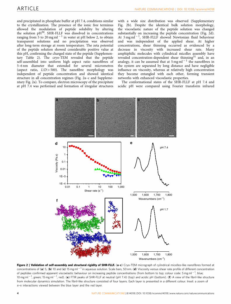
and precipitated in phosphate buffer at pH 7.4, conditions similarto the crystallization. The presence of the ionic free terminusallowed the modulation of peptide solubility by alteringthe solution pH30. SHR-FLLF was dissolved in concentrationsranging from 5 to 20 mg ml� 1 in water at pH below 2, to obtaintransparent solutions and no precipitation was observedafter long-term storage at room temperature. The zeta potentialof the peptide solution showed considerable positive value atthis pH, confirming the charged state of the peptide (Supplemen-tary Table 2). The cryo-TEM revealed that the peptideself-assembled into uniform high aspect ratio nanofibres of5–6 nm diameter that extended for several micrometres(aspect ratio, L/D4500). The nanofibre morphology wasindependent of peptide concentration and showed identicalstructure in all concentration regimes (Fig. 2a–c and Suppleme-ntary Fig. 2a). To compare, electron microscopy of the precipitateat pH 7.4 was performed and formation of irregular structures
with a wide size distribution was observed (SupplementaryFig. 2b). Despite the identical bulk solution morphology,the viscoelastic nature of the peptide solution was changedsubstantially on increasing the peptide concentration (Fig. 2d).At 5 mg ml� 1, SHR-FLLF showed Newtonian fluid behaviourand was independent of the applied shear. At higherconcentrations, shear thinning occurred as evidenced by adecrease in viscosity with increased shear rate. Manyamphiphilic molecules with cylindrical micelles assembly haverevealed concentration-dependent shear thinning31 and, in ananalogy, it can be assumed that at 5 mg ml� 1 the nanofibres inthe system are separated by long distance and have negligibleinfluence on viscosity, whereas at relatively high concentrationthey become entangled with each other, forming transientnetworks with enhanced viscoelastic properties.
The conformational states of the SHR-FLLF at pH 7.4 andacidic pH were compared using Fourier transform infrared
a
d
f
e
b c
pH 7.4
1,500 1,600 1,700
Wavenumbers (cm–1)
1,800
1,500 1,600 1,700
Wavenumbers (cm–1)
1,800
pH 1.7
10
1
0.1
0.01
Vis
cosi
ty (
Pa.
s)
1E–3
1E–40.01 0.1 1
Shear rate (s–1)10 100 1,000
Figure 2 | Validation of self-assembly and structural rigidity of SHR-FLLF. (a–c) Cryo-TEM micrograph of cylindrical micelles-like nanofibres formed at
concentrations of (a) 5, (b) 10 and (c) 15 mg ml� 1 in aqueous solution. Scale bars, 50 nm. (d) Viscosity versus shear rate profile of different concentration
of peptides confirmed apparent viscoelastic behaviour on increasing peptide concentrations (from bottom to top; colour code: 5 mg ml� 1, blue;
10 mg ml� 1, green; 15 mg ml� 1, red). (e) FTIR peaks of SHR-FLLF at neutral (pH 7.4) (top) and acidic pH (bottom). (f) A view of the fibril-like structure
from molecular dynamics simulation. The fibril-like structure consisted of four layers. Each layer is presented in a different colour. Inset: a zoom of
p–p interactions viewed between the blue layer and the red layer.
ARTICLE NATURE COMMUNICATIONS | DOI: 10.1038/ncomms14018
4 NATURE COMMUNICATIONS | 8:14018 | DOI: 10.1038/ncomms14018 | www.nature.com/naturecommunications
(FTIR) spectroscopy. The amide I and amide II bands at1,661 and 1,534 cm� 1, respectively, confirmed the presenceof helical conformation observed in the crystal structure32
(Fig. 2e). Similar amide bands were a spectral feature at pH 2and established that the conformation of SHR-FLLF didnot deviate considerably in acidic pH (Fig. 2e). Additionalsupport for helical conformations was obtained by measuringFTIR spectra in D2O solution. As shown in SupplementaryFig. 3a, the amide I bands at 1,642 and 1,649 cm� 1 indicatedthat peptide adopted both 310 and a-helical conformationsin solution, which is in agreement with the secondary structureobserved in X-ray crystallographic analysis33. This observationcan be ascribed to the rigid conformationally locked backboneof the designed peptide. Further insights about conformationalrigidity and self-assembly of SHR-FLLF were ascertainedby molecular dynamic simulation. It has been observed thatSHR-FLLF preserved its helical conformation in a simulatedmodel. To understand the supramolecular interactions innanofibres, a fibril-like structural model was constructedby forming four layers, in which each layer consisted of4 peptides by 6 peptides (that is, a total of 24 peptides), toform a diameter of B5–6 nm as observed by the cryo-TEMmeasurements (Fig. 2f and Supplementary Note 1). The fourpeptides in each layer formed p–p interactions between the twoPhe residues. The six peptides in each layer formed hydrophobicinteractions between the Aib residue and the Ala residue. Aftersimulations were performed, the diameter values of the fibril-likestructure were 6.2 nm by 5.1 nm, indicating that the diametervalues were conserved within the time scale of the simulation.In addition to intra-fibril interactions, the modelled fibrilalso formed hydrophobic interactions with neighbouring fibresand may account for a viscoelastic network at higherconcentration (Supplementary Fig. 4). The wide-angle X-rayscattering (WAXS) spectra of the nanofibre solution wererecorded at two different concentrations, to understand theabsolute molecular arrangement of peptide in nanofibresers(Supplementary Fig. 5). However, under the current experimentalconditions, the resolution of the spectra impede furtherspecific conclusion. The cumulative single crystal andFTIR studies confirmed that SHR-FLLF adopted a rigid rod-likeconformation in which the helical segment constituteda cylindrical hydrophobic environment, with the apex andbase of the cylinder being composed of hydrophilic terminalgroups and thus resembling a bolaamphiphilic structure (Fig. 1c).The arrangement of such cylindrical helical motifs in self-assembled nanofibres is better illustrated by molecular dynamicsimulation presented in Fig. 2f. The designer sequence affirmed adifferent class of self-assembling bolaamphiphilic helical peptidecomposed of natural amino acids and provides a significantstructural extension to the charged surfactant-like peptidesdesigned by the research group of Zhang and colleagues34–37
and peptide amphiphiles designed by the research group of Stuppand colleagues38–41 in which the peptides adopt b-sheetconformation and self-assemble into a lipid-like bilayer toshield the hydrophobic segments from the aqueous environment.
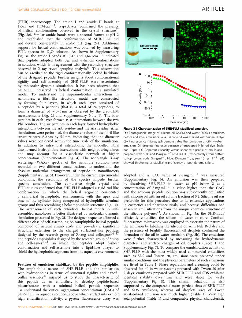
Features of emulsions stabilized by the peptide amphiphile.The amphiphilic nature of SHR-FLLF and the similaritieswith hydrophobins in terms of structural rigidity and nanofi-brillar assembly42 inspired us to study the characteristic ofthis peptide as an emulsifier, to develop peptide-basedbiosurfactants with a minimal helical peptide sequence.To understand the critical aggregation concentration (CAC) ofSHR-FLLF in aqueous solution, above which surfactants exhibithigh emulsification activity, a pyrene fluorescence assay was
adopted and a CAC value of 2.8 mg ml� 1 was measured(Supplementary Fig. 6). An emulsion was then preparedby dissolving SHR-FLLF in water at pH below 2 at aconcentration of 5 mg ml� 1, a value higher than the CAC,and the aqueous peptide solution was subsequently emulsifiedwith silicone oil with an oil volume fraction of 0.2. Silicone oil waspreferable for this procedure due to its extensive applicationsin cosmetics and pharmaceuticals, and because difficulties hadarisen in emulsifications from the unique chemical structure ofthe silicone polymer43. As shown in Fig. 3a, the SHR-FLLFefficiently emulsified the silicon oil–water mixture. Confocalfluorescence microscopy was employed to ascertain the nature ofthe emulsion by labelling the silicone oil with Nile Red dye andthe presence of brightly fluorescent oil droplets confirmed theformation of the oil-in-water emulsion (Fig. 3b). The emulsionswere further characterized by measuring the hydrodynamicdiameters and surface charges of oil droplets (Table 1 andSupplementary Fig. 7). To compare the emulsification activity ofSHR-FLLF with the most widely used commercial emulsifierssuch as SDS and Tween 20, emulsions were prepared undersimilar conditions and the physical parameters of such emulsionsare listed in Table 1. Phase separation and creaming could beobserved for oil-in-water systems prepared with Tween 20 after2 days; emulsions prepared with SHR-FLLF and SDS exhibitedidentical stability over time and were stable for weeks(Supplementary Fig. 8). This similar behaviour is alsosupported by the comparable mean particle sizes of SHR-FLLFand SDS emulsions, whereas oil droplets sizes of Tween20-stabilized emulsion was much higher (Table 1). Very highzeta potential (Table 1) and comparable physical characteristic
ba
Emulsification
100
10
1
0.1
0.01
1E–3
1E–40.01 0.1 1
Shear rate (s–1)
Vis
cosi
ty (
Pa.
s)
10 100 1,000
c
Figure 3 | Characterization of SHR-FLLF stabilized emulsion.
(a) Photographic image of silicone oil (20%) and water (80%) emulsions
before and after emulsifications. Silicone oil was stained with Sudan III dye.
(b) Fluorescence micrograph demonstrates the formation of oil-in-water
emulsion. Oil droplets fluoresce because of entrapped Nile red dye. Scale
bar, 10mm. (c) Apparent viscosity versus shear rate profile of emulsions
prepared with 5, 10 and 15 mg ml� 1 of SHR-FLLF, respectively (from bottom
to top; colour code: 5 mg ml� 1, blue; 10 mg ml� 1, green; 15 mg ml� 1, red)
showed thickening or stabilizing proficiency of peptide emulsifiers.
NATURE COMMUNICATIONS | DOI: 10.1038/ncomms14018 ARTICLE
NATURE COMMUNICATIONS | 8:14018 | DOI: 10.1038/ncomms14018 | www.nature.com/naturecommunications 5
with SDS confirmed that the SHR-FLLF was highly surface activeand formed strong interfacial networks. To understand theconformational preference of SHR-FLLF at oil–water interface,emulsions were prepared in D2O and FTIR spectra weremeasured in solution state. The bands at 1,642 and 1,649 cm� 1
demonstrated excellent correlation with the SHR-FLLFconformation observed in D2O (Supplementary Fig. 3b).Furthermore, drying of the emulsion (prepared in H2O) overKBr crystal revealed amide I band at 1,662 cm� 1, which isconsistent with helical conformation of the designed peptide(Supplementary Fig. 3c). These studies confirmed that theSHR-FLLF conserved the helical conformation at the oil–waterinterface and self-assembled to supramolecular helicalarrangement to stabilize the emulsion.
The preservation of viscoelastic properties of native SHR-FLLFat relatively high concentration would render the emulsionviscoelastic and may impart additional stability against coales-cence and creaming. Based on this supposition, SHR-FLLFstabilized emulsions were prepared by dissolving the peptide at10 and 15 mg ml� 1 concentrations, and rheological propertiesand long-term stability were studied. Zeta potential and dropletssizes of the prepared emulsions had similar characteristics as theemulsion prepared with 5 mg ml� 1 of SHR-FLLF (Table 1 andSupplementary Fig. 7). However, the formed emulsions exhibitedminimum stability at two months and several batches ofpreparations showed no signs of phase separation even after 8months of storage (Supplementary Fig. 9). All the stabilityanalysis was performed at 25 �C, to simulate a real-lifeenvironmental condition. To the best our knowledge, the long-term emulsion stability presented by SHR-FLLF is the highestamong the peptide-based emulsifiers described in the litera-ture18,20–23. Rheology measurements were performed toinvestigate the viscoelastic nature of the emulsions. As shownin Fig. 3c, at 5 mg ml� 1 of SHR-FLLF, emulsion showed Newto-nian flow behaviour as expected for an emulsion with low oilcontent. However, viscoelastic properties of the emulsionincreased dramatically at 10 and 15 mg ml� 1 of SHR-FLLF andexhibited shear thinning behaviour. The measured viscosity wasmuch higher than that of the corresponding peptide solutions andindicated that the peptide formed an assembled structure incontinuous phase and afforded transient network structures thatreduced the mobility of the oil droplets, which prohibitedcoalescence44, and consequently produced emulsion withexceptional stability. Rod-like polysaccharides and proteinfibrils are known to impart additional stability to the emulsioncompared with random coil structures owing to the higherexcluded volume45. The highly stable emulsion with SHR-FLLFsuggested that the rigid backbone may play a crucial role inself-assembly and viscoelastic emulsion formation.
Versatility of rigid minimal helical peptide as emulsifiers.To demonstrate the structural versatility of this minimalconformationally constrained heptapeptide as an emulsifierand comprehend the relationship between the structure–assembly–function, we designed two additional minimal peptidesequences SHR-FLELF and SHR-FLKLF by replacing alaninewith helix-favouring amino acids, glutamic acid and lysine(Fig. 4a). Such structural modification is expected to disruptthe bolaamphiphilic feature of SHR-FLLF and the retention of thenative functionality will prove the excellent robustness ofthe system towards structural alteration. The secondary structuralconformations of the peptides were probed by FTIR spectroscopy,both in the solution and after drying the samples over IR card.The presence of amide I peaks at 1,658 and 1,660 cm� 1 forSHR-FLELF and SHR-FLKLF, respectively, in dry samples(prepared in H2O) indicated the expected helical conformation ofthe peptides (Fig. 4b). Furthermore, the peptide was dissolved inD2O and FTIR spectra were recorded in solution state, as itprovides the information about the dynamic conformation. Thespectra of SHR-FLELF exhibited two bands at 1,644 and1,651 cm� 1, confirming the presence of 310 and a-helicalcomponents in the solution state (Supplementary Fig. 10a).Similar spectral features with amide I bands at 1,644 and1,650 cm� 1 of SHR-FLKLF indicated that the backboneconformation of the designed peptides are highly robust and havethe potential to accommodate a variety of modification(Supplementary Fig. 10d). The pyrene fluorescence assay revealedthe CAC of SHR-FLELF to be 1.5 mg ml� 1 (SupplementaryFig. 11a,b). However, no significant change in the pyrene fluor-escence peak intensity was evident in the presence of 10 mg ml� 1
of SHR-FLKLF (Supplementary Fig. 11c), a result that wasconsistent with the very high solubility of the peptide(o100 mg ml� 1) as compared with SHR-FLFLF (B15 mg ml� 1)around pH 2. This study confirmed that SHR-FLELFself-assembled into a micelle-like structure, whereas SHR-FLKLFpersisted in the non-aggregated state in solution in theconcentration range of 10 mg ml� 1. The emulsification activitiesof SHR-FLELF and SHR-FLKLF were investigated under similarconditions as used with SHR-FLLF. SHR-FLKLF stabilizedemulsions revealed a fast phase separation due to coalescence,although they also showed comparable droplet sizes andzeta potential with SHR-FLELF (Fig. 4c, Table 1 and Supple-mentary Figs 12 and 13). The backbone conformation of thepeptides at oil–water interface was assessed by FTIR spectro-scopy. The SHR-FLELF emulsions prepared in D2O revealedamide I bands at 1,645 and 1,654 cm� 1, indicating the presenceof helical conformation at the interface. The FTIR signals ofSHR-FLKLF show a single broad peak at 1,644 cm� 1 and canbe attributed to the 310 helical conformation (Supplementary
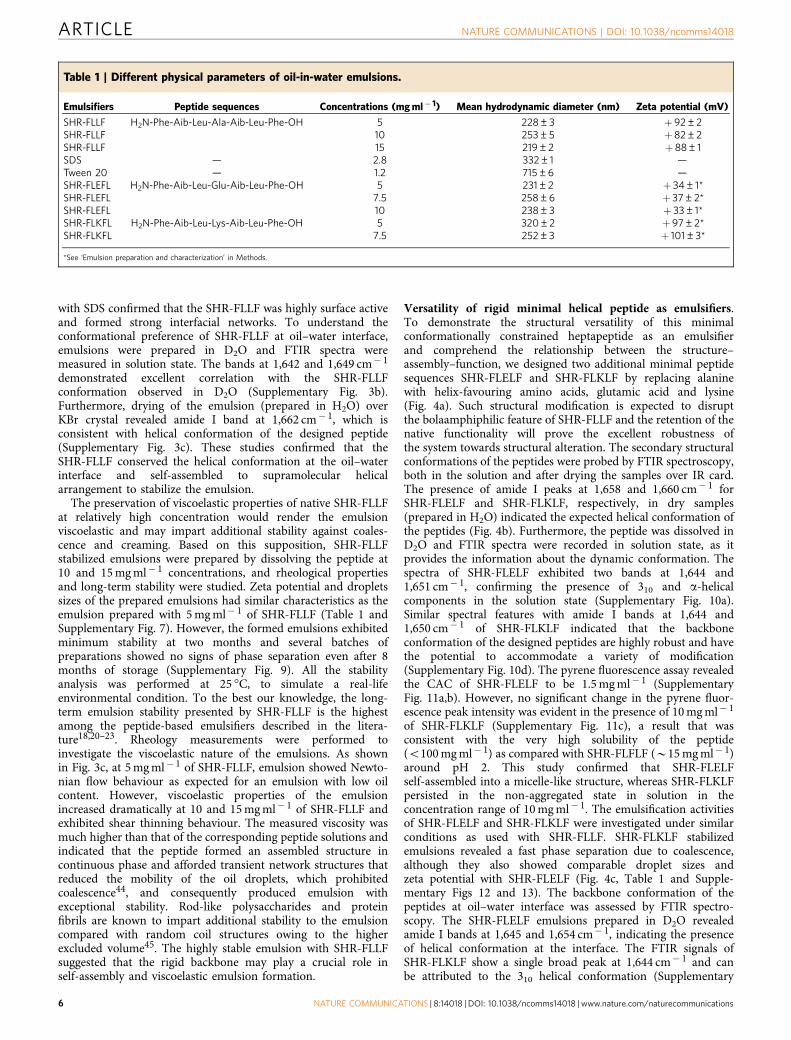
Table 1 | Different physical parameters of oil-in-water emulsions.
Emulsifiers Peptide sequences Concentrations (mg ml� 1) Mean hydrodynamic diameter (nm) Zeta potential (mV)
SHR-FLLF H2N-Phe-Aib-Leu-Ala-Aib-Leu-Phe-OH 5 228±3 þ 92±2SHR-FLLF 10 253±5 þ 82±2SHR-FLLF 15 219±2 þ 88±1SDS — 2.8 332±1 —Tween 20 — 1.2 715±6 —SHR-FLEFL H2N-Phe-Aib-Leu-Glu-Aib-Leu-Phe-OH 5 231±2 þ 34±1*SHR-FLEFL 7.5 258±6 þ 37±2*SHR-FLEFL 10 238±3 þ 33±1*SHR-FLKFL H2N-Phe-Aib-Leu-Lys-Aib-Leu-Phe-OH 5 320±2 þ 97±2*SHR-FLKFL 7.5 252±3 þ 101±3*
*See ‘Emulsion preparation and characterization’ in Methods.
ARTICLE NATURE COMMUNICATIONS | DOI: 10.1038/ncomms14018
6 NATURE COMMUNICATIONS | 8:14018 | DOI: 10.1038/ncomms14018 | www.nature.com/naturecommunications

Fig. 10d,e). The drying of the emulsions prepared withSHR-FLELF and SHR-FLKLF over KBr crystal displayed amide Ipeaks at 1,663 and 1,661 cm� 1, respectively, indicating thatthe native helical conformation of the peptides was maintainedunder this conditions (Supplementary Fig. 10c,f). This outcomedemonstrated that the designed helical peptides have excellentinterfacial adsorption characteristics but the integrity ofthe interfacial peptide network was much more robust forthe SHR-FLELF, owing to its high aggregation propensityin solution. The concentration-dependent viscoelastic nature ofthe SHR-FLELF was studied further, to gain insights about the
effect of the self-assembly of peptide with a robust backboneconformation with respect to emulsion stability (Fig. 4d).At 5 mg ml� 1, the viscosity of SHR-FLELF-stabilized emulsionwas low and had a fluid-like flow. The viscosity of the emulsionwas elevated sharply at a concentration of 7.5 mg ml� 1 witha shear thinning effect. This emulsion was stable for an exam-ination period of 2 months without any indication ofphase separation or creaming (Fig. 4c). Further increases inconcentration (10 mg ml� 1) had a minimal effect on emulsionviscosity and gave a modest rise that could be accounted forby a higher peptide concentration in continuous phase. It is
SHR-FLKLFSHR-FLELF SHR-FLKLFSHR-FLELF
2months storage
c
SHR-FLELF
SHR-FLKLF
SHR-FLLF
d
SHR-FLELF
1,500 1,600Wavenumbers (cm–1)
1,700 1,800
1,500 1,600Wavenumbers (cm–1) Shear rate (s–1)
1,700 1,800
SHR-FLKLF
10
1
0.1
Vis
cosi
ty (
Pa.
s)
0.01
1E–3
1E–40.01 0.1 1 10 100 1,000
a
b
O
O O
OH
O O
O
O
NHNH
NH
O
O
NHNH
OH
OO
O
O O
O
O O
O
NH NHNH
OH
O
NH
NH
NH
NH
O
NH
NH
H2N
H2N
H2N
NH2
NHNH
OH
O
O
NHNH
Figure 4 | Demonstration of structural modularity of conformationally constrained helical peptide emulsifiers. (a) Structural modification of SHR-FLLF
to afford SHR-FLELF and SHR-FLELF. Major residues involved in sequence modification are highlighted. (b) Truncated FTIR spectra of SHR-FLELF (top)
and SHR-FLKLF (bottom). (c) Photographic images depicting the long-term stability of emulsions prepared with SHR-FLELF (7.5 mg ml� 1) and SHR-FLELF
(7.5 mg ml� 1). (d) Profiles of apparent viscosity versus shear rate of emulsions prepared with 5, 7.5 and 10 mg ml� 1 of SHR-FLELF, respectively
(from bottom to top; colour code: 5 mg ml� 1, blue; 7.5 mg ml� 1, green; 10 mg ml� 1, red) established general thickening or stabilizing proficiency of this
class of peptide emulsifiers.
NATURE COMMUNICATIONS | DOI: 10.1038/ncomms14018 ARTICLE
NATURE COMMUNICATIONS | 8:14018 | DOI: 10.1038/ncomms14018 | www.nature.com/naturecommunications 7

important to mention that high concentrations of most proteinemulsifiers have detrimental effects on emulsion stability, andfast phase separation and creaming are common phenomena dueto depletion flocculation46.
DiscussionA direct correlation between the self-assembly features, helicalfolding pattern and emulsion stability of all the designed peptidesequences pertaining to all the tested concentrations waspresented in Table 2. The combinations of single crystal analysis,molecular dynamic simulation and FTIR spectroscopy indicatedthat the peptides adopted robust helical backbone conformation.However, as probed by the electron microscopy and pyrenefluorescence assays, only the peptides SHR-FLLF and SHR-FLELFself-assembled to supramolecular helical arrangement, whereasSHR-FLKLF exists mainly in non-aggregated states. Highly stableemulsions formed by the former two sequences can be directlycorrelated with their high self-assembly propensity in solution.The robust conformation of these peptides may permit favourableinteractions between the absorbed interfacial peptide layers andthe self-assembled structure in bulk aqueous phase, and these aremanifested by the increase in viscoelastic nature of the emulsionsat relatively high peptide concentration.
In conclusion, a group of peptide amphiphiles with a rigidbackbone conformation were designed and self-assembled intonanoscale morphology in aqueous solution. The soluble mono-disperse cylindrical micelle structures will allow molecularengineering of supramolecular helical peptide assemblies basedon minimal helical peptide sequence. The designer amphiphilicbuilding blocks demonstrated excellent surface activity towardoil-in-water emulsion formation and maintained their propensitytowards self-aggregation in emulsified continuous phase, reveal-ing an extraordinarily stable emulsion with a single componentacting as both emulsifier and stabilizer, a feature never realizedbefore in peptide emulsifiers. The engineered building blocksshowed high modularity and sequence–structure–function rela-tionship. The versatility of such amino acid toolkits point to theincorporation of different features such as the elicitation of aresponse towards external stimuli and switchable surfactantbehaviours. These materials may provide another paradigm forbiosurfactants in applications related to drug delivery and thefood and cosmetics industries.
MethodsGeneral sample preparation. All the peptides were purchased from Peptron, Inc.(South Korea). SHR-FLLF was suspended in water at desired concentration and1 N HCl was added to dissolve the peptide. Final pH was maintained in therange of 1.4–1.8. To prepare SHR-FLLF samples in buffer (pH 7.4), the peptidewas dissolved in ethanol in 100 mg ml� 1 concentration and diluted to 5 mg ml� 1
with phosphate buffer (10 mM). SHR-FLELF and SHR-FLKLF were dissolved inwater at preferred concentrations and 1 N HCl was added to adjust the pH inthe range of 1.4–1.8. All the peptides formed transparent solutions under theconditions described above. Control experiments with SDS and Tween 20 were
performed by dissolving the corresponding surfactants in water at the desiredconcentrations (SDS, 2.8 mg ml� 1 and Tween 20, 1.2 mg ml� 1) and finalsolution pH was maintained between 1.4 and 1.8.
Cryo-transmission electron microscopy. Cryo-TEM samples were preparedusing Leica EM GP cryo-preparation instrument operated at 100% relativehumidity. Samples were prepared as described in general Methods section.Peptide solution (2.5 ml) was applied on a holey carbon TEM grid (Lacey substrate,300 mesh, Ted Pella, Inc.) followed by blotting with a filter paper and plunginginto liquid ethane. The vitrified samples were kept under liquid nitrogen andsubsequently transferred to a FEI-Tecnai T12 TEM using a Gatan workstationand cryo-holder. The images were acquired at 98 K with an operatingvoltage of 120 kV in low electron dose mode. Images were recorded on aGatan 794 charge-coupled device camera.
Transmission electron microscopy. To prepare the samples for TEM imaging,peptides were dissolved in ethanol (100 mg ml� 1) and diluted by the additionof phosphate buffer (pH 7.4) to reach a final concentration of 5 mg ml� 1.A 7 ml aliquot of this solution was placed on a 400-mesh copper grid and excessfluids were removed by blotting with a filter paper after 2 min. The samples werenegative stained with 2% uranyl acetate in water and excess fluids were removedfrom the grid after 2 min. TEM images were recorded using a JEM-1400 electronmicroscope (JEOL) operating at 80 kV.
Bulk rheology. Rheological measurements of the emulsions were performedon a Discovery Hybrid Rheometer HR3 (TA Instruments, USA) equipped withcone and plate geometry (diameter, 40 mm; cone angle, 1.04�). All measurementswere conducted in a steady-state shear sweep mode at a temperature of 25±0.1 �Cafter 24 h of samples preparation.
Emulsion preparation and characterization. All the emulsions were preparedwith silicone oil (Viscosity 10 cSt; Sigma-Aldrich) with oil volume fraction of0.2. Peptide concentrations used for the emulsifications were as follows: 5, 10 and15 mg ml� 1 of SHR-FLLF; 5, 7.5 and 10 mg ml� 1 of SHR-FLFLF; and 5 and7.5 mg ml� 1 of SHR-FLKLF, respectively. Peptide solutions with desiredconcentration were prepared by the methods described in general section followedby the addition of required amount of silicone oil. The combined solution wassubjected to probe sonication (Syclon probe ultrasonicator: SKL-150-IIDN) forduration of two 5 s pulses at an output of 27%, whereas the delay between pulsesis 9 s. For long-term stability assessment, the emulsions were stored at 25 �C andstability was monitored by visual observation and capturing photography images.The droplet size distribution of the emulsions was measured by diluting thesamples by 20 times in corresponding continuous phase (Zetasizer Nano ZS,Malvern Instruments). The zeta potential measurement was performed bydiluting the emulsions in double distilled water (Zetasizer Nano ZS, MalvernInstruments). Zeta potentials were also measured by diluting the emulsionsin the corresponding acidic continuous phase and all the samples revealed zetapotential values above þ 100 mV. These high values are above the usual limit ofthe instrument and, therefore, zeta potential data were collected by diluting theemulsions in double distilled water. The SHR-FLELF has low isoelectronic pointthan the other peptides and consequently has lower zeta potential under thiscondition. For photography, Sudan III was dissolved in silicone oil before emulsionpreparation.
Confocal laser scanning microscopy. Confocal microscope images wererecorded in a Zeiss LSM 510 META. The emulsion was prepared with siliconeoil that was prestained with 1 mM Nile Red dye. Emulsion suspensions dilutedtenfold with continuous phase. A drop of emulsion was deposited on glass slideand covered with glass coverslip. The imaging was performed with � 100 objectivelens by exciting the sample at 477 nm and emission was collected in540–657 channels.
Table 2 | A comparative analysis of the different properties of SHR-FLLF, SHR-FLELF and SHR-FLKLF.
Emulsifiers Peptide sequences Concentrations (mg ml� 1) Self-assemble Emulsion stability Helical conformation
SHR-FLLF H2N-Phe-Aib-Leu-Ala-Aib-Leu-Phe-OH 5 Yes 41 Week Yes10 Yes 42 Months Yes15 Yes 42 Months Yes
SHR-FLEFL H2N-Phe-Aib-Leu-Glu-Aib-Leu-Phe-OH 5 Yes 41 Week Yes7.5 Yes 42 Months Yes10 Yes 42 Months Yes
SHR-FLKFL H2N-Phe-Aib-Leu-Lys-Aib-Leu-Phe-OH 5 No o1 Week Yes7.5 No o1 Week Yes
ARTICLE NATURE COMMUNICATIONS | DOI: 10.1038/ncomms14018
8 NATURE COMMUNICATIONS | 8:14018 | DOI: 10.1038/ncomms14018 | www.nature.com/naturecommunications
Critical aggregation concentration. The CAC value was determined by amodification of the methods described in ref. 47. Pyrene solution (5 mg) wasdissolved in methanol and diluted 20 times in methanol to prepare the stock.Fixed concentration of pyrene stock was added to different peptide solutionswith concentrations ranging from 0.1 to 10 mg ml� 1 and the total volume waskept constant at 600ml. Fluorescence spectra was recorded by exciting thesamples at 334 nm and measuring the emission from 360 to 410 nm with theuse of excitation and emission slits of 8 and 2 nm, respectively. The CAC valuewas calculated from the curve obtained by plotting the ratio of the peaks at373 and 384 nm against the peptide concentrations.
FTIR spectroscopy. FTIR spectra were collected using a Nicolet Nexus 470 FTIRspectrometer with a DTGS (deuteratedtriglycine sulfate) detector. A 30 ml aliquotof the peptides solution was deposited on a polyethylene IR card and driedunder vacuum. Measurements were taken using a 4 cm� 1 resolution and byaveraging 64 scans. The absorbance maxima values were determined using anOMNIC analysis program (Nicolet). The background was subtracted using acontrol spectrum. To record the FTIR spectra of emulsions, a 30 ml aliquotof the peptides solution was deposited on a KBr IR card and dried undervacuum. The background was subtracted using a control spectrum containingonly silicone oil on a KBr IR card.
To record the FTIR spectra in solution, peptides were lyophilized twice in0.5 M HCl solution. Subsequently, the peptides were dissolved in D2O(5–7 mg ml� 1). Emulsions were prepared in the condition described above inD2O. Peptide solutions or emulsions were sandwiched between two 25� 2 mmCaF2 windows separated with a 25 mm polytetrafluoroethylene spacer.Measurements were taken using a 4 cm� 1 resolution and by averaging128 scans. The background was subtracted using a control D2O spectrum.
Molecular dynamics simulations. All details regarding the constructionof the fibril-like model and molecular dynamics simulations procedure areprovided in the Supplementary Note 1.
X-ray crystal structure analysis and crystal data. Single crystals suitablefor X-ray diffraction were grown by slow evaporation of the peptide in ethanol(5%)—phosphate buffer (10 mM, pH 7.4) in room temperature and crystals ofdiffraction quality were obtained after 2–5 days of sample preparation. Crystalssuitable for diffraction were coated with Paratone oil (Hampton Research) andmounted on loops and flash frozen in liquid nitrogen. Diffraction data measure-ments were done on a Bruker KappaApexII system with MoKa radiation at100(2) K. Data were collected and processed with Apex2 Suite. The structures weresolved by direct methods using SHELXT-2013. The structures were refined by full-matrix least squares against F2 with SHELXL-2013. The crystallographic data aregiven in Supplementary Table 3 and Supplementary Data set 1.
Accession codes. The X-ray crystallographic coordinates for structures reportedin this study have been deposited at the Cambridge Crystallographic Data Centre(CCDC), under deposition numbers CCDC 1473157. These data can be obtainedfree of charge from the CCDCvia www.ccdc.cam.ac.uk/data_request/cif.
Wide-angle X-ray scattering. The SHR-FLLF peptide solutions at a concentra-tion of 15 and 5 mg ml� 1 were sealed in quartz capillaries with a 1.5 mm diameter.WAXS measurements were performed using an in-house X-ray scatteringsystem, with a GeniX (Xenocs) low divergence Cu Ka radiation source(wavelength of 1.54 Å) and a scatterless slits setup48. Two-dimensionalscattering data, with a momentum transfer wave vector (q) range of 0.07� 2.5 Å� 1
at a sample-to-detector distance of about 160 mm, was collected on a Pilatus300 K detector (Dectris, Baden-Daettwil, Switzerland) and radially integrated usingMatlab (MathWorks, Natick, MA, USA)-based procedures (SAXSi). Calibrationwas performed using silver behenate. The scattering data of the water at pH 2was collected as background and used to subtract the solvent and spuriousscattering from the WAXS system itself, for example, Kapton vacuum windowsand air gaps.
Data availability. The authors declare that all data supporting the findings ofthis study are available within the article and its Supplementary Informationfiles, or from the corresponding author upon request. The X-ray crystallographiccoordinates for structures that support the findings of this study have beendeposited in CCDC, with the accession code 1473157.
References1. Bibette, J., Calderon, F. L. & Poulin, P. Emulsions: basic principles. Rep. Prog.
Phys. 62, 969–1033 (1999).2. Zarzar, L. D. et al. Dynamically reconfigurable complex emulsions via tunable
interfacial tensions. Nature 518, 520–524 (2015).
3. Kislukhin, A. A. et al. Paramagnetic fluorinated nanoemulsions for sensitivecellular fluorine-19 magnetic resonance imaging. Nat. Mater. 15, 662–668(2016).
4. Malaki Nik, A., Wright, A. J. & Corredig, M. Interfacial design of protein-stabilized emulsions for optimal delivery of nutrients. Food Funct. 1, 141–148(2010).
5. Hanson, J. A. et al. Nanoscale double emulsions stabilized by single-componentblock copolypeptides. Nature 455, 85–88 (2008).
6. Rebello, S., Asok, A. K., Mundayoor, S. & Jisha, M. S. Surfactants: toxicity,remediation and green surfactants. Environ. Chem. Lett. 12, 275–287 (2014).
7. Dickinson, E. Hydrocolloids as emulsifiers and emulsion stabilizers. FoodHydrocoll. 23, 1473–1482 (2009).
8. Chassaing, B. et al. Dietary emulsifiers impact the mouse gut microbiotapromoting colitis and metabolic syndrome. Nature 519, 92–96 (2015).
9. Dickinson, E. Colloids in food: ingredients, structure, and stability. Annu. Rev.Food Sci. Technol. 6, 211–233 (2015).
10. Lam, R. S. H. & Nickerson, M. T. Food proteins: a review on their emulsifyingproperties using a structure–function approach. Food Chem. 141, 975–984(2013).
11. Dickinson, E. Exploring the frontiers of colloidal behaviour where polymersand particles meet. Food Hydrocoll. 52, 497–509 (2016).
12. McClements, D. J. Protein-stabilized emulsions. Curr. Opin. Colloid InterfaceSci. 9, 305–313 (2004).
13. Wang, L., Walsh, M. T. & Small, D. M. Apolipoprotein B is conformationallyflexible but anchored at a triolein/water interface: a possible model forlipoprotein surfaces. Proc. Natl Acad. Sci. USA 103, 6871–6876 (2006).
14. Raffaini, G., Milani, R., Ganazzoli, F., Resnati, G. & Metrangolo, P. Atomisticsimulation of hydrophobin HFBII conformation in aqueous and fluorous mediaand at the water/vacuum interface. J. Mol. Graph. Model. 63, 8–14 (2016).
15. Tucker, I. M. et al. Adsorption of hydrophobin–protein mixtures at the air–water interface: the impact of ph and electrolyte. Langmuir 31, 10008–10016(2015).
16. Cox, P. W. & Hooley, P. Hydrophobins: new prospects for biotechnology.Fungal Biol. Rev. 23, 40–47 (2009).
17. Dexter, A. F. & Middelberg, A. P. J. Peptides as functional surfactants. Ind. Eng.Chem. Res. 47, 6391–6398 (2008).
18. Dexter, A. F., Malcolm, A. S. & Middelberg, A. P. J. Reversible active switchingof the mechanical properties of a peptide film at a fluid-fluid interface.Nat. Mater. 5, 502–506 (2006).
19. Xue, Y., He, L., Middelberg, A. P. J., Mark, A. E. & Poger, D. Determining thestructure of interfacial peptide films: comparing neutron reflectometry andmolecular dynamics simulations. Langmuir 30, 10080–10089 (2014).
20. Bai, S. et al. Stable emulsions formed by self-assembly of interfacial networks ofdipeptide derivatives. ACS Nano 8, 7005–7013 (2014).
21. Scott, G. G., McKnight, P. J., Tuttle, T. & Ulijn, R. V. Tripeptide emulsifiers.Adv. Mater. 28, 1381–1386 (2016).
22. Dexter, A. F. Interfacial and emulsifying properties of designed b-strandpeptides. Langmuir 26, 17997–18007 (2010).
23. Saito, M., Ogasawara, M., Chikuni, K. & Shimizu, M. Synthesis of a peptideemulsifier with an amphiphilic structure. Biosci. Biotechnol. Biochem. 59,388–392 (1995).
24. Schafmeister, C. E., Miercke, L. J. & Stroud, R. M. Structure at 2.5 A of adesigned peptide that maintains solubility of membrane proteins. Science 262,734–738 (1993).
25. McGregor, C.-L. et al. Lipopeptide detergents designed for the structural studyof membrane proteins. Nat. Biotechnol. 21, 171–176 (2003).
26. Mondal, S. et al. Formation of functional super-helical assemblies byconstrained single heptad repeat. Nat. Commun. 6, 8615 (2015).
27. Frederix, P. W. J. M. et al. Exploring the sequence space for (tri-)peptideself-assembly to design and discover new hydrogels. Nat. Chem. 7, 30–37
(2015).28. Gazit, E. Molecular self-assembly: searching sequence space. Nat. Chem. 7,
14–15 (2015).29. Aravinda, S., Shamala, N. & Balaram, P. Aib residues in peptaibiotics and
synthetic sequences: analysis of nonhelical conformations. Chem. Biodivers. 5,1238–1262 (2008).
30. Tang, C., Smith, A. M., Collins, R. F., Ulijn, R. V. & Saiani, A. Fmoc-diphenylalanine self-assembly mechanism induces apparent pKa shifts.Langmuir 25, 9447–9453 (2009).
31. Shrestha, R. G. et al. Peptide-based gemini amphiphiles: phase behavior andrheology of wormlike micelles. Langmuir 28, 15472–15481 (2012).
32. Kong, J. & Yu, S. Fourier transform infrared spectroscopic analysis of proteinsecondary structures. Acta Biochim. Biophys. Sin. (Shanghai), 39, 549–559(2007).
33. Kennedy, D. F., Crisma, M., Toniolo, C. & Chapman, D. Studies of peptidesforming 310-helices and a-helices and b-bend ribbon structures in organicsolution and in model biomembranes by fourier-transform infrared-spectroscopy. Biochemistry 30, 6541–6548 (1991).
NATURE COMMUNICATIONS | DOI: 10.1038/ncomms14018 ARTICLE
NATURE COMMUNICATIONS | 8:14018 | DOI: 10.1038/ncomms14018 | www.nature.com/naturecommunications 9
34. Zhang, S. Lipid-like self-assembling peptides. Acc. Chem. Res. 45, 2142–2150(2012).
35. Vauthey, S., Santoso, S., Gong, H., Watson, N. & Zhang, S. Molecular self-assembly of surfactant-like peptides to form nanotubes and nanovesicles.Proc. Natl Acad. Sci. USA 99, 5355–5360 (2002).
36. Zhang, S. Fabrication of novel biomaterials through molecular self-assembly.Nat. Biotechnol. 21, 1171–1178 (2003).
37. Fatouros, D. G. et al. Lipid-like self-assembling peptide nanovesicles for drugdelivery. ACS Appl. Mater. Interfaces 6, 8184–8189 (2014).
38. Tantakitti, F. et al. Energy landscapes and functions of supramolecular systems.Nat. Mater. 15, 469–476 (2016).
39. Ortony, J. H. et al. Internal dynamics of a supramolecular nanofibre. Nat.Mater. 13, 812–816 (2014).
40. Moyer, T. J. et al. pH and amphiphilic structure direct supramolecularbehavior in biofunctional assemblies. J. Am. Chem. Soc. 136, 14746–14752(2014).
41. Hartgerink, J. D., Beniash, E. & Stupp, S. I. Self-assembly and mineralization ofpeptide-amphiphile nanofibers. Science 294, 1684–1688 (2001).
42. Kallio, J. M., Linder, M. B. & Rouvinen, J. Crystal structures of hydrophobinHFBII in the presence of detergent implicate the formation of fibrils andmonolayer films. J. Biol. Chem. 282, 28733–28739 (2007).
43. Kawaguchi, M. Silicone oil emulsions stabilized by polymers and solid particles.Adv. Colloid Interface Sci. 233, 186–199 (2016).
44. Paximada, P., Koutinas, A. A., Scholten, E. & Mandala, I. G. Effect of bacterialcellulose addition on physical properties of WPI emulsions. comparison withcommon thickeners. Food Hydrocoll. 54, 245–254 (2016).
45. Philipse, A. P. The random contact equation and its implications for (colloidal)rods in packings, suspensions, and anisotropic powders. Langmuir 12,1127–1133 (1996).
46. Dickinson, E. & Golding, M. Rheology of sodium caseinate stabilizedoil-in-water emulsions. J. Colloid Interface Sci. 191, 166–176 (1997).
47. Dominguez, A., Fernandez, A., Gonzalez, N., Iglesias, E. & Montenegro, L.Determination of critical micelle concentration of some surfactants by threetechniques. J. Chem. Educ. 74, 1227–1231 (1997).
48. Li, Y., Beck, R., Huang, T., Choi, M. C. & Divinagracia, M. Scatterless hybridmetal-single-crystal slit for small-angle X-ray scattering and high-resolutionX-ray diffraction. J. Appl. Crystallogr. 41, 1134–1139 (2008).
AcknowledgementsWe thank A. Barbul for confocal microscopy analysis, V. Holdengreber for help withTEM analysis, R. Oshri for help with photography and members of the Gazit laboratoryfor helpful discussions. We acknowledge Dr Kenno Vanommeslaeghe for obtaining theparameters of the a-aminoisobutyric acid. All simulations were performed using thehigh-performance computational facilities of the Miller Lab in BGU HPC computational
centre. The support of the BGU HPC computational centre staff is greatly appreciated.S.M. thanks the PBC Program for outstanding Postdoctoral Researchers from China andIndia, and Post-Doctoral Fellowships of the Tel Aviv University Center for Nanoscienceand Nanotechnology for scholarships. R.B. acknowledges the support from the IsraeliScience Foundation (550/15). We thank the support by grants from the Israeli NationalNanotechnology Initiative and Helmsley Charitable Trust for a focal technology area onnanomedicines for personalized theranostics and the European Research Council BISONproject (E.G.).
Author contributionsS.M., L.A.-A. and E.G. conceived and designed the experiments. S.M. planned andperformed the experiments. S.M. crystallized the peptide assemblies. M.V. and S.M.prepared the samples for cryo-TEM experiments. D.N.B., Y.A-R. and Y.M. performed themolecular dynamic simulation. L.J.W.S. gathered the diffraction data and solved thecrystal structure. G.J. and R.B. performed and analysed the WAXS experiments. S.M. andE.G. wrote the manuscript. All authors discussed the results and commented on themanuscript.
Additional informationSupplementary Information accompanies this paper at http://www.nature.com/naturecommunications
Competing financial interests: The authors declare no competing financial interests.
Reprints and permission information is available online at http://npg.nature.com/reprintsandpermissions/
How to cite this article: Mondal, S. et al. A minimal length rigid helical peptidemotif allows rational design of modular surfactants. Nat. Commun. 8, 14018doi: 10.1038/ncomms14018 (2017).
Publisher’s note: Springer Nature remains neutral with regard to jurisdictional claims inpublished maps and institutional affiliations.
This work is licensed under a Creative Commons Attribution 4.0International License. The images or other third party material in this
article are included in the article’s Creative Commons license, unless indicated otherwisein the credit line; if the material is not included under the Creative Commons license,users will need to obtain permission from the license holder to reproduce the material.To view a copy of this license, visit http://creativecommons.org/licenses/by/4.0/
r The Author(s) 2017
ARTICLE NATURE COMMUNICATIONS | DOI: 10.1038/ncomms14018
10 NATURE COMMUNICATIONS | 8:14018 | DOI: 10.1038/ncomms14018 | www.nature.com/naturecommunications