a male with a headache and a male with an ulcer on …a male with a headache and a male with an...
TRANSCRIPT

A male with a headache and a male with an ulcer on his foot
Dr. Fausto Luis Garcia Ruiz
Dr. Kabilar Venugobal
24 May 2016

Case I, History of Present Illness
•Male patient, 46 years old, HIV + (on HAART)
•Onset of symptoms:
• September 2015, Backache, headache, and difficulty bending, which was relieved by lying on his back
•On examination:
• Tumor in occipital region of patient’s head
•Headache never released

Complementary Studies
•Laboratory: No remarks
•Chest X ray: No remarks
•CT-Scan report:
• Bone destruction on left occipito-temporal area
• Irregular soft tissue mass around bone destruction area
• (January 2016)
•Biopsy by Fine Needle Aspirative Cytology was ordered

Imaging
Imaging

Imaging

Imaging

Pathology Report:
•Specimen Type: FNA
•Nature of Specimen: FNAC
•Clinical diagnosis: Painless swelling in back
•FNA Site: Occipital Region
•Macroscopy: Blood stained material aspirated

Pathology Report
•Microscopy: Smears show small to medium size clusters of large malignant cells with large nuclei and coarse chromatic texture against haemorrhagic and acute inflammatory cell background
•Diagnosis: Cytomorphologically smears are positive for malignant cells, suggestive of skin adnexed tumor. Biopsy is advised.

Pathology

Pathology
Pathology

Recent Complementaries
•Laboratory test: WBC-11,38.10⁹;Hb 12,7g/l; platelets⁹ 652.10; neutrofils 8,2
•Chest X ray: No remarks
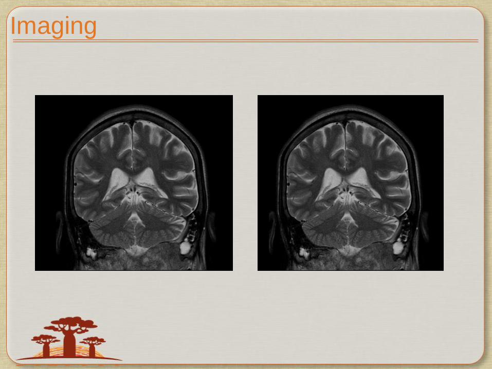
•MRI (head): A massive mass with a size of 13cmx17cm can be found in atlanto-occipital region. The lesion display is iso or high signal intensity in T1W1. Scuff muscles, bilateral masoid process, local duramatter and left neck are involved. Malignancy uinnatlanto-occipital region. Chondrosarcoma is presumed. Two cystic lesions can be found in right parietal lobe.

Any questions, please?

Case II
•A 80 years old male, a known hypertensive, presented in April 2014 with C/O- non-healing ulcer in left sole.
•O/E: Multiple inguinal Lymph nodes were palpable in left groin.
•BX from left sole lesion and inguinal lymph nodes
• S/O- Metastatic Malignant Melanoma.
•Other Metastatic Work-ups- Normal.
•Plan: Surgery, performed in May 2014. Excision of the lesion in foot and also left inguinal lymph node dissection done.

Pathology Report
•Specimen: Biopsy left foot
•Clinical History: Melanoma Left Foot. Lymph node specimen
1. With suture superficial groin left foot. Lymph node specimen
2. Without suture deep groin left
3. Skin from wedge of ulcer
Pathology Report
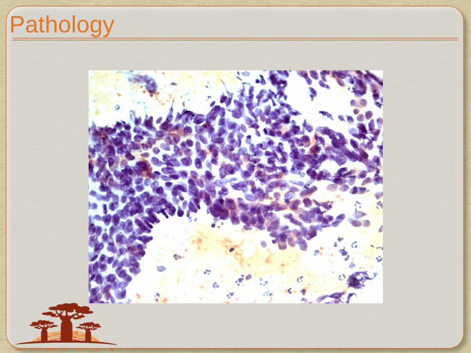
•Microscopic examination (brief):
• The tumor cells are positive for S100 and HMB 45. HHV8 is negative. The skin demonstrates ulceration with representation of ulcer base. The stroma is hyalinised.
•Conclusion: Metastatic Malignant Melanoma
• December 2014

Pathology

Pathology

Pathology

Immediate Conduct
•T4N1Mx (Stage IV)
• Referred to Princess Marina Hospital January 2015 for Radiotherapy
•Report from PMH:
• Received palliative Radiotherapy during February
• Left foot + bolus – 3000 cGy 12 MeV
• Left groin + bolus – 3000 cGy 12 MeV
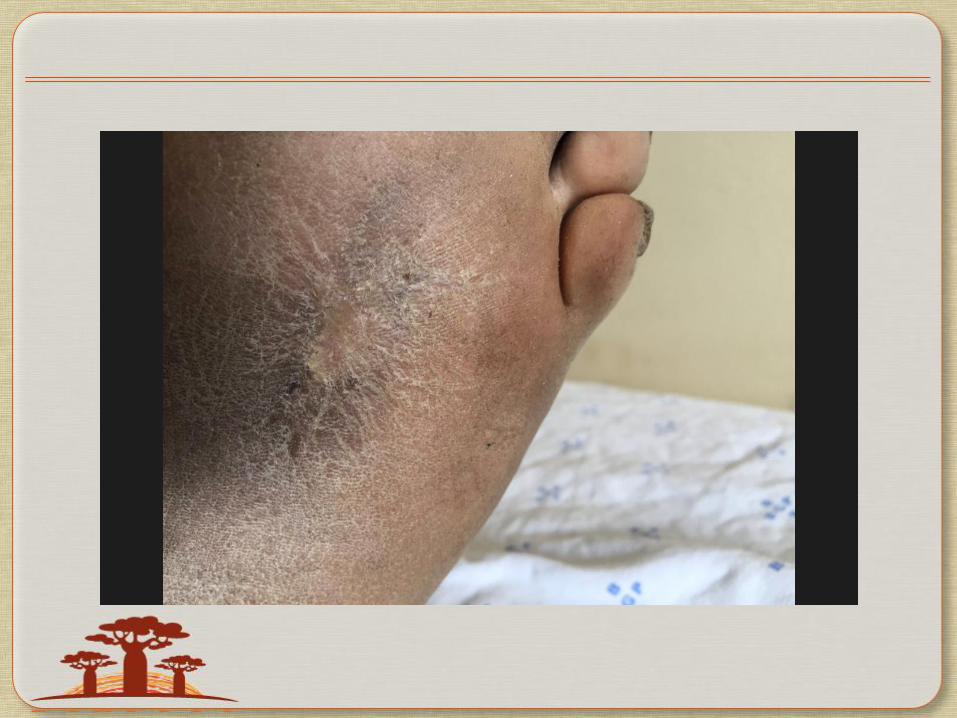
•Side effects: hyperpigmentation, oedema.

Follow up
•Follow up in October 2015
• Nil complaints
•O/E:
• Surgical scar over left leg, in plant and groin

•Chest X ray: Several opacities are seen in both pulmonary fields, specially in the left that suggest metastases
•Abdomen CT-Scan November 2015. In correspondence with Chest X Ray
•Lab Tests - wnl

Pre-chemotherapy imaging

Imaging

Plan: Systemic Chemotherapy
•Dacarbazine-220mg/m² days 1 to 3
•Cisplatin-25mg/m² days 1 to 3
•No Carmustine 150mg/m² days 1 (every 42 days)
•Cycles every 3 weeks, 6 cycles.
•From 4th cycle onwards, patient did not receive Cisplatin (since the patient developed severe delayed emesis).

CT Scan after 6 cycles Chemo, April 2016

Imaging
•Complete resolution of lung tumour after 6 cycles of chemotherapy
Follow up
•In Mid May the same patient presented with a suspicious melanoma lesion on the medial aspect of the middle of the thigh.
• Clinically the lesion appears melanoma and biopsy done
• How to approach this case, if the biopsy is proven to be melanoma


THANK YOU