a logical and sequential approaching to treating irregular astigmatism
TRANSCRIPT

A logical and sequential approaching to treating irregular astigmatism
(caused by decentered ablation): C-CAP and CustomVue – VISX system
Ming Wang, MD, PhDClinical Associate Professor of
Ophthalmology of University of Tennessee
Director, Wang Vision InstituteNashville, TN, USA
New book: “Irregular astigmatism”(Wang eds, SLACK)

Collaborators
� Tracey Swartz, OD.� Helen Boerman, O.D.� Shawna Hill, O.D.� Yangzi Jiang, M.D.,O.D.� Keming Yu, M.D.,Ph.D.� Lav Panchal, M.D.
� Financial interest: none.

Irregular astigmatism caused by decentered ablation
� A decentered ablation on corneal topography;� Increased higher order aberrations as measured using
wavefront aberrometry, predominantly coma;� The appearance of a tail on point spread functions;� Manifest refraction with reducedbest-corrected visual
acuity that improvesonly with gas permeable lenses;� A cylinder measurement on autorefraction and wavefront
that differs from manifest refraction;� A history of reduced vision immediately following surgery
that fails to improve with time.

Topo criteria for decentered ablation (heightdifference in elevationmap)
� At least 6 micronsdifference on the elevation topography, from the lowest point to the highest point, over a 6.5 mm diameter or over the patient’s pupil diameter as measured by the Zeiss Humphrey topographer, which ever is larger.

Aberrometry of decentered ablation (coma)

A sequential and logical approach to treating irregular astigmatism (caused
by decentered ablation) – VISX system
� Contour Cornea Ablation Pattern (C-CAP)
� CustomVue

A step-wise general strategy for treating decentered ablation
� If cornea is mild to moderately irregular and decentration is not too severe, WaveScancanmap it, shows coma, AND WS refraction is consistentwith MR, do CustomVue custom ablation;
� If the cornea is too irregular due to large decentration and WaveScancan notobtain any data, do C-CAP first to “pull the center of ablation back to the center first”,then, do CustomVue ablation.

FDA C-CAP indications
� Symptoms:� Reduced BSCVA
� Debilitating glare
� Monocular diplopia
� Debilitating halos

Clinical evaluation for C-CAP
� Required Information from primary Treatment and all enhancements:� BCSVA pre treatment
� Pachymetry
� Ablation depth
� Flap thickness

Clinical evaluation for C-CAP con’t
� VA: UCVA, BCSVA
� Refraction� Manifest
� Cycloplegic
� Stability
� Keratometry
� Pupillary Exam

Clinical evaluation for C-CAP con’t
� Evaluation of BSCVA loss� The etiology of the BSCVA loss or symptoms must
be the result of decentered ablation
� HCLVA: allows one to differentiate between reduced VA from irregular astimgatism vs. corneal opacification or lenticular changes

Clinical evaluation for C-CAP con’t
� Slit lamp
� Tonometry
� Dilated Fundus examination
� Pachymetry by ultrasound
� Humphrey topography

3-D stereo cornealtopographer
The first step: accurate mapping, using deviceswith the highest sensitivity for elevation

3-Stereo Cameras
Stereo Views of 8mm
Sphere
3-D stereo corneal topography:

Images (3-AstraMax camera, checker board)
@@@
@@

Case 1:Improved sensitivity using 3-D topo: Diplopia after LASIK, causes unclear, but topo measurement
inconsisent and variable due to dry eyes.
Repeated scans were highly variable and showed artifactual “steepening”, due to dry eyes. Topographic systems that require long eye exposure time are more prone to aberrant optical artifact arising from dry corneal surface.
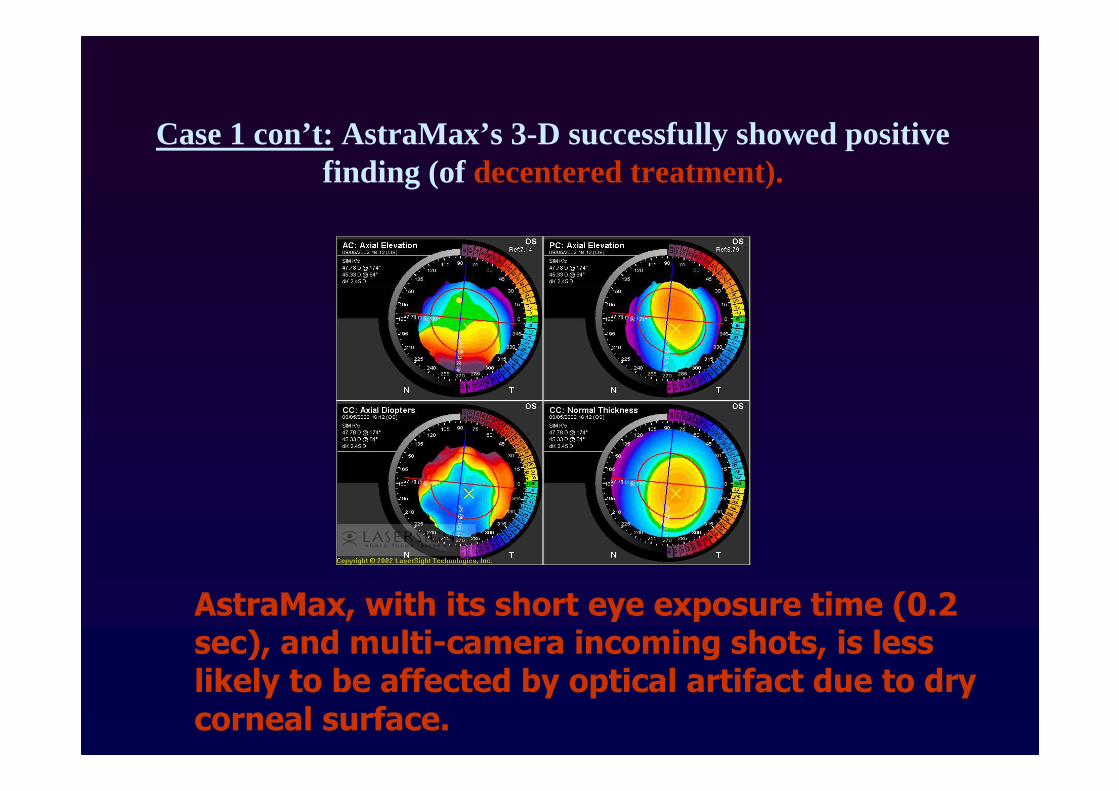
Case 1 con’t:AstraMax’s 3-D successfully showed positive finding (of decentered treatment).
AstraMax, with its short eye exposure time (0.2 sec), and multi-camera incoming shots, is less likely to be affected by optical artifact due to dry corneal surface.

Step-wise approach to C-CAP to treat decentered treatment
�Humphrey Altas topographer
�Vision Pro software� Customized ablation program
� Demonstrate the ablations effect on topography

Step 1: Elevation Map
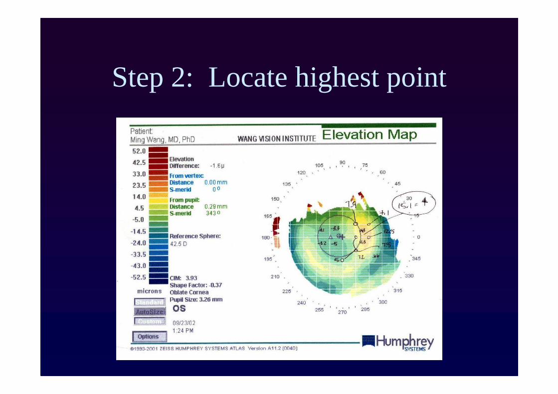
Step 2: Locate highest point

Step 3: C-CAP ablation patterns
� Myopic Sphere
� Myopic Astigmatism
� Myopic Ellipse

Spherical Treatment

Myopic Elipse Treatment

Myopic Astigmatism Treatment

Combination Treatments

Step 4: Print final pattern
� Documentation for Chart:� Includes all parameters necessary to complete
treatment
� Use this data to program the laser

Step 5: Input patterns into laser
� CAP card required
� Software update is required

Step 6: Perform treatment
� Technique identical to performing typical enhancement
� Short treatment times

C-CAP Case JC
� 47 yo Male
� S/P LASIK OU January 2001 by area surgeon with grade 4 DLK requiring flap lift OU at one week
� CC: “Visual distortion with glasses. RGP’srequired for comfortable vision, but CL’s are making my eyes dry”

C-CAP Case JC
� Unaided VA: 20/80
� MR -1.25+1.75 x 45, 20/30
� Cyclo -1.00+1.75 x 45
� RGP VA 20/20
� Ultrasound Pach 578/588/574 microns
� IOP, anterior and posterior segment healthy

Case JC “Decentration not due to primary laser treatment, but due to secondary tissue digestion
(DLK)”

C-CAP Case JC

C-CAP Case JC

C-CAP Case JC

C-CAP Case JC

C-CAP Case JC

C-CAP Case JC

C-CAP pre, post and difference elevation map (Case JC)

C-CAP Case JC
� Final treatment plan:� M Cylinder I: 3 microns x 147 (4.0x1.8 mm);
Offsets: X +0.9 mm, Y +1.00
� M Cylinder II: 5 microns x 043 (6.0x2.7 mm); Offsets: X +0.6 mm, Y -1.8mm

Case JC: 1 day s/p C-CAP
� POD #1 CC: smear is much better, equal to the other eye
� VA sc 20/60

Case JC: 1 week s/p C-CAP

Case JC: 1 week s/p C-CAP

Case JC: 1 month s/p C-CAP
Pre-Op:
Unaided VA: 20/80
MR –1.25+1.75 x 45 20/30
1 Mo PO:
Unaided VA: 20/70
MR –0.75+1.00 x 31 20/20

Case JC: 3 months s/p C-CAP
� JC returned wearing soft toric Cl for refractive correction, reporting nearly 100% resolution of the visual distortion
� VA sc 20/30, with BCVA of 20/20
� Requested refractive enhancement, which was successfully performed at 4 months

Second C-CAP Case: PG
� 49 yo Male
� S/P LASIK OU 1997 with enhancements OU 1998 by area surgeon
� CC: “Double Vision”

C-CAP Case PG
� Unaided VA: 20/60
� MR -2.75+1.75 x 135, 20/30
� Cyclo -2.75+1.00 x 135
� RGP VA 20/40 (poor fit) but subjective improvement in VA with CL noted
� Ultrasound Pach 475/480/477 microns
� IOP, anterior and posterior segment healthy

Elevation map (C-CAP Case PG)

Elevation map with height values (PG)

Treatment plan (C-CAP PG)

Treatment plan (C-CAP PG)

Treatment plan (C-CAP PG)

Treatment plan (C-CAP PG)

Treatment plan (C-CAP PG)

Treatment plan (C-CAP PG)

Final treatment plan (C-CAP PG)

C-CAP Case PG
� Final treatment plan:�M Cylinder I: 7 microns x 93 (4.5 x
2.7mm); Offsets: X +1.5 mm, Y +0.00

C-CAP Case PG
� POD #1 s/pC-CAP: Felt less double vision
� VA sc 20/200

Case PG: 1 month s/p C-CAP

Case PG: 1 month s/p CAP

Case PG: 1 month s/p CAP

Case PG: 2 mo Time Trend

Case PG
� Decentration regressed;
� More aggressiveC-CAP enhancement

Case PG (C-CAP enh)

Case PG (C-CAP enh)

Case PG (C-CAP enh)

Case PG (C-CAP enh)

Case PG (1st and 2nd C-CAP)
� First Treatment:� M Cylinder I: 7 microns x 93 (4.5 x 2.7mm);
Offsets: X +1.5 mm, Y +0.00
� Second Treatment:� M Cylinder I: 8 microns x 106 (4.0 x 1.9mm);
Offsets: X +1.6 mm, Y +0.2
� M Cylinder I: 4 microns x 96 (4.0 x 1.8mm); Offsets: X +2.3 mm, Y +0.3

Case PG (two C-CAPs)
POD #1: “less double vision OS”
Pre-C-CAP: Vsc=20/60, MR –2.75+1.75 x 45, 20/30 (diplopic)
Post-two C-CAPs: Vsc = 20/70, MR= -4.25+0.50 x 110 20/25 (“much less” diplopic)
Refractive treatment

Case PG (after two C-CAP, axial)

Case PG (after two C-CAPs, elevation)
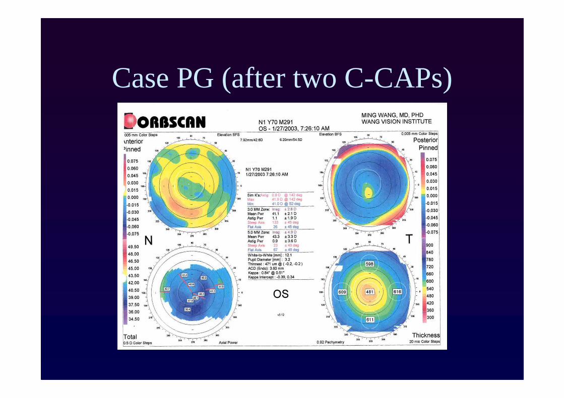
Case PG (after two C-CAPs)

C-CAP Case DC
� 48 yo male
� S/P myopic LASIK March 2002 followed by hyperopicLASIK-E OD June 2002 by area surgeon
� Complains of blurred vision even with glasses OD ever since the enhancement

C-CAP Case DC
� Unaided VA: 20/30-
� MR –0.50+2.00 x 11 (20/25-, blurred)
� Cyclo –0.25+2.00 x 11 (20/25-, blurred)
� RGP VA 20/20 (not blurred)
� Ultrasound Pachy 503 microns

Elevation map with height valuesC-CAP Case DC (s/p HL)

Treatment plan (C-CAP DC)

C-CAP Treatment plan (DC)

Treatment plan (C-CAP DC)

Final treatment plan (C-CAP DC)

Final treatment plan
� Final C-CAP treatment plan:�M Cylinder I: 7 microns x 56 (3.7 x
3.6mm); Offsets: X -0.4 mm, Y 0.6mm

C-CAP Case DC (s/p HL)
� POD #1 � CC: “Doing well”
� VA sc 20/30
� 3 mo PO
� Unaided VA: 20/70
� Symptoms of distortion resolved
� MR –1.00+1.00 x 40, 20/25+ (bluriness 90% gone)
� Patient requests refractive enhancement

Case DC: 3 mo s/p C-CAP
� Patient requested refractive enhance-ment at three months after C-CAP
� Vsc = 20/25 POD #1 After refractive treatment

Cautionary note #1 on C-CAP:Always look at the elevationmap at
the end� JJ presented with distance vision complaints
OS after having LASIK by an area surgeon
� POHx:� H-LASIK OS 11/99
� LASIK enh 4/01
� CE with IOL 3/02
� Myopic astigmatism LASIK 7/02

Case JJ: Always look at elevationmap
� MR OS -4.00+2.25 x 110, 20/100
� PH 20/40
� RGP OR VA 20/30***

Case JJ: Axial maps (OS, appeared to be inferiorly decentered)

Case JJ: Axial maps (OS, inferior decentration)

Case JJ: Elevationmap (OS nosignificant decentration, so no C-CAP
is needed)

Cautionary note #2 on C-CAP: need to wait for decentration to stablize, with full
medical theraphy, before doing any surgery such as C-CAP (ML)
� Patient presented with complaints of visual distortion and poor visual quality OD after LASIK in October 2002
� VA sc 20/60
� MR OD –2.00+1.00x10² 20/40 (blurry)
� Cyclo –1.75+0.75 x 10⁵ 20/30 (blurry)

Case ML: Decentration

Case ML: Decentered myopic LASIK (elevation map)

Case ML: Self resolution of decentration
� DES: plugged RLL;
� FU 6 wks later
� MR -1.50 +0.50x125 20/25+ (improved!)
� Decentration is improving
� Dry eye has improved OD;

Case ML: Self resolution of decentration
� 4 months PO� CC: “vision is clearing, plug helped”
� VA sc 20/40+2
� MR –1.50+0.75 x 100 20/20 No distortion! “Significant improvement in shadows with refraction”.

Case ML: Time course of self-resolution of decentration
December 2002:
-9.5microns
February 2003:
-5.4 microns

Case ML: Self resolution of decentration with dry eye treatment
� 20/20 BVA with spectacle correction with full resolution of ghosting by treating the ocular surface disease (dryness)
� No need for Custom-CAP!

CustomVue Ablation:
Wavefront-guidedUsing wavefront to correct decentered ablations,
when
1. The decentration is not too severe;
2. WaveScan can map;
3. WS indeed show high coma;
4. WS refraction is similar to MR

Case RC (Custom treatment of decentration)
42 yo Female “tired of wearing glasses”MR: OD -7.75+1.00 x 010, 20/20
OS -10.20+1.00 x 175, 20/20
Pach’s: OD 552 (ave), OS 560 (ave)Good ocular health and TPGPlan: LASIK for distance OU using
Intralase (90% OD, 80% OS)

RC: 1 day s/p ML
VA’s OD 20/30, OS 20/100
Flaps in place, no inflammation, striaeor debris
Moderate edema OS.
Tx’d with Pred (4/3/2/1 x 1 wk)

RC decentration os s/p ML
3 mos PO: slightly decentered ablations on Atlas OSVA OD 20/30, MR -0.50D 20/30+1VA OS 20/100, MR -2.00 20/30 (blurry)

RC 3 mos s/p decentered ML: Orbscan OS

RC s/p decentered ML os (high coma)

RC: CustomVue treatment for decentered ML os

RC S/P CustomVue treatment for decentered ML os
6 weeks: OS 20/25 “Doing well”AR -0.50+0.25 x 100.

OS pre and post Custom treatment for treating decentered ML

Custom treatment for decentered ML OS (DG)
45 yo Female complaining of:
Monocular diplopia, OS
“Difficulty with night glare” OU, OS > OD
“Glasses for night driving don’t help”
Original RX before ML was:
OD -7.50+1.25 x 005, 20/25
OS -9.75+2.00 x 20, 20/20
S/P Lasik May 2000
Enh OS December 2000.
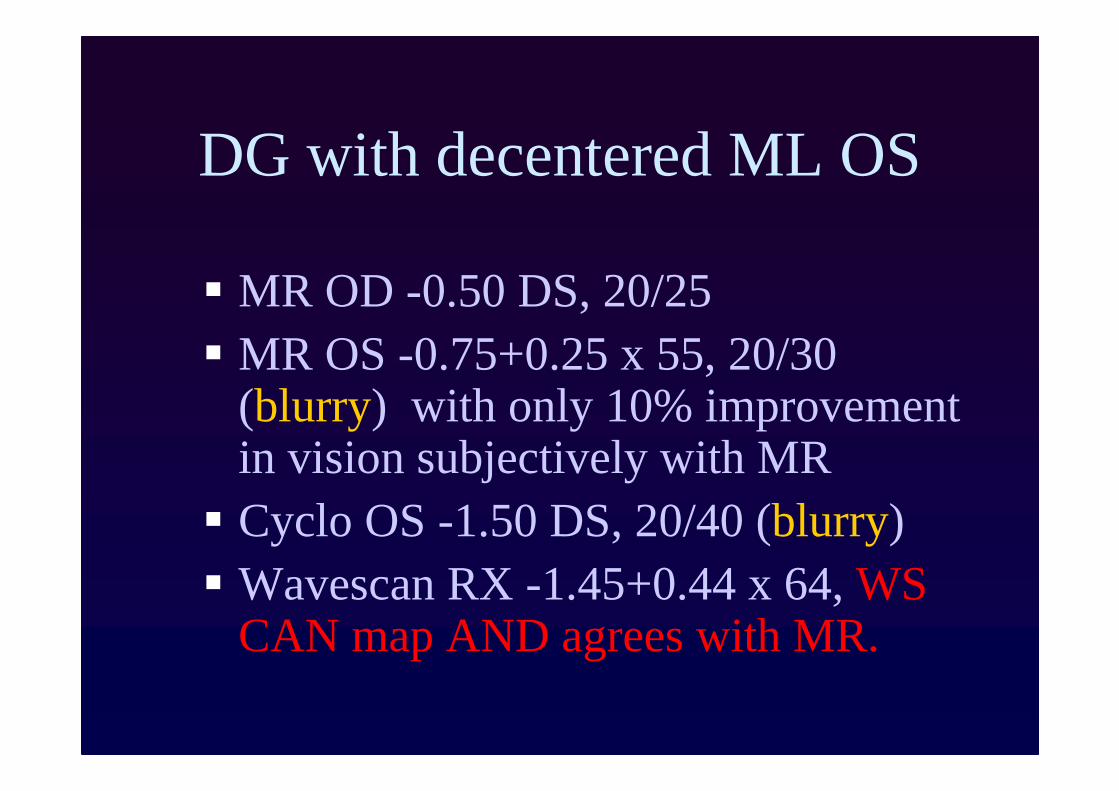
DG with decentered ML OS
� MR OD -0.50 DS, 20/25� MR OS -0.75+0.25 x 55, 20/30
(blurry) with only 10% improvement in vision subjectively with MR
� Cyclo OS -1.50 DS, 20/40 (blurry)� Wavescan RX -1.45+0.44 x 64, WS
CAN map AND agrees with MR.

DG with decentered ML os
US pach’s OD 479 (ave), OS 469 (ave)

DG superioly decentered ML OS (elevation)

DG height values OS on elevation
Values on elevation map show significant decentration

DG Wavescan map for the decenteredML OS (high coma)

DG CustomVue treatment plan for decentered ML os

DG s/p CustomVue treatment for decentered ML os
At 3 months: VAsc 20/30MR +1.75 20/25+ with no diplopia

DG with decentered ML OS, pre-custom treatment and post-custom
Pre-custom After-custom

Strength and weakness of C-CAP for treating decentered ablation (sg)
1. Strength:Large scale treatment, can “pull” the ablation back to center, in severely decenter-treated corneas in which WaveScan can’t map;
2. Weakness:Trial and error geometric shapes;Has to have another refractive treatment;
3. Cautions:Always look at the elevation map;Keeping in mind that some decentration will self resolve (with DES treatment for example).

Custom Guided Treatment for decentered ablation (pg)
1. Strength:Addresses the refractive error; more predictable (customized to the extent of decentration);
2. Weakness:For severely decentered treatment, wavefrontoften can’t map, or coma not dominant, or its refraction does not agree with MR;
3. Cautions:Custom treatment secondarily address the topographical issues – less control;Wavescan refraction is often LESS accurate in
post-keratorefractive surgery eyes.

Summary: a logical and sequental approach to treating irregular astigmatism (decentration)
using C-CAP and CustomVue – VISX� Be sure the decentered ablation is the reason
the VA is reduced. RGP VA is important!
� C–CAP treatment work well in some severelydecentered cases
� C-CAP induces refractive error changes, usually NOT in a positive way, and hence will need secondary refractive treatment;
� Wavefront CustomVue can treat, though imaging in severely decentered cases is hard. It is affected by lens HOA;
� Ideal treatment: topography-guided treatmentNew book: “Irregular Astigmatism” (Wang eds, SLACK )