a literature review: chronic inflamation and
TRANSCRIPT

A LITERATURE REVIEW: CHRONIC INFLAMATION ANDNUTRITIONAL STATUS
Item type text; Electronic Thesis
Authors RODRIGUEZ, VALERIE ALEXANDRIA
Publisher The University of Arizona.
Rights Copyright © is held by the author. Digital access to thismaterial is made possible by the University Libraries,University of Arizona. Further transmission, reproductionor presentation (such as public display or performance) ofprotected items is prohibited except with permission of theauthor.
Downloaded 3-Feb-2018 21:47:54
Link to item http://hdl.handle.net/10150/618767

A LITERATURE REVIEW: CHRONIC INFLAMATION AND NUTRITIONAL STATUS
By
VALERIE ALEXANDRIA RODRIGUEZ
____________________
A Thesis Submitted to The Honors College
In Partial Fulfillment of the Bachelor’s degree With Honors in
Nutritional Sciences
THE UNIVERSITY OF ARIZONA
AUGUST 2016
Approved by: _________________________ Dr. Mary Marian Department of Nutritional Sciences

Abstract This paper reviewed the mechanisms of systemic inflammation and the nutritional status of the individuals who suffer from chronic diseases including rheumatoid arthritis, systemic lupus erythematous, chronic obstructive pulmonary disease, irritable bowel diseases include ulcerative colitis and Crohn’s disease, asthma, and atherosclerosis. Treatment modalities such as diet regimens will also be discussed. The Anti-Inflammatory diet, Mediterranean Diet, and the Dash diet will be discussed. Nutritional status and inflammation go hand in hand according to the findings available today. There is still more research required to completely understand the mechanisms that occur in inflammation. Key words: Nutrition, nutritional status, inflammation, chronic inflammation, malnutrition, health, Mediterranean diet, dash diet, anti-inflammatory,

3
Acknowledgements Thank you Dr. Mary Marian for aiding me in selecting a topic and giving me the opportunity to complete this paper.

4
Introduction
In 2012, chronic disease had been surveyed to affect approximately 117 million adult
Americans1. In 2010, 48% of the leading causes of death were from heart disease and cancer1,
both of which are chronic diseases that have inflammation within their pathology. Within the
chronic disease spectrum, especially cardiovascular disease, obesity, cancer, systemic lupus
erythematosus (SLE), rheumatoid arthritis (RA), inflammatory bowel diseases, and sepsis,
inflammation plays a large role2. Inflammation is also crucial for the healing of wounds. During
occurrences of subclinical low to high inflammation response, found in chronic disease,
nutritional status plays a sizable part based on the status of the patient ranging from at risk of
being malnourished, malnourished, to excessive energy intake3,4,5. The purpose of this paper is to
investigate the connection between chronic inflammation and nutritional status with a focus on
diet modalities such as the Anti-Inflammatory diet, Mediterranean diet, and Dash diet to modify
the inflammatory response.
Table 1. Chronic Diseases Associated with Inflammation2
• Cardiovascular disease (specifically Atherosclerosis)
• Cancer
• Pulmonary Diseases (specifically Chronic Obstruction Pulmonary Disease and
Asthma)
• Inflammatory Bowel Diseases (specifically Ulcerative Colitis and Crohn’s
Disease)
• Obesity

5
• Autoimmune Diseases (specifically Systemic Lupus Erythematosus)
• Rheumatoid Arthritis
INFLAMMATION
There are two types of inflammation: acute and chronic inflammation; these are
interconnected. In 2010, Wong et. al. indicated that the main difference between acute and
chronic inflammation is the life span of the inflammation6. Acute inflammation is characteristic
of wound healing, responding to bacteria or pathogens, and returning the body to homeostasis2.
Acute inflammation is necessary and can last between a few minutes to several days7. Chronic
inflammation can occur for years7. There are three stages of inflammation which are the
inflammatory phase, the complement phase, and the resolution phase2. Chronic inflammation
occurs because the final stage, resolution stage cannot begin due to “failure to remove [the]
inflammatory stimulus”2 meaning the inflammatory response becomes stuck in the complement
phase. Inflammation occurs within exudate and cellular areas. The cellular component of chronic
inflammation involves monocytes, macrophages, cytokines, neutrophils, dendritic cells,
fibrocytes, and T-cells2.
As seen in Table 2, these cellular components have important functions in promoting or
ending inflammation. After the tissue becomes damaged, the first responders to the tissue site are
neutrophils and macrophages. Mast cells are activated by neuropeptides and cytokines8. Mast
cells then chemically attract other pro-inflammatory mediators such as tumor necrosis factor-a

6
(TNF- a), cytokines (specifically interleukin (IL)- 1b and IL-6), histamines, prostaglandins, and
leukotrienes2. Monocytes and neutrophils remove necrotic tissue, bacteria, and debris via
phagocytosis. In the last stage of inflammation anti-inflammatory cells enter the site such as
transforming growth factor (TGF-b), IL-10, neutrophils, and fibroblasts to complete healing2.
This physiologic response is typical in acute inflammatory processes. In chronic or systemic
inflammatory processes, the initial inflammatory trigger is unknown9.
Table 2. Cellular Components of inflammation adapted from Chronic Inflammation2
Cellular Component Function in inflammation
Monocytes Circulate to area of inflammation and become macrophages or dendritic cells associated with the tissue site to replenish tissue, phagocytosis, antigen representation which leads to an immune response, production of pro and anti-inflammatory cytokines
Macrophages Watchdog for the immune system, tissue remodeling, inflammation remodeling
Neutrophils First responders to the inflammation site, activated by chemotaxic receptors, may aide to lead to resolution phase or build up may lead to chronic inflammation
Dendritic Cells Regulate TH2-immunity
Fibrocytes Unknown.
Treg (T) Cells Limit extent and period of inflammatory response.

7
Cytokines Released by monocytes, neutrophils, and mast cells to create a pro or anti-inflammatory environment10
Black et al. 11 found that the initial trigger can occur from stress or from a malfunction in
the tissue as noted by Medzhitov et al9. This dysfunctional tissue has been seen in specific
studies related to cardiovascular disease, Type II diabetes, obesity, and stress7,9,11,12,13. A defect
in the tissue may occur from cardiac ischemia, vessels may be broken due to hypertension or a
flesh wound, and overactive adipose tissue7, 13. An increase in adipose tissue leads to decreased
insulin sensitivity as well as increased oxidative stress13. This decreased sensitivity leads to
increased pro-inflammatory mediated reactions with toll like receptor 4 (TLR-4)13,14. Toll like
receptor 4 has been associated with the release of inflammation related mediators, cardiovascular
disease, obesity, allergic response, autoimmune disorders, and inflammatory bowel diseases15.
When TLR-4 releases pro-inflammatory cytokines it has been shown to have a deteriorating
effect on blood vessels which also sends pro-inflammatory mediators to heal the area, as seen in
the study by Bhagat and Vallance14. Oxidative stress is known to increase cell death and
decrease cell tissue proliferation creating an unhealthy cellular environment13. This unhealthy
cellular environment puts an additional strain on the homeostatic environment7,13.

8
Black and fellow researchers investigated the role of the liver, endothelium, and
adipocytes in acute phase response caused by psychological stress11. This led Black et al. to
identify the liver, endothelium, and adipocytes as the key producers of the inflammatory
cytokine IL-6; which is linked to acute phase response (APR) and chronic inflammation11.
Increased levels of IL-6 are linked to pro and anti-inflammatory properties9,11,13,14,15. Acute phase
response (APR) is basically the term for the reactions that occur in order to heal during acute
inflammation11. Acute phase protein(APP), and C-reactive Protein (CRP), which are involved in
• CardiacIschemia
• Hyper-tension
Increasedpro-inflammatorymediators(e.gIL-6,TNF-α)
• APP
• ↑OxidativeStress
• ↑TLR-4
• Adiposity• InsulinResistance
Inflammation
Figure1.ContributorstoInflammation2,9,11

9
APR can lead to insulin resistance, type II diabetes, and metabolic syndrome furthering this
cycle of inflammation11. There are several studies that have investigated the effect of
psychological and physiological stressors and insulin resistance11, 16. There is data showing that
high stress both mentally and environmentally, led to insulin resistance, which headed the
physiologic response to an inflammatory cascade involving glucose 11,17. This was evidenced by
increased lab values of substance p, mast cells, corticosteroid releasing factor, catecholamine,
glucagon, and renin which induce the APR in human and animal studies11,16.
Medzhitov et al. have coined the phrase para-inflammation to describe a “state” of
inflammation in the body; specifically, an adaptive response to tissue strain or defect that has not
yet become chronic inflammation9. During para-inflammation the body is fighting for
homeostasis while combating inflammatory processes. This is determined by Medzhitov et al. by
tissue grade, or level of dead cells found within the tissue that may indicate inflammation9.
When the body remains in this state for a lengthy period of time, the para-inflammatory state
develops into chronic inflammation9. This is typical of chronic diseases such as obesity, Type 2
diabetes, atherosclerosis, asthma, and neurodegenerative diseases9. This level of para-
inflammation is related to increased adiposity as the body is trying to reach a homeostatic level
during inflammation9.
As mentioned earlier, IL-6 has a pro and anti-inflammatory effect on inflammation. IL-6
is released by multiple mediators including mast cells, macrophages, and neutrophils2. Mast cells
are immature immune cells that circulate within the blood stream until they reach the area of
inflammation2. Residential cells within the damaged tissue are the first to sense and mediate an

10
inflammatory response7. Residential cells include macrophages, mast cells, and dendritic cells7.
These cells mature in the localized tissue when activated during the inflammatory response. It is
here that mast cell function can vary depending on the tissue requiring action. Vasoactive
peptides such as substance p help to degranulate mast cells9. These vasoactive peptides can be
released during episodes of severe muscle wasting or protein break down also known as a
malnourished status9.
NUTRITIONAL STATUS AND INFLAMMATION
Nutritional status is fundamentally the assessment of nutrient levels, diet, and how well
the body is able to metabolically function18, 19, 20. Many studies have linked decreased nutritional
status (at risk of malnutrition and malnutrition) to worsening chronic disease symptoms and
increased mortality 3,4, 5, 18. Malnutrition incorporates both under and over-nutrition and is
considered a contributor to poor consequences in serious disease21. Poor nutritional status has
been shown to have an effect on specific inflammatory diseases, specifically rheumatoid arthritis
(RA), inflammatory bowel diseases such as ulcerative colitis (UC) and Crohn’s disease (CD),
systemic lupus erythematosus (SLE), atherosclerosis, chronic obstructive pulmonary disease
(COPD), obesity, and sepsis.
Nutrition and inflammation incorporates many factors other than the risk of malnutrition
or malnutrition status. There is also cachexia to consider which is common in systemic
inflammation21. Cachexia is wasting of the body due to increased catabolism due to chronic

11
illness, this can have severe consequences on the ability of the body to fight inflammation21.
Cachexia has been associated with having an effect on metabolic balance: “insulin resistance,
increased lipolysis, increased lipid oxidation, increased protein turnover, and loss of body fat and
muscle21.” A majority of what cachexia effects contributes to inflammatory processes that are
most likely already occurring21.
Obesity, can be considered an inflammatory disease due to the over active adipose tissue
creating an inflammatory cascade, which has been discussed earlier 7,13. Obesity and
malnutrition is common across the globe21. Obesity alone is associated with increased risk
factors such as hypertension, Type II diabetes, cardiovascular disease, dyslipidemia, coronary
heart disease, and respiratory disorders22. Obesity combined with malnutrition in critical care
patents has been seen to change the outcomes of mortality in patients who suffer from sepsis,
kidney injury, end stage renal disease, and acute organ failure 22. Robinson et al. noted, in obese
patients, in critical illness without malnutrition, there has been a shielding response from
inflammation, however, when malnutrition is added to the formula, there is increased mortality22.
This is interesting because it questions the inflammatory effect of the excess adipose tissue
during adequate nutritional status and critical illness. This is a clear indicator that poor
nutritional status may have an effect on inflammation.
One must also consider the effect of a poor diet on inflammatory processes because food
can have an impact on the inflammatory response 23. Poor diet, also known as the Western-style
diet, in this paper reflects the consumption of refined or processed carbohydrates, increased
saturated fatty acids, increased trans fatty acids, high omega (w) -3/omega (w)- 6 ratios, and
absent in fruits and vegetables that contain flavonoids, carotenoids, and fiber 24. When there are

12
increased refined carbohydrates in the diet, there is a spike in blood sugar and insulin which can
lead to hyperglycemia25. According to Giugliano, this increased need for glucose metabolism
leads to a large release of free radicals25. These free radicals can cause oxidative stress known to
produce damage within the tissues and activates transcription factors, which in turn activate pro-
inflammatory cytokines 25,26. When there are increased saturated fatty acids in the diet, increased
TLR-4 activation on the adipose tissue occurs24,27. TLR-4 then activates nuclear factor kB which
is a part of cytokine regulation, production, and main molecular activator of inflammation24,28.
This additionally leads to inflammatory events24.
Trans fatty acids have been associated with increased levels of TNF receptor 1 and 2 24,29.
Lopez et al., also identified a positive correlation with intake of saturated and trans fatty acids
and TNF, along with decreased endothelial function29. The decreased endothelial function is
related to symptoms of atherosclerosis, another inflammatory disorder29. A single meal with
high number and poor quality of fat can influence the acute inflammatory response29. This was
seen in the review done by Margioris, which separated factors that influence the inflammatory
response relative to diet into nutrient dependent and independent. This is shown below in Table
3. The w-3/w-6 ratio of 1:7 as seen in the Mediterranean diet has been seen to reduce 9% of
cardiovascular mortality, 6% of cancer related cases, and 13% reduction in the incidence of
Parkinson’s and Alzheimer’s24.

13
Table 3. Factors that Influence Postprandial
Inflammation23
Nutrient Independent Nutrient Dependent
Obesity Caloric Value (of meal)
Sedentary lifestyle Glycemic Index
Diabetes mellitus Lipid Profile (of meal) or
processed carbohydrates
Fruits and vegetables have shown to have an inverse correlation to CRP levels as
evidenced by Galland24. When fruits and vegetables have been added to the dietary regimen of
individuals not accustom to eating fruits and vegetables, over a 6-week period, there was a
significant decrease in the levels of CRP24.
Rheumatoid Arthritis (RA)
RA is known to be a chronic, autoimmune, systemic inflammatory disorder that does not
only attack the joints but different body systems as well30. There have been several studies that
have related adequate intake to improving nutritional status of RA patients and other studies that
have disproven this conclusion 19,20,31,32. A cross sectional observational study done by Hejazi et
al. recently assessed dietary intake and disease activity in Iranian women with RA19. Ninety
women who suffer from RA were randomly recruited between the ages of 20-70. Randomly

14
selected individuals who suffered from other diseases such as diabetes, nephrotic syndrome,
liver disease, gastrointestinal disorders and Cushing's syndrome, or were prescribed β-blockers,
oral contraceptives and angiotensin converting enzyme inhibitors drugs were excluded from the
study. The results showed a positive correlation between activity of RA and malnutrition based
on CRP and food frequency questionaires19.
In the RA patient population, muscle wasting and body mass loss is very common 19.
According to Rall et.al, this has been theorized to occur due to the overproduction of TNF-a and
IL-1 during inflammation33. When the muscle is lost in RA patients, the space is taken up by
adipose tissue, thereby making anthropometric data unusable to correlate with nutritional status
because they gain more fat mass. Limitations of the Hejazi et al. study include the lack of use of
a bio impedance analysis to determine anthropometric changes as well as the location of the
study. During the examinations in the Hejazi et al study, the patient disease activity score, global
assessment of pain using visual analog scale, and CRP were tested19. The other tests also taken
were fasting blood glucose, serum antioxidant total, malondialdehyde concentration and dietary
recalls were used to assess nutritional status19.
The results of the Hejzai et al. study showed a negative association between the disease
activity and caloric intake, but there was no statistical significance19. Of the patient population
studied 24% of the women suffered from malnutrition and inflammation defined by Hejazi et. al
as a low serum albumin level below 3.4 g/dl, decreased caloric consumption evident by twenty-
four-hour recall, and declining body composition values19. Of these women, there was a positive

15
correlation between malnutrition and disease severity. Based on Dietary Reference Intakes (DRI)
calculated from the 24-hour recall food frequency questionnaire, the patients had inadequate
intakes of calcium, folic acid, zinc, magnesium, and vitamin B619. Decreased magnesium, B6,
zinc, and folic acid been associated with increased inflammation and oxidative stress124,34,35,36.
The medications the patients used may have an effect on the DRIs caused by increased
catabolism and may have worsening effects on the inflammatory processes of RA. The following
medications that may cause such an impact are corticosteroids, methotrexate, sulfasalazine, and
D-penicillamine19. The data from this study may have also been effected by decreased
ambulation due to pain, therefore potentially causing decreased intake from patients with RA
who were required to feed themselves.
Unfortunately, in the Hejazi et al. study there was not a healthy control group to compare
the data to. The standard Iranian diet is very different to the Western diet and the deficiencies
that occur in the Iranian diet. For instance, the nutrient deficiencies found in the Hejazi et al.
study such as folic acid, are very unlikely to be seen deficient in the western diet because it is
enriched in most processed foods. In the study conducted by Gómez and colleagues, individuals
with class IV RA or end stage RA, showed no improvements in poor nutritional status even with
adequate intake20. In the Gómez et al. study, there were only 18 individuals who had been
evaluated based on stage 4 RA making it not relevant to a greater scale until it is evaluated on
with more individuals20.
Inflammatory Bowel Disorders (IBD)

16
Chronic inflammation within the gastrointestinal (GI) tract occurs in inflammatory bowel
disorders such as Crohn’s disease and ulcerative colitis (UC). Crohn’s disease can affect any
portion of the GI tract whereas UC imparts inflammation only to the lining of the colon37.
Malnutrition is common in this population, making this a high risk population that can have
nutritional deficiencies occur rapidly38. There is evidence that supports the link between
nutritional status and inflammation from chronic disease 37,38.
An analytical cross sectional study completed by Kalantari and colleagues, evaluated the
nutritional status of patients in Iran diagnosed with moderate to severe ulcerative colitis 38. Over
a five months’ time, 99 patients were evaluated between the ages of 14 and 80. Patients with
diseases such as diabetes or cancer were excluded from the study.
Body mass index, serum albumin, serum iron, hemoglobin, folic acid, vitamins A, B12,
D and E vitamins, partial thromboplastin time, international normalized ratio, IgA, and minerals
Ca, P, Mg, Zinc, Cu, K levels were evaluated as well as the severity of disease using the
Montreal classification system. Study results showed (Table 4) that patients that were at
increased risk for malnutrition had moderate to severe UC and were typically older. The
malnourished diagnosis was given based on a nutritional risk index (NRI) that takes serum
albumin and current or usual body weight in to account.
Patients with a calculated NRI of 83.5 to 97.4 were considered to be malnourished38.
Patients who suffered from moderate to severe cases of UC had a statistically significant higher
amount of serum potassium levels than those who had mild cases of UC. The significance of

17
this, is that it created a marker which indicated severe UC which often correlated with decreased
nutritional status. This may relate to malnutrition however there is not enough data to support
this claim. This study in particular had a small sample size of mild cases of UC and was not able
to compare the data to healthy individuals. Inflammatory biomarkers were not taken in this
study. However, there was evidence that connected severity of disease to increased malnutrition
risk38.
Table 4. Nutritional Risk and Patient age, disease duration, and ulcerative colitis status38
Variables Nutritional Risk Index
Non Malnourished Moderate to Severe Malnourished Risk
Age (years) 25.7 – 49.5 26.2 – 45.2
Disease duration (month)
3 – 10.4 3 -10
UC Mild 12 (100) 0
UC Moderate to Severe
78 (89.7) 9 (10.3)
In another study by Valentini et al., the nutritional status of 144 patients with CD and
UC, who were classified to be in clinical remission, were assessed by typical nutritional status
measures including subjective global assessment (SGA), BMI, serum albumin, trance elements,
bioelectrical impedance analysis, and anthropometry as well as body composition analysis and
muscle strength39. The study results reveals that most patients had been classified as well-

18
nourished based on lab values and body mass index. However, in all patients, body cell mass
(BCM) and handgrip strength were decreased. The lower values for BCM and handgrip strength
correlated with increased CRP levels. Since decreased BCM is associated with energy
expenditure and metabolism, and CRP levels were elevated, this reflects the possible link
between decreased nutritional status and inflammation40.
In the same study by Valentini, it is interesting to note that in the patients that were
identified to be malnourished in comparison to healthy counterparts, the body composition
values of malnourished CD were different than the anthropometric values of UC39. Malnourished
CD patients showed a decreased BMI and fat mass value, while the malnourished UC patients
showed decreased BCM and increased CRP. This may be due to the small sample size of
malnourished patients as only 23.7% of patients (n = 22) with CD were malnourished and 33.3%
(n =16) of patients with UC were malnourished. This spurs the question of what causes those
differences when these are both technically inflammatory diseases of the bowel?
According to the authors this was most likely due to increased malabsorption problems in
CD whereas malnourished patients with UC showed greater deterioration of muscle function39.
This is reflective in the data; CD showed decreased BMI values while UC showed decreased
BCM39. Increased pro-inflammatory mediators in UC, such as IL-6 and CRP have been seen to
cause sarcopenia in elderly individuals21. However, this was seen in patients between the ages of
18 through 70 years. This may be reflective of the lack of weight gained after weight loss in
patients suffering from CD41.

19
In another study by Reimund and associates, 40 participants (25 women and 15 men)
with CD focused on the connection between the inflammatory properties of CD, nutritional
status, and immune mediator cells including hemoglobin concentration, erythrocyte
sedimentation rate (ESR), fibrinogen, a1-acylglycoprotein, blood neopterin, IL-2 receptors,
TNF-a, and IL-1b42. These patients were compared to 26 healthy controls42. In this study disease
severity and age varied with ages ranging between 12-33 years old. Reimund et al. determined
patient nutritional status of nutritional risk or malnourished based on two abnormal values out of
the three anthropometric parameters: body weight, triceps skinfold thickness (TSF), or arm
muscle circumference (AMC). Table 5 reflects that patients with worsening symptoms of CD,
anthropometric measurements decreased and had and increased percent of malnourished patients.
Table 5. Anthropometric Parameters from Reimund study42
Controls CD (Whole
group)
Discrete
CD
Moderate
CD
Severe CD pb
Weight 63.2 55.7 62.7 54.7 52.35 0.0007
TSF 1.5 0.93 1.48 0.91 0.63 0.0001
MAC 26.7 23.8 26.55 23.3 22.6 0.0001
2/3 Items 4/26 18/40 1/7 4/15 13/18 0.0006
BMI 21/6 19.75 21.8 19.8 18.5 0.003

20
Inflammatory biomarker data CRP, Prognostic Inflammatory Nutritional Index, and
TNF-a also showed a positive correlation with severity of CD. (see Table 6). These numbers
gradually increased as the disease severity worsened. Statistical results of multivariate analysis
showed a statistically significant negative correlation between MAC, TSF, serum albumin, and
transthyretin (TTR). This shows a possible relationship between inflammatory cytokines, hepatic
transport proteins, and body composition. “Biochemical nutritional markers were positively
linked to each other and to anthropometric parameters, with a strong correlation between
Vitamin A on one hand, and TTR and retinol binding protein (RBP) on the other42.”
These binding proteins are important contributors to maintain homeostasis in the body.
When there are high levels of RBP, as seen in the Reimund et al. study (seeTable 6), these levels
appear related to insulin resistance and hyperinflammation42,43. The cofactor Vitamin A has
many functions, one of which involves immune regulation44. There is an interaction that occurs
between inflammation, immune components, and hepatic transport proteins such as albumin,
pre-albumin and RBP that are more influenced by the inflammatory response than nutritional
status. Reimund and colleagues recommend that the clinical focus should be on modifying the
patient’s nutritional status by targeting nutritional deficiencies, protein energy malnutrition, and
decreasing the inflammatory response42.

21
Table 6. Biological Parameters in Reimund et al. study42
Controls CD (Whole group)
Discrete CD
Moderate CD
Severe CD P
RBP 47 (13) 38 (17) 41 (15) 39 (19) 32 (15) 0.0032*
Albumin 46 (4) 35 (6) 42 (2.7) 37 (35) 30 (46) 0.0001*
IGF 29 (11) 19 (6) 25 (5) 23 (6) 18 (6) 0.12
Vitamin A 0.7 (0.3) 0.4 (0.2) 0.7 (0.8) 0.5 (0.2) 0.4 (0.2) 0.09
CRP 5 (2) 28 (26) 5 (0.8) 27 (27) 57 (49) 0.0001*
Fibrinogen 2.8 (0.8) 4.3 (1.5) 3.3 (1) 4 (0.9) 4.8 (1.4) 0.0001*
TNF-a 7.7 (2.5) 15.2 (14.4) 7.1 (2.2) 9.8 (8.6) 19.2 (15) 0.0001*
*reflects statistical significance
Nutritional Status and Systemic Lupus Erythematosus (SLE)
In all cases of SLE, multiple body systems are affected as SLE is a chronic autoimmune
disorder where the body’s own immune system attacks various organs; SLE can present
differently depending on the patient. In some patients there are major anthropometric changes
such as decreased body weight or changes in body composition due to anti-inflammatory drug
intake45. This may also present in a SLE cachexic related way45. Many patients are treated with
steroid medications to help decrease inflammation, but these can cause other nutritional
deficiencies such as decreased calcium and magnesium. It is common for individuals who suffer

22
from SLE to have abnormal nutritional status such as inadequate intake, mild to severe
malnutrition risk, or malnourished as seen in 45,46,47 studies.
SLE is an autoimmune inflammatory disease, the catabolism of pro-inflammatory
cytokines and tissue lead to increased levels of oxidative stress within the body. Oxidative stress
has been shown to affect the activity of SLE48. Poor nutritional status in a SLE patient does
directly affect the oxidative stress in the body, therefore worsening symptoms of SLE48. This
was shown in another study by Abou-Raya, the results showed that inflammation, stress, and
nutritional status have an effect on SLE based on malondialdehyde serum levels and
prostaglandin F2a48. Malondialdehyde serum levels are associated with oxidative stress and
prostaglandin F2a levels are associated with inflammation response48. More studies need to be
done in order to determine evidence linking poor nutritional status and inflammatory biomarkers
in the SLE patient population.
Borges et al. evaluated the nutritional status and food intake in 170 ambulatory women
between the ages of 18-60 with SLE patients cross-sectionally46. Nutritional status was
determined using the SGA and BMI, while dietary intake was determined from food frequency
questionnaires and 24-hour diet recalls. The results showed that 91% of the study participants
were well nourished, however 62% of the women were either overweight or obese. An increased
number of adipocytes can increase the inflammatory response by activating TLR-4 receptor sites
which then activate NFkB that activate an inflammatory cascade24,25. This leads to worsening
symptoms or comorbidities of dyslipidemia, cardiovascular disease, and insulin resistance46.
Insulin resistance was not measured within the study.
23
Additionally, a diet high in saturated or trans fatty acids, oils that were unspecified, and
deficient in fruit, vegetable, calcium, and iron was found in a majority of all patients 46.
Decreased iron intake has been associated with increased inflammation in obese patients 49. It
has also been previously noted in this paper that increased saturated and trans fat has a positive
correlation with pro-inflammatory markers 24,27,29. In the Borges et al. study, none of the patients
consumed fruits and vegetables. This is interesting because fruits and vegetables are known to
have an inverse correlation with the inflammatory markers CRP and IL-6 24. This means that
increased fruit and vegetable intake may help lower inflammatory status. The decreased serum
calcium value may be affected by medications, specifically with RA patients it is often
associated with long term corticosteroid use46. The data may also be skewed because a large
portion of the data was based on patient memory (food frequency questionnaires and 24-hour
recalls) which may be less telling than actual serum values. According to Borges et al., there was
no association of iron or B12 anemia with the decreased values in this study.
In another study by Noriega et al., anthropometric data such as BMI and nutritional status
were assessed for 28 study participants45. Nutritional status was determined by SGA and NRI.
Of these patients, 35% (n = 10) were considered to be at severe nutritional risk based on NRI45.
The study did not go in to detail about the results of the SGA scores which would have helped to
determine if the SLE patients where adequately nourished, moderately malnourished, or severely
malnourished. The study investigators determined from this small study that hospital risk
increased for patients with a moderate to high NRI number. A limitation of this study is that the
anthropometric and SGA data were not given in order to determine a correlation between NRI

24
and SGA. In a similar study by Abou-Raya et al. who evaluated the association between SLE
disease severity, nutritional status, and compared disease/nutrition outcome related to diet. In
this study, 121 patient’s nutritional status was assessed using BMI data and the SGA. Dietary
intake was assessed using food frequency quesitionaires47. Study results showed that a majority
of the cohort were overweight and most diets were lacking in fruits, vegetables, and dairy
products47 while most diets were high in fat and oil based foods. Patients with SLE have severe
pain which may lead some individuals to eat more high inflammatory comfort food rather than
healthy fruits and vegetables47.
Atherosclerosis
Atherosclerosis is the buildup of plaque made up of foam cells and macrophages within
the arteries50. This plaque formation occurs from various factors including genetics and lifestyle
choices such as diet and exercise50. As the plaque continues to build up, the artery lumen
becomes smaller and smaller, restricting oxygenated blood flow to the extremities50. At the core
of atherosclerosis is inflammation50. The initial attractor of foam cells and macrophages is
thought to be lymphocytes and macrophages50. Atherosclerosis has been linked to higher levels
of inflammatory biomarkers such as acute phase proteins, TNF-a, CRP, and serum amyloid A50.
There is also a connection with obesity and atherosclerosis and inflammatory biomarkers.
Increased energy intake and saturated fats, which then turns into fat is stored as fat, raises total

25
cholesterol and LDL-C levels within the bloodstream, which creates an inflammatory
atmosphere50.
People with atherosclerosis are generally overweight or obese, this was shown in the
study by Jarosz et al.51. This study evaluated the effect of weight on nutritional status in patients
who have been diagnosed with atherosclerosis. Less than two percent of the group studied,
which were of Polish descent, were underweight based on BMI and arm circumference. The
other 98% were overweight and obese. This study did not give a standard measure of unit to
determine malnourished status; malnourishment in this study was based on biochemical data
such as lymphocyte count and serum albumin51. These labs are markers of inflammation not
nutritional status. Based on anthropometric data, a very small percentage of patients were
considered to be adequately nourished according to Jarosz et al. The results of the Jarosz et al.
study showed the overweight and obese patients lost weight, had deficient serum albumin values,
and the male population studied were found to have a decreased erythrocyte and leukocyte count.
The data given from the Jarosz et al. study did not identify any changes in dietary intake. This
study did not give a standard measure of unit to determine malnourished status and
malnourishment in this study was based on biochemical data lymphocyte count and albumin51.
There is another evaluation tool for measuring nutritional status and it is called the
CONUT score which stands for controlling nutritional status3. This number is calculated based
on serum albumin, lymphocyte count, and total cholesterol. This ties in factors from immune
response, calorie depletion, and protein intake. The CONUT score has been demonstrated to be a
useful predictor of risk assessment in cancer and cardiovascular diseases 3,52. This is a different

26
way to measure nutritional status because it reduces the significance of the serum albumin
value52. This is beneficial because serum albumin levels can be affected by factors other than
nutritional status, such as fluid status, liver damage, and infection52. When poor nutritional status
and cardiovascular disease meet, the CONUT score, TNF-a, and length of prognosis help to
determine patient outcomes as evident in the study by Nakagomi et al.3.
Table 7. Nakagomi Study Results3
Univariate Multivariate
Hazard Ratio (HR)
95% Confidence Interval (C.I)
P value HR 95% C.I P value
CONUT 44.55 10.76- 184.37 <0.001 11.97 2.21-64.67 0.004
CRP 15.27 5.99 -38.50 <0.001 2.12 0.51-8.88 0.304
AGE ³ 70 3.26 1.73 -6.14 <0.001 1.69 0.44-6.45 0.442
Monocyte TNF-a
29.46 9.10-95.32 <0.001 14.10 2.55-77.92 0.002
BMI £ 21 kg/m2
7.07 3.56-14.04 <0.001 1.41 0.42-4.75 0.584
Researchers found a positive correlation between TNF-a, CRP, and CONUT score as
Cardiac Heart Failure progressed3. Out of the 114 participants, a low CONUT score equaled
poor outcomes such as a repeat cardiac event, cardiac failure leading to death, and worsening
symptoms3. Fat loss and muscle wasting from cardiac cachexia, seen in this study are linked to
inflammatory pathways including the catabolizing pro-inflammatory mediators include TNF-a,
IL-6, and IL-13. These are all seen in high ratios in patients with CHF that have cardiac cachexia

27
or malnourished status thus again reflecting a possible link between inflammatory status and
nutritional status.
Carotid intima-media thickness (CIMT) was measured in the study by Nakagomi et. al3.
The CIMT was compared with the inflammatory values CRP and TNF-a. These figures showed
a positive correlation, while serum albumin was negatively correlated with CIMT. As the CIMT
numbers increased, this was paralleled by inflammatory biomarkers. The CONUT score, that
was used to determine nutritional status was also shown to be related to the CIMT. This is
helpful to target the pathophysiology of atherosclerosis and identify future treatments for
atherosclerosis and CHF, since atherosclerosis is often an early indicator of CHF3. There may
also be a connection between inflammation and hunger as in the Nakagomi study, ghrelin and
leptin levels were abnormal possibly because of the high ratio of TNF-a.
In instances of cardiac cachexia, ghrelin has been shown to have an effect on
inflammation particularly in studies of CHF patients53. In the study by Dixit et al., pro-
inflammatory cytokines were inhibited by ghrelin and these patients did not suffer from cardiac
cachexia53. Whereas in another case, several pro-inflammatory cytokines such as IL-1b, IL-6,
and TNF-a have been indicated in increasing the symptoms of cardiac cachexia53,54. This may
indicate that without ghrelin inflammation may increase. Cytokines such as IL-1, IL-6, and TNF
have been indicated to alter the GI system in various ways. For example, a change in stomach
content movement, an altercation in the release of appetite stimulating hormones that include
cholecystokinin, leptin, and insulin from the gut mucosa55.

28
Chronic Obstructive Pulmonary Disease (COPD)
COPD is an inflammatory disease that not only effects all parts of the lung, can also
effect blood pH, heart, and, skeletal muscle56. There are increased pro-inflammatory mediators
that are seen in this disease56. Nutrition is also a concern because often COPD patients have
difficulty maintaining a healthy weight due to the high inflammatory state forcing the body into a
catabolic status57. Not only that, but COPD patients are often short of breath and fatigue easily
making day to day activities difficult to achieve56,58. In hospitalized patients, poor nutritional
status and high inflammatory biochemical values may be an indicator for increased risk for
mortality.
Table 8. COPD Mortality Patient Data58
Survived (n=212)
Died (n=49) P
BMI 19.3 - 30.4 17.1 - 28.3 0.0007
Age 56 - 80 63 - 81 0.0005
Women 54 43 0.03
Current Smokers
25 29 0.39
Diabetes 9 16 0.03
Cardiovascular Disease
43 50 0.18
Living Alone 53 51 0.74

29
One study predicted that patients who were underweight at the time of admission to the
hospital have an increased risk of mortality within the next two years, unlike patients who are
overweight58. Hallin et al. and colleagues studied 261 participants in the Nordic countries
(Denmark, Norway, Sweden, and Finland) to examine whether a relationship existed between
weight and mortality58. The participants involved were recently admitted to the hospital for an
acute exacerbation and they all suffered from stage 1 or higher of COPD based on the global
initiative for chronic obstructive disease (GOLD) scale. BMI, spirometry, psychological status,
and quality of life were also assessed. Out of the participants, 19% died within two years.
Table. 9 Risk Factors for mortality in Hallin et al. study58
Hazard Risk Ratio 95% Confidence Interval
P
BMI <20 kg/m2 12.9 2.8-59 0.001
BMI 20-25 kg/m2 6.5 1.5-28 0.01
BMI 25-30 kg/m2 1
BMI >30 kg/m2 7.7 1.5-40 0.02
Women 0.60 0.31-1.18 0.14
FEV1 per 10% predicted
0.85 0.67-1.08 0.19
Diabetes 4.2 1.7-10 0.002
The reasons for death were grouped together by the researchers in the following
categories: respiratory causes, respiratory insufficiency and pneumonia, cardiovascular diseases,

30
heart failure, stroke and rupture of aortic aneurysms, malignancy, lymphoma, and others58. The
patients who passed away had a significant negative correlation between BMI reflective of being
underweight and decreased Forced Expiratory Volume in 1 second. The majority of patients who
had died, had a BMI of 22.7 ± 5.6 as shown in Table 8. This shows that individuals who had a
decreased lung capacity were also underweight and died within two years. People with the
lowest death rate were individuals who were overweight. This is interesting because one would
think that the increased adipocytes would increase systemic inflammation and force more body
catabolism rather than increase survival rate. The disadvantages to this study are the differences
between the diets and lifestyles of people who live in Nordic countries, nutritional status was not
accounted for, and a small population sample making it difficult to relate it to the western
population. It is unclear based on the data given if the patients were continuously losing weight
or if they remained underweight for the duration of the study.
However, in another study by Coxson et. al., chronically malnourished patients were
studied to determine if any connection may exist between poor nutritional and pulmonary
emphysema59. This was a small study that only consisted of only 21 patients who suffered from
anorexia-nervosa and were compared to healthy individuals that were similar in age and sex. The
participants were evaluated based on BMI, smoking history, hemoglobin, white blood
differential cell count, serum a1-antitrypsin, spirometry, total lung capacity, residual volume,
and CT scans of the aortic arch, the tracheal carina, posterior aspect of the eighth rib, and lung
anatomy. The results of the study showed that there was a correlation between BMI and
diffusing capacity of the lungs for carbon monoxide (DlCO).
31
Table 10. Reression Analysis for Coxson et al. study59
Mean CT Density Volume of Gas per Weight of Lung Tissue
(mL gas/ g tissue)
r p r p r p
BMI (kg/m2) 0.59 <0.001 -0.60 <0.001 0.60 <0.001
DLCO/ VA
(%PcorrHGB)
0.20 0.24 -0.27 0.11 0.17 0.33
FEV1,% P 0.22 0.20 -0.19 0.26 0.22 0.19
FVC, %P 0.32 0.06 -0.31 0.06 0.32 0.06
FEV1/ FVC -0.32 0.05 0.36 0.03 -0.30 0.07
TLC, %P 0.21 0.21 -0.20 0.25 0.23 0.18
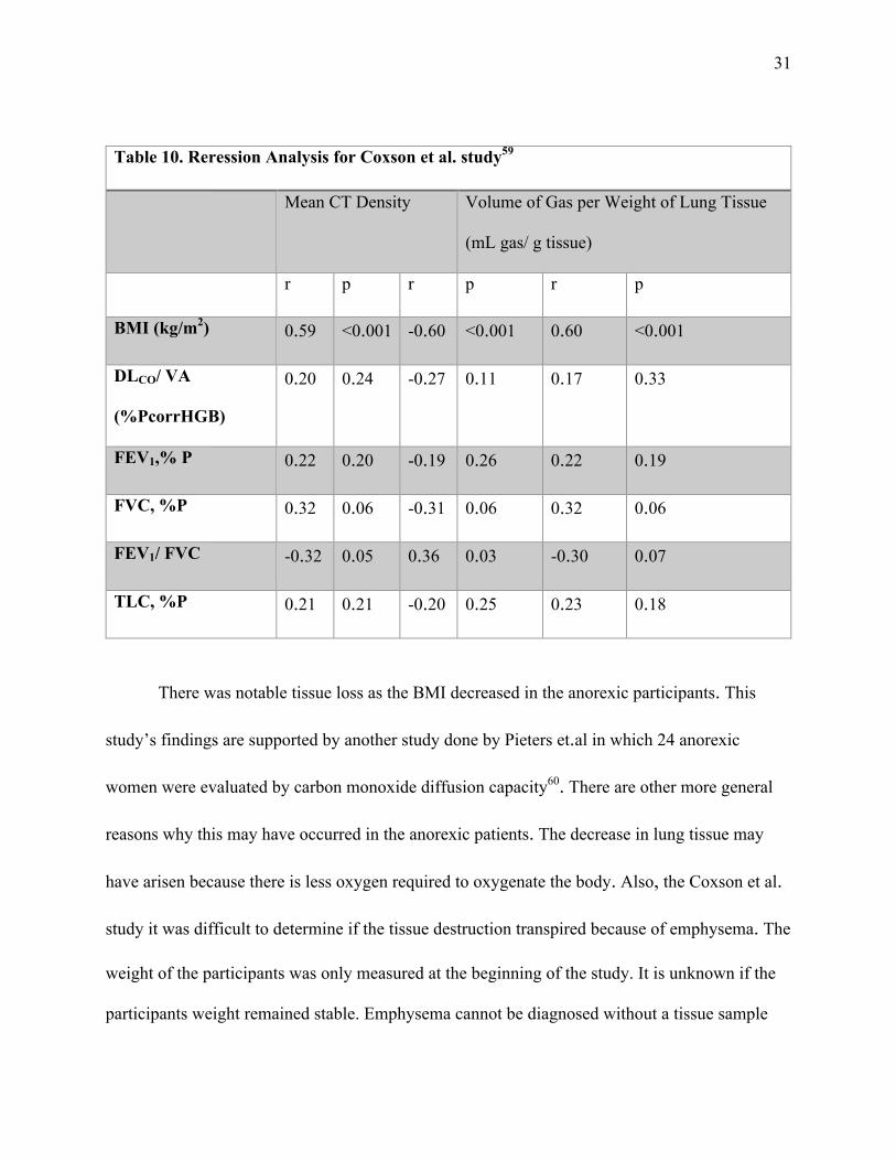
There was notable tissue loss as the BMI decreased in the anorexic participants. This
study’s findings are supported by another study done by Pieters et.al in which 24 anorexic
women were evaluated by carbon monoxide diffusion capacity60. There are other more general
reasons why this may have occurred in the anorexic patients. The decrease in lung tissue may
have arisen because there is less oxygen required to oxygenate the body. Also, the Coxson et al.
study it was difficult to determine if the tissue destruction transpired because of emphysema. The
weight of the participants was only measured at the beginning of the study. It is unknown if the
participants weight remained stable. Emphysema cannot be diagnosed without a tissue sample

32
and Coxson et.al only utilized the data from CT scans to determine destroyed lung tissue59.
However, with decreased nutritional status participants did show decreased tissue and mass loss.
Unfortunately, in the study by Coxson, inflammatory markers were not taken to compare with
declining nutritional status59.
However, in another study, Arora et al., set out to determine if a correlation existed
between inflammatory status, nutritional status, and severity of COPD61. Sixty-six patients with
COPD considered to be stable according to GOLD were enrolled with the following outcome
measures analyzed: CRP, leptin, and pre-albumin levels, BMI, mid upper arm circumference
(MUAC), skin fold thickness, and 6-minute walk test. The patients were grouped into three
categories: moderate COPD, severe COPD, and very severe COPD based on FEV1 results. As
the severity of COPD increased the following values decreased: BMI, leptin, pre-albumin,
MUAC, and walk test. Furthermore, a positive correlation with disease severity and CRP levels
was found. These values indicate that as COPD becomes more severe nutritional status declines
and inflammation increases61. There is evidence that certain dietary factors within fruits and
vegetables consistent with a healthy diet have potential beneficial characteristics for people who
suffer from COPD62. A cross sectional study done over the period of 25 years, on 12,763 men
showed improved FEV1 values in the group of men who had increased fruit, vegetable, and fish
intake in comparison to a normal diet62. Based on the study, it is uncertain what a normal diet
consisted of, however it had less fruit and vegetable intake.
Table. 11 Tabar et al. COPD Results63

33
131 patients with COPD studied
r p
BMI -0.3873 0.000
MAC -0.2555 0.0039
Malnourished patients likely to have ³1 exacerbations
0.7041 0.000
In some cases, as nutritional status declines there is an increase in COPD exacerbations63.
This is evident in the cross-sectional study of 131 patients done by Tabar et al.63. Patients who
had at least one exacerbation per year were typically of poor nutritional status, while patients
who were adequately nourished didn’t have any exacerbations63. Patients who suffer from COPD
and most likely have a decline in nutritional status will lose lean muscle tissue63. This will take a
toll on physical capacity as well. This is important to know because in some cases, where poor
nutritional status is not evident via SGA or other measures, physical capacity helped to
determine declining nutritional status63. This physical deterioration was correlated with increased
CRP levels. There is a link between lung function, nutritional status, systemic inflammation, and
physical capacity as seen in the cross sectional study done by Hallin et al.64.
TREATMENT
Anti-Inflammatory Diet

34
The Anti-inflammatory (AI) diet is designed to silence inflammatory genes that may be
activated through diet28. As mentioned earlier in the paper, highly processed refined
carbohydrates, high fat, and excess calorie consumption can increase internal inflammatory
properties28. The particular molecular targets of the AI diet are AMP kinase and NF-kB28. The
AI diet consists of carbohydrate to protein to fat ratio of 40:30:3028. This ratio is alleged to aid in
insulin and glycemic control28. The omega 3 and 6 polyunsaturated fatty acids (PUFA) play a
large role in the activation or deactivation of inflammatory properties and eicosanoid
development28. The omega 6 PUFA, also known as linoleic acid, can be broken down into
arachidonic acid which is the main component of eicosanoids leading to cellular inflammation28.
The omega 3 PUFA has eicosapentaenoic acid (EPA) or docosahexanenoic acid (DHA) which
are both anti-inflammatory28. The AI diet also incorporates the importance of a varied diet
consistent in non-starchy vegetables containing polyphenols such as asparagus, lettuce, broccoli,
and cucumber28. Polyphenols are known for their chronic disease prevention properties65.
Polyphenols are involved in biologic mechanisms that involve anti-inflammatory pathways such
as COX-1 and COX-2 inhibition, nitrous oxide synthase, NF-kB, and cytokine production65.
Table 12. Anti-Inflammatory Diet Summary28
Carbs : Protein : Fat
Ratio 40:30:30 with caloric
restriction
Omega 3 PUFA Increase

35
Omega 6 PUFA Decrease
Molecular Targets NF-KB , AMP Kinase
Focus Decrease omega 6 FA and stabilize Insulin
Eicosapentaenoic Acid (EPA)
Docosahexaenoic Acid (DHA)
2-3 g supplementation
Non-Starchy Vegetables Increase
It is important to identify the factors that can activate NF-kB because the AI diet is
supposed to inhibit NF-kB activity28. According to Marimotto et al. and Calder oxidative stress
and hormones can activate the NF-kB inflammatory switch 65,66. Oxidative stress may come
from excess energy consumption or an increase in adipocyte tissue28. “Additional dietary factors
include saturated fatty acids, advanced glycosylated end products (AGE), and inflammatory
cytokines from nearby cells all acting through specific receptors at the cell surface can also
activate NF-κB28.”

36
Figure2.ContributorstoCellularInflammation28
Nuclear factor kappa B is inhibited by polyphenols28. The polyphenols found within vegetable
and fruit coloring activate the transcription factor PPAR-g and this inhibits NF-kB. Polyphenols
also play a role in the activation of AMP kinase. AMP kinase plays a large role in glucose
metabolism and energy regulation28.
The arachidonic acid can have an impact on the cellular inflammation within the body28.
The Arachidonic acid (AA) to EPA ratio is actually considered to be a more telling indicator of
inflammation than CRP values according to Dr. Sears, the current leader of anti-inflammation
dietary research28. The typical Western diet contains over 12 times the amount of AA to EPA
recommended28. This means that there are more omega 6 components within the diet that can be
broken down by the enzymes delta 6 desaturase and delta 5 desaturase to become AA. This is
shown in Figure. 3. The enzymes in the reaction to convert linoleic acid to AA are influenced by
the glycemic load of the meal. The greater the influx of glucose, causing a rise in insulin, the
greater the amount of AA will be produced28. With increased AA intake, which is pro-
inflammatory, an increased intake of EPA is required to even out the inflammatory imbalance28.

37
The EPA helps increase the dietary potential of anti-inflammation28.
There are some areas in which there is clinical evidence that supports the use of the anti-
inflammatory diet to aid in treatment. In the study done by Adam et al., 68 patients with RA
were split into separate groups; one group consumed an anti-inflammatory diet, another
consumed an anti-inflammatory diet with fish oil pills, and the last group consumed a Western
diet67. The results showed a decrease in inflammation in both of the AI diet groups, while the
group that also consumed dietary fish oil showed a greater decrease in inflammation, nearly
double at the cellular level. This was made evident by the EPA erythrocyte lipid percentage and
decreased leukotriene B4 levels67. Based on the data given from the Adam et al. study there is no
indication of how well the patients followed the AI diet on a daily basis67.

38
In another study, the AI diet was tested on 40 individuals who struggled with IBD. The
results were not nearly as conclusive as the RA study because only eleven consistently remained
on the diet for four weeks as evident by diet history journals68. All eleven patients had results
that led to the decreased intake of an IBD medication which according to Olendzki et al.,
indicated decreased inflammation68. There were not any CRP or inflammatory lab values to base
decreased inflammation as a result of the AI diet. More research is required to identify the
clinical effectiveness of the AI diet on IBD. Typically, most IBD patients have an individualistic
diet regimen due to the individuality of the microbiota living within the gut69.
Mediterranean Diet
The Mediterranean diet is a form of anti-inflammatory diet that incorporates greater fat
intake, up to 40% of total kcals, emphasizes the importance of omega-3 fatty acids, and oleic
acid70. This is gained through increased fish intake, virgin olive oil use, legumes, complex
carbohydrates, wine, feta cheese, yogurt, and decreased red meat intake70. Virgin olive oil is a
staple in the Mediterranean diet. It is the main fat used to cook with and often used as the
dressing or dipping sauce for bread, salad, and pasta71. Virgin olive oil contains alpha-tocopherol
and phenols unlike processed olive oil that has been stripped of the healthier components72.
Supplementation of alpha-tocopherol has been related to decreasing oxidative stress and
inflammatory lab markers such as LDL, F2- isoprostanes, TNF, and CRP72. The Mediterranean
diet leads to high levels of beta-carotene, vitamins B6, B12, C and E, and polyphenols because of

39
its reliance on fresh fruit and vegetable intake70. According to Chrysohoou et al., the
Mediterranean diet has shown beneficial effects on blood pressure, BMI, and platelet aggregation
due to its ability to help increase high density lipoprotein (HDL) levels and lower total
triglyceride amounts70.
The Mediterranean diet is highly recommended for individuals who are at high risk of
cardiovascular disease by the American Stroke Association due to its anti-inflammatory
properties73. There was a Mediterranean diet trial of 7,447 high risk cardiovascular patients who
were grouped into a nut supplemented Mediterranean diet and an olive oil supplemented
Mediterranean diet groups73. The risk of stroke for the first group went from 5.0 to 3.1 per 1000
persons per year. The second group did not show as great of a decrease, coming in at 4.3 per
1000 persons per year, however, this was still a beneficial change73. This is interesting to note
because the monounsaturated fat of the olive oil is a big component of the Mediterranean diet.
Based on the current research, one would think the greater decrease of stroke risk would have
come from the group who supplemented their diet with olive oil. The Women’s Health initiative
studied the effect of a low fat diet with increased fruit and vegetable intake, increased whole
grains, and the results did not show any reduction of stroke risk73. This makes an evident point
that fat makes a difference on one’s overall health risk potential, giving rise to the Mediterranean
diet which not only incorporates fat, but incorporates monounsaturated fatty acids and omega-
3’s73.
The Mediterranean diet has also been shown to be beneficial to individuals who are obese
or diagnosed with metabolic disorders such as diabetes74. In a Spanish cohort of 41,440 subjects,
men and women were studied over the period of three years, to understand the benefits of the

40
Mediterranean diet75. Anthropometric data such as BMI, were measured once at the beginning
and then was reported by subjects at the end of the study. The results showed that people who
remained on the Mediterranean diet the best, had the lowest incidence of obesity75. The
decreased inflammatory properties of the inflammatory diet including beta-carotene, vitamins
B6, B12, C and E, and omega 3’s help to decrease the inflammatory environment of the patients
with diabetes and metabolic disorders, by decreasing weight and producing a more immune
protective environment75.
Dietary Approaches to Stop Hypertension (DASH) Diet
The DASH diet is another recommended treatment or lifestyle change for individuals
who are suffering from atherosclerosis, diabetes, hypertension, hyperlipidemia, hyperglycemia,
and obesity76. The DASH diet utilizes many of the components of the anti-inflammatory diet to
provide a protective effect against increasing obesity and metabolic disorders76. The main
components to the DASH diet are decreased sodium intake of <2,400 mg/d, low glycemic index
meals, calorie restricted, low fat dairy products, low in cholesterol, and low in saturated fats77.
There is also a focus on particular nutrients such as potassium, magnesium, and calcium because
of their known benefits to help with cardiovascular function76. “The DASH diet’s beneficial
effects are not limited to decreasing blood pressure and some studies have reported significant
improvements in insulin sensitivity, inflammation, oxidative stress, and recognized
cardiovascular risk factors including concentrations of fasting glucose and total
cholesterol76,78,79.”

41
In a randomized cross over design, 44 Type II diabetic patients were placed on an 8-week
long control or DASH diet78. The differences between the two diets were the amount of PUFA
and emphasis of low fat dairy products in the DASH diet. After 8 weeks the participants who
were on the DASH diet showed decreases in liver enzymes: alanine aminotransferase, aspartate
aminotransferase, decreased fibrinogen, and decreased CRP values78. The DASH diet decreased
participant CRP values an average of 26.9 units78. In comparison to the control diet, which
decreased the CRP values 5.1 units. This indicates there is an anti-inflammatory component to
the DASH diet that may be beneficial for individuals who are diagnosed with a chronic
inflammatory disease.
CONCLUSION
The impact of poor nutritional status on the above chronic inflammatory diseases is
evident to increase the inflammatory biomarkers of the disease and lead to increased mortality.
Inflammation is a necessary aspect of the body response that is reflective of immune response
and health. However, when the body becomes fixed in the resolution phase chronic inflammation
occurs. Chronic inflammation is a complex issue that incorporates damage from the cellular level
to the buildup of fatty tissue caused by lack of daily movement. Chronic inflammation is a series
of reactions that can contribute to a multitude of problems that become a cycle that the body
never quite adapts to and is in a constant battle to reach homeostasis. The diseases associated
with chronic inflammation are atherosclerosis, obesity, systemic lupus erythematosus, COPD,
CD, UC, RA, and cancer. The inflammatory origins of all of these diseases may be different, but
42
the overall consequence of inflammation remains the same. More research is needed to identify
the specific cellular components and how/why they work. There are mechanisms within
inflammation that are still not fully understood that may lead to better treatment plans in the
future.
In all of the mentioned diets, a consistent component in all of them is the incorporation of
fruits, vegetables, and weight loss. Weight loss overall has been shown to reduce fasting blood
glucose, total cholesterol, triglycerides, LDL, TNF-a, and IL-8 concentrations80. These are all
aspects that decrease inflammation. Particularly in people who have created an inflammatory
state within their bodies be excessive caloric intake. The goal of treatment would not only to
focus on weight loss but also to maintain the weight loss over the course of the life time to
maintain a healthier status. This is most notable of the Mediterranean diet as evidenced by the
Spanish cohort mentioned previously in the paper. Author’s stated the satiety and flavor was
increased because of the additional fat75.

43
References
1. CDC. Chronic disease prevention and health promotion. Centers for Disease Control and Prevention. Available at: http://www.cdc.gov/chronicdisease/overview/ index.htm. Updated February 23, 2016. Accessed June 11, 2016.
2. Chronic Inflammation (2013) By Roy, S., Bagchi, D., and Raychaudhuri, S.
3. Nakagomi A, Kohashi K, Morisawa T, et al. Nutritional Status is Associated with Inflammation and Predicts a Poor Outcome in Patients with Chronic Heart Failure. Journal of Atherosclerosis and Thrombosis. 2016.
4. McMillan DC. Systemic inflammation, nutritional status and survival in patients with cancer. Current Opinion in Clinical Nutrition and Metabolic Care. 2009;12:223-226.
5. Hallin R, Janson C, Arnardottir RH, et al. Relation between physical capacity, nutritional status and systemic inflammation in COPD. The Clinical Respiratory Journal. 2011;5:136-142.
6. Wong S, Devlin J. Differences between acute and chronic inflammation. Inflammation Research. 2010;59:S297-S297.
7. Kumar V, 1944, Robbins, Stanley L. 1915-2003 (Stanley Leonard). Robbins Basic Pathology. 8th ed. Philadelphia, PA: Saunders/Elsevier; 2007.
8. Theoharides TC, Alysandratos K, Angelidou A, et al. Mast cells and inflammation. Biochimica et Biophysica Acta - Molecular Basis of Disease. 2012;1822:21-33.
9. Medzhitov R. Origin and physiological roles of inflammation. Nature. 2008;454:428-435.
10. Zhang J, An J. Cytokines, inflammation, and pain. International Anesthesiology Clinics. 2007;45:27-37.
11. Black PH. The inflammatory response is an integral part of the stress response: Implications for atherosclerosis, insulin resistance, type II diabetes and metabolic syndrome X. Brain Behavior and Immunity. 2003;17:350-364.
12. Majno G, Joris I, MyiLibrary. Cells, Tissues, and Disease: Principles of General Pathology. 2nd ed. New York: Oxford University Press; 2004.
13. Crujeiras AB, Díaz-Lagares A, Carreira MC, Amil M, Casanueva FF. Oxidative stress associated to dysfunctional adipose tissue: a potential link between obesity, type 2 diabetes mellitus and breast cancer. Free Radical Research. 2013;47:243-256.
14. Bhagat K, Vallance P. Inflammatory cytokines impair endothelium-dependent dilatation in human veins in vivo. Circulation. 1997;96:3042-3047.

44
15. Yang Y, Lv J, Jiang S, et al. The emerging role of Toll-like receptor 4 in myocardial
inflammation. Cell death & disease. 2016;7:e2234.
16. Black PH. Stress and the inflammatory response: A review of neurogenic inflammation. Brain Behavior and Immunity. 2002;16:622-653.
17. Zhang H, Zhao C, Wang S, et al. Anti-dsDNA antibodies induce inflammation via endoplasmic reticulum stress in human mesangial cells. Journal of Translational Medicine. 2015;13:178.
18. Krause L, Becker MO, Brueckner CS, et al. Nutritional status as marker for disease activity and severity predicting mortality in patients with systemic sclerosis. Annals of the Rheumatic Diseases. 2010;69:1951-1957.
19. Hejazi J, Mohtadinia J, Kolahi S, Bakhtiyari M, Delpisheh A. Nutritional status of Iranian women with rheumatoid arthritis: An assessment of dietary intake and disease activity. Women's Health. 2011;7:599-605.
20. Gómez-Vaquero C, Nolla J, Fiter J et al. Nutritional status in patients with rheumatoid arthritis. Joint Bone Spine 68(5), 403-409 (2001).
21. Jensen GL. Malnutrition and Inflammation—“Burning down the house”: Inflammation as an adaptive physiologic response versus self-destruction? Journal of Parenteral and Enteral Nutrition. 2015;39:56-62.
22. Robinson M, Mogensen K, Christopher K. Obesity, malnutrition, and the response to critical illness reply. Critical Care Medicine. 2015;43:E322-E322.
23. Margioris AN. Fatty acids and postprandial inflammation. Current Opinion in Clinical Nutrition and Metabolic Care. 2009;12:129-137.
24. Galland L. Diet and inflammation. Nutrition in Clinical Practice. 2010;25:634-640.
25. Giugliano D, Ceriello A, Esposito K. the effects of diet on inflammation. emphasis on the metabolic syndrome. Journal of the American College of Cardiology. 2006;48:677-685.
26. Aruoma OI, Grootveld M, Bahorun T. Free radicals in biology and medicine: from inflammation to biotechnology. BioFactors. 2006;27:1-3.

45
27. Santos S, Oliveira A, Lopes C. Systematic review of saturated fatty acids on inflammation and circulating levels of adipokines. Nutrition Research. 2013;33:687-695.
28. Sears B. Anti-inflammatory Diets. Journal of the American College of Nutrition. 2015;34:14-21.
29. Lopez-Garcia E, Schulze MB, Meigs JB, et al. Consumption of trans fatty acids is related to plasma biomarkers of inflammation and endothelial dysfunction. Journal of Nutrition. 2005;135:562-566.
30. MCS. Rheumatoid arthritis. Overview. http://www.mayoclinic.org/diseases-conditions/rheumatoid-arthritis/home/ovc-20197388. Published March 18, 2016. Accessed June 15, 2016.
31. Hernandez-Beriain J, Segura-Garcia C, Rodriguez-Lozano B, Bustabad S, Gantes M, Gonzalez T. Undernutrition in rheumatoid arthritis patients with disability.Scand. J. Rheumatol. 25(6), 383-387 (1996).
32. Hansen G, Nielsen L, Kluger E et al. Nutritional status of Danish rheumatoid arthritis patients and effects of a diet adjusted in energy intake, fish-meal, and antioxidants. Scand. J. Rheumatol. 25(5), 325-333 (1996).
33. Rall L, Roubenoff R. Rheumatoid cachexia: metabolic abnormalities, mechanisms and interventions. Rheumatology 43(10), 1219-1223 (2004). *Describes the characteristics of rheumatoid cachexia.
34. Chiang E, Bagley P, Selhub J, Nadeau M, Roubenoff R. Abnormal vitamin B6 status is associated with severity of symptoms in patients with rheumatoid arthritis. Am. J. Med. 114(4), 283-287 (2003).
35. Kemse NG, Kale AA, Joshi SR. A combined supplementation of omega-3 fatty acids and micronutrients (folic acid, vitamin b12) reduces oxidative stress markers in a rat model of pregnancy induced hypertension: e111902. PLoS One. 2014;9.
36. Garcia O. ZINC, inflammation and type 2 diabetes. Annals of Nutrition and Metabolism. 2013;63:43-43.

46
37. CCFA. What are Crohn's & Colitis? Crohn's and Colitis Foundation of America. http://www.ccfa.org/what-are-crohns-and-colitis/what-is-ulcerative-colitis/. Published 2016. Accessed June 16, 2016.
38. Kalantari H, Barekat SM, Maracy MR, Azadbakht L, Shahshahan Z. Nutritional status in patients with ulcerative colitis in Isfahan, Iran. Advanced biomedical research. 2014;3:58-5.
39. Valentini L, Schaper L, Buning C, et al. Malnutrition and impaired muscle strength in patients with Crohn's disease and ulcerative colitis in remission. Nutrition. 2008;24:694-702.
40. Savalle M, Gillaizeau F, Maruani G, et al. Assessment of body cell mass at bedside in critically ill patients. American Journal of Physiology - Endocrinology and Metabolism. 2012;303:E389-E396.
41. Vaisman N, Dotan I, Halack A, Niv E. Malabsorption is a major contributor to underweight in Crohn's disease patients in remission. Nutrition. 2006;22:855-859.
42. Reimund J-, Arondel Y, Escalin G, Finck G, Baumann R, Duclos B. Immune activation and nutritional status in adult Crohn's disease patients. Digestive and Liver Disease. 2005;37:424-431.
43. Kraft R, Herndon DN, Kulp GA, Mecott GA, Trentzsch H, Jeschke MG. Retinol binding protein: marker for insulin resistance and inflammation postburn? Journal of Parenteral and Enteral Nutrition. 2011;35:695-703.
44. Reifen R. Vitamin A as an anti-inflammatory agent. Proceedings of the Nutrition Society.2002:61:397-400.
45. Gonzalez-Noriega M, Atisha-Fregoso Y, Romero-Diaz J, et al. Nutritional status in hospitalized patients with systemic lupus erythematosus. Annals of the Rheumatic Diseases. 2014;73:982-982.
46. Borges MC, dos Santos, Fabiana de Miranda Moura, Telles RW, Lanna CCD, Correia MITD. Nutritional status and food intake in patients with systemic lupus erythematosus. Nutrition. 2012;28:1098-1103.

47
47. Abou-Raya A, Abou-Raya S, Helmii M. Nutritional status and diet in systemic lupus erythematosus. Annals of the Rheumatic Diseases. 2013;72:264-264.
48. Abou-Raya S, Abou-Raya A, Helmii M. Nutritional status in relation to inflammatory and oxidative stress markers: association with disease activity in patients with systemic lupus erythematosus. Annals of the Rheumatic Diseases. 2014;73:186-187.
49. Chung J, Kim M, Han S. Diet-induced obesity leads to decreased hepatic iron storage associated with inflammation. FASEB JOURNAL. 2010;24.
50. McGillicuddy FC, Roche HM. Nutritional status, genetic susceptibility, and insulin resistance—important precedents to atherosclerosis. Molecular Nutrition & Food Research. 2012;56:1173-1184.
51. Jarosz M, Rychlik E, Dzieniszewski J. Nutritional status of patients with cardiovascular diseases in hospitals. Annals of Nutrition and Metabolism. 2013;63:1226-1226.
52. Iseki Y, Shibutani M, Maeda K, et al. Impact of the preoperative controlling nutritional status (CONUT) score on the survival after curative surgery for colorectal cancer: e0132488. PLoS One. 2015;10.
53. Dixit VD, Schaffer EM, Pyle RS, et al. Ghrelin inhibits leptin- and activation-induced proinflammatory cytokine expression by human monocytes and T cells. J Clin Invest. 2004;114(1):57–66.
54. Fudim M, Wagman G, Altschul R, Yucel E, Bloom M, Vittorio TJ. Pathophysiology and Treatment Options for Cardiac Anorexia. Current Heart Failure Reports. 2011;8:147-153.
55. Buchanan JB, Johnson RW. Regulation of food intake by inflammatory cytokines in the brain. Neuroendocrinology. 2007;86(3):183–90.
56. Chung KF, Adcock IM. Multifaceted mechanisms in COPD: inflammation, immunity, and tissue repair and destruction. European Respiratory Journal. 2008;31:1334-1356.
57. Ionescu AA, Nixon LS, Eid AA, et al. Body composition, inflammation and protein catabolism in clinically stable severe COPD. Thorax. 2000;55:A20-A20.

48
58. Hallin R, Gudmundsson G, Suppli Ulrik C, et al. Nutritional status and long-term mortality in hospitalised patients with chronic obstructive pulmonary disease (COPD). Respiratory Medicine. 2007;101:1954-1960.
59. Coxson H, Chan I, Mayo J,Hlynsky J, Nakano Y, Birmingham C. Early emphysema in patients with anorexia nervosa. Am J Respir Crit Care Med, 170 (2004), pp. 748–752.
60. Pieters T, Boland B, Beguin C, Veriter C, Stanescu D, Frans A, Lambert M. Lung function study and diffusion capacity in anorexia nervosa. J Intern Med 2000;248:137–142.
61. Arora S, Guleria R, Kumar G, Mohan A. Correlation of inflammatory markers and nutritional status with severity of disease in patients with stable chronic obstructive pulmonary disease (COPD). Chest. 2010;138:690A-690A.
62. Kromhout D, Heederik D, Feskens EJM, et al. Fruit and fish consumption: a possible explanation for population differences in COPD mortality (The Seven Countries Study). European Journal of Clinical Nutrition. 1998;52:819-825.
63. Tabar P, Guzman-Banzon A, Limpin M. Association of nutritional status using mini nutritional assessment short form (MNA-SF) with risk of exacerbation among elderly COPD patients. Chest. 2015;148:758.
64. Hallin R, Gudmundsson G, Suppli Ulrik C, et al. Nutritional status and long-term mortality in hospitalised patients with chronic obstructive pulmonary disease (COPD). Respiratory Medicine. 2007;101:1954-1960.
65. Calder PC. Dietary modification of inflammation with lipids. Proceedings of the Nutrition Society. 2002;61:345-358.
66. Mariotto S, Suzuki Y, Persichini T, Colasanti M, Suzuki H, Cantoni O. Cross-talk between NO and arachidonic acid in inflammation. Current Medicinal Chemistry. 2007;14:1940-1944.
67. Adam O, Beringer C, Kless T, et al. Anti-inflammatory effects of a low arachidonic acid diet and fish oil in patients with rheumatoid arthritis. Rheumatology International. 2003;23:27-36.

49
68. Olendzki B, Silverstein T, Persuitte G, Ma Y, Baldwin K, Cave D. An anti-inflammatory diet as treatment for inflammatory bowel disease: a case series report. Nutrition Journal. 2014;13:5-5.
69. Neuman MG, Nanau RM. Inflammatory bowel disease: Role of diet, microbiota, life style. Translational Research. 2012;160:29-44.
70. Chrysohoou C, Panagiotakos DB, Pitsavos C, Das UN, Stefanadis C. Adherence to the Mediterranean diet attenuates inflammation and coagulation process in healthy adults: The ATTICA study. Journal of the American College of Cardiology. 2004;44:152-158.
71. Schröder H. Protective mechanisms of the Mediterranean diet in obesity and type 2 diabetes. The Journal of Nutritional Biochemistry. 2007;18:149-160.
72. Devaraj S, Tang R, Adams-Huet B, et al. Effect of high-dose α-tocopherol supplementation on biomarkers of oxidative stress and inflammation and carotid atherosclerosis in patients with coronary artery disease. American Journal of Clinical Nutrition. 2007;86:1392-1398.
73. Meschia J, Bushnell C, Boden-Albala B, et al. Guidelines for the primary prevention of stroke a statement for healthcare professionals from the American Heart Association/American Stroke Association: The American Academy of Neurology affirms the value of these guidelines as an educational tool for neurologists. Stroke. 2014;45:3754-3754.
74. Giugliano D, Esposito K. Mediterranean diet and metabolic diseases. Current Opinion in Lipidology. 2008;19:63-68.
75. Estruch R. Anti-inflammatory effects of the Mediterranean diet: the experience of the PREDIMED study. Proceedings of the Nutrition Society. 2010;69:333-340.
76. Siervo M, Lara J, Chowdhury S, Ashor A, Oggioni C, Mathers JC. Effects of the Dietary Approach to Stop Hypertension (DASH) diet on cardiovascular risk factors: a systematic review and meta-analysis. The British journal of nutrition. 2015;113:1-15.
77. Asemi Z, Samimi M, Tabassi Z, Shakeri H, Sabihi S, Esmaillzadeh A. Effects of DASH diet on lipid profiles and biomarkers of oxidative stress in overweight and obese women with polycystic ovary syndrome: A randomized clinical trial. Nutrition. 2014;2013;30:1287-1293.
50
78. Azadbakht L, Surkan PJ, Esmaillzadeh A, Willett WC. The Dietary Approaches to Stop Hypertension eating plan affects C-reactive protein, coagulation abnormalities, and hepatic function tests among type 2 diabetic patients. The Journal of nutrition. 2011;141:1083.
79. Asemi Z, Samimi M, Tabassi Z, Sabihi S, Esmaillzadeh A. A randomized controlled clinical trial investigating the effect of DASH diet on insulin resistance, inflammation, and oxidative stress in gestational diabetes. Nutrition (Burbank, Los Angeles County, Calif.). 2013;29:619-624.
80. Pendyala S, Neff LM, Suárez-Fariñas M, Holt PR. Diet-induced weight loss reduces colorectal inflammation: implications for colorectal carcinogenesis. The American journal of clinical nutrition. 2011;93:234-242.