a journey down the cell death pathway of …...ii a journey down the cell death pathway of...
TRANSCRIPT
A Journey Down the Cell Death Pathway of Mitochondria-Targeted Chlorambucil
by
Rida Mourtada
A thesis submitted in conformity with the requirements for the degree of Master of Science
Graduate Department of Pharmaceutical Sciences University of Toronto
© Copyright by Rida Mourtada 2012
ii
A Journey Down the Cell Death Pathway of Mitochondria-Targeted
Chlorambucil
Rida Mourtada
Master of Science
Graduate Department of Pharmaceutical Sciences University of Toronto
2012
Abstract Our lab recently demonstrated that retargeting an alkylating agent, chlorambucil (Cbl), to the
mitochondrion is a viable strategy to restore drug activity and overcome drug resistance in
cancer. The mechanism of action of the mitochondria-targeted chlorambucil (mt-Cbl) was
studied using a cervical carcinoma model. It was discovered that mt-Cbl bound to mitochondrial
DNA and various mitochondrial proteins. A ρ° model revealed that the toxicity of mt-Cbl is
largely dependent on its protein targets. Damage induced by mt-Cbl was found to result in the
activation of several modes of caspase-independent cell death including necrosis. In contrast, Cbl
was found to only activate caspase-dependent cell death that is highly sensitive to caspase
inhibition. These results illustrate that the ability of mt-Cbl to activate various orthogonal cell
death pathways is what allows mt-Cbl to bypass several drug resistance mechanisms, thus
making mitochondrial retargeting a lucrative strategy for future anticancer drug development.

iii
Acknowledgements The work presented here would not have been possible without the help and contributions of
many people. To all these ladies and gentlemen who supported me throughout this journey, I
extend my sincerest gratitude and wish you all the best on your future endeavours.
First and foremost, I would like to thank my mentor and supervisor, Shana Kelley, for all the
support and guidance she has provided me with during my journey. Since I joined her lab in
2009, Shana has provided me with many opportunities to expand my horizons beyond synthetic
chemistry and delve into the world of chemical biology and drug discovery. She has given me a
great deal of intellectual freedom, that allowed me to grow immensely and to discover new
passions. And as I battled through many obstacles in the labyrinth of cell death mechanisms, she
has been my most fervent supporter and trusted guide. For all she has given me, I am truly
grateful.
I would also like to thank my committee members, Rebecca Laposa and Andrei Yudin, for all
their advice and support. In particular, I would to thank Andrei and his student, Ben Rotstein, for
the great discussions and support that resulted in a successful collaboration. I would also like to
thank Rebecca for her advice, for giving me access to her lab and for allowing me to badger
Fernando for protocols. And I would like to gratefully acknowledge the Terry Fox Foundation
Strategic Training Initiative for Excellence in Radiation Research for the 21st Century for the
funding that supported me throughout this work.
To the Kelley lab members, past and present, I extend my sincere gratitude for all the advice,
support, and fun times. In particular, I would like to thank Mark Pereira for helping me learn the
ropes when I first started and for always readily answering my questions, Sonali Fonseca for her
suggestions and for all the awesome sushi nights with Berns, and Graham Chamberlain (Sorry
but I had to do this), for cheering me on and pushing me through the tough days when science is
not kind to me. Also I would to give a shout out to Davis Holmes for being a great squash partner
(The day of reckoning is coming soon) and an awesome Unite To Revive co-founder!
To my dearest friend, Cindy Yen, I am eternally grateful for all the support, love, care, food, car
rides, etc… that you have given me. You have been with me since the beginning and have
iv
always pushed me to give my best and to strive for the highest goals. You are my guardian angel
without whom I would not be here.
To Roshan Tahavori jaan, merci! Kheili mamnoon! You have been a great friend throughout all
this, thank you for all the laughs, awkward comments, Persian dances and sage advice. I wish
you all the best on your journey to acquire Shoppers Drug Mart and become the #1 pharmacist in
North America.
To Mahnoosh Montazeri joon, merci azizam (Sorry Roshan). Thank you for being the great host
of many Persian nights and a wonderful cook! Your khoresht-e fesenjān is to die for! Good luck
in law school and work hard; I need a great lawyer once I establish my pharmaceutical empire!
I would also like to thank Jik Chin for introducing me to the world of research 5 years ago and
for always being there for me when I needed his advice. If it weren’t for him, I would have never
trodden this path. I would also like to thank Leo Mui for being a great friend and mentor, you
have taught me the ropes of the academic world and introduced me to great cuisine, I wish you
the best of luck on your journey.
I would also like to acknowledge all the friends, whom I didn’t get a chance to mention, for all
their support and contributions that helped me push through graduate school. Thank you for all
the chitchats, lunches, dinners, drinks, squash games, company-launching parties, ideas, and for
being the best friends one could ask for.
Last, but definitely not least, I would like to thank my family, Dad, Mama Reem, Mama Alia,
Grandpa Rida, Sarah, Ayla and Ghida, for their unwavering support through it all. Without them,
none of this would have been possible. They are my source of strength and with them by my
side; no obstacle is ever too great to conquer.
Rida Mourtada
July 29, 2012
v
Table of Contents
Abstract.....................................................................................................................................................ii
Acknowledgements ............................................................................................................................... iii
Table of Contents .................................................................................................................................... v
List of Figures .........................................................................................................................................vi
Abbreviations....................................................................................................................................... viii
1. Chapter 1: Introduction .................................................................................................................1 1.1 Cancer Drug Discovery .............................................................................................................................. 2 1.2 The Mitochondrion: A Therapeutic Target............................................................................................. 5 1.3 Mitochondria Drug Delivery Systems ...................................................................................................... 7 1.4 Mitochondrial Retargeting to Overcome Cancer Drug Resistance...................................................10 1.5 Rationale and Hypothesis ........................................................................................................................12
2. Chapter 2: Materials and Methods ............................................................................................. 13
3. Chapter 3: Results and Discussion .............................................................................................. 20 3.1 Second Generation of mt-Cbl ..................................................................................................................21 3.2 mt-Cbl Toxicity: A Matter of Rerouting or Cellular Uptake ..............................................................22 3.3 Mitochondrial Targets of mt-Cbl ............................................................................................................26 3.4 Caspase-Dependent Cell Death ...............................................................................................................28 3.5 Caspase-Independent Cell Death............................................................................................................32 3.6 The Necrotic Nature of mt-Cbl................................................................................................................38 3.7 Mitochondrial-Targeting of Cbl using TPPs ........................................................................................39
4. Chapter 4: Conclusion and Outlook............................................................................................ 51
6. Chapter 6: References .................................................................................................................... 54
vi
List of Figures
Figure 1.1 The structure of the mitochondrion. .............................................................................. 6
Figure 1.2. Advantages and disadvantages of available mitochondria-targeting platforms. .......... 9
Figure 3.1. MPP targets Cbl to the mitochondria and increases its activity. ................................ 23
Figure 3.2 Chemical Structure and toxicity of second-generation mt-Cbl. .................................. 24
Figure 3.3 Nuclear-targeted Cbl is less potent than mt-Cbl.......................................................... 25
Figure 3.4 mt-Cbl binds to mitochondrial DNA and proteins. ..................................................... 27
Figure 3.5. Respiration rate of 143B parental (ρ+) and ρ° cell lines. .......................................... 29
Figure 3.6. Toxicity of mt-Cbl in a ρ° cell model. ....................................................................... 30
Figure 3.7. mt-Cbl and Cbl have distinct cell death mechanisms................................................. 33
Figure 3.8. Caspase activity after Cbl and mt-Cbl treatment........................................................ 34
Figure 3.9. Effect of PARP-1 Inhibition on mt-Cbl toxicity in HeLa cells. ................................. 36
Figure 3.10. Effect of AIF knockdown on mt-Cbl toxicity in HeLa cells. ................................... 37
Figure 3.11. Effect of ROS inhibition on mt-Cbl toxicity in HeLa cells...................................... 40
Figure 3.12. Phosphatidylserine Exposure after mt-Cbl treatment in HeLa cells. ....................... 41
Figure 3.13. Phosphatidylserine Exposure after Cbl treatment in HeLa cells. ............................. 42
Figure 3.14. Effect of mt-Cbl treatment on protease activity and ATP cellular levels in HeLa
cells. .............................................................................................................................................. 43
Figure 3.15. Effect of Cbl deactivation on mt-Cbl toxicity in HeLa cells.................................... 44
vii
Figure 3.16. Effect of mitochondrial-rerouting of Cbl using triphenylphosphonium ions on Cbl
toxicity. ......................................................................................................................................... 46
Figure 3.17. Alkylation activity of Cbl, mt-Cbl and Cbl-TPP...................................................... 47
Figure 3.18. Cbl-TPP binds to mtDNA and mitochondrial proteins. ........................................... 48
Figure 3.19 Toxicity of Cbl-TPP in a ρ° cell model..................................................................... 49
Figure 3.20. Effect of Cbl-TPP treatment on protease activity and ATP cellular levels in HeLa
cells. .............................................................................................................................................. 50
viii
Abbreviations
4-NBP: 4-(4-nitrobenzyl)pyridine
AIF: Apoptosis Inducing Factor
ATP= Adenosine triphosphate
BCA: Bicinchoninic acid
Cbl: Chlorambucil
Cbl-TPP: Triphenylphosphonium-conjugated chlorambucil
DCM: Dichloromethane
DLC: Delocalized lipophilic charge
DMF: N,N-dimethylformamide
DNA: Deoxyribonucleic acid
DPQ: (3,4-dihydro-5[4-(1-piperindinyl)butoxy]-1(2H)-isoquinoline
ESI: Electrospray ionization
FBS: Fetal bovine serum
FDA: Food and Drug Administration
Fx: l-Cyclohexylalanine
GEM: Genetically-engineered mouse
HBTU: O-(benzotriazole-1-yl)-N,N,N',N'-tetramethyl-uronium-hexafluorophosphate
HTS: High-throughput screening
ix
IMM: Inner mitochondrial membrane
INH2BP: 5-iodo-6-amino-1,2-benzopyrone
MDM-2: Mouse double minute-2
MMP: Matrix metalloproteinase
MPP: Mitochondria-penetrating peptide
mt-Cbl: Mitochondria-Targeted Chlorambucil
mtDNA: Mitochondrial DNA
MtPARP-1: Intra-mitochondrial PARP-1
MTT: Mitochondria-targeted therapy
NAC: N-acetylcysteine
NAD+: Nicotinamide adenine dinucleotide
n-Cbl: Nuclear targeted chlorambucil
nDNA= Nuclear DNA
NMM: N-methylmorpholine
NMR: Nuclear Magnetic Resonance
OMM: Outer mitochondrial membrane
PARP-1: Poly[ADP-ribose] Polymerase-1
PTPC: Permeability transition pore complex
Q-VD-OPh: Q-Val-Asp(non-omethylated)-OPh
r: d-Arginine
x
RIPK1: Receptor-interacting serine/threonine-protein kinase 1
RNAi: RNA interference
ROS: Reactive oxygen species
SDS PAGE: Sodium dodecyl sulfate polyacrylamide gel electrophoresis
SEM: Standard error of the mean
siRNA: Small interfering RNA
SS-peptides: Schiller-Szeto peptides
to: Thiazole orange
TNF-α: Tumor necrosis factor-α
TPP: Triphenylphosphonium ion
tuBOOH: tert-Butyl hydroperoxide
Y: l-tyrosine
z-VAD-fmk: Benzoyloxycarbonyl-Val-Ala-Asp-fluoromethylketone
1
1. Chapter 1: Introduction
Sections of this chapter represent the following work in progress:
R. Mourtada; G. R. Chamberlain; M. P. Pereira; S. B. Fonseca; S. O. Kelley, Mitochondria-
Targeted Therapies. Manuscript in Preparation
2
1.1 Cancer Drug Discovery
Since President Nixon declared ‘the war on cancer’ forty years ago, the pharmaceutical industry
has struggled in its endeavor to find novel anticancer therapies[1]. Unlike other medical fields
such as cardiovascular diseases, oncology has one of the lowest drug development success rates
with only 5% of agents in preclinical development being approved by the Food and Drug
Administration (FDA)[2]. To make matters worse, several new anticancer drugs were recently
withdrawn from the clinic[2]. One such example is the angiogenesis inhibitor Avastin
(Bevacizumab) whose use as a breast cancer drug was revoked by the FDA in 2011. The high
rates of attrition in the oncology field have been attributed to several reasons including tumor
heterogeneity[3], poor disease models[4] and undruggable targets[5].
1.1.1 Tumor Heterogeneity
Phenotypic heterogeneity of cancer cells within patients is not a new concept. It has been long
postulated that the rise of acquired resistance and the variation in drug response in the clinical
setting could be attributed to tumor heterogeneity[1, 4]. In modern cancer drug development, small
molecule drugs or biologics are continuously selected for high specificity for a protein target that
is essential for a particular tumor survival or proliferation pathway. This kind of drug
development approach has produced remarkable results in preclinical studies. Nevertheless, the
clinical outcomes of these novel drugs have not been as encouraging with the notable exception
of imatinib[6-7]. This is not so surprising since modern anticancer drugs rely on the addiction of
tumor cells to a particular oncogene, and thus don’t take into account the slight phenotypic
changes that can occur within the tumor which taken together reduce tumor drug response and
increase the chances of acquiring drug resistance. Such changes include alterations in gene
expression in response to hypoxia reducing drug-induced senescence[8], mutations within the
target protein decreasing drug affinity[9-10] and secretion of pro-survival factors by cells in the
microenvironment that counteract drug-induced apoptotic signals[11-13]. To be able to tackle and
predict the problems associated with tumor heterogeneity, new research tools are being
developed, like single-cell polymerase chain reaction analysis[14], to create highly predictive
models of clinical outcomes. But such tools alone will not suffice if cancer researchers do not
refine their animal models to better mirror the disease in the clinic.
3
1.1.2 Cancer Animal Models
Mouse models in cancer research have been around for more than a century now. Musculus mus
is a great model for human disease because of its small size, ease of breeding, short lifespan and
many similarities to humans on both physiological and molecular levels[15]. Though standard
human cancer lines on culture plates have provided us with enormous insights on the interplay of
various biochemical pathways in cancer, they are far removed from the cellular complexity of
real tumors[5]. Mouse xenografts were developed to fill in the gap and are a step closer towards
that complexity.
Current xenograft models involve injecting human cancer cell lines either subcutaneously or
orthotopically (near the organ of origin of the tumor) into immunodeficient mice, allowing the
tumor to grow to optimal size and then initiation of treatment to determine drug efficacy.
However, one of the main drawbacks of these models is that since the cell lines were cultured on
plastic plates for many generations, the tumors grown no longer represent the tumor in its
original native state[16-17]. Also, tumor xenografts lack the architecture found in primary
tumors, exhibit diminished genetic heterogeneity and fail to reproduce important features of
tumor microenvironment[15, 18]. As a result, many drugs that show efficacy in such models fail to
reproduce similar efficacy during clinical trials. In response to these drawbacks, a new
generation of mouse models was developed, genetically engineered models (GEMs).
GEMs are mice that were genetically engineered to have specific oncogenes or to lack tumor
suppressor genes resulting in sporadic lesions that lead to tumor growth[19]. Further refinement of
GEMs has allowed the development of mouse models where somatic mutations can be induced
in both a tissue-specific and time-controlled manner[19]. The advantages of such models is that
the mice are still immunocompetent, allowing us to model the immune response, and the tumors
grow in the appropriate tissue environment, mimicking the actual tumor microenvironment[16-17,
19]. Research into GEMs has also led to the development of much-needed leukemia models, since
xenografts provided an inaccurate model of the disease[20-21]. Though GEMs mimic human
cancer quite closely, they still need to be validated using clinically-relevant anticancer drug
regimens and the outcomes compared with clinical trial patient data in order to fully comprehend
the benefits and the limitations of these models in predicting drug efficacy. In the future, GEMs
could replace xenograft models and could help us screen drugs more effectively before moving
4
into clinical trials, thus increasing the chances that a drug will work effectively and saving us
millions of dollars in potential costs.
1.1.3 Undruggable Cancer Targets
While understanding tumor heterogeneity and building accurate cancer models are crucial steps
towards enhanced cancer drug development, one main impediment that has hindered our efforts
to develop better therapies is how to target undruggable proteins.
Current cancer drugs can be broadly classified into two groups: small molecules and biologics [22]. Small molecule drugs are usually smaller than 1000 Da, can diffuse rapidly across cell
membranes and inhibit proteins by binding to hydrophobic pockets within their targets.
Biologics, on the other hand, are usually much larger and so cannot traverse the plasma
membrane. Thus they are used to inhibit cell surface proteins such as receptors through non-
covalent interactions. And so overall, the range of protein targets that can be drugged by our
current anticancer arsenal is limited to intracellular proteins with hydrophobic pockets and
proteins that are expressed on the cellular surface.[22]
Many of the crucial cancer targets that are mutated in over 50% of cancers, such as RAS, MYC
and p53, actually fall under the category of intracellular proteins with no hydrophobic pockets[22-
23]. Hence the problem of drugging these targets with traditional small molecule drugs has so far
seemed impossible. However recent advances in x-ray crystallography and nuclear magnetic
resonance (NMR) spectroscopy have given risen to a new drug discovery methodology known as
fragment-based drug discovery[22, 24]. This technique relies on screening small molecular
fragments for moieties that bind to neighboring sites on a protein surface and then linking these
fragments chemically to produce a compound that can inhibit protein-protein interactions[22, 24].
One notable example that has demonstrated the potential of fragment-based drug development is
ABT-737, a drug developed by Abbott Laboratories targeting Bcl-XL, a member of the BCL-2
protein family[25]. For many years, members of the BCL-2 protein family were deemed
undruggable due to their lack of hydrophobic pockets. Some proteins in this family, like Bcl-XL,
are involved in anti-apoptotic signaling by directly binding to certain proteins through a shallow
groove found on the protein’s surface[26]. Exploiting the presence of the groove on the surface of
Bcl-XL, ABT-737 was developed from two molecular compounds that bound to adjacent pockets
5
within the groove, resulting in nanomolar affinity to Bcl-XL[26]. Preclinical data shows that ABT-
737 is cytotoxic towards various cancers and can also sensitize drug resistant cancer cells to
various chemotherapeutics[25]. The lessons learned from ABT-737 are being applied by the
pharmaceutical industry to a wide range of novel protein targets, such as the matrix
metalloproteinase (MMP) and mouse double minute-2 (MDM-2) [24]. Owing to its great success
so far, we will likely witness a shift from high-throughput screening (HTS) drug discovery to
fragment-based screening drug discovery in the near future.
The problem with undruggable proteins is not limited to the presence of hydrophobic pockets; it
is also a matter of target accessibility. Components of eukaryotic cells are organized into various
compartments. Each compartment is segregated from the rest of the cell by a single or double
membrane layer. These membranes provide another barrier that drug molecules have to
overcome, aside from the plasma membrane, in order to reach their desired targets at the required
concentration. In an effort to overcome this setback, researchers have devised various methods to
engineer compartment-targeting properties into their drug molecules. One notable compartment
that has recently gained a lot of attention due to the crucial role its protein components play in
regulating cancer cell homeostasis is the mitochondrion [27].
1.2 The Mitochondrion: A Therapeutic Target
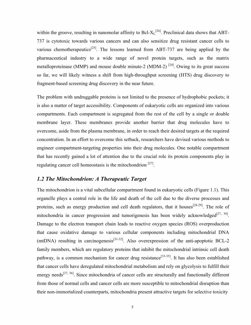
The mitochondrion is a vital subcellular compartment found in eukaryotic cells (Figure 1.1). This
organelle plays a central role in the life and death of the cell due to the diverse processes and
proteins, such as energy production and cell death regulators, that it houses[28-29]. The role of
mitochondria in cancer progression and tumorigenesis has been widely acknowledged[27, 30].
Damage to the electron transport chain leads to reactive oxygen species (ROS) overproduction
that cause oxidative damage to various cellular components including mitochondrial DNA
(mtDNA) resulting in carcinogenesis[31-32]. Also overexpression of the anti-apoptotic BCL-2
family members, which are regulatory proteins that inhibit the mitochondrial intrinsic cell death
pathway, is a common mechanism for cancer drug resistance[33-35]. It has also been established
that cancer cells have deregulated mitochondrial metabolism and rely on glycolysis to fulfill their
energy needs[27, 36]. Since mitochondria of cancer cells are structurally and functionally different
from those of normal cells and cancer cells are more susceptible to mitochondrial disruption than
their non-immortalized counterparts, mitochondria present attractive targets for selective toxicity
6
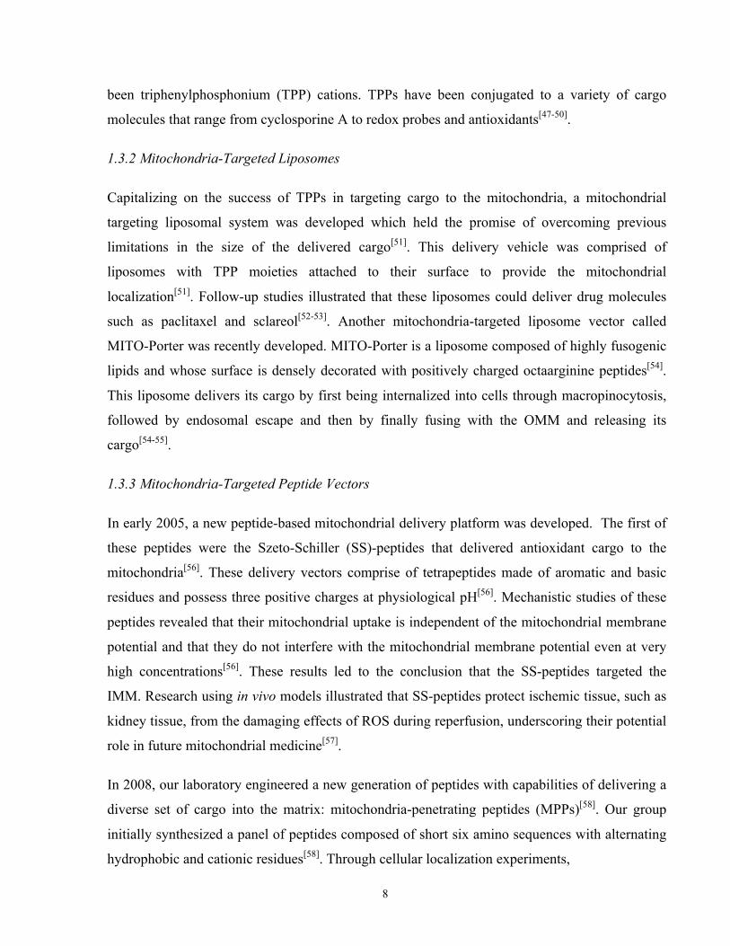
Figure 1.1 The structure of the mitochondrion.
The mitochondrion is a cellular organelle that is surrounded by a double membrane layer. The
outer mitochondrial membrane (OMM) is a porous structure while the inner mitochondrial
membrane (IMM) is densely packed with a high protein:lipid ratio and a very negative
membrane potential (- 180mV). The IMM forms cristae within the mitochondrial matrix that are
studded with respiratory chain protein complexes that utilize the IMM potential to generate ATP
through oxidative phosphorylation. The mitochondrial matrix contains several copies of the
mitochondrial genome and houses various processes that are needed for energy storage and
production.
7
towards tumor cells and effective cancer therapy[27, 36-39].
Unfortunately, mitochondrial targets are difficult to access due to the nature of the mitochondrial
double membrane. The inner mitochondrial membrane (IMM) is highly hydrophobic with a high
cardiolipin and protein content excluding many molecules from the mitochondrial matrix. This
difficulty in accessing the matrix meant that much of the initial mitochondria-targeted drug
discovery was serendipitous. One example is betulinic acid, a pentacyclic triterpene that targets
mitochondria and activates apoptosis, which was discovered through a chemical library screen
and has shown activity against drug resistant cancer lines[40-44]. However the cost and difficulty
of developing new small molecule therapies, through screening of large chemical libraries, has
lead to a major interest in developing targeting vectors in order to provide access to
mitochondrial drug targets.
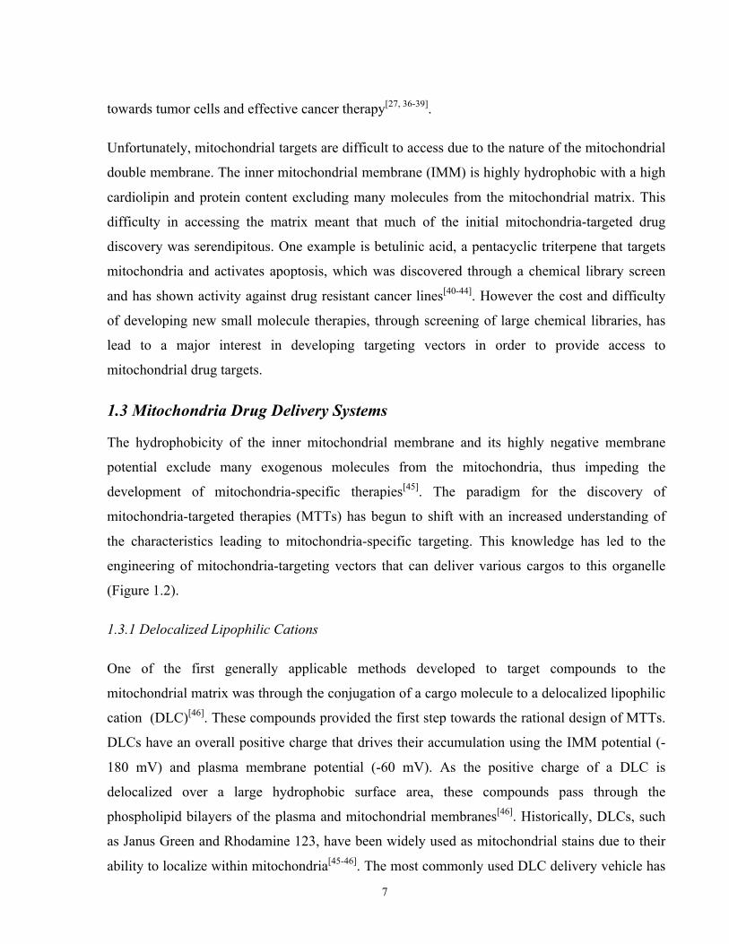
1.3 Mitochondria Drug Delivery Systems
The hydrophobicity of the inner mitochondrial membrane and its highly negative membrane
potential exclude many exogenous molecules from the mitochondria, thus impeding the
development of mitochondria-specific therapies[45]. The paradigm for the discovery of
mitochondria-targeted therapies (MTTs) has begun to shift with an increased understanding of
the characteristics leading to mitochondria-specific targeting. This knowledge has led to the
engineering of mitochondria-targeting vectors that can deliver various cargos to this organelle
(Figure 1.2).
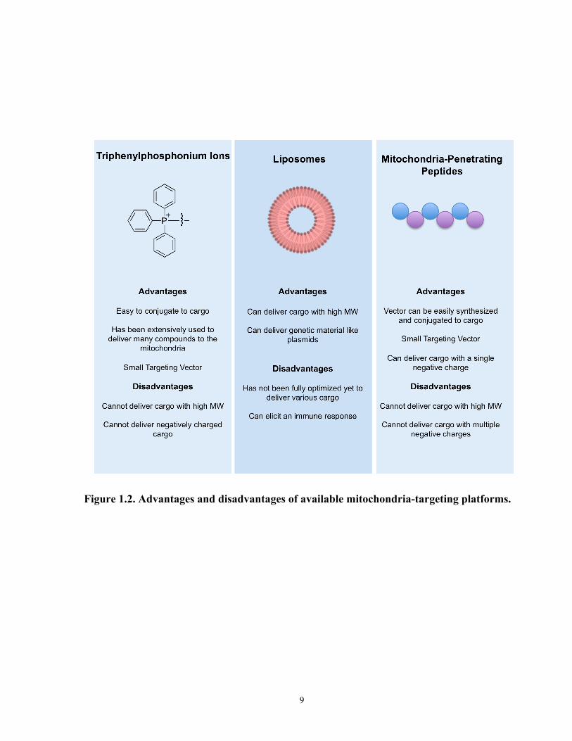
1.3.1 Delocalized Lipophilic Cations
One of the first generally applicable methods developed to target compounds to the
mitochondrial matrix was through the conjugation of a cargo molecule to a delocalized lipophilic
cation (DLC)[46]. These compounds provided the first step towards the rational design of MTTs.
DLCs have an overall positive charge that drives their accumulation using the IMM potential (-
180 mV) and plasma membrane potential (-60 mV). As the positive charge of a DLC is
delocalized over a large hydrophobic surface area, these compounds pass through the
phospholipid bilayers of the plasma and mitochondrial membranes[46]. Historically, DLCs, such
as Janus Green and Rhodamine 123, have been widely used as mitochondrial stains due to their
ability to localize within mitochondria[45-46]. The most commonly used DLC delivery vehicle has
8
been triphenylphosphonium (TPP) cations. TPPs have been conjugated to a variety of cargo
molecules that range from cyclosporine A to redox probes and antioxidants[47-50].
1.3.2 Mitochondria-Targeted Liposomes
Capitalizing on the success of TPPs in targeting cargo to the mitochondria, a mitochondrial
targeting liposomal system was developed which held the promise of overcoming previous
limitations in the size of the delivered cargo[51]. This delivery vehicle was comprised of
liposomes with TPP moieties attached to their surface to provide the mitochondrial
localization[51]. Follow-up studies illustrated that these liposomes could deliver drug molecules
such as paclitaxel and sclareol[52-53]. Another mitochondria-targeted liposome vector called
MITO-Porter was recently developed. MITO-Porter is a liposome composed of highly fusogenic
lipids and whose surface is densely decorated with positively charged octaarginine peptides[54].
This liposome delivers its cargo by first being internalized into cells through macropinocytosis,
followed by endosomal escape and then by finally fusing with the OMM and releasing its
cargo[54-55].
1.3.3 Mitochondria-Targeted Peptide Vectors
In early 2005, a new peptide-based mitochondrial delivery platform was developed. The first of
these peptides were the Szeto-Schiller (SS)-peptides that delivered antioxidant cargo to the
mitochondria[56]. These delivery vectors comprise of tetrapeptides made of aromatic and basic
residues and possess three positive charges at physiological pH[56]. Mechanistic studies of these
peptides revealed that their mitochondrial uptake is independent of the mitochondrial membrane
potential and that they do not interfere with the mitochondrial membrane potential even at very
high concentrations[56]. These results led to the conclusion that the SS-peptides targeted the
IMM. Research using in vivo models illustrated that SS-peptides protect ischemic tissue, such as
kidney tissue, from the damaging effects of ROS during reperfusion, underscoring their potential
role in future mitochondrial medicine[57].
In 2008, our laboratory engineered a new generation of peptides with capabilities of delivering a
diverse set of cargo into the matrix: mitochondria-penetrating peptides (MPPs)[58]. Our group
initially synthesized a panel of peptides composed of short six amino sequences with alternating
hydrophobic and cationic residues[58]. Through cellular localization experiments,
9
Figure 1.2. Advantages and disadvantages of available mitochondria-targeting platforms.
10
the optimal requirements needed for efficient mitochondrial localization of MPPs were
elucidated. It was found that MPPs needed a minimum of three positive charges and a
lipophilicity above a certain threshold in order to localize within mitochondria with high
specificity[58]. Mechanistic studies revealed that, unlike their SS-peptide counterpart, MPP uptake
is mitochondrial membrane potential-dependent with a decrease in potential resulting in
decreased peptide accumulation[58]. The dependence of MPP uptake on mitochondrial membrane
potential provides important therapeutic benefits since tumor pathology is often characterized by
an upregulated mitochondrial membrane potential, which can allow selective accumulation of
MPP-drug conjugates within malignant cells compared to healthy cells[59-61]. Further work
demonstrated that the alternating scaffold was not necessary for mitochondrial access as long as
all the cationic residues in the sequence were not grouped together[62]. Moreover, delocalizing
residue cationic charges over a larger surface area, appeared to result in higher mitochondrial
specificity[63]. MPPs have been successfully used in delivering different types of cargo including
fluorescent dyes and therapeutic drugs[59, 62-64].
1.4 Mitochondrial Retargeting to Overcome Cancer Drug Resistance
With the advent of mitochondria-targeting delivery vectors, a new therapeutic strategy known as
mitochondrial retargeting has emerged. By using mitochondria-specific vectors, drug targets
within this important organelle are now accessible, allowing a rational development of
mitochondria-targeted drugs. Mitochondrial retargeting results in a dramatic change in the
intracellular localization profile of a drug with high accumulation in the mitochondria, whereas
previously there was little to no mitochondrial penetration by the drug alone. This specific
mitochondrial accumulation of a drug causes the mitochondria to become the drug’s main site of
action. Ultimately, targeting the mitochondria allows finer control over a drug’s mechanism of
action by closely controlling its localization.
Our lab recently retargeted a DNA alkylating agent to the mitochondria to damage mtDNA.
DNA damaging agents are one of the most common cancer therapeutic drugs on the market and
are designed to target nuclear DNA (nDNA)[65]. However, cancer resistance to these kinds of
drugs has become a common problem[66-67]. Using chlorambucil (Cbl) as a model alkylating
drug, we attached it to an MPP sequence using simple solid-phase chemistry to generate a
mitochondria-targeted DNA damaging agent, mt-Cbl[59]. In vitro studies showed that by
11
changing Cbl’s target from nDNA to mtDNA, there was a 5-30 fold increase in cytotoxicity in a
variety of leukemia cell lines including Cbl-resistant cell lines[59]. Thus by targeting Cbl to the
mitochondria, drug potency is not only increased but drug resistance mechanisms are also
bypassed.
The increase in potency and overcoming of resistance is attributable to several factors. Firstly,
mtDNA is more prone to damage than nDNA since it is not packaged around histones[68].
Secondly, mtDNA repair pathways are less efficient than their nuclear counterparts[68]. And
thirdly, by retargeting Cbl to the mitochondria, the drug is protected from cytoplasmic
glutathione S-transferase, a known resistance mechanism for Cbl[69]. Our in vitro results also
pointed out that resistance pathways that involve the overexpression of anti-apoptotic BCL-2
proteins do not affect the cytotoxicity of mt-Cbl, unlike Cbl[59, 70]. Moreover, when the selectivity
of mt-Cbl toxicity was tested, mt-Cbl was found to be more toxic toward leukemia patient blood
cells than normal blood cells. These results confirmed that a therapeutic window was maintained
after retargeting[59]. This could be attributed to the fact that the mitochondrial membrane
potential of cancer cells is more elevated than normal cells, leading to a greater accumulation of
mt-Cbl in cancer cells[59, 71]. Indeed, when the mitochondrial membrane potential and MPP
uptake was compared in peripheral bloods, mononuclear cells and CLL patient cells, we found
that CLL patient cells had a higher mitochondrial membrane potential than the normal blood
cells and this resulted in higher MPP uptake[59]. This study emphasized the value of
mitochondrial retargeting and showed that resistance pathways against FDA-approved drugs, like
Cbl, could be easily overcome through the use of mitochondrial delivery vectors.
12
1.5 Rationale and Hypothesis
Currently, it takes around 10-15 years and close to a billion dollars to discover a novel anticancer
drug and bring it from the research lab to a patient’s bedside. New methodologies of drug
discovery and development could help immensely reduce the timeframe and costs of this
process. With the advent of mitochondrial drug targeting platforms, mitochondrial rerouting of
FDA-approved drugs could prove to be a highly valuable methodology for future cancer drug
discovery, allowing researchers to regain the therapeutic power of anticancer drugs already on
the market.
Our recent study of rerouting chlorambucil to mitochondria is, to our knowledge, the very first
successful attempt of targeting mtDNA for cancer therapy. However, before applying this
technique to other cancer drugs, it is vital that the mechanism of action of mt-Cbl is elucidated.
By understanding how mt-Cbl induces cell death, novel cellular stress signaling and apoptotic
pathways could be discovered. This could allow us to better understand how cell death is induced
due to mitochondrial stress and could shed light on new proteins involved in these processes that
could be then targeted for cancer therapy. More importantly, unraveling the mechanism of action
of mt-Cbl will lead to further drug refinement and development of a second generation with
higher efficacy and tumor selectivity.
The central hypothesis of this project is that mitochondria-targeted chlorambucil induces a
cell death pathway that is different from untargeted chlorambucil. In order to validate this
hypothesis, we attempted to answer the following questions:
1- What are the mitochondrial targets of mt-Cbl?
2- After mitochondrial damage is induced by mt-Cbl, how is cell death initiated and what
pathway is activated?
13
2. Chapter 2: Materials and Methods
A section of this chapter (mtDNA labeling with mt-Cbl) represents the following published work:
S. B. Fonseca, M. P. Pereira, R. Mourtada, M. Gronda, K. L. Horton, R. Hurren, M. D. Minden,
A. D. Schimmer and S. O. Kelley. Chem. Biol. 2011, 18(4), 445-453
Data contributions:
First-generation mt-Cbl synthesis and purification was performed by Sonali Fonseca and Mark
Pereira. Cytotoxicity studies of first-generation mt-Cbl in HeLa cells were performed by Mark
Pereira (Figure 3.1). Localization experiments of first-generation mt-Cbl in HeLa cells were
performed by Sonali Fonseca (Figure 3.1). Cbl-TPP and TPP antibody were kindly provided by
Prof. Michael P. Murphy (University of Cambridge) and Prof. Robin A. J. Smith (University of
Otago).

14
Peptide Synthesis: Peptide scaffolds were synthesized at a 50 µmol scale on Rink amide MBHA
resin (0.56 mmol/g, 100-200 mesh) (NovaBiochem) using a Prelude automated peptide
synthesizer (Protein Technologies, Inc). For couplings, Fmoc-D-Arg(pbf)-OH or Fmoc-L-
cyclohexylalanine-OH amino acids (4 equiv., Advanced ChemTech, Fmoc= 9-
fluorenylmethyloxycarbonyl), HBTU (4 equiv., Protein Technologies Inc., HBTU= O-
(benzotriazol-l-yl)-N,N,N’,N’-tetramethyl-uronium hexafluorophosphate), and NMM (8 equiv.,
Protein Technologies, Inc., NMM= N-methylmorpholine) were stirred in N,N-dimethyl
formamide (DMF) for 1 hour. Arginine residues were added using double couplings. The Fmoc
protecting group on the N termini was removed using piperidine (20% v/v) in DMF (2x15 min).
Synthesis and characterization of peptide conjugates: Thiazole orange (to) was synthesized as
described previously[72] and coupled to peptides using HBTU (4 equiv.), and DIPEA (8 equiv.) in
DMF for 3 hours. Cbl (Oakwood Products, Inc.) was coupled to peptides using HBTU (4 eq) and
DIPEA (4 eq) in DMF. For biotin-labeled peptide conjugates, resin was coupled to Fmoc-
Lys(Biotin)-OH (Anaspec, Inc.) using HBTU (4 equiv.), and DIPEA (8 equiv.) in DMF for 2
hours. The N terminus of unconjugated peptides was capped using acetic anhydride, pyridine,
and dichloromethane (DCM) (1:5:10) (2x10 min). Peptides were deprotected and cleaved using
trifluoroacetic acid:triisopropylsilane (TFA/TIPS, 95:5 v/v) for 2 hours and precipitated and
washed using cold ether. Peptides were purified using reverse-phase HPLC on a C18 column
using MeCN/0.1% TFA and H2O/0.1% TFA as mobile phases. Peptide conjugate identity was
then confirmed by electrospray ionization (ESI) mass spectroscopy. To reduce Cbl degradation,
purified Cbl-peptide conjugates were immediately flash-frozen and lyophilized. Quantification of
to-labeled peptides and Cbl-conjugated peptides was done using an extinction coefficient of
63,000 M−1 cm−1 at 500 nm and 15,200 M−1 cm−1 at 258 nm, respectively, in H2O. A
bicinchoninic acid (BCA) assay (Thermo Scientific) was used to quantify unlabeled peptides.
Cell Culture: HeLa cells were cultured in Minimum Essential Medium alpha (Invitrogen)
supplemented with 10% (v/v) fetal bovine serum (FBS, Sigma-Aldrich) on 75 cm2 cell culture
plates with vent caps. HL60 cells and K562 cells were cultured in RPMI 1640 (Invitrogen) plus
10% FBS using suspension flasks with vent caps. 143B cells were cultured in Dulbecco’s
Modified Eagle Medium with high glucose (DMEM, Invitrogen) plus 10% FBS while 143B ρ°
cells were cultured in DMEM high glucose plus 10% FBS supplemented with 100 mM sodium
15
pyruvate and 5 mg/ml uridine. All cell lines were incubated in a humidified incubator at 37°C
with 5% CO2
Localization of to-(Yr)3 peptide HeLa cells were seeded in 8-well ibidis at a concentration of
25,000 cells/well 1 day before experiments. Cell culture medium was removed and cells were
washed with Dulbecco’s phosphate-buffered saline (DPBS, Invitrogen) and then incubated with
to-(Yr)3 peptide (8 µM) in MEM-alpha (No phenol red, invitrogen) for 45 minutes. After which
4',6-diamidino-2-phenylindole (DAPI, 300 ng/mL) was added to the cells and incubated for 10
minutes Cells were then washed thrice with DPBS and imaged using an inverted Zeiss LSM 510
confocal microscope with a C-APO 63x water immersion lens. The excitation wavelength used
for to was 488 nm and emission was collected at 505 nm using a long-pass filter. Differential
intereference contrast (DIC) images were also taken.
Uptake Measurement by Flow Cytometry: HeLa cells were plated at 100,000 cells/well in 12-
well flat-bottom plates 1 day prior to the experiment. Cells were washed with FBS-free MEM-
alpha and then incubated with to-labeled peptide in FBS-free MEM-alpha for 45 minutes Then
the medium was removed, cells were washed with PBS, and were enzymatically harvested. From
this point on, the samples were maintained on ice until analysis. The samples were transferred to
sterile tubes, pelleted by centrifugation (3 min at 700 x g), and washed with 0.7 mL PBS,
pelleted by centrifugation and resuspended in 300 µL PBS containing 5 nM SYTOX Red
(Invitrogen). Samples were then analyzed by flow cytometry on a BD FACSCanto flow
cytometer (BD Biosciences). A minimum of 10,000 cells was analyzed per sample. Those
staining positive for Sytox Red were excluded from analysis. The fluorescence median of the live
population was used for statistical analysis.
CCK8 Cell Viability Assay: Adherent cell lines (HeLa, 143B and 143B ρ° cells) were seeded in
96-well flat-bottom tissue culture plates (Sarstedt) the day before treatment at a density of 12,000
cells per well. Suspension cell lines (K562 cells) were seeded in 96-well flat-bottom tissue
culture plates at a density of 25,000 cells per well just before treatment. Adherent cell lines were
washed with serum-free media before treatment. Cells were preincubated with various inhibitors
at the indicated concentrations for 1 hour before the addition of various concentrations of
chlorambucil and chlorambucil conjugates prepared by serial dilution. The plates were incubated
for 18 hrs at 37°C in a humidified incubator and then 10 µL of CCK-8 viability dye (Dojindo,
16
Rockville) was added to each well and the plates were further incubated till color development
occurred. Absorbance at 450 nm was then measured and statistical analysis was done using
GraphPad Prism software (GraphPad). For caspase inhibition, 100 µM z-VAD-fmk
(Benzoyloxycarbonyl-Val-Ala-Asp-fluoromethylketone, Calbiochem) or 20 µM Q-VD-OPh (Q-
Val-Asp(non-omethylated)-OPh, R&D Systems) was used. For PARP-1 inhibition, 50 µM DPQ
(3,4-Dihydro-5[4-(1-piperindinyl)butoxy]-1(2H)-isoquinoline, Santa Cruz Biotech.) or 100 µM
INH2BP ( 5-Iodo-6-amino-1,2-benzopyrone, Santa Cruz Biotech.) was used. For ROS inhibition,
cells were preincubated with 10 mM NAC (N-acetylcysteine, Sigma-Aldrich).
Measurement of Endogenous Respiration: The experimental procedure was previously
described[73]. Cells were grown to 90% confluency, then collected by trypsinization, centrifuged,
washed and resuspended to 4,000,000 cells/mL in Tris-based Mg2+-, Ca2+-deficient buffer (0.137
M NaCl/5 mM KCl/0.7 mM Na2HPO4/ 25 mM Tris.HCl, pH 7.4 at 25oC). The cell suspension
was then transferred to a water-jacketed chamber (Mitocell, MT200, Strathkelvin) containing a
magnetic stir bar (37oC) and a Mitocell Strathkelvin oxygen monitor recorded the oxygen
consumption over a 6 minute time interval.
Annexin-V Apoptosis Assay: HeLa cells were seeded at 100,000 cells/well the day prior to an
experiment, while K562 cells were seeded at 200,000 cells/well on the day of the experiment in a
12-well flat-bottom tissue culture plate (BD Falcon). Chlorambucil and chlorambucil-conjugate
at indicated concentrations were added to the cells in triplicate in serum-free cell-appropriate
media. For mtCbl experiments, cells were incubated for 90 minutes and for Cbl experiments,
cells were incubated for 9 hours at 37°C with 5% CO2. Cells were later harvested and washed
with ice cold PBS, then washed again with annexin V binding buffer (50 mM HEPES, 700 mM
NaCl, 12.5 mM CaCl2, pH 7.4). Cells were resuspended in annexin V binding buffer plus
annexin V-FITC (1:20, Invitrogen) and incubated for 15 minutes at room temperature. Later, cell
suspensions were diluted with more annexin V binding buffer plus SYTOX Red (5 nM) and
incubated for a further 15 minutes. Flow cytometry was then performed on a FACSCanto flow
cytometer (BD Biosciences). A minimum of 10,000 cells were analyzed for every sample.
Western blots: After treatment, cells were harvested and washed with ice cold PBS prior to lysis
with RIPA buffer plus protease inhibitor cocktail (Santa Cruz Biotech) at 4°C for 20 minutes
Cells were then centrifuged at 16,000 g at 4°C for 10 minutes and the supernatant was collected.
17
Total protein concentrations were quantified using BCA assay and then 40 µg of total protein in
each sample was diluted with 2X sample buffer and boiled for 5 minutes before loading onto
10% or 15% gels. Gels were run at 80 V for 2 hours using tris-glycine buffer (25 mM TrisCl, 250
mM glycine, 0.1% SDS, pH 8.3) and then transferred onto nitrocellulose of PVDF membranes
(Pall Corporation) for 1-2 hrs at 100 V at 4°C. Membranes were then blocked with 5% BSA-
TBST for 1 hour at room temperature and then probed with primary antibody (1:1000 AIF
antibody (Santa Cruz Biotech), 1:400 Cytochrome C antibody (Santa Cruz Biotech), 1:5000 β-
actin antibody (Abcam), 1:1000 COX IV antibody (Mitosciences), 1:3000 GAPDH antibody
(Abcam), 1:500 PAR antibody (BD biosciences), 1:1000 PARP-1 antibody (Cell Signaling),
1:1000 cleaved Caspase-3 antibody (Cell Signaling), 1:5000 Biotin antibody (Jackson
ImmunoResearch), 1:1000 TBP antibody (Abcam) and 1:1000 Histone H3 antibody (Abcam)).
Membranes were then washed with TBST and incubated with 1:5000 donkey anti-mouse (Santa
Cruz Biotech) or goat anti-rabbit (Jackson ImmunoResearch) IgG-HRP secondary Ab for 1 hour
at room temperature in 2% milk-TBST prior to chemiluminescence detection (GE Amersham).
Cbl-conjugate Labeling of mtDNA: HL-60 cells (25 million cells) were incubated with biotin-
labeled mt-Cbl peptide (Biotin-mt-Cbl) (0, 1.5 µM) or Cbl-TPP (10 uM) for 30 minutes, after
which the cells were collected. The cells were then washed with ice-cold PBS, and their
mitochondria were isolated using Mitochondrial Isolation Kit for Mammalian Cells (Thermo
Scientific). The mtDNA was then extracted from the mitochondrial pellets using AllPrep
DNA/RNA Mini Kit (QIAGEN). DNA concentration was measured using a NanoDrop 1000
Spectrophotometer (Thermo Scientific) and then normalized (10 ng/µl). After this, a
nitrocellulose membrane was spotted with untreated and treated DNA and UV crosslinked using
UV Stratalinker 1800 (Stratagene) The membrane was blocked with 5% BSA-TBST for 1 hr at
room temperature and then probed with streptavidin-HRP (Sigma-Aldrich) in 5% BSA-TBST
(1:5000) or TPP antibody (1:5000) at 4°C overnight. Then the membrane was washed and
visualized with chemiluminescence detection (GE Amersham).
Cbl-conjugate labeling of mitochondrial proteins: HL-60 cells (25 million cells) or HeLa cells
(6 million cells) were incubated with biotin-labeled mt-Cbl peptide (Biotin-mt-Cbl) (0, 1.5 µM)
or Cbl-TPP (10 uM) for 30 minutes, after which the cells were collected. The cells were then
washed with ice-cold PBS, and their mitochondria were isolated using Mitochondrial Isolation
Kit for Mammalian Cells (Thermo Scientific). The mitochondrial pellets were then lysed using
18
RIPA buffer plus protease inhibitors for 30 minutes at 4°C and the total protein concentration
determined using BCA assay. Lysates (10 µg of total protein) were then loaded onto 7% gels and
run at 30 V for 30 minutes and 150 V for 1 hour using a tris-tricine buffer system (Cathode
buffer: 0.1M Tris, 0.1M Tricine, and 0.1% SDS, Anode buffer: 0.2M Tris-Cl, pH8.9).
Immunoblotting was continued as described previously using biotin antibody to detect the biotin
tag on mt-Cbl or TPP antibody to detect Cbl-TPP.
Subcellular Fractionation: HeLa cells (7 million cells) or K562 cells (10 million cells) were
treated with Cbl or Cbl conjugates at indicated concentrations and time intervals using serum-
free cell-appropriate media. Cells were then collected, washed with ice cold PBS and pelleted at
300 g to room temperature. Pellets were then fractionated using Cell Fractionation Kit-Standard
(Mitosciences) as per manufacturer’s instructions. For nuclear fraction, the final pellet obtained
was lysed using RIPA buffer plus protease inhibitors. Since kit produces diluted fractions, the
cytoplasmic and mitochondrial fractions were concentrated using Amicon Ultra-0.5 10kDa filters
(Millipore) before proceeding to immunoblotting as described above.
Analysis of Mitochondrial Superoxide Levels. HeLa cells were plated at 50,000 per well of a
24-well plate 24 hr prior to experiment and treated with Cbl or mt-Cbl in OPTI-MEM
(Invitrogen) for 1 hr. Media were removed, and cells were incubated with MitoSOX (Invitrogen)
according to manufacturer's instructions. Cells were washed with PBS, trypsinized, and analyzed
via flow cytometry with FACSCanto (BD Biosciences).
Assessment of protease activity and ATP cellular levels: HeLa cells were seeded at 7500
cells/well a day prior to experiments while K562 cells were seeded at 15,000 cells/well on the
day of experiment. The plates used were white flat clear-bottom 96-well plates (Greiner Bio
one). Cells were incubated with Cbl, Cbl conjugates and digitonin at indicated concentrations for
90 minutes and then protease activity and ATP cellular levels were measured using
Mitochondrial ToxGlo assay (Promega) as per manufacturer’s instructions.
Assessment of caspase 3/7 activity: HeLa cells were seeded at 7500 cells/well a day prior to
experiments while K562 cells were seeded at 15,000 cells/well on the day of experiment. The
plates used were white flat clear-bottom 96-well plates (Greiner Bio one). Cells were incubated
Cbl, Cbl conjugates and staurosporine at indicated concentrations for 6 hours and then caspase

19
3/7 activity was measured using Caspase-Glo® 3/7 Assay (Promega) as per manufacturer’s
instructions.
RNA interference: HeLa cells were seeded at 100,000 cells/well in a six-well plate and
incubated at 37oC with 5% CO2 overnight. The cells were then treated with ON-Targetplus
SMARTpool siRNA, Human PDCD8 (Target sequences: GCUGCAUGCUUCUACGAUCA;
GGUAGAAACUGACCACAUA;AGUCAGCAGUGGCAAGUUA;GUCCGAGGCCUCAGAA
AUU, Dharmacon) and Dharmafect 1 using manufacturer’s protocol. After 48 hours incubation,
cells were washed with DPBS, trypsinized and then seeded at a density of 12,000 cells/well in a
96-well plate for cell viability assay and also a six-well plate was seeded with 100,000 cells/well
to check for AIF silencing using immunoblotting.
Colorimetic Alkylation Assay: A modified version of the 4-(4-nitrobenzyl)pyridine (4-NBP)
assay was conducted to measure the alkylating activity of each compound. Briefly, 100 µL of
compound (200 µM) was mixed with 100 µL of sodium acetate buffer (pH 4.0) and 75 µL of
3.3% (w/v) 4-NBP in acetone. Mixture was incubated at 37°C for a certain time interval.
Reaction was terminated by freezing in a dry ice/ethanol bath. To develop the sample, 250 µL of
50% (v/v) Et3N in acetone was added. The mixture was shaken vigorously and then read at 575
nm against a blank. This was repeated for various time intervals (30, 60, 90, 120 min) in
triplcate.
20
3. Chapter 3: Results and Discussion
A section of this chapter represents the following published work:
S. B. Fonseca, M. P. Pereira, R. Mourtada, M. Gronda, K. L. Horton, R. Hurren, M. D. Minden,
A. D. Schimmer and S. O. Kelley. Chem. Biol. 2011, 18(4), 445-453
Data contributions:
First-generation mt-Cbl synthesis and purification was performed by Sonali Fonseca and Mark
Pereira. Cytotoxicity studies of first-generation mt-Cbl in HeLa cells were performed by Mark
Pereira (Figure 3.1). Localization experiments of first-generation mt-Cbl in HeLa cells were
performed by Sonali Fonseca (Figure 3.1). Cbl-TPP and TPP antibody were kindly provided by
Prof. Michael P. Murphy (University of Cambridge) and Prof. Robin A. J. Smith (University of
Otago).
21
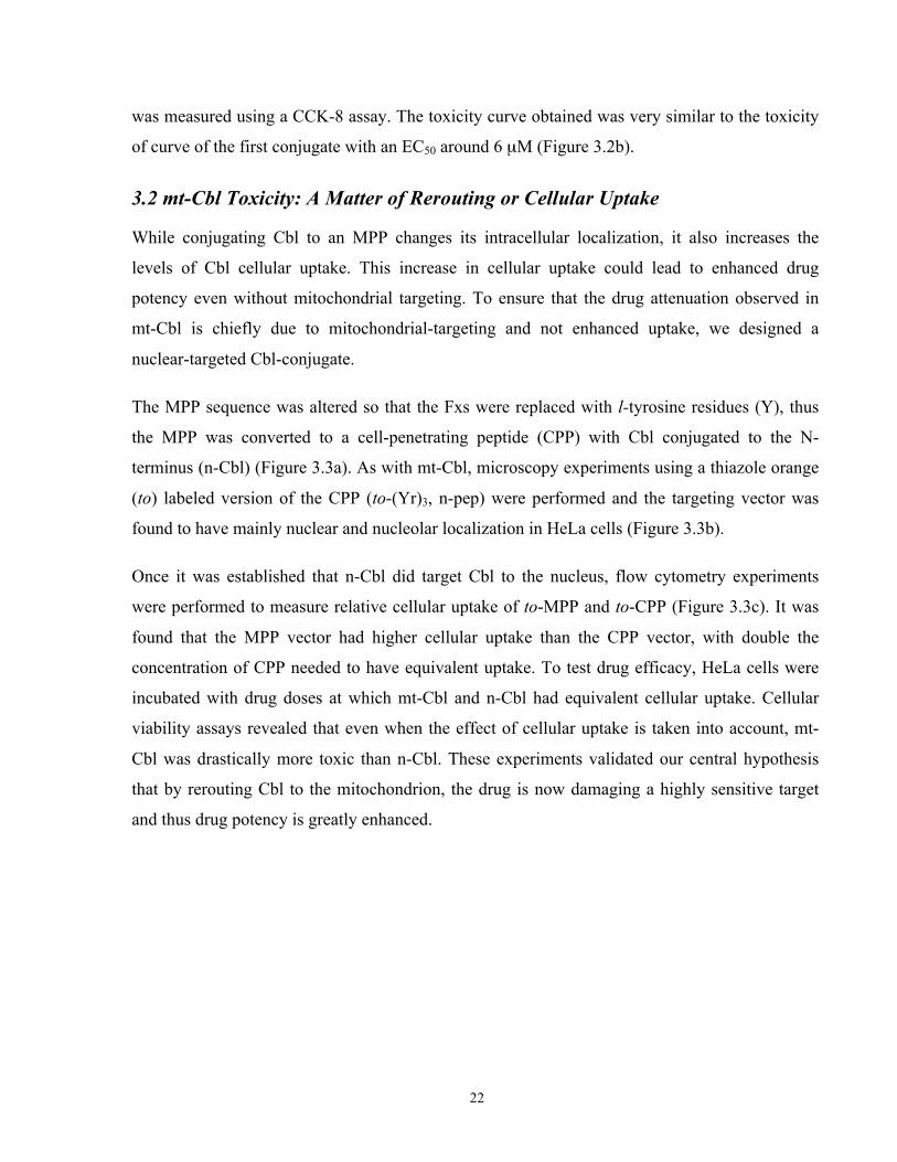
In our previous study[59], our laboratory generated the first, to our knowledge, mitochondria-
targeted chlorambucil (Cbl) conjugate (Figure 3.1a). The drug-conjugate rerouted Cbl from its
usual nuclear destination to the mitochondria. This was confirmed through colocalization
experiments in a cervical carcinoma cell line, HeLa cells, using a mitochondrial dye,
Mitotracker, and a fluorophore-labeled version of the mitochondria-targeted Cbl (mt-Cbl)
(Figure 3.1b). Drug efficacy tests in HeLa cells revealed that by retargeting Cbl to the
mitochondrion, drug potency was increased by around 100-fold compared to untargeted Cbl
(Figure 3.1c). This increase in potency was reproduced in a variety of leukemia cell lines,
including several Cbl-resistant cancer cell lines. The study unveiled the therapeutic potential that
the strategy of rerouting DNA damaging agents to mitochondria holds for future cancer drug
discovery. However, the mechanism of action of mt-Cbl needed scrutinizing to fully understand
how a DNA damaging agent, like Cbl, triggers cell death from within the mitochondria.
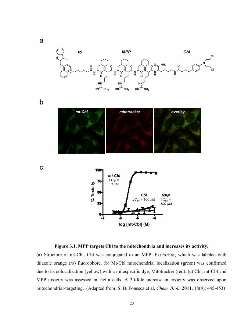
3.1 Second Generation of mt-Cbl
In order to perform our mechanistic studies, we opted to refine the chemical structure of mt-Cbl
in order to simplify the synthetic methodology. In the first generation of mt-Cbl, Cbl was
attached to a mitochondria-penetrating peptide (MPP) sequence, with an alternating l-
cyclohexylalanine (Fx) and d-arginine (r) sequence (Fx-r-Fx-r-Fx-r), through the amine group on
a lysine residue attached to the C-terminus of the MPP vector (Figure 3.2a). This kind of
attachment meant that the functional group on the lysine group needed to be protected during
peptide synthesis, and then the N-terminus was capped with an acetyl group before deprotecting
the lysine residue to attach Cbl. These intermediate steps complicated the synthesis and
decreased overall yield.
To simplify the synthesis of mt-Cbl, we opted to changing the point of attachment of Cbl from a
lysine reside to the N-terminus of the MPP vector (Figure 3.2a). This allowed us to synthesize
the peptide on resin using a peptide synthesizer and then Cbl was attached immediately after
deprotecting the N-terminus before cleaving the Cbl-conjugate off the resin and purifying it
using HPLC. The new synthetic route helped us remove three unnecessary steps and improved
overall yield.
To ensure that the second-generation mt-Cbl had similar potency to the first-generation mt-Cbl,
HeLa cells were incubated with the drug-conjugate at various doses overnight and cell viability
22
was measured using a CCK-8 assay. The toxicity curve obtained was very similar to the toxicity
of curve of the first conjugate with an EC50 around 6 µM (Figure 3.2b).
3.2 mt-Cbl Toxicity: A Matter of Rerouting or Cellular Uptake
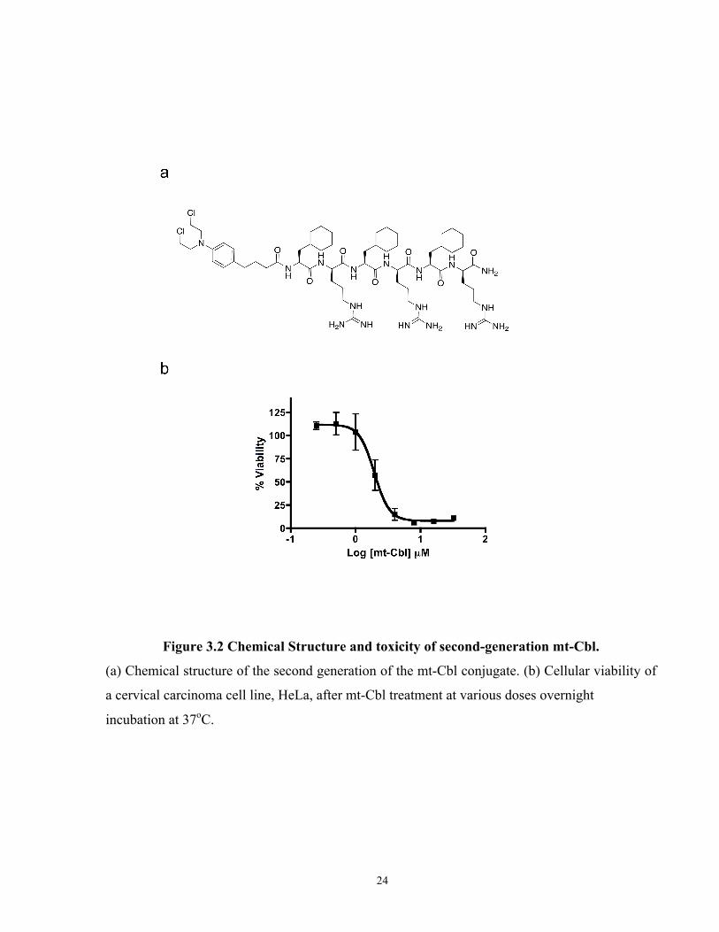
While conjugating Cbl to an MPP changes its intracellular localization, it also increases the
levels of Cbl cellular uptake. This increase in cellular uptake could lead to enhanced drug
potency even without mitochondrial targeting. To ensure that the drug attenuation observed in
mt-Cbl is chiefly due to mitochondrial-targeting and not enhanced uptake, we designed a
nuclear-targeted Cbl-conjugate.
The MPP sequence was altered so that the Fxs were replaced with l-tyrosine residues (Y), thus
the MPP was converted to a cell-penetrating peptide (CPP) with Cbl conjugated to the N-
terminus (n-Cbl) (Figure 3.3a). As with mt-Cbl, microscopy experiments using a thiazole orange
(to) labeled version of the CPP (to-(Yr)3, n-pep) were performed and the targeting vector was
found to have mainly nuclear and nucleolar localization in HeLa cells (Figure 3.3b).
Once it was established that n-Cbl did target Cbl to the nucleus, flow cytometry experiments
were performed to measure relative cellular uptake of to-MPP and to-CPP (Figure 3.3c). It was
found that the MPP vector had higher cellular uptake than the CPP vector, with double the
concentration of CPP needed to have equivalent uptake. To test drug efficacy, HeLa cells were
incubated with drug doses at which mt-Cbl and n-Cbl had equivalent cellular uptake. Cellular
viability assays revealed that even when the effect of cellular uptake is taken into account, mt-
Cbl was drastically more toxic than n-Cbl. These experiments validated our central hypothesis
that by rerouting Cbl to the mitochondrion, the drug is now damaging a highly sensitive target
and thus drug potency is greatly enhanced.
23
Cbl MPP to
a
mt-Cbl mitotracker overlay
b
mt-Cbl LC50 = 3 µM
Cbl LC50 > 100 µM %
Tox
icity
log [mt-Cbl] (M)
c
MPP LC50 >
100 µM
Figure 3.1. MPP targets Cbl to the mitochondria and increases its activity.
(a) Structure of mt-Cbl. Cbl was conjugated to an MPP, FxrFxrFxr, which was labeled with
thiazole orange (to) fluorophore. (b) Mt-Cbl mitochondrial localization (green) was confirmed
due to its colocalization (yellow) with a mitospecific dye, Mitotracker (red). (c) Cbl, mt-Cbl and
MPP toxicity was assessed in HeLa cells. A 30-fold increase in toxicity was observed upon
mitochondrial-targeting. (Adapted from: S. B. Fonseca et al. Chem. Biol. 2011, 18(4): 445-453)
24
Figure 3.2 Chemical Structure and toxicity of second-generation mt-Cbl.
(a) Chemical structure of the second generation of the mt-Cbl conjugate. (b) Cellular viability of
a cervical carcinoma cell line, HeLa, after mt-Cbl treatment at various doses overnight
incubation at 37oC.
25
b
c
a
d
CPP DAPI merged
8 160
2000
4000
6000
8000
10000
12000
14000mt-pepn-pep
[to-peptide] µM
Figure 3.3 Nuclear-targeted Cbl is less potent than mt-Cbl.
(a) Chemical structure of nuclear-targeted Cbl (n-Cbl). (b) Cellular Localization of to-labeled
nuclear-targeted peptide (n-pep) in live HeLa cells compared with DAPI. Replacement of
cyclohexylalanines in the MPP with tyrosines results in nuclear targeting of the Cbl-peptide
conjugate. (c) Cellular uptake of to-labeled n-pep and mt-pep in HeLa cells using flow
cytometry. The n-pep exhibits equal levels of uptake relative to the mt-pep at double the
concentration. (d) Toxicity profiles of n-Cbl and mt-Cbl in live HeLa cells following
overnight incubation. mt-Cbl is more potent that n-Cbl at concentrations with equal cellular
uptake.

26
3.3 Mitochondrial Targets of mt-Cbl
Due to its mechanism of action, Cbl can bind to various targets including DNA, proteins and
lipids. Cbl is a nitrogen mustard, thus when in solution, the lone pair of electrons of the nitrogen
attacks the carbon bound to the chlorine atom in an electrophilic manner. This results in the
expulsion of the chlorine as a chloride ion and the formation of a highly reactive aziridinium
moiety. Because of its electron deficiency, the aziridinium moiety is highly susceptible to a
nucleophilic attack from the nitrogen in guanine groups on DNA strands, amine groups on
proteins and lipids and other electron-rich groups found on various compounds within a cell.
In order to track down the various targets that mt-Cbl could bind to in the mitochondrion, we
generated a biotin-labeled version of mt-Cbl by conjugating a biotin moiety to the functional
group of a lysine residue and then attaching the labeled residue to the C-terminus of mt-Cbl. A
leukemia cell line, HL60 cells, was then treated with the biotin-labeled mt-Cbl (bt-mt-Cbl) and
their mitochondria isolated. Using a DNA prep column, we isolated the mtDNA and probed them
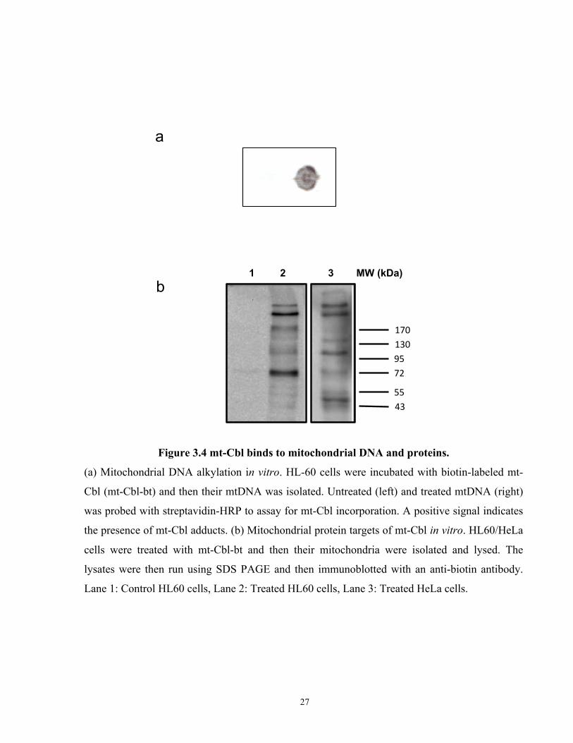
using streptavidin-HRP, a protein that binds to biotin with very high affinity. As anticipated, the
mtDNA from treated HL60 cells gave a positive signal indicating the presence of mt-Cbl
covalently bound to the DNA (Figure 3.4a). These results confirmed that after rerouting, Cbl
maintained its alkylating activity and was able to form mtDNA lesions.
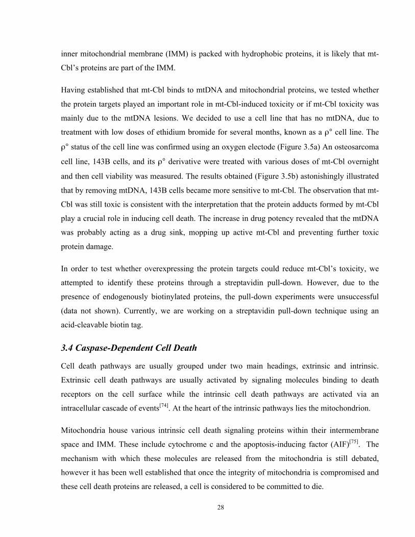
The mitochondrion is filled with proteins that play crucial functions in regulating overall cellular
homeostasis. Some proteins are involved in the production of ATP, such as the ATP synthase,
while others are involved in activating the intrinsic cell death pathway, such as the permeability
transition pore complex (PTPC)[71]. If mt-Cbl bound to such proteins, the damage induced could
lead to enhanced cellular toxicity due to the vital role these proteins play within a cell[27]. To
visualize mt-Cbl’s protein targets, we performed western blots on the mitochondrial lysates of
HL60 cells and HeLa cells treated with bt-mt-Cbl. The results obtained revealed that mt-Cbl did
bind to mitochondrial proteins (Figure 3.4b). However, it was quite interesting to see that mt-Cbl
had specific protein targets to which it preferentially bound. Comparing the protein targets found
in HeLa cells and HL60 cells, there was significant overlap in the targets bound indicating that
the MPP vector maybe acting as a protein targeting sequence as well. Due to the hydrophobic
nature of the MPP sequence, it would probably prefer to bind to proteins with deep hydrophobic
pockets. Since the
27
b
a
!"#$!%#$$&'$$"($$
''$)%$
1 2 3 MW (kDa)
Figure 3.4 mt-Cbl binds to mitochondrial DNA and proteins.
(a) Mitochondrial DNA alkylation in vitro. HL-60 cells were incubated with biotin-labeled mt-
Cbl (mt-Cbl-bt) and then their mtDNA was isolated. Untreated (left) and treated mtDNA (right)
was probed with streptavidin-HRP to assay for mt-Cbl incorporation. A positive signal indicates
the presence of mt-Cbl adducts. (b) Mitochondrial protein targets of mt-Cbl in vitro. HL60/HeLa
cells were treated with mt-Cbl-bt and then their mitochondria were isolated and lysed. The
lysates were then run using SDS PAGE and then immunoblotted with an anti-biotin antibody.
Lane 1: Control HL60 cells, Lane 2: Treated HL60 cells, Lane 3: Treated HeLa cells.
28
inner mitochondrial membrane (IMM) is packed with hydrophobic proteins, it is likely that mt-
Cbl’s proteins are part of the IMM.
Having established that mt-Cbl binds to mtDNA and mitochondrial proteins, we tested whether
the protein targets played an important role in mt-Cbl-induced toxicity or if mt-Cbl toxicity was
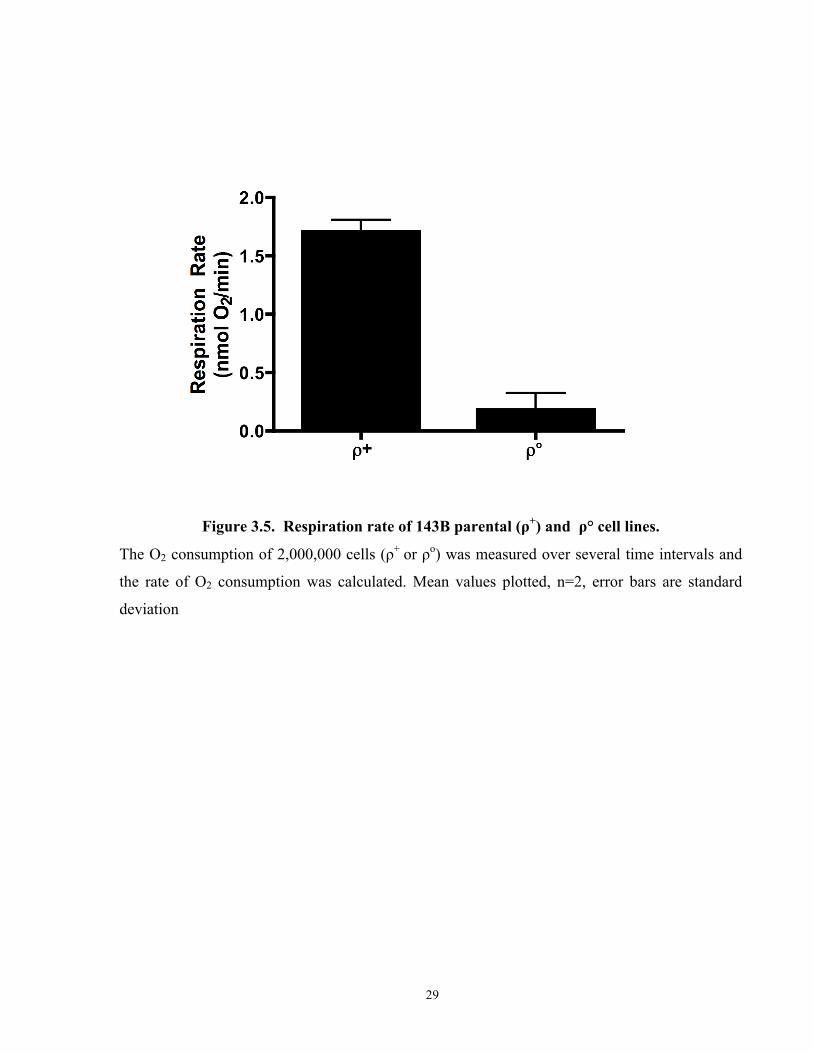
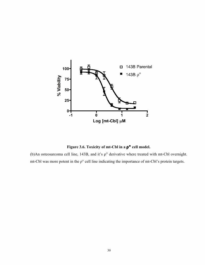
mainly due to the mtDNA lesions. We decided to use a cell line that has no mtDNA, due to
treatment with low doses of ethidium bromide for several months, known as a ρ° cell line. The
ρ° status of the cell line was confirmed using an oxygen electode (Figure 3.5a) An osteosarcoma
cell line, 143B cells, and its ρ° derivative were treated with various doses of mt-Cbl overnight
and then cell viability was measured. The results obtained (Figure 3.5b) astonishingly illustrated
that by removing mtDNA, 143B cells became more sensitive to mt-Cbl. The observation that mt-
Cbl was still toxic is consistent with the interpretation that the protein adducts formed by mt-Cbl
play a crucial role in inducing cell death. The increase in drug potency revealed that the mtDNA
was probably acting as a drug sink, mopping up active mt-Cbl and preventing further toxic
protein damage.
In order to test whether overexpressing the protein targets could reduce mt-Cbl’s toxicity, we
attempted to identify these proteins through a streptavidin pull-down. However, due to the
presence of endogenously biotinylated proteins, the pull-down experiments were unsuccessful
(data not shown). Currently, we are working on a streptavidin pull-down technique using an
acid-cleavable biotin tag.
3.4 Caspase-Dependent Cell Death
Cell death pathways are usually grouped under two main headings, extrinsic and intrinsic.
Extrinsic cell death pathways are usually activated by signaling molecules binding to death
receptors on the cell surface while the intrinsic cell death pathways are activated via an
intracellular cascade of events[74]. At the heart of the intrinsic pathways lies the mitochondrion.
Mitochondria house various intrinsic cell death signaling proteins within their intermembrane
space and IMM. These include cytochrome c and the apoptosis-inducing factor (AIF)[75]. The
mechanism with which these molecules are released from the mitochondria is still debated,
however it has been well established that once the integrity of mitochondria is compromised and
these cell death proteins are released, a cell is considered to be committed to die.
29
Figure 3.5. Respiration rate of 143B parental (ρ+) and ρ° cell lines.
The O2 consumption of 2,000,000 cells (ρ+ or ρo) was measured over several time intervals and
the rate of O2 consumption was calculated. Mean values plotted, n=2, error bars are standard
deviation
30
Figure 3.6. Toxicity of mt-Cbl in a ρ° cell model.
(b)An osteosarcoma cell line, 143B, and it’s ρ° derivative where treated with mt-Cbl overnight.
mt-Cbl was more potent in the ρ° cell line indicating the importance of mt-Cbl’s protein targets.
31
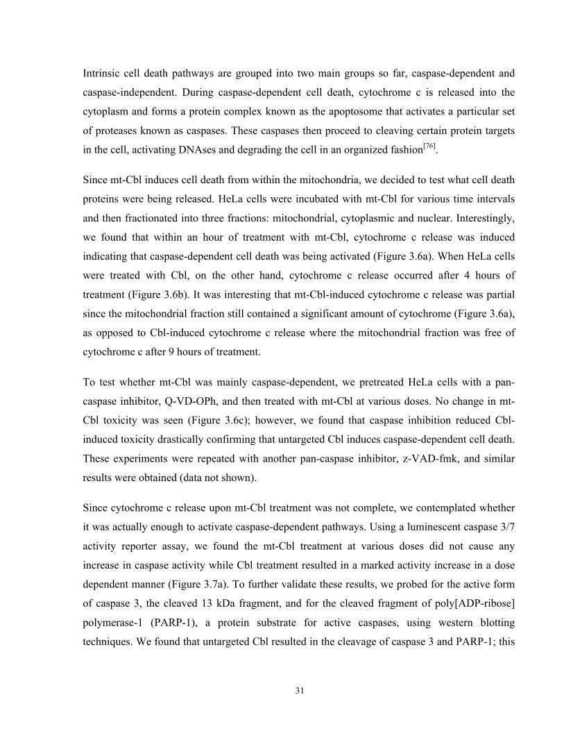
Intrinsic cell death pathways are grouped into two main groups so far, caspase-dependent and
caspase-independent. During caspase-dependent cell death, cytochrome c is released into the
cytoplasm and forms a protein complex known as the apoptosome that activates a particular set
of proteases known as caspases. These caspases then proceed to cleaving certain protein targets
in the cell, activating DNAses and degrading the cell in an organized fashion[76].
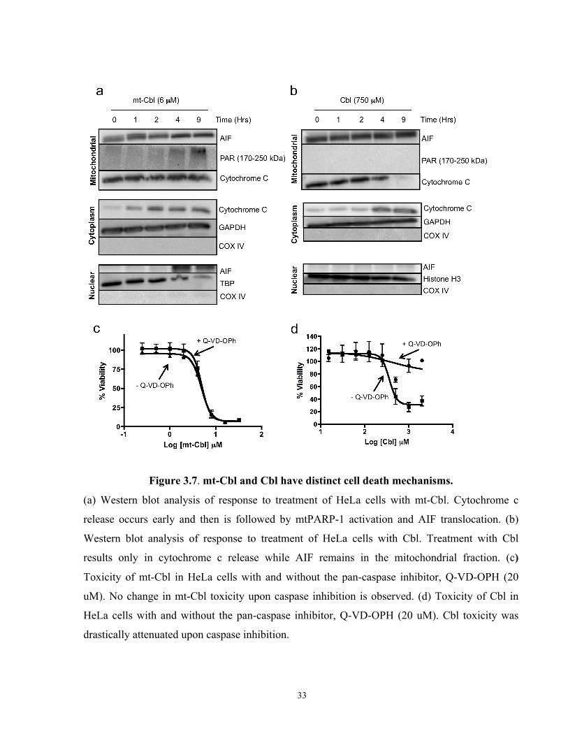
Since mt-Cbl induces cell death from within the mitochondria, we decided to test what cell death
proteins were being released. HeLa cells were incubated with mt-Cbl for various time intervals
and then fractionated into three fractions: mitochondrial, cytoplasmic and nuclear. Interestingly,
we found that within an hour of treatment with mt-Cbl, cytochrome c release was induced
indicating that caspase-dependent cell death was being activated (Figure 3.6a). When HeLa cells
were treated with Cbl, on the other hand, cytochrome c release occurred after 4 hours of
treatment (Figure 3.6b). It was interesting that mt-Cbl-induced cytochrome c release was partial
since the mitochondrial fraction still contained a significant amount of cytochrome (Figure 3.6a),
as opposed to Cbl-induced cytochrome c release where the mitochondrial fraction was free of
cytochrome c after 9 hours of treatment.
To test whether mt-Cbl was mainly caspase-dependent, we pretreated HeLa cells with a pan-
caspase inhibitor, Q-VD-OPh, and then treated with mt-Cbl at various doses. No change in mt-
Cbl toxicity was seen (Figure 3.6c); however, we found that caspase inhibition reduced Cbl-
induced toxicity drastically confirming that untargeted Cbl induces caspase-dependent cell death.
These experiments were repeated with another pan-caspase inhibitor, z-VAD-fmk, and similar
results were obtained (data not shown).
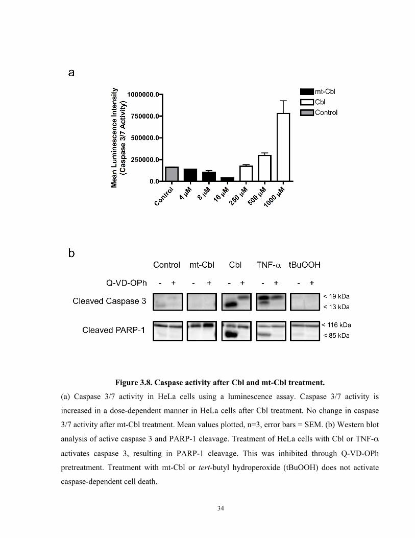
Since cytochrome c release upon mt-Cbl treatment was not complete, we contemplated whether
it was actually enough to activate caspase-dependent pathways. Using a luminescent caspase 3/7
activity reporter assay, we found the mt-Cbl treatment at various doses did not cause any
increase in caspase activity while Cbl treatment resulted in a marked activity increase in a dose
dependent manner (Figure 3.7a). To further validate these results, we probed for the active form
of caspase 3, the cleaved 13 kDa fragment, and for the cleaved fragment of poly[ADP-ribose]
polymerase-1 (PARP-1), a protein substrate for active caspases, using western blotting
techniques. We found that untargeted Cbl resulted in the cleavage of caspase 3 and PARP-1; this
32
cleavage could be inhibited by pretreating with Q-VD-Oph. As expected, mt-Cbl treatment did
not result in any changes in cleaved caspase 3 levels or PARP-1 cleavage (Figure 3.7b).
Overall, the results from these experiments illustrate that by retargeting Cbl to the
mitochondrion, the mechanism of cell death has been changed. Untargeted Cbl relies on a
caspase-dependent mechanism, which is susceptible to inhibition. mt-Cbl induces cytochrome c
release but no caspase activity is elicited indicating that caspase-independent mechanisms were
being activated.
3.5 Caspase-Independent Cell Death
In the past decade, caspase-independent cell death mechanisms have emerged as an important
alternative to caspase-dependent cell death. These cell death signaling pathways have not been
elucidated fully yet, however several new pathways have been classified recently.
One important caspase-independent cell death pathway that has been discovered to be activated
when an alkylating agent causes extensive DNA damage is known as PARP-1 mediated cell
death or parthanatos. PARP-1 is a nuclear protein that is activated in response to DNA strand
breaks[77]. Studies have shown that PARP-1 activation leads to the production of poly [ADP-
ribose] (PAR) polymers that then initiate various DNA repair pathways and cell death[78-79].
Parthanatos is triggered in response to PARP-1 hyperactivation from extensive DNA damage,
which leads to depletion of cellular nicotinamide adenine dinucleotide (NAD+) and ATP
reserves[80]. One important feature of the parthanatos is the nuclear translocation of the
mitochondrial intermembrane protein, AIF[81]. A recent study has revealed that AIF translocation
occurs due to the binding of PAR polymers to AIF[82].
Recent studies have shown that PARP-1 is not only localized in the nucleus but it can translocate
to the mitochondrion by interacting with mitofilin, a transmembrane protein in the IMM [83-85].
Moreover intra-mitochondrial PARP-1 (mtPARP-1) was found to be activated by oxidative stress
leading to cell death. Further work revealed that mtPARP-1 binds to mtDNA and is involved in
recruiting DNA ligase III during mtDNA repair[85]. To test whether mtPARP-1 was being
activated upon the formation of mt-Cbl-induced DNA lesions, the presence of PAR polymers in
the mitochondrial fractions from the mt-Cbl treated HeLa cells was examined. PAR polymer
33
Figure 3.7. mt-Cbl and Cbl have distinct cell death mechanisms.
(a) Western blot analysis of response to treatment of HeLa cells with mt-Cbl. Cytochrome c
release occurs early and then is followed by mtPARP-1 activation and AIF translocation. (b)
Western blot analysis of response to treatment of HeLa cells with Cbl. Treatment with Cbl
results only in cytochrome c release while AIF remains in the mitochondrial fraction. (c)
Toxicity of mt-Cbl in HeLa cells with and without the pan-caspase inhibitor, Q-VD-OPH (20
uM). No change in mt-Cbl toxicity upon caspase inhibition is observed. (d) Toxicity of Cbl in
HeLa cells with and without the pan-caspase inhibitor, Q-VD-OPH (20 uM). Cbl toxicity was
drastically attenuated upon caspase inhibition.
34
Figure 3.8. Caspase activity after Cbl and mt-Cbl treatment.
(a) Caspase 3/7 activity in HeLa cells using a luminescence assay. Caspase 3/7 activity is
increased in a dose-dependent manner in HeLa cells after Cbl treatment. No change in caspase
3/7 activity after mt-Cbl treatment. Mean values plotted, n=3, error bars = SEM. (b) Western blot
analysis of active caspase 3 and PARP-1 cleavage. Treatment of HeLa cells with Cbl or TNF-α
activates caspase 3, resulting in PARP-1 cleavage. This was inhibited through Q-VD-OPh
pretreatment. Treatment with mt-Cbl or tert-butyl hydroperoxide (tBuOOH) does not activate
caspase-dependent cell death.
35
formation was observed after 4 hours of treatment indicating the activation of mtPARP-1 (Figure
3.6a). Interestingly, around the same time point, AIF translocation from the mitochondrial
fraction to the nuclear fraction was observed (Figure 3.6a). These results pointed towards the
activation of parthanatos upon mt-Cbl treatment.
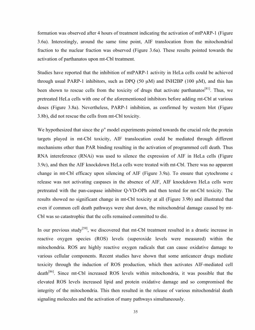
Studies have reported that the inhibition of mtPARP-1 activity in HeLa cells could be achieved
through usual PARP-1 inhibitors, such as DPQ (50 µM) and INH2BP (100 µM), and this has
been shown to rescue cells from the toxicity of drugs that activate parthanatos[81]. Thus, we
pretreated HeLa cells with one of the aforementioned inhibitors before adding mt-Cbl at various
doses (Figure 3.8a). Nevertheless, PARP-1 inhibition, as confirmed by western blot (Figure
3.8b), did not rescue the cells from mt-Cbl toxicity.
We hypothesized that since the ρ° model experiments pointed towards the crucial role the protein
targets played in mt-Cbl toxicity, AIF translocation could be mediated through different
mechanisms other than PAR binding resulting in the activation of programmed cell death. Thus
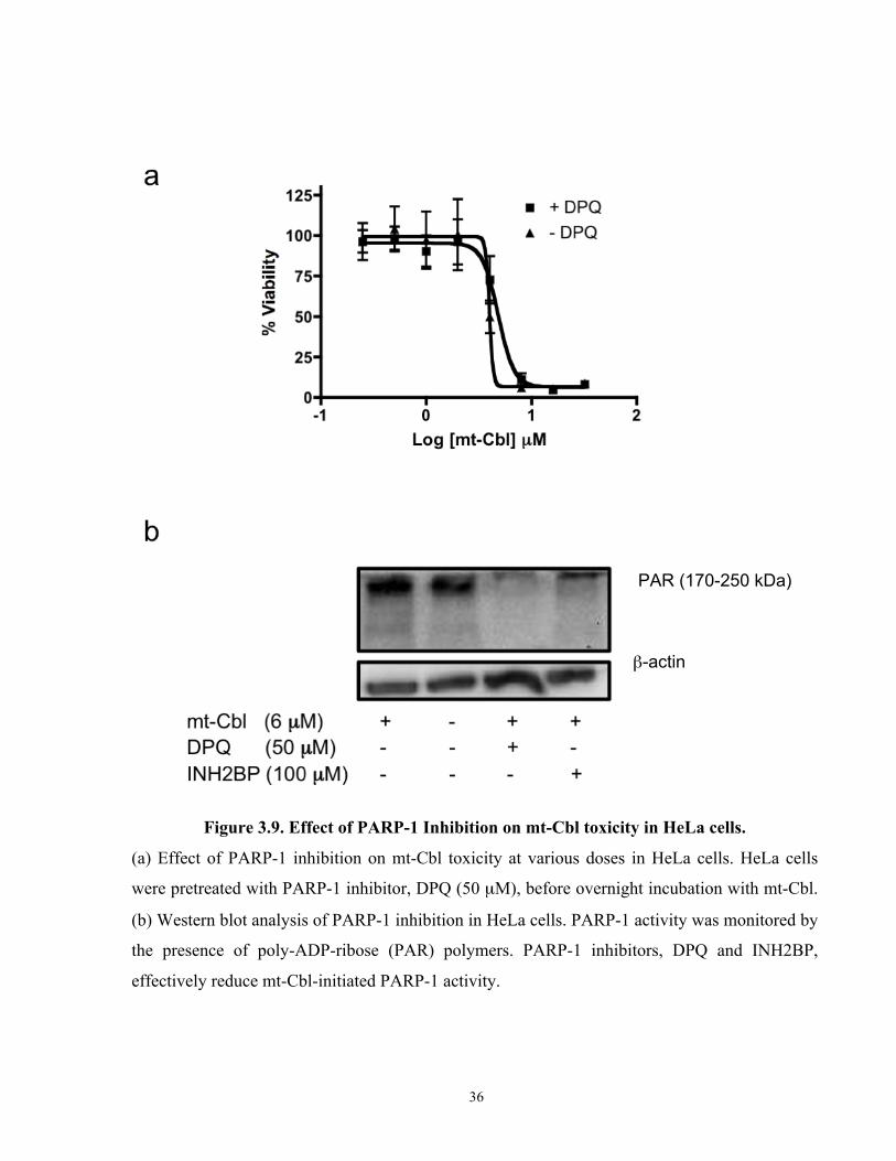
RNA intereference (RNAi) was used to silence the expression of AIF in HeLa cells (Figure
3.9c), and then the AIF knockdown HeLa cells were treated with mt-Cbl. There was no apparent
change in mt-Cbl efficacy upon silencing of AIF (Figure 3.9a). To ensure that cytochrome c
release was not activating caspases in the absence of AIF, AIF knockdown HeLa cells were
pretreated with the pan-caspase inhibitor Q-VD-OPh and then tested for mt-Cbl toxicity. The
results showed no significant change in mt-Cbl toxicity at all (Figure 3.9b) and illustrated that
even if common cell death pathways were shut down, the mitochondrial damage caused by mt-
Cbl was so catastrophic that the cells remained committed to die.
In our previous study[59], we discovered that mt-Cbl treatment resulted in a drastic increase in
reactive oxygen species (ROS) levels (superoxide levels were measured) within the
mitochondria. ROS are highly reactive oxygen radicals that can cause oxidative damage to
various cellular components. Recent studies have shown that some anticancer drugs mediate
toxicity through the induction of ROS production, which then activates AIF-mediated cell
death[86]. Since mt-Cbl increased ROS levels within mitochondria, it was possible that the
elevated ROS levels increased lipid and protein oxidative damage and so compromised the
integrity of the mitochondria. This then resulted in the release of various mitochondrial death
signaling molecules and the activation of many pathways simultaneously.
36
Figure 3.9. Effect of PARP-1 Inhibition on mt-Cbl toxicity in HeLa cells.
(a) Effect of PARP-1 inhibition on mt-Cbl toxicity at various doses in HeLa cells. HeLa cells
were pretreated with PARP-1 inhibitor, DPQ (50 µM), before overnight incubation with mt-Cbl.
(b) Western blot analysis of PARP-1 inhibition in HeLa cells. PARP-1 activity was monitored by
the presence of poly-ADP-ribose (PAR) polymers. PARP-1 inhibitors, DPQ and INH2BP,
effectively reduce mt-Cbl-initiated PARP-1 activity.
β-actin
PAR (170-250 kDa)
37
Figure 3.10. Effect of AIF knockdown on mt-Cbl toxicity in HeLa cells.
(a) Effect of AIF knockdown on mt-Cbl toxicity in HeLa cells. HeLa cells were pretreated with
AIF siRNA for 48 hrs. After which they were treated with mt-Cbl overnight. (b) Effect of AIF
silencing and caspase inhibition on mt-Cbl toxicity in HeLa cells. (c) Western blot analysis of
AIF expression after siRNA pretreatment in HeLa cells. AIF siRNA efficiently reduced AIF
expression by more than 80%.
38
To scavenge the ROS generated through mt-Cbl treatment, HeLa cells were pretreated with the
ROS scavenger n-acetylcysteine (NAC, 10 mM) before adding mt-Cbl. Cell viability assays
indicated that NAC pretreatment did not alter mt-Cbl toxicity (Figure 3.10a). We measured ROS
levels generated after NAC pretreatment using flow cytometry, and the results showed that NAC
pretreatment reduced mt-Cbl-induced ROS levels by over 50 % (Figure 3.10b) indicating that
mt-Cbl-induced toxicity was not fully mediated by ROS damage. However, these experiments do
not rule out the contribution of the elevated ROS levels to mt-Cbl’s cell death mechanism.
3.6 The Necrotic Nature of mt-Cbl
Necrosis is a passive cell death process that has been considered, until recently, as a largely
unregulated mode of cell death characterized by the loss of plasma membrane integrity and a fast
drop in cellular ATP levels. With the discovery of the role of receptor-interacting
serine/threonine-protein kinase 1 (RIPK1) during a certain form of necrosis induced by TNF-α
under conditions of caspase inhibition, a paradigm shift has occurred[87]. Current research efforts
are focused on differentiating between the different kinds of necrosis and on providing a set of
signature molecular events to define each mode[88].
Our inability to rescue cells from mt-Cbl toxicity by inhibiting various programmed cell death
mechanisms prompted us to consider necrosis as a possible mechanism of action. Initially, time
course cell death experiments were performed to determine how fast mt-Cbl kills HeLa cells.
The results revealed that mt-Cbl-induced toxicity was occurring within 1 hour of treatment
resulting in a 50% decrease in viability at EC50 (data not shown). The fast kinetics of mt-Cbl-
induced cell death pointed towards a necrotic mechanism.
Further analysis using an annexin V/sytox red assay revealed that within 90 minutes of treatment,
plasma membrane integrity was lost in treated cells at doses higher than EC50 (Figure 3.11). This
meant that above EC50, necrosis became the dominant mechanism of cell death which explained
why cells could no longer be rescued from mt-Cbl-induced cell death. Conversely, at doses lower
than EC50, the ratio of apoptotic to necrotic cells increased indicating that apoptotic cell death
mechanisms were more dominant at these concentrations. A comparison with untargeted Cbl-
induced cell death revealed that Cbl cell death kinetics were much slower. The onset of Cbl
toxicity occurred around 9 hours after treatment with the exposure of phosphatidylserine on the
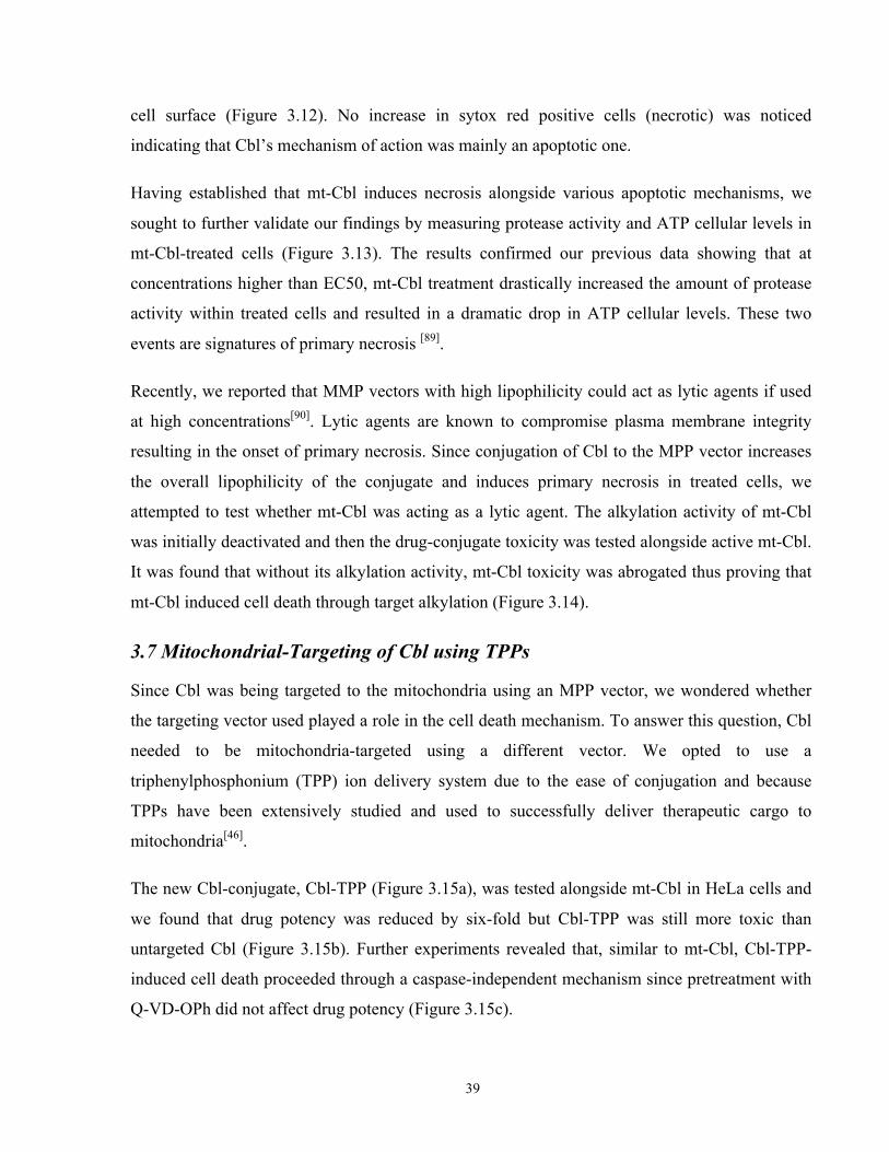
39
cell surface (Figure 3.12). No increase in sytox red positive cells (necrotic) was noticed
indicating that Cbl’s mechanism of action was mainly an apoptotic one.
Having established that mt-Cbl induces necrosis alongside various apoptotic mechanisms, we
sought to further validate our findings by measuring protease activity and ATP cellular levels in
mt-Cbl-treated cells (Figure 3.13). The results confirmed our previous data showing that at
concentrations higher than EC50, mt-Cbl treatment drastically increased the amount of protease
activity within treated cells and resulted in a dramatic drop in ATP cellular levels. These two
events are signatures of primary necrosis [89].
Recently, we reported that MMP vectors with high lipophilicity could act as lytic agents if used
at high concentrations[90]. Lytic agents are known to compromise plasma membrane integrity
resulting in the onset of primary necrosis. Since conjugation of Cbl to the MPP vector increases
the overall lipophilicity of the conjugate and induces primary necrosis in treated cells, we
attempted to test whether mt-Cbl was acting as a lytic agent. The alkylation activity of mt-Cbl
was initially deactivated and then the drug-conjugate toxicity was tested alongside active mt-Cbl.
It was found that without its alkylation activity, mt-Cbl toxicity was abrogated thus proving that
mt-Cbl induced cell death through target alkylation (Figure 3.14).
3.7 Mitochondrial-Targeting of Cbl using TPPs
Since Cbl was being targeted to the mitochondria using an MPP vector, we wondered whether
the targeting vector used played a role in the cell death mechanism. To answer this question, Cbl
needed to be mitochondria-targeted using a different vector. We opted to use a
triphenylphosphonium (TPP) ion delivery system due to the ease of conjugation and because
TPPs have been extensively studied and used to successfully deliver therapeutic cargo to
mitochondria[46].
The new Cbl-conjugate, Cbl-TPP (Figure 3.15a), was tested alongside mt-Cbl in HeLa cells and
we found that drug potency was reduced by six-fold but Cbl-TPP was still more toxic than
untargeted Cbl (Figure 3.15b). Further experiments revealed that, similar to mt-Cbl, Cbl-TPP-
induced cell death proceeded through a caspase-independent mechanism since pretreatment with
Q-VD-OPh did not affect drug potency (Figure 3.15c).
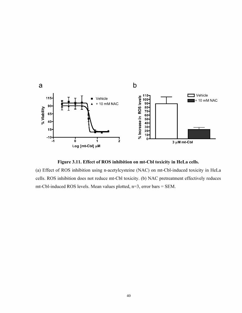
40
Figure 3.11. Effect of ROS inhibition on mt-Cbl toxicity in HeLa cells.
(a) Effect of ROS inhibition using n-acetylcysteine (NAC) on mt-Cbl-induced toxicity in HeLa
cells. ROS inhibition does not reduce mt-Cbl toxicity. (b) NAC pretreatment effectively reduces
mt-Cbl-induced ROS levels. Mean values plotted, n=3, error bars = SEM.
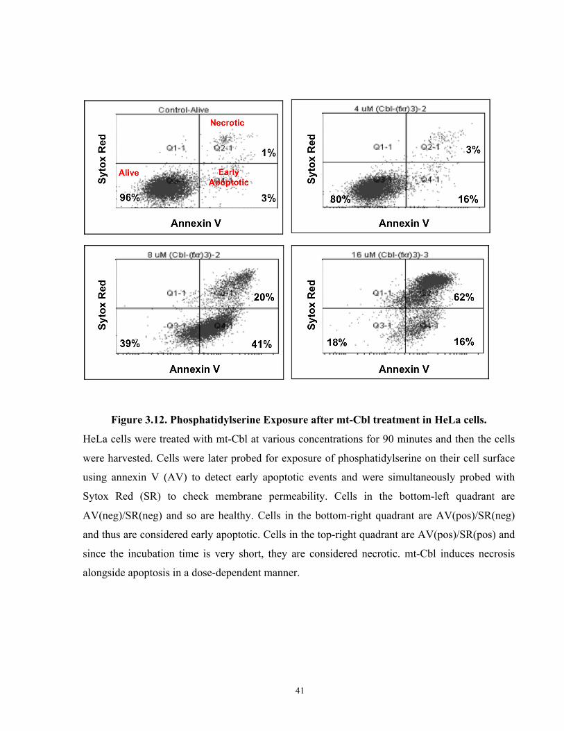
41
Figure 3.12. Phosphatidylserine Exposure after mt-Cbl treatment in HeLa cells.
HeLa cells were treated with mt-Cbl at various concentrations for 90 minutes and then the cells
were harvested. Cells were later probed for exposure of phosphatidylserine on their cell surface
using annexin V (AV) to detect early apoptotic events and were simultaneously probed with
Sytox Red (SR) to check membrane permeability. Cells in the bottom-left quadrant are
AV(neg)/SR(neg) and so are healthy. Cells in the bottom-right quadrant are AV(pos)/SR(neg)
and thus are considered early apoptotic. Cells in the top-right quadrant are AV(pos)/SR(pos) and
since the incubation time is very short, they are considered necrotic. mt-Cbl induces necrosis
alongside apoptosis in a dose-dependent manner.
42
Figure 3.13. Phosphatidylserine Exposure after Cbl treatment in HeLa cells.
Treatment of HeLa cells with Cbl at various concentrations was done for 9 hours and then the
cells were harvested. Cells were later probed for exposure of phosphatidylserine on their cell
surface using annexin V (AV) to detect early apoptotic events and were simultaneously probed
with Sytox Red (SR) to check membrane permeability. Cbl induces apoptosis in a dose-
dependent manner.
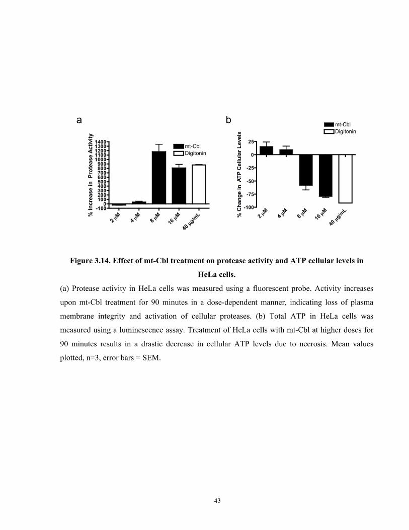
43
Figure 3.14. Effect of mt-Cbl treatment on protease activity and ATP cellular levels in
HeLa cells.
(a) Protease activity in HeLa cells was measured using a fluorescent probe. Activity increases
upon mt-Cbl treatment for 90 minutes in a dose-dependent manner, indicating loss of plasma
membrane integrity and activation of cellular proteases. (b) Total ATP in HeLa cells was
measured using a luminescence assay. Treatment of HeLa cells with mt-Cbl at higher doses for
90 minutes results in a drastic decrease in cellular ATP levels due to necrosis. Mean values
plotted, n=3, error bars = SEM.
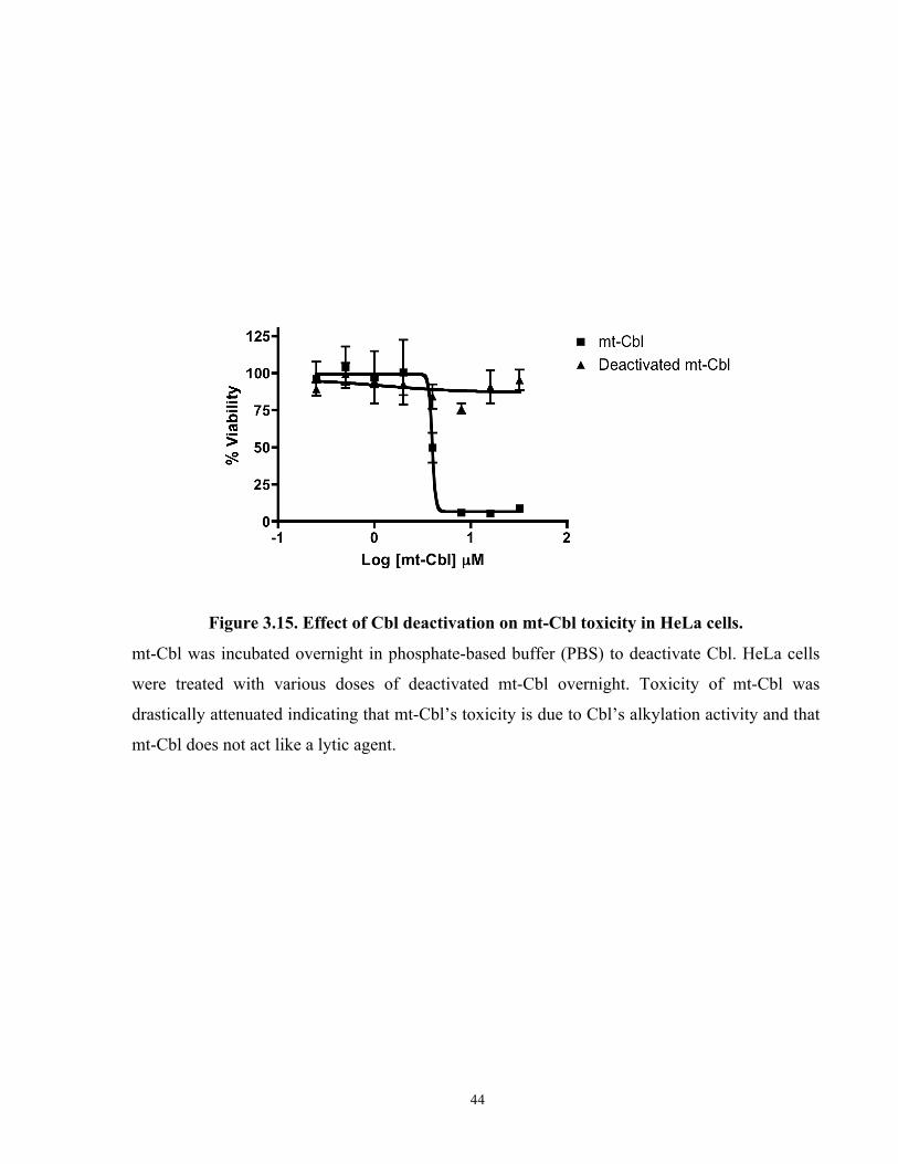
44
Figure 3.15. Effect of Cbl deactivation on mt-Cbl toxicity in HeLa cells.
mt-Cbl was incubated overnight in phosphate-based buffer (PBS) to deactivate Cbl. HeLa cells
were treated with various doses of deactivated mt-Cbl overnight. Toxicity of mt-Cbl was
drastically attenuated indicating that mt-Cbl’s toxicity is due to Cbl’s alkylation activity and that
mt-Cbl does not act like a lytic agent.
45
In our previous study, we noticed that MPP conjugation to Cbl resulted in a two-fold decrease in
the alkylation activity of the drug. Since Cbl-TPP was less potent than mt-Cbl, we tested the
alkylation activity of the new drug-conjugate and compared it to that of Cbl and mt-Cbl (Figure
3.16). The results showed that TPP conjugation also resulted in a two-fold decrease in alkylation
activity. However Cbl-TPP’s alkylation activity was slightly higher than that of mt-Cbl ruling
out that the decrease in drug potency was due to a reduction in the alkylation ability of Cbl.
We then proceeded to testing whether Cbl-TPP formed mtDNA adducts and bound to protein
targets like mt-Cbl. Using immunoblotting, we observed that Cbl-TPP successfully alkylated
both mtDNA and proteins (Figure 3.17). Interestingly, Cbl-TPP seemed to have no preferred
protein target unlike mt-Cbl, which alkylated a certain set of mitochondrial proteins
preferentially. This observation confirmed our previous hypothesis that the MPP vector
conferred protein target selectivity due to its peptide sequence.
Since Cbl-TPP alkylated mitochondrial proteins with no specificity, we sought to test if this
meant that the mtDNA lesions played a more important role in the Cbl-TPP-induced cell death
mechanism. Cbl-TPP was tested in a ρ° model and the results clearly showed that was not the
case (Figure 3.18). The presence or absence of mtDNA was discovered to have little effect on
Cbl-TPP toxicity. These results firmly illustrated that by rerouting Cbl to the mitochondria, the
onset of extensive mitochondrial protein damage causes severe cellular stress and activates cell
death.
Finally we tested whether Cbl-TPP also induced a necrotic form of cell death. It was found that
upon Cbl-TPP treatment at concentrations higher than EC50, protease activity was drastically
increased (Figure 3.19a) and a dramatic drop in ATP cellular levels occurred (Figure 3.19b)
confirming the onset of primary necrosis.
Overall, these results confirmed that the cell death mechanism of mitochondria-targeted Cbl is
not dependent on mtDNA damage . We also found that the targeting vector of mitochondria-
targeted Cbl has no effect on the cell death pathway being activated. The shift in potency seen
with Cbl-TPP could be attributed to differences in cellular uptake, mitochondrial targeting
specificity, and protein target specificity.
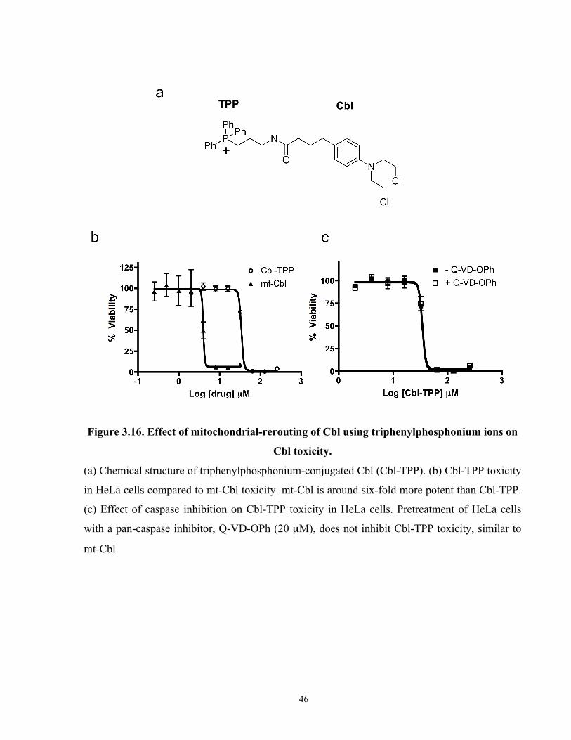
46
Figure 3.16. Effect of mitochondrial-rerouting of Cbl using triphenylphosphonium ions on
Cbl toxicity.
(a) Chemical structure of triphenylphosphonium-conjugated Cbl (Cbl-TPP). (b) Cbl-TPP toxicity
in HeLa cells compared to mt-Cbl toxicity. mt-Cbl is around six-fold more potent than Cbl-TPP.
(c) Effect of caspase inhibition on Cbl-TPP toxicity in HeLa cells. Pretreatment of HeLa cells
with a pan-caspase inhibitor, Q-VD-OPh (20 µM), does not inhibit Cbl-TPP toxicity, similar to
mt-Cbl.
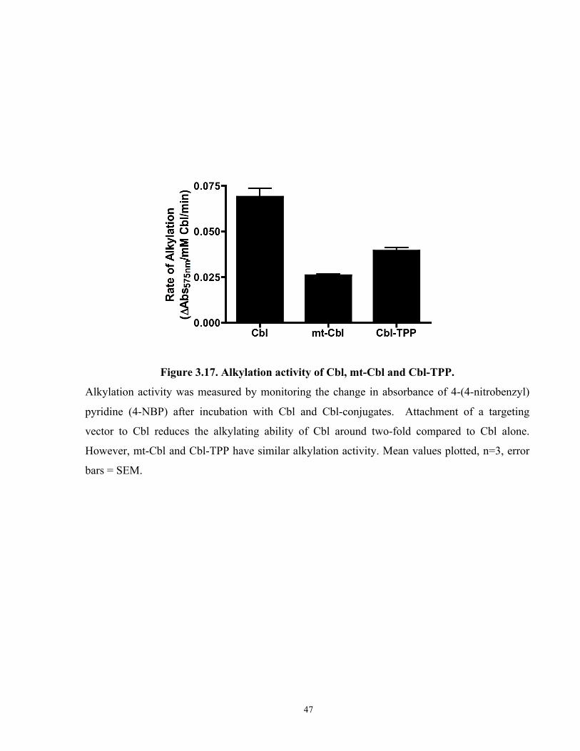
47
Figure 3.17. Alkylation activity of Cbl, mt-Cbl and Cbl-TPP.
Alkylation activity was measured by monitoring the change in absorbance of 4-(4-nitrobenzyl)
pyridine (4-NBP) after incubation with Cbl and Cbl-conjugates. Attachment of a targeting
vector to Cbl reduces the alkylating ability of Cbl around two-fold compared to Cbl alone.
However, mt-Cbl and Cbl-TPP have similar alkylation activity. Mean values plotted, n=3, error
bars = SEM.
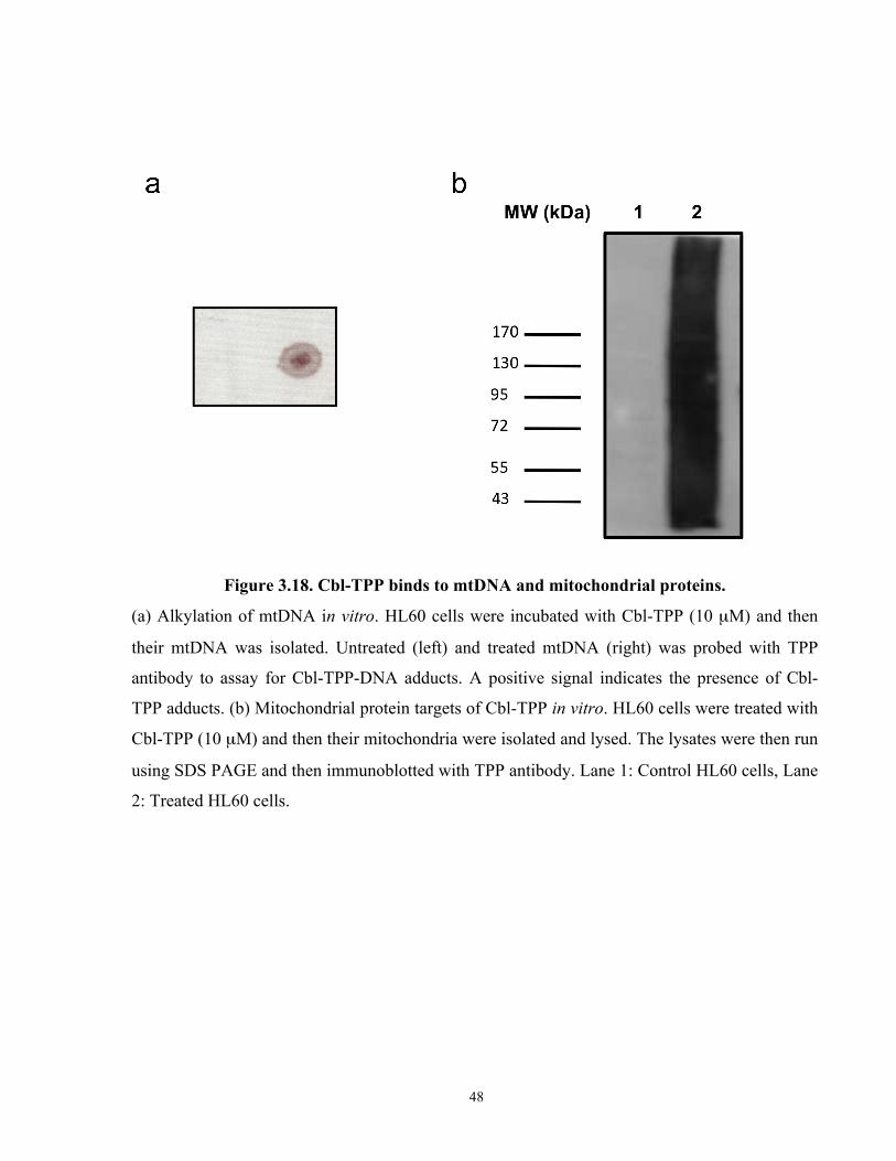
48
Figure 3.18. Cbl-TPP binds to mtDNA and mitochondrial proteins.
(a) Alkylation of mtDNA in vitro. HL60 cells were incubated with Cbl-TPP (10 µM) and then
their mtDNA was isolated. Untreated (left) and treated mtDNA (right) was probed with TPP
antibody to assay for Cbl-TPP-DNA adducts. A positive signal indicates the presence of Cbl-
TPP adducts. (b) Mitochondrial protein targets of Cbl-TPP in vitro. HL60 cells were treated with
Cbl-TPP (10 µM) and then their mitochondria were isolated and lysed. The lysates were then run
using SDS PAGE and then immunoblotted with TPP antibody. Lane 1: Control HL60 cells, Lane
2: Treated HL60 cells.
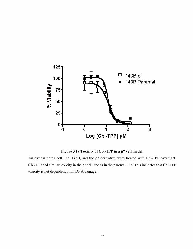
49
Figure 3.19 Toxicity of Cbl-TPP in a ρ° cell model.
An osteosarcoma cell line, 143B, and the ρ° derivative were treated with Cbl-TPP overnight.
Cbl-TPP had similar toxicity in the ρ° cell line as in the parental line. This indicates that Cbl-TPP
toxicity is not dependent on mtDNA damage.
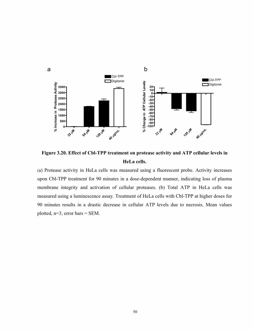
50
Figure 3.20. Effect of Cbl-TPP treatment on protease activity and ATP cellular levels in
HeLa cells.
(a) Protease activity in HeLa cells was measured using a fluorescent probe. Activity increases
upon Cbl-TPP treatment for 90 minutes in a dose-dependent manner, indicating loss of plasma
membrane integrity and activation of cellular proteases. (b) Total ATP in HeLa cells was
measured using a luminescence assay. Treatment of HeLa cells with Cbl-TPP at higher doses for
90 minutes results in a drastic decrease in cellular ATP levels due to necrosis. Mean values
plotted, n=3, error bars = SEM.

51
4. Chapter 4: Conclusion and Outlook
52
In summary, the cell death mechanism of mt-Cbl was studied using a cervical carcinoma cell
model. Our results support our initial hypothesis that by rerouting Cbl from its usual nuclear
target to the mitochondrion, novel cell death pathways are activated that are different from the
cell death mechanism of untargeted Cbl.
Our experiments revealed that mt-Cbl induces two kinds of damage within mitochondria,
mtDNA lesions and protein damage. Surprisingly, we found that the mitochondrial protein
damage induced by mt-Cbl was the main source of cellular stress that triggers the initiation of
various cell death pathways. A possible explanation is that since the mitochondrion is a tightly
regulated organelle, the rapid damage of its protein components, especially the proteins that are
within the IMM, compromises the integrity of the IMM leading to the dysregulation of various
mitochondrial processes and the overproduction of ROS. The loss of IMM integrity results in the
leakage of various cell death signaling molecules, like cytochrome c and AIF, and thus activates
several cell death pathways.
Though mt-Cbl was found to also activate various levels of necrosis at different concentrations,
this property should not exclude it from being used as an anticancer therapeutic. With the advent
of cancer immunotherapy, researchers are currently looking into harnessing the inflammatory
properties of necrosis to induce an immune response within a targeted tumor[91]. Mounting an
immune attack against a tumor would enhance drug efficacy and would help eliminate the tumor
with fewer drug doses.
In terms of cancer drug resistance, the necrotic nature of mt-Cbl-induced cell death should be
viewed as an asset. The majority of cancer drug resistance pathways block apoptotic cell death
induced by current chemotherapeutics. However, no resistance pathway has been discovered, as
of yet, that can block a necrotic signal. Recent studies have shown that by taking advantage of
necrosis, apoptosis-resistant tumor cells could be eliminated[92-94].
Drug toxicity is an important issue to consider for further development of mt-Cbl. It has been
shown that certain chemotherapeutics, such as adriamycin, that damage mitochondria can cause
cardiotoxicity[95-96]. Since mt-Cbl induces its toxicity through mitochondrial damage, we plan to
test the toxicity of this novel compound by treating mice at therapeutic doses for several weeks
and then isolating their cardiac mitochondria and examining their structure and function.
53
Since this study has revealed that mt-Cbl has certain preferred protein targets, future efforts will
involve isolating mt-Cbl's mitochondrial protein targets and studying their role in triggering cell
death. If a certain target is found to preferentially activate novel caspase-independent cell death,
then further drug development could generate a third-generation mt-Cbl that acts as a potent
mitochondrial protein inhibitor. Future work will also involve rerouting various anticancer
agents, such as cisplatin and temozolomide, and studying the effects of rerouting on drug
potency.
The implications of this study go beyond cancer therapy. With the emerging evidence linking
mitochondrial dysfunction to type 2 diabetes[97-98] and the role that mitochondria play in
neurodegenerative diseases such as Parkinson’s[99-100], the need for mitochondria-targeted drugs
is rapidly rising. Thus the understanding and optimization of mitochondria-targeted small
molecules is crucial for fast tracking the development of much-needed mitochondrial
therapeutics. Aside from their potential use in therapy, mitochondria-targeted compounds could
also be employed as probes to understand important biological functions. For instance,
mitochondria-targeted DNA damaging agents could be used to probe for novel mtDNA repair
pathways.
On the whole, this study has confirmed that rerouting anticancer drugs to the mitochondrion for
the sake of repurposing ‘FDA-approved’ drugs for the treatment of drug-resistant cancers is a
viable strategy for drug development. This strategy allows the activation of multiple cell death
pathways that overwhelms cancer drug resistance pathways and pushes cancer cells to the point
of no return.

54
6. Chapter 6: References
55
[1] A. Kamb, S. Wee, C. Lengauer, Nature Reviews Drug Discovery 2007, 6, 115-120.
[2] L. Hutchinson, R. Kirk, Nature Reviews Clinical Oncology 2011, 8, 189-190.
[3] A. Marusyk, V. Almendro, K. Polyak, Nat Rev Cancer 2012, 12, 323-334.
[4] A. Kamb, Nature Reviews Drug Discovery 2005, 4, 161-165.
[5] E. Iorns, C. J. Lord, N. Turner, A. Ashworth, Nature Reviews Drug Discovery 2007, 6, 556-568.
[6] T. Lahaye, B. Riehm, U. Berger, P. Paschka, M. C. Muller, S. Kreil, K. Merx, U. Schwindel, C. Schoch, R. Hehlmann, A. Hochhaus, Cancer 2005, 103, 1659-1669.
[7] H. Kantarjian, C. Sawyers, A. Hochhaus, F. Guilhot, C. Schiffer, C. Gambacorti-Passerini, D. Niederwieser, D. Resta, R. Capdeville, U. Zoellner, M. Talpaz, B. Druker, I. S. C. S. Grp, New England Journal of Medicine 2002, 346, 645-652.
[8] R. Sullivan, G. C. Pare, L. J. Frederiksen, G. L. Semenza, C. H. Graham, Molecular Cancer Therapeutics 2008, 7, 1961-1973.
[9] C. H. Yun, K. E. Mengwasser, A. V. Toms, M. S. Woo, H. Greulich, K. K. Wong, M. Meyerson, M. J. Eck, Proc Natl Acad Sci U S A 2008, 105, 2070-2075.
[10] F. Heidel, F. K. Solem, F. Breitenbuecher, D. B. Lipka, S. Kasper, M. H. Thiede, C. Brandts, H. Serve, J. Roesel, F. Giles, E. Feldman, G. Ehninger, G. J. Schiller, S. Nimer, R. M. Stone, Y. F. Wang, T. Kindler, P. S. Cohen, C. Huber, T. Fischer, Blood 2006, 107, 293-300.
[11] Y. Nefedova, T. H. Landowski, W. S. Dalton, Leukemia 2003, 17, 1175-1182.
[12] P. M. Voorhees, Q. Chen, D. J. Kuhn, G. W. Small, S. A. Hunsucker, J. S. Strader, R. E. Corringham, M. H. Zaki, J. A. Nemeth, R. Z. Orlowski, Clinical Cancer Research 2007, 13, 6469-6478.
[13] M. B. Meads, R. A. Gatenby, W. S. Dalton, Nature Reviews Cancer 2009, 9, 665-A674.
[14] P. Dalerba, T. Kalisky, D. Sahoo, P. S. Rajendran, M. E. Rothenberg, A. A. Leyrat, S. Sim, J. Okamoto, D. M. Johnston, D. L. Qian, M. Zabala, J. Bueno, N. F. Neff, J. B. Wang, A. A. Shelton, B. Visser, S. Hisamori, Y. Shimono, M. van de Wetering, H. Clevers, M. F. Clarke, S. R. Quake, Nature Biotechnology 2011, 29, 1120-U1111.
[15] K. K. Frese, D. A. Tuveson, Nature Reviews Cancer 2007, 7, 645-658.
[16] O. J. Becher, E. C. Holland, Cancer Research 2006, 66, 5526-5526.
[17] N. Cook, D. I. Jodrell, D. K. Tuveson, Drug Discovery Today 2012, 17, 253-260.
56
[18] H. Sikder, D. L. Huso, H. Zhang, B. H. Wang, B. Ryu, S. T. Hwang, J. D. Powell, R. M. Alani, Cancer Cell 2003, 4, 291-299.
[19] J. Jonkers, A. Berns, Nature Reviews Cancer 2002, 2, 251-265.
[20] E. M. Rego, D. Ruggero, C. Tribioli, G. Cattoretti, S. Kogan, R. L. Redner, P. P. Pandolfi, Oncogene 2006, 25, 1974-1979.
[21] E. M. Rego, L. Z. He, R. P. Warrell, Z. G. Wang, P. P. Pandolfi, Proceedings of the National Academy of Sciences of the United States of America 2000, 97, 10173-10178.
[22] G. L. Verdine, L. D. Walensky, Clinical Cancer Research 2007, 13, 7264-7270.
[23] I. Collins, P. Workman, Nature Chemical Biology 2006, 2, 689-700.
[24] P. J. Hajduk, J. Greer, Nature Reviews Drug Discovery 2007, 6, 211-219.
[25] T. Oltersdorf, S. W. Elmore, A. R. Shoemaker, R. C. Armstrong, D. J. Augeri, B. A. Belli, M. Bruncko, T. L. Deckwerth, J. Dinges, P. J. Hajduk, M. K. Joseph, S. Kitada, S. J. Korsmeyer, A. R. Kunzer, A. Letai, C. Li, M. J. Mitten, D. G. Nettesheim, S. Ng, P. M. Nimmer, J. M. O'Connor, A. Oleksijew, A. M. Petros, J. C. Reed, W. Shen, S. K. Tahir, C. B. Thompson, K. J. Tomaselli, B. L. Wang, M. D. Wendt, H. C. Zhang, S. W. Fesik, S. H. Rosenberg, Nature 2005, 435, 677-681.
[26] N. N. Danial, Clinical Cancer Research 2007, 13, 7254-7263.
[27] S. Fulda, L. Galluzzi, G. Kroemer, Nat Rev Drug Discov 2010, 9, 447-464.
[28] T. Althoff, D. J. Mills, J. L. Popot, W. Kuhlbrandt, EMBO J 2011.
[29] Y. P. Ow, D. R. Green, Z. Hao, T. W. Mak, Nat Rev Mol Cell Biol 2008, 9, 532-542.
[30] K. K. Singh, L. Costello, Mitochondria and cancer, Springer Verlag, 2009.
[31] S. J. Ralph, S. Rodriguez-Enriquez, J. Neuzil, E. Saavedra, R. Moreno-Sanchez, Mol Aspects Med 2010, 31, 145-170.
[32] R. W. Taylor, D. M. Turnbull, Nat Rev Genet 2005, 6, 389-402.
[33] T. N. Chonghaile, A. Letai, Oncogene 2008, 27 Suppl 1, S149-157.
[34] V. Labi, F. Grespi, F. Baumgartner, A. Villunger, Cell Death Differ 2008, 15, 977-987.
[35] N. N. Danial, Clin Cancer Res 2007, 13, 7254-7263.
[36] R. H. Xu, H. Pelicano, Y. Zhou, J. S. Carew, L. Feng, K. N. Bhalla, M. J. Keating, P. Huang, Cancer Res 2005, 65, 613-621.
[37] V. Gogvadze, S. Orrenius, B. Zhivotovsky, Trends Cell Biol 2008, 18, 165-173.
57
[38] R. Rossignol, R. Gilkerson, R. Aggeler, K. Yamagata, S. J. Remington, R. A. Capaldi, Cancer Res 2004, 64, 985-993.
[39] G. Kroemer, J. Pouyssegur, Cancer Cell 2008, 13, 472-482.
[40] E. Pisha, H. Chai, I. S. Lee, T. E. Chagwedera, N. R. Farnsworth, G. A. Cordell, C. W. Beecher, H. H. Fong, A. D. Kinghorn, D. M. Brown, et al., Nat Med 1995, 1, 1046-1051.
[41] S. Fulda, K. M. Debatin, Med Pediatr Oncol 2000, 35, 616-618.
[42] R. D. Meng, W. S. El-Deiry, Exp Cell Res 2001, 262, 154-169.
[43] V. Zuco, R. Supino, S. C. Righetti, L. Cleris, E. Marchesi, C. Gambacorti-Passerini, F. Formelli, Cancer Lett 2002, 175, 17-25.
[44] S. Fulda, G. Kroemer, Drug Discov Today 2009, 14, 885-890.
[45] L. F. Yousif, K. M. Stewart, S. O. Kelley, Chembiochem 2009, 10, 1939-1950.
[46] R. A. Smith, R. C. Hartley, M. P. Murphy, Antioxid Redox Signal 2011.
[47] A. Manetta, G. Gamboa, A. Nasseri, Y. D. Podnos, D. Emma, G. Dorion, L. Rawlings, P. M. Carpenter, A. Bustamante, J. Patel, D. Rideout, Gynecol Oncol 1996, 60, 203-212.
[48] A. M. James, H. M. Cocheme, M. P. Murphy, Mech Ageing Dev 2005, 126, 982-986.
[49] G. F. Kelso, C. M. Porteous, C. V. Coulter, G. Hughes, W. K. Porteous, E. C. Ledgerwood, R. A. Smith, M. P. Murphy, J Biol Chem 2001, 276, 4588-4596.
[50] R. A. Smith, G. F. Kelso, A. M. James, M. P. Murphy, Methods Enzymol 2004, 382, 45-67.
[51] S. V. Boddapati, P. Tongcharoensirikul, R. N. Hanson, G. G. D'Souza, V. P. Torchilin, V. Weissig, J Liposome Res 2005, 15, 49-58.
[52] N. R. Patel, S. Hatziantoniou, A. Georgopoulos, C. Demetzos, V. P. Torchilin, V. Weissig, G. G. D'Souza, J Liposome Res 2010, 20, 244-249.
[53] S. Biswas, N. S. Dodwadkar, R. R. Sawant, A. Koshkaryev, V. P. Torchilin, J Drug Target 2011, 19, 552-561.
[54] Y. Yamada, H. Akita, H. Kamiya, K. Kogure, T. Yamamoto, Y. Shinohara, K. Yamashita, H. Kobayashi, H. Kikuchi, H. Harashima, Biochim Biophys Acta 2008, 1778, 423-432.
[55] Y. Yasuzaki, Y. Yamada, H. Harashima, Biochem Biophys Res Commun 2010, 397, 181-186.
[56] K. Zhao, G. M. Zhao, D. Wu, Y. Soong, A. V. Birk, P. W. Schiller, H. H. Szeto, J Biol Chem 2004, 279, 34682-34690.
58
[57] H. H. Szeto, P. W. Schiller, Pharm Res 2011, 28, 2669-2679.
[58] K. L. Horton, K. M. Stewart, S. B. Fonseca, Q. Guo, S. O. Kelley, Chem Biol 2008, 15, 375-382.
[59] S. B. Fonseca, M. P. Pereira, R. Mourtada, M. Gronda, K. L. Horton, R. Hurren, M. D. Minden, A. D. Schimmer, S. O. Kelley, Chem Biol 2011, 18, 445-453.
[60] B. G. Heerdt, M. A. Houston, L. H. Augenlicht, Cancer Research 2005, 65, 9861-9867.
[61] M. G. Tan, L. L. Ooi, S. E. Aw, K. M. Hui, Journal of Biological Chemistry 2004, 279, 45235-45244.
[62] L. F. Yousif, K. M. Stewart, K. L. Horton, S. O. Kelley, Chembiochem 2009, 10, 2081-2088.
[63] S. O. Kelley, K. M. Stewart, R. Mourtada, Pharm Res 2011, 28, 2808-2819.
[64] M. P. Pereira, S. O. Kelley, J Am Chem Soc 2011, 133, 3260-3263.
[65] J. K. Horton, D. K. Srivastava, B. Z. Zmudzka, S. H. Wilson, Nucleic Acids Res 1995, 23, 3810-3815.
[66] C. Stecca, G. B. Gerber, Biochem Pharmacol 1998, 55, 941-951.
[67] D. Michod, C. Widmann, Crit Rev Oncol Hematol 2007, 63, 160-171.
[68] F. M. Yakes, B. Van Houten, Proc Natl Acad Sci U S A 1997, 94, 514-519.
[69] W. Z. Yang, A. Begleiter, J. B. Johnston, L. G. Israels, M. R. Mowat, Mol Pharmacol 1992, 41, 625-630.
[70] C. Pepper, A. Thomas, T. Hoy, P. Bentley, Br J Haematol 1999, 104, 581-588.
[71] I. Samudio, M. Fiegl, M. Andreeff, Cancer Res 2009, 69, 2163-2166.
[72] J. R. Carreon, K. M. Stewart, K. P. Mahon, Jr., S. Shin, S. O. Kelley, Bioorg Med Chem Lett 2007, 17, 5182-5185.
[73] G. Villani, G. Attardi, Proc Natl Acad Sci U S A 1997, 94, 1166-1171.
[74] S. Elmore, Toxicologic Pathology 2007, 35, 495-516.
[75] S. W. G. Tait, D. R. Green, Nature Reviews Molecular Cell Biology 2010, 11, 621-632.
[76] Y. L. P. Ow, D. R. Green, Z. Hao, T. W. Mak, Nature Reviews Molecular Cell Biology 2008, 9, 532-542.
[77] M. Rouleau, A. Patel, M. J. Hendzel, S. H. Kaufmann, G. G. Poirier, Nat Rev Cancer 2010, 10, 293-301.
59
[78] B. W. Durkacz, O. Omidiji, D. A. Gray, S. Shall, Nature 1980, 283, 593-596.
[79] S. Shall, G. de Murcia, Mutation Research-DNA Repair 2000, 460, 1-15.
[80] S. J. Berger, D. C. Sudar, N. A. Berger, Biochem Biophys Res Commun 1986, 134, 227-232.
[81] S. W. Yu, H. Wang, M. F. Poitras, C. Coombs, W. J. Bowers, H. J. Federoff, G. G. Poirier, T. M. Dawson, V. L. Dawson, Science 2002, 297, 259-263.
[82] Y. F. Wang, N. S. Kim, J. F. Haince, H. C. Kang, K. K. David, S. A. Andrabi, G. G. Poirier, V. L. Dawson, T. M. Dawson, Sci Signal 2011, 4.
[83] L. O. Burzio, L. Saez, R. Cornejo, Biochem Biophys Res Commun 1981, 103, 369-375.
[84] E. Pankotai, Z. Lacza, M. Muranyi, C. Szabo, Mitochondrion 2009, 9, 159-164.
[85] M. N. Rossi, M. Carbone, C. Mostocotto, C. Mancone, M. Tripodi, R. Maione, P. Amati, Journal of Biological Chemistry 2009, 284, 31616-31624.
[86] D. Trachootham, J. Alexandre, P. Huang, Nature Reviews Drug Discovery 2009, 8, 579-591.
[87] L. Galluzzi, G. Kroemer, Cell 2008, 135, 1161-1163.
[88] T. Vanden Berghe, N. Vanlangenakker, E. Parthoens, W. Deckers, M. Devos, N. Festjens, C. J. Guerin, U. T. Brunk, W. Declercq, P. Vandenabeele, Cell Death Differ 2010, 17, 922-930.
[89] A. L. Niles, R. A. Moravec, P. Eric Hesselberth, M. A. Scurria, W. J. Daily, T. L. Riss, Anal Biochem 2007, 366, 197-206.
[90] K. L. Horton, M. P. Pereira, K. M. Stewart, S. B. Fonseca, S. O. Kelley, Chembiochem 2012, 13, 476-485.
[91] R. K. Amaravadi, C. B. Thompson, Clinical Cancer Research 2007, 13, 7271-7279.
[92] G. Guidicelli, B. Chaigne-Delalande, M. S. Dilhuydy, B. Pinson, W. Mahfouf, J. M. Pasquet, F. X. Mahon, P. Pourquier, J. F. Moreau, P. Legembre, PLoS One 2009, 4, e5493.
[93] M. Naito, C. Hashimoto, S. Masui, T. Tsuruo, Cancer Sci 2004, 95, 361-366.
[94] X. Hu, Y. Xuan, Cancer Lett 2008, 259, 127-137.
[95] L. Chaiswing, M. P. Cole, D. K. St Clair, W. Ittarat, L. I. Szweda, T. D. Oberley, Toxicol Pathol 2004, 32, 536-547.
[96] S. Zhou, A. Starkov, M. K. Froberg, R. L. Leino, K. B. Wallace, Cancer Res 2001, 61, 771-777.

60
[97] B. B. Lowell, G. I. Shulman, Science 2005, 307, 384-387.
[98] M. E. Patti, S. Corvera, Endocr Rev 2010, 31, 364-395.
[99] N. Exner, A. K. Lutz, C. Haass, K. F. Winklhofer, EMBO J 2012, 31, 3038-3062.
[100] M. Filosto, M. Scarpelli, M. S. Cotelli, V. Vielmi, A. Todeschini, V. Gregorelli, P. Tonin, G. Tomelleri, A. Padovani, J Neurol 2011, 258, 1763-1774.