a journey down the canal: radiological assessment of spinal
TRANSCRIPT

A Journey Down The CanalA Journey Down The Canal
Radiological Assessment of Radiological Assessment of Spinal Cord MassesSpinal Cord Masses
John BerryJohn Berry--CandelarioCandelario HMS IIIHMS IIIGillian Lieberman, MD BIDMCGillian Lieberman, MD BIDMC

Objectives
Patient reviewPatient review
Anatomy of the spineAnatomy of the spine
Imaging techniquesImaging techniques
Classification of spinal massesClassification of spinal masses
Patients revisited!Patients revisited!

Let’s meet our 2 patients

Patient #1 – History and presentationHD is a 67 year old female who presents with HD is a 67 year old female who presents with
lower extremity weakness for the past six lower extremity weakness for the past six months It worsened in the last two months months It worsened in the last two months resulting in frequent nonresulting in frequent non--traumatic falls. traumatic falls.
Pertinent positive is urinary urgency.Pertinent positive is urinary urgency.PMH includes previous back surgery of unknown PMH includes previous back surgery of unknown
reasons.reasons.Received Received myelogrammyelogram, however results unavailable., however results unavailable.PE remarkable for decreased sensation from T7 PE remarkable for decreased sensation from T7
down.down.
Patient #2 – History and presentationCR is a 40 year old male who reported CR is a 40 year old male who reported
atypical leg soreness after riding of an allatypical leg soreness after riding of an all-- terrain vehicle. Soreness was greater than terrain vehicle. Soreness was greater than after similar episodes of riding. after similar episodes of riding. Progressive weakness developed in the Progressive weakness developed in the right leg.right leg.
Pertinent negatives are no difficulty with Pertinent negatives are no difficulty with bowel or bladder function.bowel or bladder function.
PMH and PE unremarkable.PMH and PE unremarkable.

Indications for imaging the spineIndications for imaging the spine
Increased weakness in the lower Increased weakness in the lower extremitiesextremities
Changes in bowel or bladder functionChanges in bowel or bladder function
Saddle anesthesia Saddle anesthesia –– Sensory loss Sensory loss occurring over the buttocks, posterioroccurring over the buttocks, posterior-- superior thigh and superior thigh and perianalperianal regionregion

Imaging TechniquesImaging Techniques

Imaging Modalities of the SpineImaging Modalities of the SpineIMAGING IMAGING MODALITYMODALITY
ADVANTAGESADVANTAGES DISADVANTAGESDISADVANTAGES
Plain filmPlain film Modest visualization of the Modest visualization of the vertebrae, low cost, fastvertebrae, low cost, fast
No clarity of the fluids and soft No clarity of the fluids and soft tissue, radiation exposuretissue, radiation exposure
CTCT Clear visualization of bony Clear visualization of bony structures, some visualization of structures, some visualization of soft tissue structuressoft tissue structures
Limited visualization of soft tissue, Limited visualization of soft tissue, radiation exposure,radiation exposure,
MRIMRI Best modality for soft tissue Best modality for soft tissue visualization, no radiation visualization, no radiation exposure. Test of choice for exposure. Test of choice for lower extremity weakness and lower extremity weakness and bowel/bladder dysfunction.bowel/bladder dysfunction.
Difficult to evaluate cortical bone Difficult to evaluate cortical bone and calcifications, expensive and calcifications, expensive relative to other modalitiesrelative to other modalities
CT CT MyelographyMyelography
Used when MRI is Used when MRI is contraindicated, allows contraindicated, allows visualization of spinal cord and visualization of spinal cord and nerve roots, can evaluate for nerve roots, can evaluate for lesions within the spinal canallesions within the spinal canal
Invasive, involves the injection of Invasive, involves the injection of contrast into the contrast into the thecalthecal sacsac

LetLet’’s review spine s review spine anatomy!anatomy!

Anatomy Review: Basics 31 Pairs of Spinal Nerves
8 cervical12 thoracic5 lumbar5 sacral1 coccygeal
Conus tapers at ~L1/2
Cauda equina falls freely at this level

Anatomy Review: Vertebrae and Spinal nerves
http://www.jefferson.edu/neurosurgery/documents/Spinal%20Cord%20Tumors.pdfhttp://www.backpain-guide.com/Chapter_Fig_folders/Ch05_Anatomy_Folder/
Ch5_Images/05-4_Overall_Spine.jpg
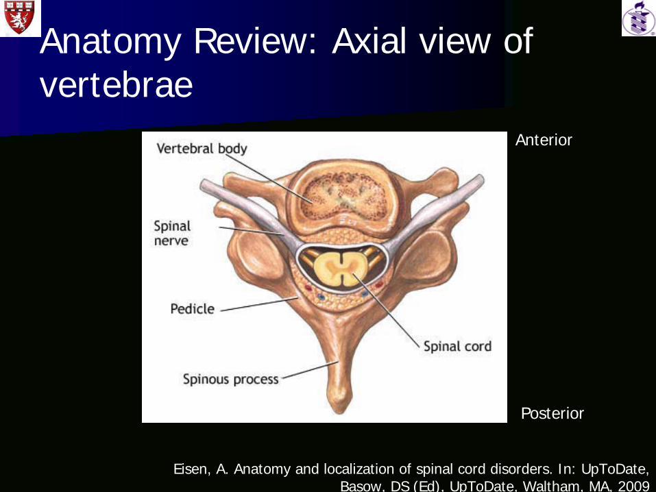
Anatomy Review: Axial view of vertebrae
Eisen, A. Anatomy and localization of spinal cord disorders. In: UpToDate, Basow, DS (Ed), UpToDate, Waltham, MA, 2009
Anterior
Posterior

Anatomy Review: Cross section of Anatomy Review: Cross section of spinal cord spinal cord –– Spinal nervesSpinal nerves
Eisen, A. Anatomy and localization of spinal cord disorders. In: UpToDate, Basow, DS (Ed), UpToDate, Waltham, MA, 2009
Anterior
Posterior

Anatomy Review: Cross section of spinal cord – White matter tracts
Eisen, A. Anatomy and localization of spinal cord disorders. In: UpToDate, Basow DS (Ed) UpToDate Waltham MA 2009
Posterior
Anterior

Spinal Cord MassesSpinal Cord Masses

Spinal Cord MassesAnatomic Classification
1. Extradural
Arise outside the dura
From the osseous spine, intervertebral disc and adjacent soft tissue
Relevant imaging findings: Focal, exophytic mass displacing the thecal sac
2. Intradural extramedullary
Arise inside the dura but outside the spinal cord
DISPLACES THE SPINAL CORD
3. Intradural intramedullary
Arise from the spinal cord
Most common lesions are malignant neoplasms in the form of glioma

IntraspinalIntraspinal MassesMasses
IntramedullaryIntradural
Extramedullary Extradural
http://www.mayoclinic.org/spinal-cord-tumors/types.html
Key:
3a-Develops outside spinal cord but potentially compresses nerves and blood vessels
3b-Invades from the bone. Typical of metastatic tumors
3c-Develops along soft tissue between the vertebrae.

Spinal Cord Masses Spinal Cord Masses -- SubcategoriesSubcategories
BenignBenign
Cystic (and other Cystic (and other tumorliketumorlike masses)masses)
MalignantMalignant
MetastasesMetastases

LetLet’’s identify a list of s identify a list of specific masses in these specific masses in these
subcategories.subcategories.

Spinal TumorsSpinal TumorsExtraduralExtradural IntraduralIntradural
ExtramedullaryExtramedullaryIntraduralIntraduralIntramedullaryIntramedullary
Benign HemangiomaHemangiomaOsteoidOsteoid osteomaosteomaOsteochondromaOsteochondroma
MeningiomaMeningiomaNerve sheath tumorsNerve sheath tumors
RareRare
Cystic Synovial cystSynovial cystArachnoidArachnoid cystcystEosinophilicEosinophilic granulomagranuloma
EpidermoidEpidermoidDermoidDermoid
Multiple SclerosisMultiple SclerosisSyringomyeliaSyringomyeliaTransverse Transverse myelitismyelitis
Malignant ChordomaChordomaLymphomaLymphomaSarcomaSarcoma
RareRare EpendymomaEpendymomaAstrocytomaAstrocytomaHemangioblastomaHemangioblastoma
Metastases BreastBreastLungLungProstateProstate
Seeding from the CNSSeeding from the CNS BreastBreastLungLungLymphomaLymphomaLeukemiaLeukemia

ExtraduralExtradural
Benign
HemangiomaHemangioma
OsteoidOsteoid osteomaosteoma
OsteochondromaOsteochondroma

PACS, BIDMC
Companion Patient #1: Hemangioma on CT
CT with IV and oral contrast – Revealed a mass with low attenuation. Further work up only necessary if the patient was symptomatic.

Cysts and others tumorlike masses
Synovial cysts
Arachnoid cysts
Eosinophilic granuloma
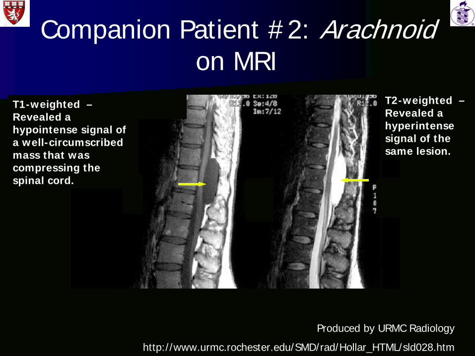
Companion Patient #2: Arachnoid on MRI
Produced by URMC Radiology
http://www.urmc.rochester.edu/SMD/rad/Hollar_HTML/sld028.htm
T1-weighted – Revealed a hypointense signal of a well-circumscribed mass that was compressing the spinal cord.
T2-weighted – Revealed a hyperintense signal of the same lesion.

Malignant
ChordomaChordoma
LymphomaLymphoma
SarcomaSarcoma–– OsteosarcomaOsteosarcoma–– ChondrosarcomaChondrosarcoma–– Multiple MyelomaMultiple Myeloma

Companion Patient #3: Chondrosarcoma on MRI
T2 Sagittal with contrast– Demonstrates a large, well- circumscribed mass compressing the spinal cord. It enhances with a mixed signal but is mostly hyperintense.
Produced by URMC Radiology
http://www.urmc.rochester.edu/SMD/rad/Hollar_HTML/sld040.htm

Metastases
MOST COMMON EXTRADURAL MOST COMMON EXTRADURAL MALIGNANT CANCERMALIGNANT CANCER
Breast, lung, and prostate cancer are the Breast, lung, and prostate cancer are the most prevalentmost prevalent
Initial site of lesion are the vertebral Initial site of lesion are the vertebral bodiesbodies

MetastasesMetastases
T2 Sagittal with contrast – Demonstrates an enhancing hyperintense signal. It is a poorly-marginated mass compressing the spinal cord.
Produced by URMC
http://www.urmc.rochester.edu/SMD/rad/Hollar_HTML/sld048.htm

IntraduralIntradural ExtramedullaryExtramedullary

Benign
MeningiomaMeningioma
Nerve sheath tumorsNerve sheath tumors–– NeurofibromaNeurofibroma–– SchwannomaSchwannoma

Meningioma
Produced by URMC
http://www.urmc.rochester.edu/SMD/rad/Hollar_HTML/sld057.htm
T1 AXIAL with contrast – Demonstrates displacement of the spinal cord
T1 SAGITTAL with contrast – Demonstrates a moderately enhancing, well-circumscribed mass.

Cysts and other masses
EpidermoidEpidermoid
DermoidDermoid

Dermoid
T2 Sagittal T1 SagittalProduced by URMC
http://www.urmc.rochester.edu/SMD/rad/Hollar_HTML/sld064.htm
Metastases
Can arise from inside or outside the CNSCan arise from inside or outside the CNS
Usually findings are discovered in the Usually findings are discovered in the lumbosacrallumbosacral regionregion

IntraduralIntradural IntramedullaryIntramedullary

Cysts and other masses
Multiple SclerosisMultiple Sclerosis
SyringomyeliaSyringomyelia
Transverse Transverse MyelitisMyelitis

Syringomyelia
Produced by URMC
http://www.urmc.rochester.edu/SMD/rad/Hollar_HTML/sld074.htm
T1 Sagittal with contrast T2 Sagittal

Malignant
EpendymomaEpendymoma
AstrocytomaAstrocytoma
HemangioblastomaHemangioblastoma

Astrocytoma
Produced by URMC
http://www.urmc.rochester.edu/SMD/rad/Hollar_HTML/sld087.htm
T2 Sagittal T2 Sagittal T1 Sagittal

Metastases
RareRare
Primary malignancies include: breast, Primary malignancies include: breast, lung, lymphoma, leukemia, and malignant lung, lymphoma, leukemia, and malignant melanomamelanoma
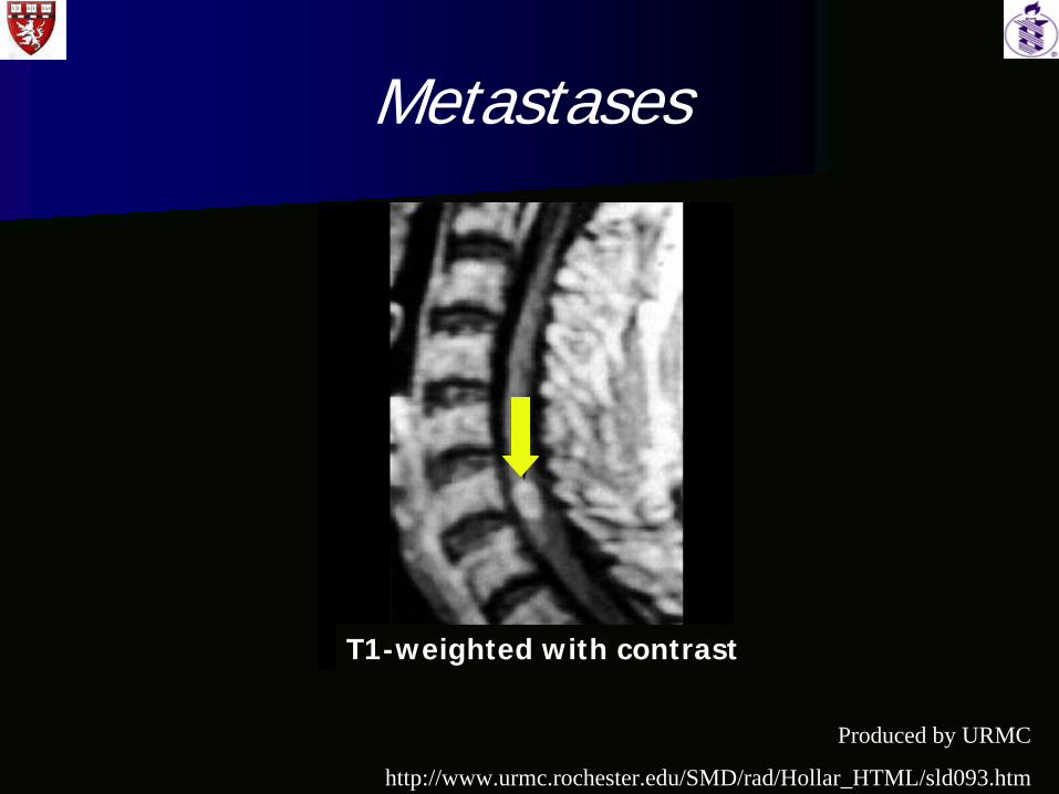
Metastases
Produced by URMC
http://www.urmc.rochester.edu/SMD/rad/Hollar_HTML/sld093.htm
T1-weighted with contrast

Patients RevisitedPatients Revisited

Patient HDPatient HD
T1 Sagittal Pre-contrast T2 Sagittal Pre-contrast
PACS, BIDMC
1.6x2.4cm mass lesion in the thoracic spinal canal at T6-T7.

Patient HDPatient HD
What is happening to the spinal cord?
Displacement
PACS, BIDMC
T2 Axial No Contrast – This image demonstrates the intradural, extramedullary location of the mass.

Patient HDPatient HD
PACS, BIDMC
What is that?
T2 Axial Post-gadolinium contrast

Patient HDPatient HD
PACS, BIDMC
DURAL TAIL
T2 Axial Post-gadolinium contrast – Indicates an enhancing lesion with a dural tail.

Patient HDPatient HD
CT Sagittal Reformatted – Calicifed component of the intradural mass. Notice the lack of bone involvement above and below the lesion.
PACS, BIDMC

Patient HDPatient HD
Differential Diagnosis includes:Differential Diagnosis includes:–– Nerve Sheath TumorNerve Sheath Tumor–– MeningiomaMeningioma–– MetastasesMetastases
WhatWhat’’s next?s next?–– Biopsy specimensBiopsy specimens
Patient HDPatient HD
Pathology ReportPathology Report–– Multiple fragments of soft tissue in one Multiple fragments of soft tissue in one
sectionsection–– Multiple fragments of white and Multiple fragments of white and erythematouserythematous
soft and bony tissuesoft and bony tissue–– No atypical featuresNo atypical features

Patient HD Patient HD -- definitive diagnosisdefinitive diagnosis
MENINGIOMAMENINGIOMA

Patient HD - Epilogue
SY was discharged home after surgery SY was discharged home after surgery under normal instructions. under normal instructions.
Appointment made for surgical follow up Appointment made for surgical follow up visit in 7visit in 7--10 days.10 days.
Brain tumor clinic visit set for July.Brain tumor clinic visit set for July.

Patient CRPatient CR
PACS, BIDMC
T2 Sagittal without contrast – Indicates an intramedullary mass at ~T11/T12 that is nodular yet irregular, measuring 2.1x1.0cm AP x 1.3 cm transverse.

Patient CRPatient CR
PACS, BIDMC
T1 Axial and Sagittal with contrast – Indicates an enhancing intramedullary mass at ~T11/T12.

Patient CRPatient CR
Differential Diagnosis includes:Differential Diagnosis includes:–– Primary Spinal Primary Spinal intramedullaryintramedullary massesmasses
EpendymomaEpendymoma
AstrocytomaAstrocytoma
HemangioblastomaHemangioblastoma
WhatWhat’’s next?s next?–– Biopsy specimenBiopsy specimen

Patient CRPatient CR
Pathology ReportPathology Report–– IntraoperativeIntraoperative smears revealed a smears revealed a gliomaglioma
producing many producing many glialglial processes. processes. –– Nuclear Nuclear pleomorphismpleomorphism–– Permanent Section revealed infiltration and Permanent Section revealed infiltration and
significant significant anaplasiaanaplasia–– Mitotic figures identifiedMitotic figures identified

Patient CR Patient CR -- definitive diagnosisdefinitive diagnosis
ANAPLASTIC ASTROCYTOMAANAPLASTIC ASTROCYTOMA

Patient CR - Epilogue
RBRB’’ss diagnosis resulted in nonsurgical diagnosis resulted in nonsurgical management management –– chemotherapy.chemotherapy.
Despite a stable recovery and no growth to the Despite a stable recovery and no growth to the masses at 3 month follow up, masses at 3 month follow up, RBRB’’ss condition condition began to deteriorate. Neurological deficits began to deteriorate. Neurological deficits increased. Numerous increased. Numerous comorbiditiescomorbidities developed.developed.
RB was discharged from BIDMC to palliative care; RB was discharged from BIDMC to palliative care; therapy was ceased.therapy was ceased.
RB receives pain management.RB receives pain management.
Recap
Review of the anatomy of the spine and Review of the anatomy of the spine and its functional correlatesits functional correlates
Explored the different anatomical Explored the different anatomical classification of spinal massesclassification of spinal masses
Identified MRI with IVIdentified MRI with IV--contrast as the test contrast as the test of choice for spinal massesof choice for spinal masses
Utilized this imaging modality to Utilized this imaging modality to demonstrate spinal masses for patients demonstrate spinal masses for patients reporting lower extremity weakness reporting lower extremity weakness and/or bowel/bladder dysfunctionand/or bowel/bladder dysfunction

Acknowledgments
JeanJean--Marc Marc GauguetGauguet, MD PhD, MD PhD
Alice Fisher, MDAlice Fisher, MD
Jay Jay PahadePahade, MD, MD
NagamaniNagamani PeriPeri, MD, MD
Rafael Rojas, MDRafael Rojas, MD
Gillian Lieberman, MDGillian Lieberman, MD
Maria Maria LevantakisLevantakis