a heat-shock-like response with cytoskeletal disruption...
TRANSCRIPT

A heat-shock-like response with cytoskeletal disruption occurs following hydrostatic pressure in MG-63 osteosarcoma cells
CHRISTINE L. HASKIN
Department of Cellular and Structural Biology, University of Texas Health Science Center at Sun Antonio, Sun Antonio, TX 78284- 7762, U.S.A.
KYRIACOS A. ATHANASIOU Department of Orthopaedics, University of Texas Health Science Center at Sun Antonio, Sun Antonio,
TX U.S.A.
AND
ROBERT KLEBE AND IVAN L. CAMERON Department of Cellular and Structural Biology, University of Texas Health Science Center at Sun Antonio,
Sun Antonio, TX 78284-7762, U.S.A.
Received May 14, 1993
HASKIN. C.L., ATHANASIOU, K.A., KLEBE, R., and CAMERON, I.L. 1993. A heat-shock-like response with cytoskeletal disruption occurs following hydrostatic pressure in MG-63 osteosarcoma cells. Biochem. Cell Biol. 71: 361-371.
Human osteosarcoma cells. MG-63, were exposed to a hydrostatic pressure shock of 4.0 MPa for 20 min. Changes in subcellular distribution of the cytoskeletal elements and heat shock protein 70 (hsp70) were followed by indirect immunofluorescence and by avidin-biotin-peroxidase protocols. During recovery, total cellular RNA was determined and actin and aldolase mRNA content was followed using reverse transcription - polymerase chain reaction techniques. Hydrostatic pressure caused cell rounding (but not cell death), disruption of microtubules, collapse of intermediate filaments to a perinuclear location, collapse of actin stress fibers into globular aggregates in the cytoplasm, and the formation of several large elongated intranuclear actin inclusions. During recovery, the cells flattened, reorganized microtubules, and redistributed intermediate filaments prior to the reappearance of actin stress fibers. At 20 and 60 min following the initiation of hydrostatic pressure, there was increased anti-hsp70 staining at the nuclear membrane and concentration of hsp70 in four to six granules in the nucleus. At 120 min following the hydrostatic pressure, hsp70 showed intense staining in the cytoplasm and hsp70-containing granules in the nucleus disappeared. Cellular RNA decreased during the first 120-min posthydrostatic pressure shock and then recovered to near prehydrostatic pressure treatment levels by 240 min. Actin mRNA abundance, in relation to aldolase mRNA abundance, showed the same temporal pattern of initial decrease, followed by increase as did total RNA. Review of the literature indicated that eukaryotic cells respond to heat shock and to hydrostatic pressure by disruption of the cytoskeletal elements and by similar modifications in genetic expression. In this study, the observed responses of MG-63 cells to a 4-MPa hydrostatic pressure shock was like the reported response of mammalian cells to a 43°C heat shock.
Key words: heat shock response, hydrostatic pressure, cytoskeleton.
HASKIN, C.L., ATHANASIOU, K.A., KLEBE, R., et CAMERON, I.L. 1993. A heat-shock-like response with cytoskeletal disruption occurs following hydrostatic pressure in MG-63 osteosarcoma cells. Biochem. Cell Biol. 71 : 361-371.
Des cellules MG-63 d'osteosarcome humain ont ete soumises a une pression hydrostatique de 4,O MPa pendant 20 min. Les modifications de la distribution cellulaire des elements du cytosquelette et de la proteine du choc thermique hsp70 ont ete suivies par immunofluorescence indirecte et par la technique de I'avidine-biotine-peroxydase. Pendant la pkriode de recuperation, I'ARN cellulaire total a ete mesurt et les quantites des ARNm de l'actine et de I'aldolase ont t t t deter- mintes par des techniques de transcription inverse en chaine. La pression hydrostatique entraine un arrondissement des cellules (mais non leur mort), une dtpolymerisation des microtubules, une accumulation des filaments intermediaires autour du noyau, une contraction des fibres de stress d'actine qui forment alors des agregats globulaires cytoplasmiques et la formation de plusieurs grosses inclusions d'actine allongees intranucltaires. Pendant la recuperation, les cellules s'aplatissent, les microtubules se reorganisent et les filaments intermediaires se redistribuent avant la reapparition des fibres de stress d'actine. Vingt et soixante minutes ap rb le debut de I'exposition a la pression hydrostatique, le marquage avec un anticorps anti-hsp70 augmente prts de la membrane nucltaire et l'hsp70 est concentrk dans quatre ti six granules intranucltaires. Puis, 120 min aprb I'exposition a la pression hydrostatique, il y a un marquage t r b prononce de I'hsp70 dans le cytoplasme et les granules intranuclkaires contenant I'hsp70 disparaissent. Le taux d'ARN diminue dans les cellules durant les premitres 120 min a p r b le choc de pression hydrostatique, puis il augmente de nouveau et atteint presque le taux antkrieur a l'exposition a la pression hydrostatique ap rb 240 min. La quantitk de I'ARNm de I'actine, relativement a celle de I'ARNm de I'aldolase, varie en fonction du temps comme le taux d'ARN total : elle diminue initialement puis elle augmente. Une revue de la litterature indique que les cellules eucaryotes repondent A un choc thermique et a la pression hydrostatique par une depolymCrisation des elements du cytosquelette et par des modifica- tions similaires de l'expression gknique. La reponse des cellules MG-63 a l'exposition a une pression hydrostatique de 4 MPa, observee dans cette etude, est similaire a la reponse des cellules de mammifhre A un choc thermique de 43°C dkcrite dans la litttrature.
Mots clPs : reponse a un choc thermique, pression hydrostatique, cytosquelette. [Traduit par la redaction]
ABBREVIATIONS: hsp70, heat shock protein 70; kDa, kilodalton(s); HP, hydrostatic pressure; BSA, bovine serum albumin; PBS, phosphate-buffered saline; FITC, fluorescein isothiocyanate; IgG, immunoglobulin G; DAB, diaminobenzidine; OD, optical density; PCR, polymerase chain reaction; RT, reverse transcription; dNTP, deoxynucleotide triphosphate; DEPC, diethyl pyrocarbonate; HSFI, heat shock factor 1; hnRNA, heterogeneous nuclear ribonuclear acid; HSE, heat shock element. Printed in Canada / Imprim6 au Canada
Bio
chem
. Cel
l Bio
l. D
ownl
oade
d fr
om w
ww
.nrc
rese
arch
pres
s.co
m b
y C
alif
Dig
Lib
- D
avis
on
09/0
6/13
For
pers
onal
use
onl
y.

362 BIOCHEM. CELL BIOL. VOL. 71, 1993
Introduction Ritossa (1962) first observed a heat-induced alteration in
the pattern of chromosome puffs in the giant salivary gland cells of Drosophila. Exposure to supraoptimal temperatures resulted in the loss of most of the existing puffs and the rapid appearance of seven to nine new puffs (Ashburner and Bonner 1979). The induced expression of a special set of heat-response proteins in Drosophila was first linked to Ritossa's observations by Tissieres et al. (1974). Heat shock proteins have subsequently been found to be induced in cells of all types tested when stressed by supraoptimal tempera- tures or various other stressors. It is now known that there are multiple heat shock protein families (Southgate et al. 1985). Heat shock proteins with a mass of about 70 kDa constitute a family of highly conserved proteins. Some hsp70 proteins are expressed constitutively, while others show indu- cible gene expression, in response to various stress conditions including heat shock, heavy metals (Welch 1990), oncogene and protooncogene activation, fever, inflammation, neu- ronal injury, aging (Morimoto et al. 1992), and increased hydrostatic pressure (Haskin et al. 1993). A major role of hsp70 is to bind to and refold partially denatured or mis- folded proteins, and to help disassociate abnormal protein aggregates. In addition, hsp70 plays a role as a chaperone that linearizes proteins for translocation across membranes such as endoplasmic reticulum, mitochondria, or other organelles (Langer and Neupert 1991), and for protein import into the nucleus (Dingwall and Laskey 1992; Shi and Thomas 1992). Hsp70 also appears to aid in the assembly of microtubules (Gupta 1990).
The heat shock response includes (i) cell cycle arrest (Rao and Engelberg 1965; Zeuthen 1971); (ii) decreases in intra- cellular pH and ATP levels (Leenders et al. 1974; Findly et al. 1983; Weitzel et al. 1985; Drummond et al. 1986); (iii) disruption of RNA processing (Muhich et al. 1989; Yost et al. 1990; Welch 1990); (iv) increased perichromatin granules due to accumulation of aggregates of unprocessed RNA (Heine et al. 1971 ; Mayrand and Pederson 1983; Yost and Lindquist 1986; Welch and Mizzen 1988; Welch 1990 and references therein); (v) a reduction in the synthesis of large ribosomal RNAs and 5S RNAs, a process dependent on continuous synthesis of ribosomal proteins (Simard and Bernhard 1967; Ellgaard and Clever 1971; Rubin and Hogness 1975; Yost et al. 1990); (vi) selective degradation of mRNAs (Lindquist 1980); (vii) suppression of normal protein synthesis (Yost et al. 1990 and references therein); (viii) the appearance of actin-containing inclusions within the nucleus (Pekkala et al. 1984; Welch and Suhan 1985; Iida et al. 1986); (ix) loss of actin filament organization in the cytoplasm (Glass et al. 1985; van Bergen en Henegouwen et al. 1985; van Bergen en Henegouwen and Linnemans 1987; Shyy et al. 1989; Kitano and Okada 1990); (x) micro-
Zimmerman 1970; Reed et al. 1993); (iii) disruption of RNA synthesis and processing (Landau 1970; Zimmerman et al. 1987); (iv) suppression of normal protein synthesis (Landau 1970; Zimmerman et al. 1987); (v) disassembly of cytoskele- tal components, inhibition of cytokinetic activity, and cell rounding (Marsland 1970; Zimmerman and Zimmerman 1970; Swezey and Somero 1985; Otter et al. 1987; Bourns et al. 1988; Haskin et al. 1993); (vi) loss of actin filament organization in the cytoplasm (Somero 1992; Haskin et al. 1993); (vii) microtubule disassembly (Zimmerman and Zimmerman 1970; Haskin et al. 1993); (viii) collapse of intermediate filaments into a perinuclear network (Haskin
.
et al. 1993); and (ix) relocalization of organelles (Zimmerman and Zimmerman 1970).
We report further evidence that hydrostatic pressure induces a heat shock type response in MG-63 osteosarcoma cells and that this response appears to be linked to the state of cytoskeletal disassembly. Briefly, cells that exhibited a greater cytoskeletal disassembly demonstrated a more pronounced heat shock response than cells that underwent a minimal loss of cytoskeletal organization. That both hydrostatic pressure and heat shock are shown to result in a parallel sequence of cytoskeletal and hsp70 responses suggests that the cytoskeletal elements, particularly actin, play an essential role in the cell's transcriptional regulation of hsp70 genes when exposed to environmental stressors.
Methods Tissue culture
MG-63 cells (a human osteosarcoma cell line obtained from American Type Culture) were maintained in Dulbecco's modified essential media (Sigma, D-1152) supplemented with 10% fetal bovine serum (Sigma, F 4 1 0 ) and glutamine (Sigma, G-7513). Cells to be used in immunohistochemistry and immunofluorescence studies were seeded onto uncoated masked glass slides (Carlson Scientific, 100808), cultured for 24 h in a high humidity chamber with 5% COz at 37"C, and then treated with a single 20-min hydrostatic pressure pulse of 4.0 MPa. Cell culture slides were fiied in ice-cold acetone for 5 min immediately following the pressure pulse and at 15, 30, 45, 60, and 120 min after the pressure pulse. Fixed slides were stored at - 20°C until the immunofluorescence assay. MG-63 cells (1 x lo6 cells/60-mm plate) used for RNA iso- lation were grown on uncoated glass Petri dishes and treated with a single 20-min pressure pulse of 4.0 MPa. Total RNA was iso- lated from control and hydrostatic pressure treated cultures using the single-step protocol of Chomczynski and Sacchi (1987), as modified and described below.
Pressure treatments Prior to the selected pressure treatment, MG-63 cells were
incubated in a high humidity chamber with 5% COz at 37°C and then transferred to the pressure cytochamber as previously described (Haskin et al. 1993). This closed-loop, computer- controlled cvtochamber system allows accurate pressurization
tubule disassembly (van Bergen en Henegouwen et al. 1985; without osciilations at constant temperature (~thanasiou et al. van Bergen en Henegouwen and Linnemans 1987; Kitan0 1993). A single 4.0-MPa pressure pulse of 20 min of duration was and Okada, 1990); (xi) collapse of intermediate filaments used for both the RNA and the immunohistochemistry studies. into a perinuclear network (shyy et al. 1989; Welch 1990); Experimental controls included cell cultures maintained in the high
and (xii) relocalization of organelles (welch and suhan humidity incubator and cell cultures treated at ambient atmospheric r nor\ pressure in the cvtochamber for the same length of time as the 170J).
experimental gro;ps. The pressure pulse curves-were symmetrical, Several to the heat shock have i.e., the rate at which the pressure was increased was equal to the also been reported following exposure of cells to hydrostatic rate at which the pressure was released at the end of the 20-min Pressures in the 2-20 MPa range, including (i) cell cycle pulse. The variation in temperature during the 20-min pulse was arrest and decreased DNA synthesis; (ii) disruption of poly- + 1°C. Because the process of transferring cell cultures to the somes (Marsland 1970; Murakami 1970; Zimmerman and cytochamber briefly exposes the cultures to the air, antibiotics
Bio
chem
. Cel
l Bio
l. D
ownl
oade
d fr
om w
ww
.nrc
rese
arch
pres
s.co
m b
y C
alif
Dig
Lib
- D
avis
on
09/0
6/13
For
pers
onal
use
onl
y.

TABLE 1. Changes in area and morphology of MG-63 osteosarcoma cells in response to hydrostatic pressure
Time Average % partially after HP area (pm2) % rounded rounded % flattened
(min) (n) (length = width) (length > width) (length >>> width)
20 0.1812k0.05* 37 40 23 (72)
80 0.2192k 0.07 27 48 25 (75)
1 40 0.2402 k 0.07 13 40 47 (73)
Control 0.4573 * 0.13 05 10 85 (68)
* & standard deviation.
(penicillin-streptomycin, Sigma P-0781) were added to the media low GC content (35%), and appropriate melting points. PCR following the pressure pulse. primers were synthesized by the Biopolymer Sequencing and
Indirect immunofluorescence A panel of antibodies was selected to demonstrate changes in
cytoskeletal organization in response to physiological levels of pressure. A polyclonal anti-actin antibody (Sigma, A2668) that recognizes multiple epitopes was used to assay actin organization so that different forms of actin, including f-actin and g-actin, could be visualized. Additional antibodies, including monoclonal anti- bodies against a-tubulin (Sigma, T9026). P-tubulin (Sigma, T0426), and vimentin (Oncogene Science, lFOl), were also used to evaluate the cytoskeletal organization. Anti-hsp (Oncogene Science, HSPOI) monoclonal antibody was used to evaluate the stress response to HP. Fixed slides stored at - 20°C were rehydrated, blocked using 5% BSA in Dulbecco's PBS (Sigma, D-7030), and incubated for 45-60 min at room temperature with one of the antibodies. Prep- arations were rinsed three times in PBS-BSA, blocked with 1% goat serum (Sigma, S-6898), and then incubated with FITC-labeled secondary anti-IgG antibodies (Sigma, F5387 or FO511) for 30 min at 4OC. Preparations were counterstained with propidium iodide, as needed, to locate nuclei, rinsed three times in PBSiBSA, and prepared for microscopic evaluation. Photomicrographs were taken using a Zeiss fluorescent photomicroscope using ASA 1600 35-mm film.
Zmmunohistochemistry and morphometry Localization of hsp70 was also assayed by an avidin - biotin -
horseradish peroxidase and DAB protocol as described by Stewart et al. (1986). Briefly, slides were fixed in ice-cold acetone for 5 min, air dried, rehydrated with PBS-BSA, treated with H202 to inac- tivate endogenous peroxidase activity, incubated with the anti-hsp70 monoclonal antibody, rinsed in PBS-BSA, incubated with biotinylated anti-IgG, rinsed, treated with avidin - biotin - horse- radish peroxidase complex, rinsed, incubated with DAB substrate, and washed. Photomicrographs were taken using a Zeiss photo- microscope using Kodak high-contrast copy film.
Morphometric measurements were carried out using a digitizing table and the Bioquant I1 image analysis system (R & M Biometrics, Nashville, Tenn.) on a Leitz Diaplan microscope. Measurements consisted of (i) area seen by transillumination of the stained cells; (ii) maximum length of cell drawn through the cell nucleus, and (iii) maximum width of cell drawn through the cell nucleus.
RNA isolation and polymerase chain reaction Total RNA was isolated from 1 x lo6 cells from control and
hydrostatic pressure treated cultures using the single-step protocol of Chomczynski and Sacchi (1987), quantitated by OD at 260 and 280 nm. The relative abundance of actin and aldolase mRNAs was assayed by reverse transcription - polymerase chain reaction tech- niques. Primer sequences of the desired size were selected based upon their lack of homology with other known DNA sequences,
Synthesis Facility in the Department of Biochemistry (University of Texas Health Science Center at San Antonio, Tex.) on a Cyclone DNA synthesizer (Milligen/Biosearch, Burlington, Mass.) and purified by OPC cartridge. One microgram of total RNA isolated from each condition was used in single-tube reverse transcription reactions to obtain cDNAs for use in PCR amplification.
The one-tube RT-PCR method used (Conboy et al. 1988) was modified to yield free MgCI, (rather than dNTP bound), 2 mM Taq DNA polymerase, and 6 mM avian myeloblastosis virus reverse transcriptase. Actin and aldolase cDNAs were generated by using specific oligonucleotide primers to prime reverse transcription in a 20-pL reaction solution as described below. RNA and primer annealing was done in DEPC-treated H 2 0 at 68OC for 15 min. followed by cooling on wet ice. Following annealing, the reaction solution (40 mM KCI, 50 mM Tris-HC1 (pH 8.3). 8 mM MgCl,, 0.5 mM of each dNTP for a total of 2 mM dNTPs (Pharmacia, Piscataway, N.J.), 200 ng of antisense primer, 10 U of avian myeloblastosis virus reverse transcriptase (Life Sciences, St. Petersburg, Fla.), 20 U of RNAsin (Promega, Madison, Wis.), 5 mM dithiothreitol, and 5 pg BSA) was added to a final volume of 20 pL and the reaction was then incubated at 42°C for 1 h.
Following reverse transcription, PCRs were carried out as follows. The 20 pL reverse transcription reaction solution was diluted with 1 x PCR buffer (50 mM KCI, 10 mM Tris-HC1, 1.5 mM MgCl,, 0.1% gelatin; United States Biochemicals, Cleveland, Ohio) to a final volume of 100 pL and a final MgCI, concentration of 3 mM (2 mM free MgCI,), with 100 ng of "~-5'-end labeled sense oligonucleotide primer, 100 ng of cold sense primer, additional dNTPs to a concentration of 0.2 mM, and 2.5 U of Taq DNA polymerase (Perkin-Elmer Cetus, Norwalk, Va.). Amplification was performed on an Ericomp thermocycler (San Diego, Calif.). The reaction mixture was initially denatured at 94°C for 1.5 min and then annealed at 54OC for 1 min, extended at 72°C for 1 min 50 s, and denatured at 94OC for 20 s. The chain reaction was terminated with a final cycle of annealing at 54OC to 56OC for 1 min, and extension at 72°C for 7 min. The resulting DNA fragments were analyzed by electrophoresis on 5% polyacryl- amide gels and quantitated by scintillation counting of excised bands.
Results Zrnrnunohistochernistry a n d imrnunofluorescence
Hydrostatic pressure caused loss of cytoskeletal organi- zation and resulted in increased cell rounding and decreased cell area (Table 1). Indirect immunofluorescence staining with monoclonal antibodies to actin, vimentin, and a-tubulin demonstrated loss of organization of cytoskeletal proteins in response to 4.0-MPa H P . Because the polyclonal
Bio
chem
. Cel
l Bio
l. D
ownl
oade
d fr
om w
ww
.nrc
rese
arch
pres
s.co
m b
y C
alif
Dig
Lib
- D
avis
on
09/0
6/13
For
pers
onal
use
onl
y.

364 BIOCHEM. CELL BIOL. VOL. 71. 1993
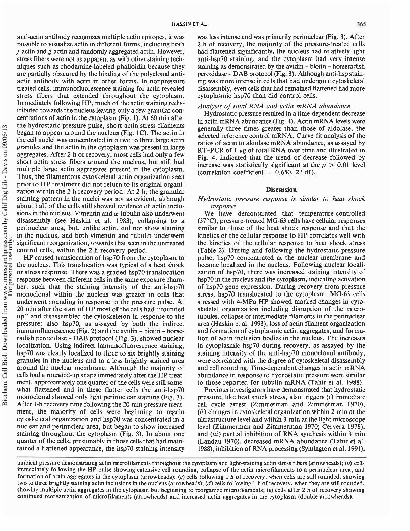
FIG. 1. Indirect immunofluorescence analysis demonstrates time-dependent changes in the distribution of actin stress filaments in MG-63 osteosarcoma cells following a 20-min, 4-MPa H P pulse. MG-63 cells were fixed, processed, and stained using a polyclonal anti-actin antibody and a FITC-conjugated secondary as described in Methods. Each scale bar is 10 pm. (a) Control cultures kept at
Bio
chem
. Cel
l Bio
l. D
ownl
oade
d fr
om w
ww
.nrc
rese
arch
pres
s.co
m b
y C
alif
Dig
Lib
- D
avis
on
09/0
6/13
For
pers
onal
use
onl
y.
HASKIN ET AL. 365
anti-actin antibody recognizes multiple actin epitopes, it was possible to visualize actin in different forms, including both f-actin and g-actin and randomly aggregated actin. However, stress fibers were not as apparent as with other staining tech- niques such as rhodamine-labeled phalloidin because they are partially obscured by the binding of the polyclonal anti- actin antibody with actin in other forms. In nonpressure treated cells, immunofluorescence staining for actin revealed stress fibers that extended throughout the cytoplasm. Immediately following HP, much of the actin staining redis- tributed towards the nucleus leaving only a few granular con- centrations of actin in the cytoplasm (Fig. 1). At 60 min after the hydrostatic pressure pulse, short actin stress filaments began to appear around the nucleus (Fig. 1C). The actin in the cell nuclei was concentrated into two to three large actin granules and the actin in the cytoplasm was present in large aggregates. After 2 h of recovery, most cells had only a few short actin stress fibers around the nucleus, but still had multiple large actin aggregates present in the cytoplasm. Thus, the filamentous cytoskeletal actin organization seen prior to HP treatment did not return to its original organi- zation within the 2-h recovery period. At 2 h, the granular staining pattern in the nuclei was not as evident, although about half of the cells still showed evidence of actin inclu- sions in the nucleus. Vimentin and a-tubulin also underwent disassembly (see Haskin et al. 1983), collapsing to a perinuclear area, but, unlike actin, did not show staining in the nucleus, and both vimentin and tubulin underwent significant reorganization, towards that seen in the untreated control cells, within the 2-h recovery period.
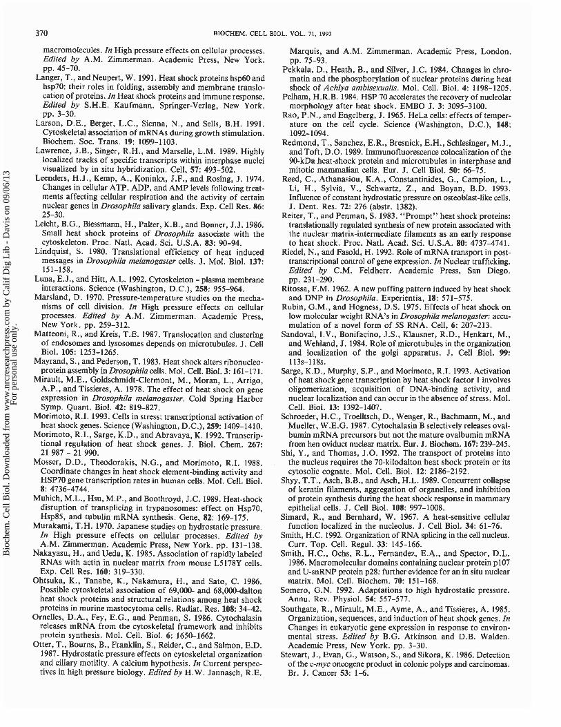
HP caused translocation of hsp70 from the cytoplasm to the nucleus. This translocation was typical of a heat shock or stress response. There was a graded hsp70 translocation response between different cells in the same exposure cham- ber, such that the staining intensity of the anti-hsp70 monoclonal within the nucleus was greater in cells that underwent rounding in response to the pressure pulse. At 20 min after the start of HP most of the cells had "rounded up" and disassembled the cytoskeleton in response to the pressure; also hsp70, as assayed by both the indirect immunofluorescence (Fig. 2) and the avidin - biotin - horse- radish peroxidase - DAB protocol (Fig. 3), showed nuclear localization. Using indirect immunofluorescence staining, hsp70 was clearly localized to three to six brightly staining granules in the nucleus and to a less brightly stained area around the nuclear membrane. Although the majority of cells had a rounded-up shape immediately after the HP treat- ment, approximately one quarter of the cells were still some- what flattened and in these flatter cells the anti-hsp70 monoclonal showed only light perinuclear staining (Fig. 3). After 1-h recovery time following the 20-min pressure treat- ment, the majority of cells were beginning to regain cytoskeletal organization and hsp70 was concentrated in a nuclear and perinuclear area, but began to show increased staining throughout the cytoplasm (Fig. 3). In about one quarter of the cells, presumably in those cells that had main- tained a flattened appearance, the hsp70-staining intensity
was less intense and was primarily perinuclear (Fig. 3). After 2 h of recovery, the majority of the pressure-treated cells had flattened significantly, the nucleus had relatively light anti-hsp70 staining, and the cytoplasm had very intense staining as demonstrated by the avidin - biotin - horseradish peroxidase - DAB protocol (Fig. 3). Although anti-hsp stain- ing was more intense in cells that had undergone cytoskeletal disassembly, even cells that had remained flattened had more cytoplasmic hsp70 than did control cells.
Analysis of total RNA and actin mRNA abundance Hydrostatic pressure resulted in a time-dependent decrease
in actin mRNA abundance (Fig. 4). Actin mRNA levels were generally three times greater than those of aldolase, the selected reference control mRNA. Curve-fit analysis of the ratios of actin to aldolase mRNA abundance, as assayed by RT-PCR of 1 pg of total RNA over time and illustrated in Fig. 4, indicated that the trend of decrease followed by increase was statistically significant at the p > 0.01 level (correlation coefficient = 0.650, 22 df).
Discussion Hydrostatic pressure response is similar to heat shock
response We have demonstrated that temperature-controlled
(37"C), pressure-treated MG-63 cells have cellular responses similar to those of the heat shock response and that the kinetics of the cellular response to HP correlates well with the kinetics of the cellular response to heat shock stress (Table 2). During and following the hydrostatic pressure pulse, hsp70 concentrated at the nuclear membrane and became localized in the nucleus. Following nuclear locali- zation of hsp70, there was increased staining intensity of hsp70 in the nucleus and the cytoplasm, indicating activation of hsp70 gene expression. During recovery from pressure stress, hsp70 translocated to the cytoplasm. MG-63 cells stressed with 4-MPa HP showed marked changes in cyto- skeletal organization including disruption of the micro- tubules, collapse of intermediate filaments to the perinuclear area (Haskin et al. 1993), loss of actin filament organization and formation of cytoplasmic actin aggregates, and forma- tion of actin inclusion bodies in the nucleus. The increases in cytoplasmic hsp70 during recovery, as assayed by the staining intensity of the anti-hsp70 monoclonal antibody, were correlated with the degree of cytoskeletal disassembly and cell rounding. Time-dependent changes in actin mRNA abundance in response to hydrostatic pressure were similar to those reported for tubulin mRNA (Tahir et al. 1988).
Previous investigators have demonstrated that hydrostatic pressure, like heat shock stress, also triggers (i) immediate cell cycle arrest (Zimmerman and Zimmerman 1970), (ii) changes in cytoskeletal organization within 2 min at the ultrastructure level and within 3 min at the light microscope level (Zimmerman and Zimmerman 1970; Cervera 1978), and (iii) partial inhibition of RNA synthesis within 3 min (Landau 1970), decreased mRNA abundance (Tahir et al. 1988), inhibition of RNA processing (Syrnington et al. 1991),
-
ambient pressure demonstrating actin microfilaments throughout the cytoplasm and light-staining actin stress fibers (arrowheads); (6) cells immediately following the HP pulse showing extensive cell rounding, collapse of the actin microfilaments to a perinuclear area, and formation of actin aggregates in the cytoplasm (arrowheads); (c) cells following 1 h of recovery, when cells are still rounded, showing two to three brightly staining actin inclusions in the nucleus (arrowheads); (d) cells following 1 h of recovery, when they are still rounded, showing multiple actin aggregates in the cytoplasm but beginning to reorganize microfilaments; (e) cells after 2 h of recovery showing continued reorganization of microfilaments (arrowheads) and increased actin aggregates in the cytoplasm (double arrowheads).
Bio
chem
. Cel
l Bio
l. D
ownl
oade
d fr
om w
ww
.nrc
rese
arch
pres
s.co
m b
y C
alif
Dig
Lib
- D
avis
on
09/0
6/13
For
pers
onal
use
onl
y.

366 BIOCHEM. CELL BIOL. VOL. 71, 1993
FIG. 2. Indirect immunofluorescence analysis demonstrates time-dependent changes in hsp70 localization following a 20-min, 4-MPa H P pulse. MG-63 cells were fixed, processed, and stained using a monoclonal anti-hsp7O antibody and a FITC-conjugated secondary as described in Methods. Each scale bar is 10 pm. (a) Control cultures kept at ambient pressure demonstrating hsp70 in the cytoplasm and the nucleus; (6) cells immediately following H P pulse showing extensive relocalization of hsp7O to the nucleus (double arrowheads) and perinuclear area (arrowhead); (c) cells following 1 h of recovery showing increased hsp70 levels in the nucleus and cytoplasm; (d ) cells following 2 h of recovery showing increased cytoplasmic concentration of hsp70 relative to nuclear hsp70 concentration.
Bio
chem
. Cel
l Bio
l. D
ownl
oade
d fr
om w
ww
.nrc
rese
arch
pres
s.co
m b
y C
alif
Dig
Lib
- D
avis
on
09/0
6/13
For
pers
onal
use
onl
y.

HASKIN ET AL. 3 67
I
D
fl B
1 -
0 30 60 90 120 150 180 210 240 270 300
Time (min)
vv I
FIG 4. The abundance of actin and aldolase mRNAs in MG-63 osteosarcoma cells as assayed by RT-PCR techniques as described
1 in Methods. Curve analysis of the actin/aldolase ratios of MG-63 e 1% osteosarcoma cells, prior to and following a 20-min, 4-MPa HP
pulse indicates that the trend of decrease followed by increase was statistically significant at the p > 0.01 level.
:i and partial inhibition of protein synthesis within 6 min (Landau 1970), followed by resumption of normal protein
- 4 synthesis within 2-3 h of pressure stress.
* Ab The temporal responses to heat shock have been reviewed , r by Welch (1990). Briefly, normal protein synthesis was
inhibited within 3-5 min after a heat shock temperature shift, changes in cytoskeletal organization of mammalian cells were observed within 5-15 min after the temperature shift to 43°C (Pekkala et al. 1984; Glass et al. 1985; van I!# 7 ; '6 : Bergen en Henegouwen et al. 1985; Welch and Suhan 1985;
i - Iida et al. 1986; van Bergen en Henegouwen and Linnemans 'I- *
1987; Mosser et al. 1988; Shyy et al. 1989; Kitano and Okada F~ 1990), and increased heat shock proteins were detected after
8-10 min (Mirault et al. 1978). Hsp70 is normally found in * ,gG c - - the cytoplasm at low levels prior to heat shock. After heat
- . :I l a # $ - A, Lm shock, hsp70 concentrates at the nuclear membrane, moves
into the nucleus, and is either complexed with denatured or aggregated ribosomal proteins (Welch and Suhan 1985; Welch 1990), or with HSFl and the hsp70 promoter (Morimoto et al. 1992; Morimoto 1993). In addition, there are time-dependent changes in mRNA abundance and dis- ruption of RNA processing (Muhich et al. 1989; Yost et al. 1990). During release from heat shock and as the heat- disrupted nucleoli begin to recover, the hsp70 concentrates in the cytoplasm and is colocalized with translation machinery (Pelham 1984; Velazquez and Lindquist 1984; Welch 1990). In addition to the changes in hsp70 localiza- tion, changes in actin and microtubule organization were reported in response to heat shock (Welch and Suhan 1985; Iida et al. 1986; Shyy et al. 1989; Kitano and Okada 1990).
FIG. 3. Avidin - biotin - horseradish peroxidase - DAB anal- Thus, the kinetics of the cellular responses to heat shock ysis demonstrates time-dependent changes in hsp70 localization and and pressure stress are very similar. concentration of anti-hsp70 following a 20-min, 4-MPa HP pulse. The baroprotective effect of heat shock provides addi- Each scale bar is 10 Pm. (a) Control cultures kept at ambient tional evidence that the heat shock response and the pres- Pressure demonstrating low levels of h s ~ 7 0 in the cytoplasm and sure response are closely related. Although the effect of heat the nucleus; (b) cells immediately following HP pulse showing relocalization and increased concentration of hsp70 in the nucleus synchronization was not reported as being baroprotective,
(arrowheads) and perinuclear area (double arrowheads); (c) cells Zimmerman and Zimmerman (1970) reported that logarith- following 1 h of recovery showing increased hsp70 levels in the mically growing cells were more Sensitive to high pressure nucleus (arrowheads) and cytoplasm (compare with cytoplasmic than were the heat-synchronized cells and recovery from the staining in a; (d) cells following 2 h of recovery showing redistri- effects of pressure was slower in the nonsynchronized cells bution of hsp70 from the nucleus (arrowheads) to the cytoplasm. than in the heat-synchronized cells. This baroprotective
Bio
chem
. Cel
l Bio
l. D
ownl
oade
d fr
om w
ww
.nrc
rese
arch
pres
s.co
m b
y C
alif
Dig
Lib
- D
avis
on
09/0
6/13
For
pers
onal
use
onl
y.

368 BIOCHEM. CELL BIOL. VOL. 71, 1993
TABLE 2. Sequence of reported events following exposure of eukaryotic cells to hydrostatic pressure or heat shock stress
Time (min) Hydrostatic pressure Heat shock
0 Beginning of HP pulse Beginning of heat shock stress
Cell cycle arrest (1-3) Loss of cytoskeletal organization (1, 2, 6-8) Cell rounding (1, 8) Inhibition of cytokinetic activity (2, 10) Disruption of organelle location (1)
Increased hsp70 product in nucleus (12) Disruption of polysomes (1) Inhibition of most RNA synthesis (3, 18, 19) Inhibition of RNA processing (20)
Inhibition of normal protein synthesis (18, 19)
Lowest total RNA yield Increased hsp70 perinuclear
Loss of cytoskeletal organization (4, 5) Disruption of organelle location (5, 9) Activation, translocation. DNA binding of HSFl to
HSE (11-15)
Increased hsp70 in nucleus (12) Inhibition of transcription of most genes (16) 'Disruption of RNA processing (16, 17)
Maximum HSFl levels in nucleus (1 1, 13) Maximum hsp70 transcription (1 1, 13) Increase hsp70 product (1 1, 13, 21) Inhibition of normal cellular protein synthesis (5, 22)
Decline in HSFl levels and hsp70 transcription (1 1) Hsp70 mRNA remains stable for rest of heat txt (16) Increased levels of hsp70 in cytoplasm (1 1)
Lowest actin mRNA level Cytoskeletal reorganization begins (4, 5) Increased levels of hsp70 in cytoplasm Resumption of normal protein synthesis
Cytoskeletal reorganization begins (4, 5, 8) HSF 1 levels at basal (1 1, 13) Actin dots and filaments in cytoplasm (4, 8) Hsp70 transcription at basal (1 1) Appearance of actin inclusion bodies in nucleus
Appearance of actin inclusion bodies in nucleus 260 Total RNA yield at control levels (9, 23, 24)
NOTES: Numbers in parentheses represent references: 1, Zimmerman and Zimmerman 1970; 2, Marsland 1970; 3, Tahir et al. 1988; 4, Kitano and Okada 1990; 5, Shyy et al. 1989; 6, Bourns et al. 1988; 7, Swezey and Somero 1985; 8, Haskin et al. 1993; 9, Welch and Suhan 1985; 10, Otter et al. 1987; 11, Abravaya et al. 1991; 12, Sargeet al. 1993; 13, Mosser et al. 1988; 14, Morimoto et al. 1992; 15, Morimoto 1993; 16, Yost et al. 1990; 17, Muhich et al. 1989; 18, Landau 1970; 19, Zimmerman et al. 1987; 20, Symington et al. 1991; 21, Craig and Gross 1991; 22, Lindquist 1981; 23, Iida et al. 1986; 24, Pekkala et al. 1984.
effect of heat shock was confirmed and extended by Komatsu et al. (1991), who reported that preincubation of Saccharomyces cerevisiae at 40 or 43OC gave significant protection against lethal levels of hydrostatic pressures.
Cytoskeletal organization may be a critical element in adaptation of eukaryotic cells to environmental stress
Many cellular functions that are affected by heat stress or pressure stress depend on the integrity of the cytoskeleton, including (i) protein synthesis (Cervera et al. 1981; Ornelles et al. 1986; Hesketh 1991; Vedeler et al. 1991), (ii) organelle position (Ball and Singer 1982; Sandoval et al. 1984; Eckert 1986; Matteoni and Kreis 1987), (iii) cell shape (Stossel 1984; Luna and Hitt 1992), and (iv) mRNA translation and stability (Hesketh 1991; Larson et al. 1991; Vedeler et al. 1991). Additional cellular functions that are affected by heat stress or pressure stress depend on the integrity of the nuclear matrix, including mRNA posttranscriptional processing, transport, translation, and stability (Nakayasu and Ueda 1985; Smith et al. 1986; Zeitlin et al. 1987; Lawrence et al. 1989; Agutter 1991; Smith 1992). Actin, intermediate fila- ment, and nuclear matrix organization are important regu- lators of RNA processing and transport. The binding of hnRNAs is specific for actin and can be abolished by anti- actin antibodies (Schroeder et al. 1987), and release and export of hnRNAs, but not mature mRNA, occurs in response to depolymerization of actin filaments in the presence of cytochalasin B (Riedel and Fasold 1992 and
references therein). Pre-mRNA, pre-rRNA, and rRNA have also been shown to be associated with the nuclear matrix (Herlan et al. 1979; Smith et al. 1986). Electron microscopy and biochemical studies have demonstrated links between intermediate filaments and the nuclear pores (Franke et al. 1981; Georgatos and Blobel 1987a, 1987b) and that the export of mRNA transcripts occurs along linear tracts at the nuclear pore complexes (Feldherr et al. 1984; Lawrence et al. 1989).
The importance of the cytoskeleton to the heat shock response is also suggested by the evidence that some heat shock proteins are associated with cytoskeletal elements (Reiter and Penman 1983; Leicht et al. 1986; Ohtsuka et al. 1986; Redmond et al. 1989; Tomasovic et al. 1989; Karasev et al. 1992). Although alterations in the cytoskeleton due to heat shock vary from cell type to cell type, some type of cytoskeletal disruption appears to be a common denomi- nator in heat shock response. Reorganization of the cytoskeleton during recovery requires synthesis of new proteins (van Bergen en Henegouwen and Linnemans 1987), resumption of normal protein synthesis is coincident with cytoskeletal reorganization, and prevention of heat shock protein synthesis during stress treatment does not alter the rapid loss of stress fibers (van Bergen en Henegouwen and Linnemans 1987). These observations suggest that cytoskele- tal organization may be a critical element in adaptation to environmental stress.
Using indirect immunofluorescence, Sarge et al. (1993)
Bio
chem
. Cel
l Bio
l. D
ownl
oade
d fr
om w
ww
.nrc
rese
arch
pres
s.co
m b
y C
alif
Dig
Lib
- D
avis
on
09/0
6/13
For
pers
onal
use
onl
y.

HASKIN ET AL. 369
have shown that prior to heat shock, HSFl , a transcriptional activator for hsp70, is present in low levels in both the cytoplasm and the nucleus of HeLa cells, and that following heat shock, HSFl has a nuclear localization. Sarge et al. (1993) suggested that HSFl activation was normally repressed by a titratable negative regulatory factor and demonstrated that recombinant eukaryotic H S F l expressed in Escherichia coli constitutively bound DNA. Since E. coli d o not have a cytoskeleton, the titratable negative factor may be a binding site on the cytoskeleton that stabilizes the HSFl monomers and prevents activation, unless HSFl undergoes a conformational change and (or) release from cytoskeletal elements in response t o stress.
We propose that the stress response following H P o r heat shock is related t o the disruption of cytoskeletal elements. The depolymerization of the cytoskeletal elements may sustain the heat shock response by several possible mecha- nisms, including (i) increased release of HSFl from depolymerized cytoskeletal components; (ii) increased sub- strates, such as disassembled tubulin or actin, that temporar- ily deplete the free pool of hsp70; and (iii) disassembly of actin in the nuclear matrix in such a way that RNA splicing is disrupted, requiring nuclear localization of hsp70 to repair the damage.
Evidence already exists that supports the role of tubulin dimers as a pool of substrates (Gupta 1990) that depletes cytoplasmic hsp70 pools, and substantial evidence supports the proposal that actin in the nuclear matrix stabilizes RNA processing. However, additional research is needed to deter- mine if HSFl binds t o actin o r other cytoskeletal components.
Acknowledgements This research was supported, in part, by a National Insti-
tutes of Health grant DEOO512, an Orthopedic Research and Education Foundation grant, and a Whitaker Foundation grant.
Abravaya, K., Phillips, B., and Morimoto, R.I. 1991. Heat shock- induced interactions of heat shock transcription factor and the human hsp70 promoter examined by in vivo footprinting. Mol. Cell. Biol. 11: 586-592.
Agutter. P.S. 1991. Role of the cytoskeleton in nucleocytoplasmic RNA and protein distributions. Biochem. Soc. Trans. 19: 1094- 1098.
Ashburner, M., and Bonner, J.J. 1979. The induction of gene activity in Drosophila by heat shock. Cell, 17: 241-254.
Athanasiou, K.A., Lanctot, D., and Constantinides, G. 1993. Design and validation of a closed loop, variable hydrostatic pressure cytochamber. Adv. Bioeng. In press.
Ball, E.H., and Singer, S.J. 1982. Mitochondria are associated with microtubules and not with intermediate filaments in cultured fibroblasts. Proc. Natl. Acad. Sci. U.S.A. 79: 123-126.
Bourns, B., Franklin, S., Cassimeris, L., and Salmon, E.D. 1988. High hydrostatic pressure effects in vivo: changes in cell mor- phology, microtubule assembly and actin organization. Cell Motil. Cytoskeleton, 10: 380-390.
Cervera, J. 1978. Effects of thermal shock on Hep-2 cells. An ultrastructural and high resolution autoradiographic study. J. Ultrastruct. Res. 63: 51-63.
Cerveza, M., Dreyfuss, G., and Penman, S. 1981. Messenger RNA is translated when associated with the cytoskeletal framework in normal and VSV-infected HeLa cells. Cell, 23: 113-120.
Chomczynski, P., and Sacchi, N. 1987. Single-step method of RNA isolation by acid guanidinium thiocyanate - phenol - chloroform extraction. Anal. Biochem. 162: 156-159.
Conboy, J., Chan, J., Mohandas, N., and Kan, Y. 1988. Multiple protein 4.1 isoforms produced by alternative splicing in human erythroid cells. Proc. Natl. Acad. Sci. U.S.A. 85: 9062-9065.
Craig, E.A., and Gross, C.A. 1991. Is hsp7O the cellular thermom- eter? Trends Biochem. Sci. 16: 135-140.
Dingwall, C., and Laskey, R. 1992. The nuclear membrane. Science (Washington, D.C.), 258: 942-947.
Drummond, I.A., McClure, S.A., Poenie, M., Tsien, R.Y., and Steinhardt, R.A. 1986. Large changes in intracellular pH and calcium observed during heat shockare not responsible for the induction of heat shock proteins in Drosophila melanogaster. Mol. Cell. Biol. 6: 1767-1775.
Eckert, B.S. 1986. Alteration of the distribution of intermediate filaments in PtKl cells by acrylamide. 11. Effect on the organi- zation of cytoplasmic organelles. Cell Motil. Cytoskeleton, 6: 15-24.
Ellgaard, E.G., and Clever, U. 1971. RNA metabolism during puff induction in Drosophila melanogaster. Chromosoma, 36: 60-78.
Feldherr, C.M., Kallenbach, E., and Schultz, N. 1984. Movement of a karyophilic protein through the nuclear pores of oocytes. J. Cell Biol. 99: 2216-2222.
Findly, R.C., Gillies, R.J., and Shulman, R.G. 1983. In vivo phosphorous-31 nuclear magnetic resonance reveals lowered ATP during heat shock of tetrahymena. Science (Washington, D.C.), 219: 1223-1225.
Franke, W.W., Scheer, U., Krohne, G., and Jarasch, E.D. 1981. The nuclear envelope and the architecture of the nuclear periph- ery. J. Cell Biol. 91: 39s-50s.
Georgatos, S.D., and Blobel, G. 1987a. Lamin B constitutes an intermediate filament attachment site at the nuclear envelope. J. Cell Biol. 105: 117-125.
Georgatos, S.D., and Blobel, G. 1987b. Two distinct attachment sites for vimentin along the plasma membrane and the nuclear envelope in avian erythrocytes: a basis for a vectorial assembly of intermediate filaments. J. Cell Biol. 105: 105-1 15.
Glass, J.R., DeWitt, R.G., and Cress, A.E. 1985. Rapid loss of stress fibers in Chinese hamster ovary cells after hyperthermia. Cancer Res. 45: 258-262.
Gupta, R.S. 1990. Microtubules, mitochondria, and molecular chaperones: a new hypothesis for in vivo assembly of microtu- bules. Biochem. Cell Biol. 68: 1352- 1363.
Haskin, C., Cameron, I., and Athanasiou, K. 1993. Physiological levels of hydrostatic pressure alter morphology and organization of cytoskeletal and adhesion proteins in MG-63 osteosarcoma cells. Biochem. Cell Biol. 71: 27-35.
Heine, V., Sverkak, L., Kondratuck, J., and Bonar, R.A. 1971. The behavior of Hela S3 cells under the influence of supranormal temperatures. J. Ultrastruct. Res. 34: 375-396.
Herlan, G., Eckert, W.A., Kaffenberger, W., and Wunderlich, F. 1979. Isolation and characterization of an RNA-containing nuclear matrix from Tetrahymena macronuclei. Biochemistry, 18: 1782-1787.
Hesketh, J. 1991. Association of ribosomes with myofibrils and microfilaments: a role in the spatial organization of protein synthesis. Biochem. Soc. Trans. 19: 1103-1 107.
Iida, K., Iida, H., and Yahara, I. 1986. Heat shock induction of intranuclear actin rods in cultured mammalian cells. Exp. Cell Res. 165: 207-215.
Karasev, A.V., Kashina, A.S., Gelfand, V.I., and Dolja, V.V. 1992. HSP70-related 65 kDa protein of beet yellows closterovirus is a microtubule-binding protein. FEBS Lett. 304: 12-114.
Kitano, Y., and Okada, N. 1990. Organization and disorganization of actin filaments in human epidermal keratinocytes: heat-shock treatment and recovery process. Cell Tissue Res. 261: 269-274.
Komatsu, Y., Obuchi, K., Iwahashi, H., Kaul, S.C., Ishimura, M., Fahy, G.M., and Rall, W.F. 1991. Deuterium oxide, dimethylsulfoxide and heat shock confer protection against hydrostatic pressure damage in yeast. Biochem. Biophys. Res. Commun. 174: 1141-1 147.
Landau, J.V. 1970. Hydrostatic pressure on the biosynthesis of
Bio
chem
. Cel
l Bio
l. D
ownl
oade
d fr
om w
ww
.nrc
rese
arch
pres
s.co
m b
y C
alif
Dig
Lib
- D
avis
on
09/0
6/13
For
pers
onal
use
onl
y.
370 BIOCHEM. CELL BIOL. VOL. 71, 1993
macromolecules. In High pressure effects on cellular processes. Edited by A.M. Zimmerman. Academic Press, New York. pp. 45-70.
Langer, T., and Neupert, W. 1991. Heat shock proteins hsp60 and hsp70: their roles in folding, assembly and membrane translo- cation of proteins. In Heat shock proteins and immune response. Edited by S.H.E. Kaufmann. Springer-Verlag, New York. pp. 3-30.
Larson, D.E., Berger, L.C., Sienna, N., and Sells, B.H. 1991. Cytoskeletal association of mRNAs during growth stimulation. Biochem. Soc. Trans. 19: 1099-1 103.
Lawrence, J.B., Singer, R.H., and Marselle, L.M. 1989. Highly localized tracks of specific transcripts within interphase nuclei visualized by in situ hybridization. Cell, 57: 493-502.
Leenders, H. J., Kemp, A., Koninkx, J.F., and Rosing, J. 1974. Changes in cellular ATP, ADP, and AMP levels following treat- ments affecting cellular respiration and the activity of certain nuclear genes in Drosophila salivary glands. Exp. Cell Res. 86: 25-30.
Leicht, B.G., Biessmann, H., Palter, K.B., and Bonner, J. J. 1986. Small heat shock proteins of Drosophila associate with the cytoskeleton. Proc. Natl. Acad. Sci. U.S.A. 83: 90-94.
Lindquist, S. 1980. Translational efficiency of heat induced messages in Drosophila melanogaster cells. J. Mol. Biol. 137: 151-158.
Luna, E.J., and Hitt, A.L. 1992. Cytoskeleton - plasma membrane interactions. Science (Washington, D.C.), 258: 955-964.
Marsland, D. 1970. Pressure-temperature studies on the mecha- nisms of cell division. In High pressure effects on cellular processes. Edited by A.M. Zimmerman. Academic Press, New York. pp. 259-312.
Matteoni, R., and Kreis, T.E. 1987. Translocation and clustering of endosomes and lysosomes depends on microtubules. J. Cell Biol. 105: 1253-1265.
Mayrand, S., and Pederson, T. 1983. Heat shock alters ribonucleo- protein assembly in Drosophila cells. Mol. Cell. Biol. 3: 161-171.
Mirault, M.E., Goldschmidt-Clermont, M., Moran, L., Arrigo, A.P., and Tissieres, A. 1978. The effect of heat shock on gene expression in Drosophila melanogaster. Cold Spring Harbor Symp. Quant. Biol. 42: 819-827.
Morimoto, R.I. 1993. Cells in stress: transcriptional activation of heat shock genes. Science (Washington, D.C.), 259: 1409-1410.
Morimoto, R.I., Sarge, K.D., and Abravaya, K. 1992. Transcrip- tional regulation of heat shock genes. J. Biol. Chem. 267: 21 987 - 21 990.
Mosser, D.D., Theodorakis, N.G., and Morimoto, R.I. 1988. Coordinate changes in heat shock element-binding activity and HSP70 gene transcription rates in human cells. Mol. Cell. Biol. 8: 4736-4744.
Muhich, M.L., Hsu, M.P., and Boothroyd, J.C. 1989. Heat-shock disruption of transplicing in trypanosomes: effect on Hsp70, Hsp85, and tubulin mRNA synthesis. Gene, 82: 169-175.
Murakami, T.H. 1970. Japanese studies on hydrostatic pressure. In High pressure effects on cellular processes. Edited by A.M. Zimmerman. Academic Press, New York. pp. 131-138.
Nakayasu, H., and Ueda, K. 1985. Association of rapidly labeled RNAs with actin in nuclear matrix from mouse L5178Y cells. Exp. Cell Res. 160: 319-330.
Ohtsuka, K., Tanabe, K., Nakamura, H., and Sato, C. 1986. Possible cytoskeletal association of 69,000- and 68,000-dalton heat shock proteins and structural relations among heat shock proteins in murine mastocytoma cells. Radiat. Res. 108: 34-42.
Ornelles, D.A., Fey, E.G., and Penman, S. 1986. Cytochalasin releases mRNA from the cytoskeletal framework and inhibits protein synthesis. Mol. Cell. Biol. 6: 1650-1662.
Otter, T., Bourns, B., Franklin, S., Reider, C., and Salmon, E.D. 1987. Hydrostatic pressure effects on cytoskeletal organization and ciliary motility. A calcium hypothesis. In Current perspec- tives in high pressure biology. Edited by H.W. Jannasch, R.E.
Marquis, and A.M. Zimmerman. Academic Press, London. pp. 75-93.
Pekkala, D., Heath, B., and Silver, J.C. 1984. Changes in chro- matin and the phosphorylation of nuclear proteins during heat shock of Achlya ambisexualis. Mol. Cell. Biol. 4: 1198-1205.
Pelham, H.R.B. 1984. HSP 70 accelerates the recovery of nucleolar morphology after heat shock. EMBO J. 3: 3095-3100.
Rao, P.N., and Engelberg, J. 1965. HeLa cells: effects of temper- ature on the cell cycle. Science (Washington, D.C.), 148: 1092-1094.
Redmond, T., Sanchez, E.R., Bresnick, E.H., Schlesinger, M.J., and Toft, D.O. 1989. Immunofluorescence colocalization of the 90-kDa heat-shock protein and microtubules in interphase and mitotic mammalian cells. Eur. J. Cell Biol. 50: 66-75.
Reed, C., Athanasiou, K.A., Constantinides, G., Campion, L., Li, H., Sylvia, V., Schwartz, Z., and Boyan, B.D. 1993. Influence of constant hydrostatic pressure on osteoblast-like cells. J. Dent. Res. 72: 276 (abstr. 1382).
Reiter, T., and Penman, S. 1983. "Prompt" heat shock proteins: translationally regulated synthesis of new protein associated with the nuclear matrix-intermediate filaments as an early response to heat shock. Proc. Natl. Acad. Sci. U.S.A. 80: 4737-4741.
Riedel, N., and Fasold, H. 1992. Role of mRNA transport in post- transcriptional control of gene expression. In Nuclear trafficking. Edited by C.M. Feldherr. Academic Press, San Diego. pp. 231-290.
Ritossa, F.M. 1962. A new puffing pattern induced by heat shock and DNP in Drosophila. Experientia, 18: 571-575.
Rubin, G.M., and Hogness, D.S. 1975. Effects of heat shock on low molecular weight RNA's in Drosophila melanogaster: accu- mulation of a novel form of 5S RNA. Cell, 6: 207-213.
Sandoval, I.V., Bonifacino, J.S., Klausner, R.D., Henkart, M., and Wehland, J. 1984. Role of microtubules in the organization and localization of the golgi apparatus. J. Cell Biol. 99: 113s-118s.
Sarge, K.D., Murphy, S.P., and Morimoto, R.I. 1993. Activation of heat shock gene transcription by heat shock factor 1 involves oligomerization, acquisition of DNA-binding activity, and nuclear localization and can occur in the absence of stress. Mol. Cell. Biol. 13: 1392-1407.
Schroeder, H.C., Troelltsch, D., Wenger, R., Bachrnann, M., and Mueller, W.E.G. 1987. Cytochalasin B selectively releases oval- bumin mRNA precursors but not the mature ovalbumin mRNA from hen oviduct nuclear matrix. Eur. J. Biochem. 167: 239-245.
Shi, Y ., and Thomas, J.O. 1992. The transport of proteins into the nucleus requires the 70-kilodalton heat shock protein or its cytosolic cognate. Mol. Cell. Biol. 12: 2186-2192.
Shyy, T.T., Asch, B.B., and Asch, H.L. 1989. Concurrent collapse of keratin filaments, aggregation of organelles, and inhibition of protein synthesis during the heat shock response in mammary epithelial cells. J. Cell Biol. 108: 997-1008.
Simard, R., and Bernhard, W. 1967. A heat-sensitive cellular function localized in the nucleolus. J. Cell Biol. 34: 61-76.
Smith, H.C. 1992. Organization of RNA splicing in the cell nucleus. Curr. Top. Cell. Regul. 33: 145-166.
Smith, H.C., Ochs, R.L., Fernandez. E.A., and Spector. D.L. 1986. Macromolecular domains containing nuclear protein p107 and U-snRNP protein p28: further evidence for an in situ nuclear matrix. Mol. Cell. Biochem. 70: 151-168.
Somero, G.N. 1992. Adaptations to high hydrostatic pressure. Annu. Rev. Physiol. 54: 557-577.
Southgate, R., Mirault, M.E., Ayme, A., and Tissieres, A. 1985. Organization, sequences, and induction of heat shock genes. In Changes in eukaryotic gene expression in response to environ- mental stress. Edited by B.G. Atkinson and D.B. Walden. Academic Press, New York. pp. 3-30.
Stewart, J., Evan, G., Watson, S., and Sikora, K. 1986. Detection of the c-myc oncogene product in colonic polyps and carcinomas. Br. J. Cancer 53: 1-6.
Bio
chem
. Cel
l Bio
l. D
ownl
oade
d fr
om w
ww
.nrc
rese
arch
pres
s.co
m b
y C
alif
Dig
Lib
- D
avis
on
09/0
6/13
For
pers
onal
use
onl
y.

HASKIN ET AL. 371
Stossel, T.P. 1984. Contribution of actin to the structure of the cytoplasmic matrix. J. Cell Biol. 99: 15s-21s.
Swezey, R.R., and Somero, G.N. 1985. Pressure effects on actin self assembly: interspecific differences in the equilibrium and kinetics of the G to F transformation. Biochemistry, 24: 852-860.
Symington, A.L., Zimmerman, S., Stein, J., Stein, G., and Zimmerman, A.M. 1991. Hydrostatic pressure influences histone mRNA. J. Cell Sci. 98: 123-129.
Tahir, S.K., Symington, A.L., and Zimmerman, A.M. 1988. Pressure sensitivity of tubulin expression in Tetrahymena. Cell Biol. Int. Rep. 12: 1005-1009.
Tissieres, A., Mitchell, H.K., and Tracy, U.M. 1974. Protein synthesis in salivary glands of Drosophila melanogaster: relation to chromosome puffs. J. Mol. Biol. 84: 389-398.
Tomasovic, S.P., Simonette, R.A., Wolf, D.A., Kelley, K.L., and Updyke. T.V. 1989. Co-isolation of heat stress and cvtoskeletal proteins with plasma membrane proteins. Int. J. ~yperthermia, 5: 173-190.
van Bergen en Henegouwen, P.M.P.. and Linnemans, W.A.M. 1987. Heat shock gene expression and cytoskeletal alterations in mouse neuroblastoma cells. Exp. Cell Res. 171: 367-375.
van Bergen en Henegouwen, P.M.P., Jordi, W.J.R.M., van Dongen, G., Ramackers, F.C.S., Amesz, H., and Linnemans, W.A.M. 1985. Studies on a possible relationship between alter- ations in the cytoskeleton and induction of heat shock protein synthesis in mammalian cells. Int. J. Hyperthermia, 1: 69-83.
Vedeler, A., Pryme, I.F., and Hesketh, J.E. 1991. Compartmen- talization of polysomes into free, cytoskeletal-bound and membrane-bound populations. Biochem. Soc. Trans. 19: 1108-1111.
Velazquez, J.M., and Lindquist, S. 1984. Hsp 70: nuclear con- centration during environmental stress; cytoplasmic storage during recovery. Cell, 36: 655-663.
Weitzel, G., Pilatus, U., and Rensing, L. 1985. Similar dose response of heat shock protein synthesis and intracellular pH change in yeast. Exp. Cell Res. 159: 252-256.
Welch, W.J. 1990. The mammalian stress response: cell physiology and biochemistry of stress proteins. In Stress proteins in biology and medicine. Edited by R. Morimoto, A. Tissieres, and C. Georgopoulos. Cold Spring Harbor Laboratory Press, Cold Spring Harbor. pp. 223-278.
Welch, W.J., and Mizzen, L.A. 1988. Characterization of the thermotolerant cell. 11. Effects on the intracellular distribution of heat shock protein 70, intermediate filaments and small ribonucleoprotein complexes. J. Cell Biol. 106: 11 17-1 130.
Welch, W.J., and Suhan, J.P. 1985. Morphological study of the mammalian stress response: characterization of changes in cytoplasmic organelles, cytoskeleton, and nucleoli, and appear- ance of intranuclear actin filaments in rat fibroblasts after heat- shock treatment. J. Cell Biol. 101: 1198-121 1.
Yost, H.J., and Lindquist, S. 1986. RNA splicing is interrupted by heat shock and is rescued by heat shock protein synthesis. Cell, 45: 185-193.
Yost, H.J., Petersen, R.B., and Lindquist, S. 1990. RNA metabolism: strategies for regulation in the heat shock response. Trends Genet. 6: 223-227.
Zeitlin, S., Parent, A., Silverstein, S., and Efstratiadia, A. 1987. Pre-mRNA splicing and the nuclear matrix. Mol. Cell. Biol. 7: 111-120.
Zeuthen, E. 1971. Synchrony in Tetrahymena by heat shocks spaced a normal cell generation apart. Exp. Cell Res. 68: 49-60.
Zimmerman, S.B., and Zimmerman, A.M. 1970. Biostructural, cytokinetic, and biochemical aspects of hydrostatic pressure on protozoa. In High pressure effects on cellular processes. Edited by A.M. Zimmerman. Academic Press, New York. pp. 179-210.
Zimmerman, A.M., Tahir, S., and Zimmerman, S. 1987. Macro- molecular synthesis under hydrostatic pressure. In Current perspectives in high pressure biology. Edited by H.W. Jannasch, R.E. Marquis, and A.M. Zimmerman. Academic Press, London. pp. 49-63.
Bio
chem
. Cel
l Bio
l. D
ownl
oade
d fr
om w
ww
.nrc
rese
arch
pres
s.co
m b
y C
alif
Dig
Lib
- D
avis
on
09/0
6/13
For
pers
onal
use
onl
y.
This article has been cited by:
1. Sulagna Banerjee, Venugopal Thayanithy, Veena Sangwan, Tiffany N. Mackenzie, Ashok K. Saluja, Subbaya Subramanian. 2013.Minnelide reduces tumor burden in preclinical models of osteosarcoma. Cancer Letters 335:2, 412-420. [CrossRef]
2. A.C. Smriti Aryal, Kentaro Miyai, Tadayoshi Hayata, Takuya Notomi, Tetsuya Nakamoto, Tony Pawson, Yoichi Ezura, MasakiNoda. 2013. Nck1 deficiency accelerates unloading-induced bone loss. Journal of Cellular Physiology 228:7, 1397-1403. [CrossRef]
3. Anu Olkku, Jarkko J. Leskinen, Mikko J. Lammi, Kullervo Hynynen, Anitta Mahonen. 2010. Ultrasound-induced activation ofWnt signaling in human MG-63 osteoblastic cells. Bone 47:2, 320-330. [CrossRef]
4. Linda Saxe Einbond, Ye Wen-Cai, Kan He, Hsan-au Wu, Erica Cruz, Marc Roller, Fredi Kronenberg. 2008. Growth inhibitoryactivity of extracts and compounds from Cimicifuga species on human breast cancer cells. Phytomedicine 15:6-7, 504-511.[CrossRef]
5. Toshiro Yamamoto, Masakazu Kita, Isao Kimura, Fumishige Oseko, Takeshi Amemiya, Akira Nakanishi, Kei Sakao, KenjiTakahashi, Toshikazu Kubo, Narisato Kanamura. 2006. Hydrostatic Pressure Induces Cytokine Production in Human PeriodontalLigament Cells. Oral Science International 3:2, 64-71. [CrossRef]
6. Jerry C. Hu, Kyriacos A. Athanasiou. 2006. The Effects of Intermittent Hydrostatic Pressure on Self-Assembled ArticularCartilage Constructs. Tissue Engineering, ahead of print060510114812001. [CrossRef]
7. Jerry C. Hu, Kyriacos A. Athanasiou. 2006. The Effects of Intermittent Hydrostatic Pressure on Self-Assembled ArticularCartilage Constructs. Tissue Engineering 12:5, 1337-1344. [CrossRef]
8. Ashish Agar, Shaojuan Li, Neeraj Agarwal, Minas T. Coroneo, Mark A. Hill. 2006. Retinal ganglion cell line apoptosis inducedby hydrostatic pressure. Brain Research 1086:1, 191-200. [CrossRef]
9. T Yamamoto, M Kita, I Kimura, F Oseko, R Terauchi, K Takahashi, T Kubo, N Kanamura. 2006. Mechanical stress inducesexpression of cytokines in human periodontal ligament cells. Oral Diseases 12:2, 171-175. [CrossRef]
10. Ranjan Gupta, Laura Rummler, Oswald Steward. 2005. Understanding the Biology of Compressive Neuropathies. ClinicalOrthopaedics and Related Research &NA;:436, 251-260. [CrossRef]
11. Eric M. Darling, Kyriacos A. Athanasiou. 2003. Articular Cartilage Bioreactors and Bioprocesses. Tissue Engineering 9:1, 9-26.[CrossRef]
12. D.H. Bartlett. 2002. Pressure effects on in vivo microbial processes. Biochimica et Biophysica Acta (BBA) - Protein Structure andMolecular Enzymology 1595:1-2, 367-381. [CrossRef]
13. Irene Fialka, Christian Pasquali, Robert Kurzbauer, Friedrich Lottspeich, Lukas A. Huber. 1999. Loss of epithelial polarity isaccompanied by differential association of proteins with intracellular membranes. Electrophoresis 20:2, 331-343. [CrossRef]
14. M Hamamura, H Ozawa, Y Kimuro, J Okouchi, K Higasa, A Iwaki, Y Fukumaki. 1999. Differential decreases in c-fos and aldolaseC mRNA expression in the rat cerebellum after repeated administration of methamphetamine. Molecular Brain Research 64:1,119-131. [CrossRef]
15. Toshikazu Kubo, Isao Kitajima, Kenji Takahashi, Yuji Arai, Toshihiro Ishida, Takumi Ikeda, Suzuyo Ohashi, Jiro Imanishi,Yasusuke Hirasawa. 1998. Interleukin 8 is produced by hydrostatic pressure in human osteoblast cell line, MG-63. Pathophysiology5:3, 199-204. [CrossRef]
16. K. Takahashi, T. Kubo, Y. Arai, J. Imanishi, M. Kawata, Y. Hirasawa. 1997. Localization of Heat Shock Protein in OsteoarthriticCartilage. Scandinavian Journal of Rheumatology 26:5, 368-375. [CrossRef]
17. G.W. Arnett, S.B. Milam, L. Gottesman. 1996. Progressive mandibular retrusion—idiopathic condylar resorption. Part II.American Journal of Orthodontics and Dentofacial Orthopedics 110:2, 117-127. [CrossRef]
18. G.W. Arnett, S.B. Milam, L. Gottesman. 1996. Progressive mandibular retrusion—Idiopathic condylar resorption. Part I. AmericanJournal of Orthodontics and Dentofacial Orthopedics 110:1, 8-15. [CrossRef]
19. D.H. Bartlett, E. Chi, T.J. WelchHigh pressure sensing and adaptation in the deep-sea bacterium Photobacterium species strainSS9 13, 29-36. [CrossRef]
20. Stephen B Milam, John P Schmitz. 1995. Molecular biology of temporomandibular joint disorders: Proposed mechanisms ofdisease. Journal of Oral and Maxillofacial Surgery 53:12, 1448-1454. [CrossRef]
21. D.H. Bartlett, C. Kato, K. Horikoshi. 1995. High pressure influences on gene and protein expression. Research in Microbiology146:8, 697-706. [CrossRef]
Bio
chem
. Cel
l Bio
l. D
ownl
oade
d fr
om w
ww
.nrc
rese
arch
pres
s.co
m b
y C
alif
Dig
Lib
- D
avis
on
09/0
6/13
For
pers
onal
use
onl
y.