a guide to overnight orthokeratology · • importance and use of topography ... practice...
TRANSCRIPT

A Guide to Overnight
ORTHOKERATOLOGY
Polymer Technology,
a Bausch & Lomb company
Third Edition, 2004
International English

All rights reserved. No part of this book may be reproduced or transmitted in any
form by any means, electronic or mechanical, including photocopying, recording,
or by any information storage and retrieval system, without express written
permission from Polymer Technology, a Bausch & Lomb company, except for
the inclusion of brief quotations in a review.
Copyright © 2002, 2004 by Polymer Technology, a Bausch & Lomb company
TABLE OF CONTENTS
Foreword . . . . . . . . . . . . . . . . . . . . . . . . . . . . . . . . . . . . . . . . . . . . 5
I. Background . . . . . . . . . . . . . . . . . . . . . . . . . . . . . . . . . . . . . . . . . . 5
• About Myopia . . . . . . . . . . . . . . . . . . . . . . . . . . . . . . . . . . . . . . . . 5
• History . . . . . . . . . . . . . . . . . . . . . . . . . . . . . . . . . . . . . . . . . . . . . 6
• Ortho-k Designs, Then . . . . . . . . . . . . . . . . . . . . . . . . . . . . . . . . . . 8
II. Modern Orthokeratology . . . . . . . . . . . . . . . . . . . . . . . . . . . . . . 12
• Theory/Mechanisms . . . . . . . . . . . . . . . . . . . . . . . . . . . . . . . . . . 12
• Ortho-k Designs, Now . . . . . . . . . . . . . . . . . . . . . . . . . . . . . . . . . 15
• Daily Wear Orthokeratology Versus Overnight Orthokeratology . . . . 16
• Is Ortho-k Myopia Control?. . . . . . . . . . . . . . . . . . . . . . . . . . . . . . 18
III. The Modern Ortho-k Fitting Process . . . . . . . . . . . . . . . . . . . . 19
• Alternate Ways to Correct Nearsightedness . . . . . . . . . . . . . . . . . . 22
• Patient Interview and Selection. . . . . . . . . . . . . . . . . . . . . . . . . . . 22
• Pre-Fitting Examination . . . . . . . . . . . . . . . . . . . . . . . . . . . . . . . . 23
• Instruments Required/Suggested . . . . . . . . . . . . . . . . . . . . . . . . . 24
• The General Fitting Process . . . . . . . . . . . . . . . . . . . . . . . . . . . . . 25
• Shaping Lens Adaptation and Follow-Up . . . . . . . . . . . . . . . . . . . . 25
• The Importance of Using High Dk Materials . . . . . . . . . . . . . . . . . 28
• Treatment Considerations . . . . . . . . . . . . . . . . . . . . . . . . . . . . . . 29
• Importance and Use of Topography . . . . . . . . . . . . . . . . . . . . . . . . 31
IV. Practice Management. . . . . . . . . . . . . . . . . . . . . . . . . . . . . . . . . 34
• Fees and Costs . . . . . . . . . . . . . . . . . . . . . . . . . . . . . . . . . . . . . . 34
• How to Incorporate Ortho-k into the Practice . . . . . . . . . . . . . . . . . 36
• Ortho-k Lens Care and Handling . . . . . . . . . . . . . . . . . . . . . . . . . . 36
• Sample Fitting and Care Agreement . . . . . . . . . . . . . . . . . . . . . . . 40
V. Glossary . . . . . . . . . . . . . . . . . . . . . . . . . . . . . . . . . . . . . . . . . . . . 42
VI. Frequently Asked Questions (FAQs) . . . . . . . . . . . . . . . . . . . . . 49
VII. Suggested Reading . . . . . . . . . . . . . . . . . . . . . . . . . . . . . . . . . . . 54
VIII. Useful Ortho-k Web sites. . . . . . . . . . . . . . . . . . . . . . . . . . . . . . 55
IX. Bibliography. . . . . . . . . . . . . . . . . . . . . . . . . . . . . . . . . . . . . . . . . 56
A Guide to Overnight ORTHOKERATOLOGY 3

Background I.5
FOREWORD
This publication is an educational tool intended to provide an overviewfor the use of specialized contact lenses for overnight orthokeratology(corneal reshaping) treatment for the temporary reduction of myopia.
Presented is a general foundation behind the history and principles of the orthokeratology process. Training for fitting specific ortho-kdesigns can be obtained from individual ortho-k lens designers and/ormanufacturing laboratories.
I . BACKGROUND
IN THIS CHAPTER…
• This section discusses the worldwide incidence of myopia and itspotential impact on ocular health and vision.
• A brief history of orthokeratology is presented as it began in the 1960s right up to the present-day evolution.
• Early ortho-k lens designs are reviewed with regard to theiradvantages over the earlier systems and their shortcomings.
About Myopia
Myopia (nearsightedness) has been labeled as one of the mostcommon ocular disorders affecting human beings—and is increasingworldwide. The increased incidence is occurring worldwide ascountries industrialize and education levelsrise. The percentage varies from country tocountry from as little as 25% in the UnitedStates to as much as 90% in some parts ofChina (16) (Figure 1). This has caused concernamong some governments and their healthagencies with regard to the ability of citizens to function in times of emergency and naturaldisasters.
Figure 1. Epidemiology
of myopia in some countries

This concern has sparked an interest in a variety of methods toattempt to control or reduce myopia including:
1. Contact lens fitting methods (orthokeratology) to temporarilyreduce myopic correction requirements
2. Surgical techniques (RK, PRK, LASIK, etc.) to permanentlyalter the corneal shape, reducing myopic correctionrequirements
History
Orthokeratology is the temporary reduction of myopia achieved by theprogrammed application of contact lenses to reshape the cornea.
Modern orthokeratology achieves this using specially designed reversegeometry gas permeable (GP) ortho-k shaping lenses worn duringsleep. This overnight wearing process provides a faster, morepredictable result than early ortho-k attempts as practiced in the1960s using conventional, rigid contact lenses fitted incrementallyflatter. Modern 4-, 5-, and 6-zone designs speed corneal reshapingand the myopic reduction process while the wearer sleeps. Using thistechnique, properly selected patients can go through the day wearingno correction and usually enjoy excellent visual acuity.
Orthokeratology, or ortho-k fitting, has been employed since the early 1960s in one form or another in the United States. George Jessen first attempted to deliberately change refractive myopic errorusing rigid contact lenses using a technique he named “Orthofocus.” (13)
Ziff, May, Grant, Fontana, Tabb, Carter, and Kerns are names that figure prominently among the early researchers and proponents ofortho-k fitting.
Much of this early work in orthokeratology arose from myopia controlstudies such as those performed by Robert Morrison in 1956. His study showed that 1,000 teenagers wearing PMMA rigid lenses fitted1.50 to 2.50D flatter than the flattest corneal curvature (20) (Figures 3
and 4) had no myopic progression over a two-year period. In similarcircumstances, practitioners found that corneal curvatures hadchanged, refractive errors had decreased, and unaided visual acuitieshad improved in these myopic patients wearing rigid contact lenses.
A Guide to Overnight ORTHOKERATOLOGYI.6

For more than two decades, orthokeratology did not gain widespreadacceptance, partly due to resistance from the scientific communitywho maintained that altering the central cornea would not be safe.Optometry and ophthalmology did not accept the procedure as beingsound in the absence of clinical evidence that this procedure would not interfere with the structure and function of the cornea. The factthat only keratometry was available to evaluate, demonstrate, andmonitor corneal topographical changes limited its use to a body offitters who had ample anecdotal evidence, yet little scientific data. For this reason, orthokeratology was classified as a “fringe” science atbest. The introduction of corneal mapping instruments allowed a morescientific approach to employing this procedure. Studies were carriedout using standard PMMA lens designs to test the theory. (2, 3, 4, 14, 15)
These studies showed a certain reduction in myopia during treatmentbefore a “plateau effect” limited further myopic reduction. Thesereductions in myopia varied from 0.30 to 1.52D in subjects with 2.50 to4.00D of myopia.
The time it took to achieve these changes ranged anywhere from three to ten months, with varied myopia reduction rates reportedamong individual patients during the treatment time. On average,most myopia reduction occurred during the first six months. The earlymethods of fitting progressively flatter lenses also led to an increasein “with-the-rule” corneal astigmatism of as much as 0.80D.
This was most likely caused by lenses fitted with very flat base curvesthat centered very high, causing pressure on the superior cornea and remolding it to be steeper inferiorly (Figure 5). This phenomenongives credence to a later theory that “corneal power can neither bedestroyed or created, it is simply redistributed.” The problems with
Background I.7
Figure 3 Figure 4

these early methods were that theamount of myopia reduction wasdifficult to predict and visual acuityoften fluctuated greatly during thecourse of the treatment.
Predicting ortho-k success using the early fitting methods was dependent on the initial shapeof the cornea even though themethod of determining this wasbased on an inherently inaccuratecorneal measurement system(keratometry). The theory at thistime was that the more spherical
the cornea and the lower the eccentricity, the smaller the ortho-keffect. As a result, corneas that had steeper corneal curves and highereccentricities were believed to have a better chance of experiencingreduced myopia. During this process, the cornea became morespherical (“sphericalization”) as the difference between the flatter and steeper corneal meridians became more similar and theeccentricity became lower.
Early corneal measurements were taken as they still are today, using the keratometer. Later studies (3) claimed that if the temporalhorizontal meridian of the cornea is flatter than the central horizontalcurvature, the chance for myopia reduction would be excellent.
Ortho-k Designs, Then
The reasons for early failures and a general lack of acceptance oforthokeratology among the ophthalmic community are now moreobvious. The fitting philosophies of altering the cornea as little aspossible with contact lenses prevailed. The early ortho-k lenses werestill conventionally designed with peripheries flatter than the centralbase curves. They were simply conventional rigid contact lenses fit as flat as possible, while still maintaining acceptable lens position onthe cornea.
These designs were flat-fitting, commonly de-centering up or down.This resulted in corneal distortion and problems such as increased
A Guide to Overnight ORTHOKERATOLOGYI.8
Figure 5

astigmatism. Another factor in early failureswas due to the use of PMMA lenses. Theycaused corneal edema, thereby exacerbatingcorneal distortion. However, despite theacknowledged physiological disadvantages of PMMA contact lenses, no other significantlasting effects were noted as a result of ortho-k lens wear.
The procedure involved making very smallincremental lens design changes. The processwas very slow, costly, and tedious for fitter andpatient alike.
Myopia reduction did not last very long whenlenses were worn occasionally on a daily wearbasis. The lack of high-permeability GPmaterials did not allow for safe overnight wearof a retainer lens to maintain corneal shape.
The second generation of ortho-k lenses addressed the problem ofcontrolling and increasing the amount of myopic reduction that couldbe achieved. Pioneers like Nick Stoyan, who patented designs usingreverse curve configurations (base curve flatter than the centralcornea with a secondary curve of steeper radius) specifically fororthokeratology (Figure 6); Dr. Sami El Hage (who was the first to usetopography to fit ortho-k lenses); (7) and Dr. El Hageand Dr. Tom Reim (who independently andseparately developed different aspheric andspherical 4-zone ortho-k design prototypes), andothers such as Al Blackburn, ushered in this nextorthokeratology era.
The Contex OK®-3 lens utilized three distinct zones(Figure 7) to effect a more controlled and profoundflattening of the central cornea. Use of this designalso shortened the time in which myopia reductioncould be achieved versus fitting conventional rigidlenses incrementally flatter.
Background I.9
Figure 6. Contex OK-3 Lens
Ortho-k Design, Then:
1. It took a long time toachieve only a short-term reduction ofmyopia.
2. The amount of myopiareduction was limited to1.00 or 2.00 diopters.
3. The number of triallenses needed to fitortho-k were many,making the process timeconsuming and expensive.
4. It was difficult to predicta result because the lensfit was unstable over thecourse of the treatment.
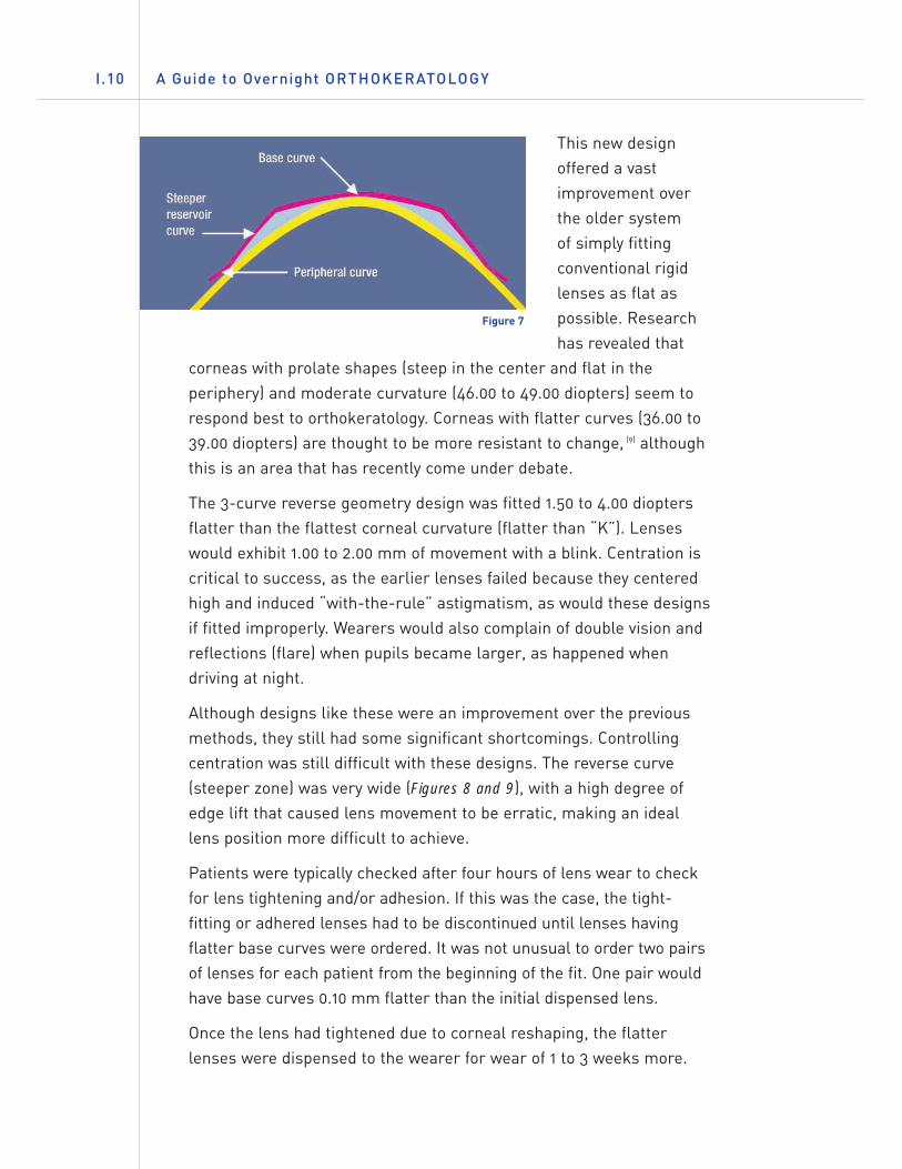
This new designoffered a vastimprovement overthe older system of simply fittingconventional rigidlenses as flat aspossible. Researchhas revealed that
corneas with prolate shapes (steep in the center and flat in theperiphery) and moderate curvature (46.00 to 49.00 diopters) seem torespond best to orthokeratology. Corneas with flatter curves (36.00 to39.00 diopters) are thought to be more resistant to change, (9) althoughthis is an area that has recently come under debate.
The 3-curve reverse geometry design was fitted 1.50 to 4.00 dioptersflatter than the flattest corneal curvature (flatter than “K”). Lenseswould exhibit 1.00 to 2.00 mm of movement with a blink. Centration iscritical to success, as the earlier lenses failed because they centeredhigh and induced “with-the-rule” astigmatism, as would these designsif fitted improperly. Wearers would also complain of double vision andreflections (flare) when pupils became larger, as happened whendriving at night.
Although designs like these were an improvement over the previousmethods, they still had some significant shortcomings. Controllingcentration was still difficult with these designs. The reverse curve(steeper zone) was very wide (Figures 8 and 9), with a high degree ofedge lift that caused lens movement to be erratic, making an ideallens position more difficult to achieve.
Patients were typically checked after four hours of lens wear to checkfor lens tightening and/or adhesion. If this was the case, the tight-fitting or adhered lenses had to be discontinued until lenses havingflatter base curves were ordered. It was not unusual to order two pairsof lenses for each patient from the beginning of the fit. One pair wouldhave base curves 0.10 mm flatter than the initial dispensed lens.
Once the lens had tightened due to corneal reshaping, the flatterlenses were dispensed to the wearer for wear of 1 to 3 weeks more.
A Guide to Overnight ORTHOKERATOLOGYI.10
Figure 7

A third pair of lenses would then beordered in preparation for the nextlens change. It was essential thateach flatter lens also have theappropriate power compensation toassure optimum vision during dailylens wear.
The method of making four to five small incremental (flatter) base curve changes allowed better control of lens centration.Initial reduction in myopia afterapproximately two to seven dayswould be approximately 1.00diopter, with additional myopicreduction occurring over theremainder of the three- to six-month treatment period, reaching a maximum of 2.00 to 3.00 dioptersmyopic reduction.(8)
Background I.11
Figures 8 & 9.
Steep-wide reverse curve on 3-zone designs
KEY POINT SUMMARY…
• Myopia is one of the most common ocular disorders affectinghumans.
• Orthokeratology has been used in some form since the 1960s in the USA.
• Early ortho-k fitting was time consuming and expensive, withunpredictable results.
• Early reverse geometry ortho-k shaping lens designs made myopicreduction more predictable and achievable.

A Guide to Overnight ORTHOKERATOLOGYII.12
I I . MODERN ORTHOKERATOLOGY
IN THIS CHAPTER…
• Modern ortho-k designs are discussed and compared to theirpredecessors.
• Theories for how ortho-k treatment effects myopic reduction arepresented.
• This chapter discusses the advantages of overnight (accelerated)orthokeratology versus previous methods.
Theory/Mechanisms
The new orthokeratology designs have allowed the reshaping processto take place rather quickly. This accelerated form of ortho-k (alsoknown as AOK) offers some immediate change after one-night wear of the shaping lens, with the remainder usually occurring over atreatment period of about 10–30 days.
There is debate regarding the actual mechanismby which orthokeratology works. Some believethat the rigid shaping lenses actually bend thecornea to reshape it and thereby reduce myopia. (23)
Other studies seem to indicate that the cornealshape changes in orthokeratology are temporary,demonstrating that the cornea is elastic and has a“memory” (the cornea will resume its pre-ortho-kshape, once lens wear has been discontinued).This is contrasted to a “plastic” change (one wherethe cornea is permanently molded into a differentshape by wear of a rigid lens). Polse’s result inthis study showed that whatever the mechanism,
Figure 10. Example, before
ortho-k treatment
Figure 11. Example, after
ortho-k treatment

the changes to the shape of the cornea and resultant myopic reductionwere temporary and reversed themselves once rigid contact lens wearwas discontinued. The question remained whether this was due to abending of the corneal surface or by another mechanism.
Newer studies suggest that ortho-k shaping lens fitting using reversegeometry GP designs may compress corneal tissue (in some fashion)rather than changing refractive error by bending the cornea, at leastafter initial adaptation. The hypothesis is that a thin layer of tear filmexists between the back of the ortho-k shaping lens and the centralcornea. These tear film “shear” forces act hydraulically to force acompression and possibly subsequent redistribution of very anteriorepithelial cells under the shaper from the center toward the periphery (29)
(Figures 10 & 11). This seems to refute the old theory that ortho-k lenswear changes myopic correction by permanently bending the cornea. Italso may explain why these patients can wear shaping lenses whosebase curves are flatter than “K” and still experience no central cornealstaining or irritation with well-fitted ortho-k shaping lenses.
A study conducted by Helen Swarbrick et al., of The University of New South Wales, Sydney,Australia in 1998 (32) evaluated topography andpachometry changes in accelerated ortho-k lenswearers over a 30-day period. Their studyconcluded that:
1. The corneal epithelial cell layer wasredistributed in some manner, leading to a thinning of the central cornea tosignificant levels across the cornealsurface.
2. There was concurrent thickening of the mid-peripheral cornea, particularly in the stromal layer.
These changes occurred with no apparent changeto the posterior cornea curvature . It was thecorneal epithelial cells that were affected insome manner by the fluid pressure effect of theaforementioned tear film “forces.”
Modern Orthokeratology II.13
Figure 12. Pre- and post-ortho-k
lens wear topography.
Pre-treatment cornea with steep
center and gradually flattening
periphery.
Post-treatment cornea with flat
center and steeper mid-periphery.

In theory, these tear film forces cause a compression that results in a redistribution of the epithelial cells (and possibly some stromal)toward the corneal periphery. (1, 32) This process of compression andredistribution is thought to produce a reduction in corneal sagittal
height, which results in a change(flattening) in corneal curvature ofthe eye (Figures 12 and 13). Thiscorneal shape change results inthe refocusing of the light rays onthe retina (macula) of the eye,reducing or eliminating the needfor myopic correction.
It must be remembered that thereduction of myopia, whether donepermanently by means of removingtissue by use of a laser (LASIK and
PRK) or by changing corneal shape by use of ortho-k shaping lenses,is measured in microns or thousandths of a millimeter. For example,the cornea is estimated to be approximately 540 microns in thicknessor about 0.54 millimeter (Figure 14).
Comparatively, a human hair is approximately 50 microns in thickness,the same thickness as the human corneal epithelium. So, the gradualredistribution of corneal mass in orthokeratology that takes placeunder the shaping lens is what accounts for the reduction in thesagittal depth and thickness of the cornea, and the resultant reductionin myopia.
A Guide to Overnight ORTHOKERATOLOGYII.14
Figure 13. Corneal flattening resulting in correct focus
of light rays on the retina
Figure 14. Corneal thickness by layer

Ortho-k Designs, Now
Depending on the fitting philosophy ofthe design being used, an initial basecurve is chosen that is 0.30 mm to 1.40 mm flatter than the flattestcorneal curvature (flat “K”). Thisoptical zone width may vary from 6.0 mm to 8.0 mm. Commonly aposterior optical zone diameter of 6.0 to 6.5 mm is most often used.
The secondary (reverse) lens curve of the shaping lens is chosensteeper than the base curve radius. This “reservoir” zone is commonly3.00 to 5.00 diopters steeper than the base curve radius, but may alsobe 9.00 D or more steeper than the base curve in some designs. Thewidth of the reverse curve ranges from 0.6 mmto 1.0 mm (Figures 15 and 16). All theseparameters can be manipulated individually to reach an optimal shaping lens fit and myopicreduction effect. In most cases, making achange in one parameter will also requiremaking a compensatory change in one or moreother parameters. In some fitting systems, theactual shaper parameter combinations areproprietary and protected, leaving the fitter torelate poor fitting characteristics to the lensmanufacturer who then provides another lenswith the parameters necessaryto improve the fit.
Peripheral curve radius isslightly steeper than what isused for conventional GP lensfits, producing an edge (edgelift) clearance of 60 to 70microns (0.06 mm to 0.07 mm).Edge clearance on conventionalGP lenses is typically 80 to 120microns (0.08 mm to 0.12 mm).
Modern Orthokeratology II.15
Figure 15
Figure 16. Reverse curve tear film profile
Modern four-zone ortho-k shapinglenses typically consist of:
• base curve
• reverse (steeper) curve
• fitting (alignment) curve
• peripheral curve

Modern three-zone ortho-k shaping lens designs were madeavailable by Roger Tabb, Jim Day (Fargo™), Donald Harris, and NickStoyan (Contex OK®). Modern four-zone designs were introduced bySami El Hage (CKR), Tom Reim (Dreimlens®/DreamLens™), Euclid(Emerald™), Don Noack and John Mountford (BE Retainer™), Rinehart-Reeves, Paragon CRT®, and others.
Daily Wear Orthokeratology Versus OvernightOrthokeratology
The limitations imposed by the poor physiologic effects of PMMA lenswear dictated that early ortho-k lens wear be limited to daily lenswear. The same is true today of ortho-k shaping lenses that are madeof gas permeable plastics of medium and low permeabilities. LikePMMA, these materials are not optimal for safe overnight ortho-kwear, since the new generation of ortho-k lens designs are typicallylarger and thicker than conventional gas permeable lenses.
Being limited to daily wear made the early ortho-k process moredifficult for the patient to endure in terms of comfort and consistentvision as compared to wearing conventional rigid lenses for visioncorrection alone. Added to this was the cost of this procedure in terms of the number of lenses required (eight pairs or more) and thetreatment period (nine to twelve months), with no way to accuratelypredict the visual result.
Ortho-k of decades past simplyemployed conventional rigid contactlens designs, progressively fit as flatas possible to reduce the height of the central cornea, thereby reducingmyopia. During the treatment, visionat times became worse due to poorlens positioning (high-riding lenses)that caused an increase in “with-the-rule” astigmatism or worse, cornealdistortion (Figure 17).
A Guide to Overnight ORTHOKERATOLOGYII.16
Figure 17

Comparing the ortho-k methods of the ’60s with the designs andtechniques of today is like comparing a 1930 Model A Ford to the latestmodel Mercedes Benz. This brings up the issue of what to call thismodality. In its beginnings in the 1960s, George Jessen called it“Orthofocus.” It soon came to be known simply as “orthokeratology.”Looking at the PDR Medical Dictionary definition, orthokeratology isdefined as: “A method of molding the cornea with contact lenses toimprove unaided vision.” And in fact, that’s exactly what it is. Theprocess involves the programmed application of a contact “shaping”lens for the purpose of systematically and predictably reshaping thecorneal surface to temporarily reduce the need for myopic correction.
Today, other names have been coined for the more modern processutilizing four and five zone reverse geometry designs, such as“accelerated orthokeratology” (AOK), “Ortho-K,” “AdvancedOrthokeratology,” Corneal Refractive Therapy (CRT ®), etc.
The advent of new high-permeability GP materials (ISO/Fatt Dk of 85or more) has allowed overnight wear of these ortho-k shaping lensesinstead of during the day.* This provides easy and fast lens adaptationfor the patient.
New innovative four, five, and six curve reverse geometry designs in large diameters have not only allowed for better control of position of the shaping lens, but have also provided ortho-k fitterswith a scientific and more accurate means to control and predictmyopic reduction. These modern ortho-k shaping lenses allow forrapid myopia reduction as well. What took nine to twelve months to achieve in the ’60s now will occur usually within 30 days.Approximately 70 to 80% of the patients treated with modern ortho-kshaping lenses achieve their desired myopia reduction with only onepair of shapers, as compared to the old process that often took eightor more pairs of conventional rigid contact lenses to achieve myopiareduction. The first approvals for overnight orthokeratology in theU.S. were obtained by Paragon Vision Sciences (CRT®) and EuclidSystems Inc. (Emerald Lens).
Modern Orthokeratology II.17
* Overnight orthokeratology should only be performed using GP lens materials and designsapproved for overnight use.

Is Ortho-k Myopia Control?
Orthokeratology treatment effects a rapid reduction in myopia. Thisreduction is temporary in nature and is maintained by regular nightlywear of the shaping lens as prescribed.
Since orthokeratology treatment results in this myopic reduction, it islogical that questions have arisen regarding the effect this processmight have on myopic progression. Among these question are:
• Would the systematic reshaping of the cornea to reducemyopia also slow its progression?
• If ortho-k treatment is discontinued, what (if any) is the effecton myopic progression? Will there be a “rebound” effect thatresults in an increased amount and rate of progression ofmyopia or will it slow?
There are no definitive answers or scientific evidence to address thesequestions at this point. The role that orthokeratology may play inaffecting myopic progression is currently under investigation.
A Guide to Overnight ORTHOKERATOLOGYII.18
KEY POINT SUMMARY…
• Overnight (accelerated) orthokeratology allows for treatment resultswithin approximately 30 days.
• Reverse geometry designs utilize tear film forces to effect cornealshape changes.
• Modern four and five zone designs allow better control of treatmentprocess.
• Patients easily accept overnight ortho-k because adaptation is rapidwith minimal awareness of the shaping lens.
• The role that orthokeratology may play in myopia control is currentlyunder investigation.

The Modern Ortho-k Fitting Process III.19
I I I . THE MODERN ORTHO-K
FITTING PROCESS
IN THIS CHAPTER…
• Modern orthokeratology involves the combination of reversegeometry designs, high permeability GP materials, and cornealtopography to understand the relationship of the shaping lenses tothe cornea.
• This chapter provides guidance to practitioners to help select andinterview potential ortho-k wearers.
• The pre-fitting screening examination is outlined with tips on how toevaluate patients as good candidates.
• A list of essential equipment to fit, evaluate, and manage ortho-kpatients in the practice is provided.
• An overview of the general fitting process is provided.
• Emphasized is the importance of using high Dk GP materials forovernight lens wear of orthokeratology shaping lenses.
• Possible complications are presented with a discussion on how touse corneal topography to assess fit, follow progress, and avoidunfavorable results.
For the purpose of explaining the ortho-k fitting process and theprogress of myopicreduction, we will speakin general termsregarding the use of four-zone ortho-k shapinglenses (Figure 18).
The base curve of theshaping lens (which is
Figure 18

purposely chosen to be flatter than the flattest central apical radius)applies pressure on the thin layer of tear film that lies between theback of the ortho-k shaper and the corneal surface. Even though theuse of fluorescein to observe the lens fit gives the appearance ofapical touch, a tear film layer of less than 10 microns exists.
Theoretically, these “tear film forces” cause the apical epithelial cellsto compress and possibly “migrate” toward the periphery. This cornealreshaping process creates a decrease in corneal sagittal height andcauses the cornea to become more spherical and flatter, therebyreducing or eliminating the need for myopic correction.
As epithelial mass is shifted toward the periphery, the steepersecondary curve (reverse curve) forms a tear reservoir where excesstear and the displaced corneal cells may form.
The mid-peripheral or fitting curve (also known as alignment curve) isactually the curve that allows the shaping lens to center and positionproperly on the eye. This curve is calculated to be in alignment(parallel) with the mid-periphery of the cornea.
The purpose of the peripheral curve on an ortho-k shaping lens is thesame as on any GP lens, to allow for tear circulation under the shaper,allow for its easy removal, and provide a means to remove debris fromunder the shaper.
Generally, an ortho-k fitter will calculate the parameters of the trialshaping lens with a design-specific nomogram or computer program.In some cases, “K”s and refraction are given to the laboratory tocalculate the shaping lens parameters. The trial shaping lens or theshaper to be dispensed is then placed on the patient’s eye and allowedto settle before it is evaluated to ensure that it does not tighten
A Guide to Overnight ORTHOKERATOLOGYIII.20
Three major developments changed not only the processby which ortho-k shaping lenses were fit, but also howthey are perceived. These are:
1. reverse geometry designs
2. the availability of high Dk GP lens materials
3. a better understanding of the relationship of theseshaping lenses to the cornea through the use ofcorneal topography.

excessively. There have been cases of “corneal responders” who will display corneal shape and refractive changes anywhere from 10 to30 minutes. (14, 33) This allows the fitter to decide whether the patient willrespond successfully to the treatment.
Once it is decided to allow the patient to wear the ortho-k shapinglenses overnight, the patient is instructed to return to the office asearly as possible (within a few hours of awakening) in the morning.Prior to wearing the shaping lenses overnight, the patient must beinstructed how to insert, remove, and care for the shapers. Under nocircumstances should patients be allowed to leave the office and wearthe shaping lenses overnight if they cannot confidently remove them.Patients should be instructed in shaper removal using the eyelids(blink/scissor method). At the practitioner’s discretion, the patient mayalso be supplied with and instructed in the use of the silicone rubber(DMV) lens remover.
Probably the most critical step in the fitting process is to examine thepatient early the first morning after the first night of ortho-k shapinglens wear. It is important for the fitter to assess position of theshaping lenses and the location of the flattened zone on the cornea for centration. This is accomplished by observing the shapers on theeyes and evaluating corneal topography after removal of the shapinglenses. Unaided visual acuity is also checked at this time. De-centeredshaping lenses will not produce the desired myopia reduction and mayeven cause corneal distortion. Shapers must also be checked foradherence and corneal integrity must be determined.
A significant degree of myopic reduction (1.00 to2.00 diopters) often occurs after the first nightof ortho-k shaper lens wear. If the initial shaperappears to have tightened, a shaping lens witha lower sagittal height should be fitted.
The patient should not be allowed to wear atight-fitting shaper in order to avoid metabolicand corneal distortion problems. Some fitters order a second shaperthat has a lower sagittal height at the same time that the initial shaperis ordered to facilitate this change easily.
The Modern Ortho-k Fitting Process III.21
A significant degree ofmyopic reduction (asmuch as 2.00 diopters)often occurs after thefirst night of lens wear.

As mentioned, a large part of the ortho-k effect occurs in the first sevendays of shaping lens wear, with maximum results usually achieved in one month. (19, 22, 30) In some cases, myopia reduction may take up to threemonths.
The initial goal in ortho-k shaping lens fitting is to achieve the desiredamount of myopia reduction. Having reached that point, the fitter’sgoal is then to try to reduce overnight shaper wear to a frequency thatwill still maintain the patient’s cornea in the desired shape and visualacuity level.
One benefit to the ortho-k treatment is that the process is reversible.Studies have shown that regression will usually occur in a period ofapproximately 30–90 days, (12, 25, 26) with most wearers showing completereversal in less time. While this factor represents an advantage oversurgical refractive correction, it is a disadvantage at the same time. For most, nightly wear of the shaping lens will be required to maintainmyopic reduction. For others, lens wear may be required only everysecond or third night. The last shaping lens worn that produced theoptimum corneal shape change is typically used for nightly wear tomaintain reduction.
Alternative Ways to Correct Nearsightedness
Myopia (nearsightedness) can be corrected by any method that reducesthe focusing power of the eye. The most common methods of correctionutilize eyeglasses or regular standard daily, extended, or continuouswear contact lenses. These represent a means of correcting myopia onlyduring the time that the eyeglasses or regular contact lenses are worn,with no lasting effect on the myopia. Other methods of correcting myopiainvolve various surgical procedures such as LASIK.
Patient Interview and Selection
Patient selection criteria will depend on the approach and philosophyof the fitter. Patient selection criteria are presented here using a“broad-range” approach.
A Guide to Overnight ORTHOKERATOLOGYIII.22

Orthokeratology Candidate Profile
• Age: juvenile to adult myopes
• Spherical refractive error: -1.00 D to -5.00 D spherical power correction
• Cylindrical refractive error: - 1.50 D or less “with-the-rule” corneal astigmatism - 0.75 D or less “against-the-rule” astigmatism
• Recreational and sports activities where periods withoutwearing visual correction are beneficial
• Those whose vocation requires unaided visual acuity forcertain periods, such as police, firemen, military, oroccupations where refractive surgery may be a cause forexclusion (deep-sea divers, high altitude pilots, etc.)
• Free of corneal dystrophies (e.g. keratoconus), oculardiseases, or any condition that may preclude the patient fromwearing any type of GP lens
• Motivated to undergo full or partial myopia reduction andwilling to return to the office for two to three months of activetreatment and every six months for passive treatment
• Committed to the initial and ongoing cost of ortho-ktreatment (See section on fees and costs)
• Practitioners should consult fitting information provided byspecific design/fitting systems.
Pre-Fitting Examination
This should include:
• Refraction (with dilation)
• Baseline topography (keratometry optional, but topography is a must)
• Tear film analysis
a. Schirmer test (quantitative)
b. Tear Break-Up Time or TBUT (qualitative)
• Biomicroscopy
The Modern Ortho-k Fitting Process III.23

Several fitting systems advocate the use of corneal shape factors or eccentricity comparison of central corneal curvature to temporalcurvature to predict which wearers might have good myopic reductionpotential. Other studies have not been able to verify this procedure asbeing indicative of myopic reduction. (12) However, these studies did findthat there was a correlation between the amount of refractive errorand the amount of myopia reduction that was achieved.
These were categorized by Carkeet, Mountford, and Carney (4) as:
Horner and Bryant (12) categorized the results of their retrospective study as:
Instruments Required/Suggested
The instruments required for ortho-k shaping lens fitting are the samethat are required for fitting conventional contact lenses.
• Topographer (preferred) or keratometer (minimal)*
• Slit lamp (biomicroscope)
• Autorefractor, phoropter, or universal trial frame
• Visual acuity charts
• Contrast sensitivity charts (may be helpful to quantify visualacuity)
* Certain shaping lens designs (DreamLens and Contex) utilize either corneal topography orkeratometry information to determine the initial diagnostic shaping lens.
A Guide to Overnight ORTHOKERATOLOGYIII.24
Very low (< 2.00 D): Decrease of 1.12 diopters myopia
(spherical equivalent)
Low (2.00 to 3.00 D): Decrease of 1.86 diopters myopia
(spherical equivalent)
Middle (3.00 to 5.00 D): Decrease of 2.15 diopters myopia
(spherical equivalent)
High (> 5.00 D): Decrease of 2.48 diopters myopia
(spherical equivalent)
Good responders: Decrease of > 1.50 diopters myopia
(spherical equivalent)
Moderate responders: Decrease of 0.75 to 1.50 diopters myopia
(spherical equivalent)
Poor responders: Decrease of < 0.75 diopter myopia
(spherical equivalent)
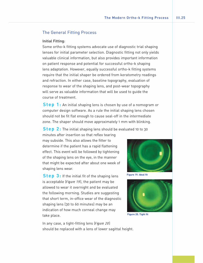
The General Fitting Process
Initial Fitting:
Some ortho-k fitting systems advocate use of diagnostic trial shapinglenses for initial parameter selection. Diagnostic fitting not only yieldsvaluable clinical information, but also provides important informationon patient response and potential for successful ortho-k shaping lens adaptation. However, equally successful ortho-k fitting systemsrequire that the initial shaper be ordered from keratometry readingsand refraction. In either case, baseline topography, evaluation ofresponse to wear of the shaping lens, and post-wear topography will serve as valuable information that will be used to guide the course of treatment.
Step 1: An initial shaping lens is chosen by use of a nomogram orcomputer design software. As a rule the initial shaping lens chosenshould not be fit flat enough to cause seal-off in the intermediatezone. The shaper should move approximately 1 mm with blinking.
Step 2: The initial shaping lens should be evaluated 10 to 30minutes after insertion so that reflex tearingmay subside. This also allows the fitter todetermine if the patient has a rapid flatteningeffect. This event will be followed by tighteningof the shaping lens on the eye, in the mannerthat might be expected after about one week ofshaping lens wear.
Step 3: If the initial fit of the shaping lens is acceptable (Figure 19), the patient may beallowed to wear it overnight and be evaluatedthe following morning. Studies are suggestingthat short term, in-office wear of the diagnosticshaping lens (30 to 60 minutes) may be anindication of how much corneal change maytake place.
In any case, a tight-fitting lens (Figure 20)should be replaced with a lens of lower sagittal height.
The Modern Ortho-k Fitting Process III.25
Figure 19. Ideal fit
Figure 20. Tight fit

Shaping Lens Adaptation and Follow-up
Step 4: Once the optimal initial shaping lens is chosen, the patientis instructed on insertion, removal, and care. Accelerated ortho-ktreatment allows patients to wear their shaping lenses overnightimmediately. For this reason, these patients should be seen in the office (wearing their shapers) the morning after the first night of wear.Shaping lenses are checked for:
• Centration: Centration of the shaping lens is critical to theortho-k effect. De-centered shapers do not produce the desiredmyopia reduction. The result will not only be poor visual acuity,but may also cause localized corneal distortion.
• Movement: Shaping lenses should move approximately 1 mmwith blinking. Shapers that are adhered during wear should bereplaced with shaping lenses of lower sagittal height.
• Fluorescein pattern: Note tear reservoirsize and shape, shaper lens movement;look for signs of adherence, air bubbles inthe reverse curve (reservoir) zone (Figures
21 through 23), change in central bearing(steeper appearance), staining (well-fittedshaping lenses should not cause staining).There should be no evidence of debristrapped behind the shaping lens.
Fluorescein pattern evaluation in ortho-kfitting has limited value. Since tear filmthicknesses under these lenses vary by only microns, it is almost impossible todifferentiate between acceptable andunacceptable fits using fluorescein.Therefore, the value of fluorescein evaluationis limited to observing the position of theshaping lens on the open eye, detectingadherence of the shaping lens, evaluatingcorneal integrity, and assessing reverse zonewidth and depth (Figures 21, 22, 23).
A Guide to Overnight ORTHOKERATOLOGYIII.26
Figure 21. Flat fit
Figure 22. Ideal fit
Figure 23. Steep fit

Since the fluorescein pattern in ortho-k shaping lens fitting is unusualand has many subtle nuances, use of a yellow Wratten filter isrecommended to accurately evaluate the fine detail of these fits(Figures 24 and 25).
Step 5:
• Remove shaping lenses: to check unaided vision. Perform aslit lamp examination of the cornea.
• Perform refraction: (or perform refractometry)
• Perform topography: to determine and record corneal shapechanges.
Characteristics of acceptable and unacceptable fitting of the ortho-k
shaping lens are as follows:
The Modern Ortho-k Fitting Process III.27
Figure 25.
Filter in front of slit lamp objective
Figure 24.
Boston® Wratten Yellow Slit Lamp Filter
Insufficient Ideal ExcessiveSagittal Height Sagittal Height Sagittal Height
Centration: Usually Well-centered Well-centered superior vertically and or inferior(may also horizontallybe inferior)
Central > 3 mm 3–5 mm < 3–5 mmalignment zone:
Reverse curve zone: Wide Wide, but Deep bubbles (Tear reservoir) tapered in reverse zone
approximately 50 μm deep
Mid-peripheral Reduced or Alignment Wide zone ofcurve: absent (uniform) heavy bearing
fluorescein 360°pattern 360°
Periphery: > 0.70 μm axial edge lift < 0.70 μm axial edge lift approx. 0.70 μm axial edge lift
Movement: > 2.0 mm 0.5–1.0 mm < 0.5 mm

The Importance of Using High Dk Materials
Questions have arisen regarding the safety of fitting rigid lenses forthe purpose of intentionally changing the shape (flattening) of thecornea. However, studies over time indicate that ortho-k shaping lens wear appears to be safe, with no permanent adverse visual andcorneal physiological effects.(8, 23, 27, 32) Even early studies using PMMAlenses reported no significant adverse effects that could be attributedto the disruption of the integrity and/or function of the cornealepithelium or any other ocular adverse effects. (27)
More conservative fitters may think orthokeratology opposes theprinciple of conventional rigid lens fitting—that contact lenses shouldnot alter the corneal surface when fitted “properly.” Yet, it is interestingto note that properly fitted reverse geometry shaping lens designsexhibit only 0.5 mm to 1.0 mm movement, similar to a soft lens, whilestill employing the “tear pump” mechanism for debris removal.
Most of the studies published to date agree that the myopic reductionresulting from wear of ortho-k shaping lenses is temporary. (3, 9, 12, 14, 25, 26)
That is, the myopic reduction effect lasts only as long as the patientwears the shaping lenses. Therefore, it has a “built-in” safeguard: if adverse reactions are noted, wear of the shaping lenses isdiscontinued and the cornea begins to revert back to its baselineshape. This process may take from 20 hours to 95 days. (12, 25, 26) Studiesindicate that recovery time for the cornea is based upon the individualshape of each cornea, as well as the amount of time ortho-k shapinglenses are worn (months, years, etc). It appears that the longer theseshapers are worn, the longer the recovery time required. (12, 25)
The advent of today’s techniques of accelerated orthokeratologyrequires that strict attention be paid to the GP lens material used tomake these specialized shaping lenses. Use of high permeabilitymaterials such as Boston ® XO or Boston ® Equalens ® II can reducehypoxic stress levels during overnight wear of these shapers.
Holden and Mertz found that limiting corneal edema levels to 4% orless during sleep requires a lens capable of 87 Dk/t using conventionalsoft contact lenses. (11) This conclusion was based on using thetransmissability formula x10-9(cm2/sec)(mlO2/ml x mmHg). It shouldalso be taken into consideration that GPs cover a smaller area of the
A Guide to Overnight ORTHOKERATOLOGYIII.28

cornea and the Dk ratings are generally higher than with conventionalsoft contact lenses.
There is another reason why use of high Dk materials is critical.Ortho-k shaping lenses generally have center thicknesses which aregreater than conventional GP lenses,(19) to avoid flexure and producethe desired, controlled corneal flattening.
Ortho-k Treatment Considerations
One of the most critical fitting criteria is centration of the ortho-kshaping lens (See “The Importance and Use of Topography”). Withoutgood centration (both vertical and lateral), visual acuity will not beoptimal and may be unacceptable to the wearer because the“treatment” zone will not be centered over the pupil and optical axis ofthe eye. This is why observation of the fit of the shaping lens,movement, visual acuity, and cornealtopography are so important and why the fittermust see the patient within several hours ofawakening in the morning after the first night of overnight wear of the shaping lens. Patientsmust not be allowed to continue to wearshapers that are misfit in any way.
Modern orthokeratology studies have shownthat the complication rate associated with thismodality is within the confines of acceptableclinical sequelae as seen in fitting conventionalGP lenses. (23, 32) Adaptation may even be easierand faster since the lenses are of largediameter and worn only at night during sleep,so issues of lid sensation are minimized.
Adherence (binding) of the shaping lenses isalways a possibility when a large diametershaping lens is worn overnight (Figures 26
through 28). Fitters often advise wearers toinstill several drops of the recommendedwetting/rewetting solution in each eye justbefore going to sleep. Upon awakening, severalmore drops should be instilled in each eye.
The Modern Ortho-k Fitting Process III.29
Figure 26. Slit lamp white light view
of adhered lens
Figure 27. Adhered lens viewed with
fluorescein
Figure 28. Epithelial indentation
(fluorescein staining) following lens
removal
Another option is not to remove the shaping lenses immediately uponarising. Oftentimes an adhered lens will begin to move spontaneouslyafter instillation of drops and a few minutes of blinking. Patients canalso be taught how to observe binding of a shaping lens with aflashlight and how to loosen a bound shaper manually, prior toremoval (See page 38). Patients should contact their eye carepractitioner for instructions if a bound lens does not move freely after 30 minutes.
Corneal staining levels are equivalent to those that are clinicallyacceptable for wear of conventional rigid lenses. Patients withinsufficient tear quantity and/or quality are not good candidates for ortho-k treatment, since the amount of corneal staining may rise to unacceptable levels in these patients.(17) It is also important todistinguish true corneal epithelial staining from that of mucus bindingto the corneal surface.
Corneal epithelial fluorescein staining may be noted centrally as theresult of either mechanical irritation (e.g. deposits on posterior side of lens) or corneal physiology (high oxygen demand cornea). Centralcorneal apical staining should not be present with a properly designedand fit ortho-k shaping lens. This underscores the need to use a Dkmaterial that offers reliable stability of the ortho-k shaper.
Dimple veiling (fluorescein staining of indentations in the epitheliumdue to air bubbles between the shaping lens and the cornea) occurs ifthe base curve, or depth of the reverse or reservoir curve is too steep.
Other complications such as irritation or infection may be associatedwith a lack of patient education and/or compliance with wearing and careinstructions (See page 36).
The two most common side effects that occur in any rigid contact lens wearer are corneal edema and corneal staining. It is anticipatedthat the same side effects may also occur in some wearers oforthokeratology shaping lenses to some degree. Other side effectsthat sometimes occur in all rigid contact lens wearers are extremediscomfort, redness, tearing, irritation, discharge, corneal abrasion or distortion of vision. These are usually temporary conditions if the shaping lenses are removed promptly and professional care isobtained. The wearer should be advised to remove the shaping lenses
A Guide to Overnight ORTHOKERATOLOGYIII.30

and not to re-insert them if any of these symptoms are present and tocontact the eye care practitioner immediately.
In rare instances, there may occur permanent corneal scarring,decreased vision, and infections of the eye, corneal ulcer, iritis, orneovascularization. The occurrence of these side effects should beminimized or completely eliminated if a proper schedule of shapinglens care and professional follow-up is exercised.
Importance and Use of Topography
The appearance of the cornea changes over time from the start of the treatment (Figure 29) until the desired result is attained. Thesechanges may occur rapidly at first (Figure 30), then slow as the corneaadjusts to its new shape and reaches the point where the desiredeffect is achieved (See Figures 31 and 32).
Topography is important to ensure that the treatment is being applied tothe center of the cornea. Ortho-k shaping lenses that position too high cancause a flattening of the superior cornea and may actually induce localizedcorneal distortion. These can be recognized by the typical “Smiley Face”inferior corneal steepening pattern (Figure 33) or “Frowny Face” superiorcorneal steepening (Figure 35). Shaping lenses that are fitted too steeply cancause certain areas of the central cornea to protrude forming small islandswhich will negatively impact visual acuity (Figure 34).
Shaping lenses that position too low will present a “Frowny Face”pattern indicating superior corneal steepening caused by pressure fromthe shaper on the lower cornea (Figure 35). The “Bull’s Eye” patternindicates that the shaping lens is centering acceptably both verticallyand horizontally (Figure 36). Lateral decentration is usually resolved byincreasing the overall diameter of the ortho-k shaping lens.
The Modern Ortho-k Fitting Process III.31

A Guide to Overnight ORTHOKERATOLOGYIII.32
Figure 29. Before OK treatment Figure 30. One day
Figure 31. One week Figure 32. Two weeks
Figure 33. “Smiley Face” pattern (left) caused by shaping lens positioned high (right)
Figure 34. Central Island (left) caused by steep-fitting shaping lens (right)

The Modern Ortho-k Fitting Process III.33
Figure 36. Bull’s Eye pattern indicating perfect centration of the shaping lens
Figure 35. “Frowny Face” pattern (left) caused by shaping lens that is positioning low (right)
KEY POINT SUMMARY…
• The combination of modern reverse geometry designs, high Dk GP materials, and corneal topographers make orthokeratologyfitting fast, safe, and more predictable.
• One of the most critical visits for both the patient and thepractitioner is the first morning after the shaping lenses are worn at night.
• An advantage of orthokeratology is that it is reversible.
• Both objective and subjective patient information must beconsidered when presenting options and evaluating a potentialwearer for ortho-k shaping lenses.
• Pre-fitting examination does not vary much from those routineexams done in the office.
• It has been suggested that patients who may expect the best chance for success with ortho-k treatment are those candidateswho have steep corneas with high eccentricity, although this is notalways the case.

A Guide to Overnight ORTHOKERATOLOGYIV.34
IV. PRACTICE MANAGEMENT
IN THIS CHAPTER…
• This section offers examples of fee structures for ortho-k shapinglenses and services.
• Follow-up programs are discussed for continued wear of ortho-kshaping lenses.
• Care and handling of these special designs is reviewed.
• Important points on incorporating orthokeratology into the practiceare presented.
• Examples of pre-treatment permission forms are presented.
• Helping the patient to decide if ortho-k treatment is the right visioncorrection modality.
Fees and Costs
Fitting of ortho-k shaping lenses involves higher costs for the fitter,both in terms of materials (shaping lenses) and increased time forfitting and follow-up. Therefore, fees for ortho-k treatment aresignificantly higher than for conventional GP lens fitting. That said,ortho-k fitting competes to some extent with refractive surgery andother vision correction modalities, such as conventional contact lensesand eyeglasses. As the number of laser surgical centers and LASIKprocedures increases, so does the competition for the same adultmyopic patients. Fees for ortho-k fitting vary from place to place andmay be comprehensive (all-inclusive) or based on the duration of thetreatment. Generally, all-inclusive fees are between $750 and $2,500,depending on the length of treatment required and the number of

Practice Management IV.35
shaping lenses needed. However, the average fees range from $1100.00to $1400.00 (US) for ortho-k shaping lenses and three months of visits.
Sample Fitting Fee Structure
Practitioner Cost Patient Fees Practice Income
Initial ocular exam/ —— $175 $175
consultation
Diagnostic fitting/ —— $200 $200
dispensing shaping lenses
Shaping lenses (4@$70/lens) $280 $350 $70
8 visits (@$75visit) —— $600 $600
TOTALS $280 $1325 $1045
Some fitters may also offer “Shaping Lens Service” Programs toprovide a yearly package for patient exams and shaping lensmaintenance.
Sample Shaping Lens Retainer Program Fees
Practitioner Costs Patient Fees Practice Income
Annual fee (1 yr/2 yrs) —— $200/300 $200/300
Includes 2 visits per year
Shaping lens replacement $70/lens $100/lens $30/lens
(lost, broken, parameter changes)
All ortho-k lens patients should be encouraged to have a spare pair ofshaping lenses in case they should break or lose one. Failure to wearortho-k shaping lenses regularly will lead to a regression of treatmentthat will result in a return to pre-treatment myopic state. Replacementof shaping lenses may be an additional cost to the wearer or may beincluded in the Shaping Lens Service Program fee. Since maintenanceof corneal shape is dependent on the ortho-k shaping lens remainingconstant in micron accuracy, it is strongly recommended that ortho-kshapers be replaced on a yearly basis.

How to Incorporate Ortho-k into the Practice
The introduction and incorporation of orthokeratology to the practicerequires a change in mindset for not only the practitioner, but also forthe office staff. The following steps are recommended prior tobeginning to offer orthokeratology:
1. Fitter research into the various ortho-k shaping lens designoptions.
2. Upgrade or refresh GP fitting skills as needed.
3. Education and training for the fitter in using the designchosen by attending courses, seminars and home study.
4. Training for the office staff on discussing the ortho-k optionwith patients, handling patient questions, and instructingortho-k wearers on lens care, insertion, removal, etc. of theortho-k shaping lenses.
5. Establishing office fees and policies for ortho-k treatment. Thisshould include development of ortho-k information sheets(FAQs), ortho-k shaping lens wear and care instructions, andPatient Fitting and Care Agreements for patients or parents ofminors to read and sign (See pages 40 and 41).
Ortho-k Shaping Lens Care and Handling
In order to minimize the potential for wearing complications such as eye irritations or serious infections, patients must be thoroughlytrained in the proper way to wear and care for their ortho-k shapinglenses, and using good hygienic methods whenever they handle theirortho-k shapers.
Preparing the Lens for Wearing
Cleanliness is a very important aspect of proper care of the shapinglenses. Hands should be clean and free of any foreign substanceswhenever the shaping lenses are handled.
• Always wash, rinse, and dry hands thoroughly beforehandling the shaping lenses.
A Guide to Overnight ORTHOKERATOLOGYIV.36

• Avoid soaps containing cold cream, lotions, or oily cosmeticsprior to handling shapers. These substances can adhere tothe surface of the shaping lens and be difficult to remove.
• Handle shaping lenses with the fingertips, avoiding use offingernails that can scratch or chip them.
• Always start with the same shaping lens first to avoid mix-ups.
• Remove the shaping lens from its storage case and examineit. Be sure it is clean, moist, and free of any nicks or cracks.
Placing the Lens on the Eye
After thoroughly washing and rinsing hands, follow these steps toinsert the shaping lens on the eye:
• Remove shaping lens from case.
• Rinse shaping lens with fresh conditioning solution.
• Inspect shaping lens for cleanliness, uniform wetness, andunwanted debris.
• Rub several drops of fresh conditioning solution over thesurface of the shaping lens.
• Place shaper on the top of index finger ofdominant hand.
• Hold down lower lid and lift upper lid upwith other hand (Figure 37).
• Gently place shaping lens on the center ofthe eye. It is not necessary to press theshaper on the eye.
• Gently release lids and blink. The shapinglens should center automatically.
• Use the same technique to insert the other lens.
• The wearer should be instructed to place two or three drops of the recommended rewetting solution in each eyeprior to wear.
Practice Management IV.37
Figure 37
Removing the Shaping Lenses
Before attempting to remove a shaping lens, it is very important thatthe wearer verify that it is moving. If the shaping lens is not moving,instill 5 drops of the recommended rewetting solution. Oftentimes anadhered lens will begin to move spontaneously after instillation ofdrops and a few minutes of blinking. Wait until the shaper begins tomove freely with the blink before attempting to remove it.
From the office, the practitioner should instruct the patient as follows:While looking upwards, a finger is placed at the lower eyelid margin atthe edge of the shaper to gently but firmly apply pressure. Lookingdownward, the process is repeated using the fingertip placed on theupper eyelid at the shaper edge. The patient should then look straightahead and blink several times.
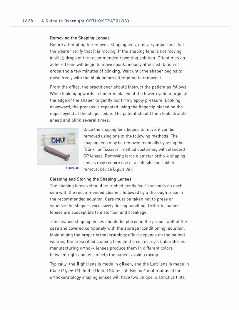
Once the shaping lens begins to move, it can beremoved using one of the following methods: Theshaping lens may be removed manually by using the“blink” or “scissor” method customary with standardGP lenses. Removing large diameter ortho-k shapinglenses may require use of a soft silicone rubberremoval device (Figure 38).
Cleaning and Storing the Shaping Lenses
The shaping lenses should be rubbed gently for 20 seconds on eachside with the recommended cleaner, followed by a thorough rinse inthe recommended solution. Care must be taken not to press orsqueeze the shapers excessively during handling. Ortho-k shapinglenses are susceptible to distortion and breakage.
The cleaned shaping lenses should be placed in the proper well of thecase and covered completely with the storage (conditioning) solution.Maintaining the proper orthokeratology effect depends on the patientwearing the prescribed shaping lens on the correct eye. Laboratoriesmanufacturing ortho-k lenses produce them in different colorsbetween right and left to help the patient avoid a mixup.
Typically, the Right lens is made in gReen, and the Left lens is made inbLue (Figure 39). In the United States, all Boston® material used fororthokeratology shaping lenses will have two unique, distinctive tints,
A Guide to Overnight ORTHOKERATOLOGYIV.38
Figure 38

red and yellow, for the same purposes.The red and yellow tints are availableexclusively in the U.S.
The ortho-k shaping lenses should beallowed to soak for a minimum of fourhours or as recommended on solutionlabel. If a multi purpose solution (such as Boston® Simplus™ or Boston®
Simplicity™) is used, all steps (includingprotein removal) are performed with theone solution only. If a two-bottle care system is used, a weekly enzymaticcleaner (such as the Boston® One Step Liquid Enzyme Cleaner) may berecommended forweekly use to ensurethat stubborn depositsare removed. In allcases, the wearershould be instructed tofollow the directionsfor use on eachsolution package.
Practice Management IV.39
Figure 39
The major benefits of orthokeratology are:
1. It is a non-surgical alternative for myopiamanagement.
2. Orthokeratology appears to be safe with few, if any, complications.
3. It is reversible with corneal shape returning tobaseline within about 90 days after discontinuingshaping lens wear.
4. Both juveniles and adults can benefit from thevisual freedom that ortho-k offers.
5. Very easy adaptation with large diameter ortho-kshaping lenses and overnight wear.
6. Limited or no daytime wear of correction lensesneeded once treatment correction is achieved.
KEY POINT SUMMARY…
• A complete ortho-k program that includes developing fee structures, refund policies, and staff training is essential for success of the modality.
• “Preparing the office” for orthokeratology includes thoroughtraining of the office staff.
• Thorough education of the ortho-k lens wearer on lens care andhandling of shaping lenses is essential for a successful outcome.
• A thorough discussion of the benefits that orthokeratology offerswill help the patient to make an intelligent decision about visioncorrection modalities.

Below is an example of an agreement form that also contains treatment fee information:
Hartsdale Vision Health Center
1355 West Main Street · Anywhere, USA 12345
(555) 123–4567
ORTHOKERATOLOGY FITTING AND CARE AGREEMENT
This document is supplemented by an ortho-k pre-treatment evaluation, aboutorthokeratology and care of ortho-k shaping lenses, which I have read andunderstood. All questions that I had were answered by Dr. (Name). Thisprogram involves me wearing specially designed gas-permeable shapinglenses overnight (while sleeping) that reshape my corneas in order to provideacceptable unaided distance vision during my waking hours. I understand thatthe ortho-k effect is temporary and reversible and that it may be necessary towear my shaping lenses longer to maintain satisfactory distance vision,especially if I failed to wear these shaping lenses as advised. I also understandthat the quality of my unaided vision is dependent on wearing these shapinglenses as prescribed by my doctor and on how much internal astigmatism ispresent in my eyes, which is not always predictable. If I do not find the resultsacceptable, the process will be reversed by my wearing rigid gas permeable orsoft contact lenses for about one to three weeks.
BENEFITS: These orthokeratology shaping lenses have been designed toprovide excellent visual acuity and oxygen transmission to the eye. The designof the shaping lens is believed to provide a reduction in the refractive error of atreated eye with a resultant improvement in the unaided distance visual acuity.This change is believed to be completely reversible and temporary in nature.
RISKS: While no harmful health risks to your eyes are anticipated from usingthe ortho-k shaping lenses, as with any contact lens there are potential risks of irritation to the eye, infections, or corneal ulcers. Transient distorted visionthat is not corrected with spectacle lenses may occur after removal of thelenses. No harmful effects are expected from any of the examinationprocedures used in the fitting process. If you develop any unusual symptoms or prolonged discomfort, removing the shaping lens should, in most cases,provide immediate relief. In addition, you should contact your eye carepractitioner immediately.
In the event it is determined that use of shaping these lenses presents newrisks or the possibility of undesirable side effects, you will be advised of thisinformation so that you may determine whether or not you wish to continueortho-k treatment.
ALTERNATIVES: Currently available alternatives to ortho-k shaping lensesare spectacles or other types of soft, conventional hard or gas permeablecontact lenses or surgical vision correction. Dr. (Name) or his/her staff candiscuss these alternatives with you.
A Guide to Overnight ORTHOKERATOLOGYIV.40
SAMPLE

FEE SCHEDULE
Initial consultation: Includes comprehensive evaluation of refractivestatus, corneal topography, and determinationof diagnostic ortho-k shaping lens parameters.$175.00 (payable at time of examination).
Diagnostic shaping lens trial: Includes evaluating the refractive changes overa period of three to five hours resulting fromthe wearing of diagnostic ortho-k shapinglenses $200 (payable at time of examination)
Treatment Program: $625 (Includes all visits for three months)
Retainer lenses: $300 $925 ($300 payable in advance and $625payable by fourth week scheduled visit.)
Refund policy: Should you wish to discontinue the treatmenton or before the fourth week’s scheduled visit,the balance of $625 will be credited or, if paidby cash or check, refunded.
I have read all of the above information regarding ortho-k shaping lenses. I understand what I have read and the circumstances have been explained tome. Although it is impossible for my eye care practitioner to inform me of everypossible complication, he/she has answered all my questions to my satisfactionand has assured me that he will advise me of new risks if they develop and willanswer any further inquiries I may have about this treatment or wearing thistype of lens. Should any complications occur, I agree to contact Dr. (Name)immediately at: (555) 123–2020 daytime or (555) 123–4321 after hours.
(please print)
Name:
Practitioner:
Address:
Date: Phone:
Signature:
If patient is under 18 years old, parent or guardian signature required.
Signature, Parent/Guardian:
Relationship to minor:
Signature, Witness:
Practice Management IV.41
SAMPLE
A Guide to Overnight ORTHOKERATOLOGYV.42
V. GLOSSARY
Ablate/Ablation The process of using a laser light to vaporize corneal
epithelial cells in a specific area (treatment zone) during a
LASIK or PRK refractive surgical treatment.
Accelerated orthokeratology The use of specially designed 4, 5, and 6 zone lens designs
that employ base curves calculated to be flatter than the
flat corneal curvature, reverse zones of steeper radii, and
alignment zones that provide improved lens positioning
and the positive-negative pressures between lens and
cornea necessary to effect desired myopic reduction
quickly and systematically, while worn during sleep.
“Against-the-rule” astigmatism The flattest (longest) radius of corneal curvature lies
vertically at 90° and the steepest (shortest) curve of the
cornea is horizontal at 180°.
Alignment Curve (AC) Based on specific design, this curve may also be called
Anchor Curve, Landing Zone, or Fitting Curve. Area of
posterior lens surface that aligns lens with mid-periphery
of cornea to control lens position and centration during
lens wear.
Anterior optical zone (AOZ) Front, central lens curve that (combined with the base
curve and refractive index) determines lens power.
Apical/Apex The extreme top or tip of a curve (e.g. corneal apex: tip of
the cornea).
Apical bearing Posterior lens curvature lightly touches the corneal apex.
Apical touch Posterior lens curvature rests on corneal apex.
Aspheric Not spherical. A posterior or anterior lens surface design
which progressively flattens at a given rate (eccentricity) as
the curve progresses toward the periphery.
Astigmatism Refractive defect where the refractive components of the
eye have differing powers in different meridians. The result
is that light rays will focus at more than one point inside
the eye.
Axial edge lift Vertical distance from the lens edge to an extension of the
base curve of a lens.

Back Optic Zone diameter Chord length measurement across back optic zone.
(BOZD)/Optic Zone diameter In ortho-k, determines size of central corneal
(OZD) treatment area.
Back Optic Zone radius The major central posterior curve of the lens that fits over
(BOZR)/Base curve (BC)/ the apex of the cornea.
Posterior optical zone (POZ)
Biomicroscopy/slit lamp exam Use of high power microscope and light source to examine
the anterior segments of the eye using various amounts of
magnification.
Bowman’s Membrane Second layer of the cornea located between the epithelium
and the stroma, approximately 10 microns in thickness.
“Bull’s Eye” topography Corneal topography map that shows acceptable centration
and central corneal flattening with an ortho-k shaping
lens fit.
“Central Island” topography Topography maps that show areas of steepness in the
central cornea caused by an ortho-k shaping lens that is
fitted too steeply, causing a squeezing of the cornea and
central steepening.
Contrast sensitivity test Contrast sensitivity tests plot the lowest contrast level at
which a person can detect an object of a given size to
detect variations in the quality of the image on the retina.
Cornea Transparent, small dome-shaped anterior portion of the
eye through which light rays pass into the eye.
Corneal astigmatism Condition where the toroidal (oval) shape of the cornea is
the reason that light rays are caused to focus at various
points within the eye.
Corneal staining Healthy corneal epithelial cells normally will not take
up fluorescein dye. Damaged or dead cells will allow
fluorescein dye to enter. The extent and location of this
staining is an indication of corneal irritation (mechanical
or chemical) and/or insufficient oxygen (hypoxia).
Cylindrical refractive Requiring a lens whose correcting power is located in one
error/power specific meridian.
Descemet’s Membrane Thin elastic fourth layer of the cornea that adds to the
flexibility of the cornea, approximately 10 microns in
thickness.
Dk Refers to the inherent permeability of a lens material to
allow the passage of gases through it. D = diffusion
coefficient, k = degree of material solubility.
Dk/L or Dk/t Refers to the amount of oxygen (gases) that pass through
a lens material of a specified thickness (“D” and “k”
references are same as above. “L” or “t” refers to specific
lens average thickness).
Glossary V.43
Dimple veiling Fluorescein staining of indentations made in the corneal
epithelium by trapped air bubbles under the lens.
Diopter Unit of measurement of light ray convergence or
divergence equal to the reciprocal focal length of a lens
in meters (e.g. a 2.00 diopter lens brings light rays into
convergence at 0.5 meter).
Eccentricity The rate of flattening of an aspheric curve measured as an
“e” value.
Edema (corneal) Condition where the corneal water content increases
causing a loss of clarity.
Edge lift (clearance) Distance between lens edge and the corneal surface.
Created by the width and radii of the peripheral curves.
Allows for tear circulation under the lens and aids in lens
removal.
Endothelium Fifth (innermost) layer of the cornea, responsible for
corneal metabolism and maintaining the water content of
the cornea. Approximately 5 microns in thickness.
Epithelial flap Use of a microkeratome instrument to incise and lift an
epithelial layer of the central cornea prior to applying laser
ablation.
Epithelium The outermost layer of the cornea comprised of
approximately five layers of cells that is approximately 50
microns in thickness.
Fitting curve (FC) Depending on specific ortho-k design, this may also be
called Alignment curve or Reverse curve.
Flat meridian/Flat “K” Meridian of the cornea having the longer radius of
curvature.
Fluorescein pattern The appearance of the tear film distribution and thickness
between the posterior of a rigid lens and the anterior
corneal curvature, as viewed with fluorescent dye that
stains the tear film.
Fluoro-silicone acrylate (F–S/A) An oxygen permeable rigid contact lens material
developed in the 1980s that combines fluorine for lens
stability, wettability, and added oxygen transmission; with
silicone for oxygen permeability; and methyl methacrylate
for stability, machinability, durability, and optical quality.
“Frowny Face” topography Topography map that shows a steepened area of the
superior cornea caused by an ortho-k lens that centers too
low on the cornea causing inferior flattening.
GP/RGP Gas permeable (rigid) contact lens.
High Dk Having permeability ratings 31 to 60 (ISO/Fatt).
Hyper Dk Having permeability ratings greater than 100 (ISO/Fatt).
A Guide to Overnight ORTHOKERATOLOGYV.44

Hyperopia Farsightedness; condition where the eye is underpowered
causing light rays to focus at a point behind the retina.
Correction is supplied using “plus” or “positive” powered
optical lenses.
Hypoxic stress Refers to corneal oxygen uptake rates on a given eye with
a lens of a given permeability or the lack thereof.
ISO/Fatt International Standard Organization (ISO) method for
measuring and stating oxygen permeability and other
values for all contact lens materials.
Keratoconus Degenerative corneal disease characterized by thinning
and cone-shaped protrusion of the central cornea.
Keratometry (K-readings) Use of a keratometer to measure central apical cornea
curvature zone of 3 to 4 millimeters in diameter.
LASEK Laser Assisted Subepithelial Keratectomy is a refractive
surgery procedure where a thin flap of epithelium only is
created using an alcohol solution to soften the epithelium
and a thin surgical blade to lift it. This process is said to be
beneficial for corneas too thin to have LASIK or PRK. The
underlying cornea is then treated with a laser to reshape
the central cornea. The flap is then replaced to heal.
LASIK Laser Assisted In-Situ Keratomileusis; same as LASEK
except a thicker hinged flap consisting of the epithelium
and some stroma is created using a microkeratome
instrument. The central cornea is then treated by laser
as in LASEK and PRK and the flap replaced to heal.
Low permeability GP materials having Dk ratings of less than 15 (ISO/Fatt).
Macular degeneration Degeneration of the macular portion of the retina which
leads to permanent loss of central vision.
Medium Dk Having Dk ratings between 15 and 30 (ISO/Fatt).
Micron One-thousandth of a millimeter. One average human cell
is 10 microns in height. One human hair is approximately
50 microns thick.
Myopia (nearsightedness) Nearsightedness; condition where the eye is over powered
causing light rays to focus at a point in front of the retina.
Correction is supplied using “minus” or “negative”
powered optical lenses.
Myopic progression Process by which myopia in an individual gradually
increases over a period of time, requiring the need for
increased myopic prescription changes.
Nomogram Calculation system (charts, tables, computer programs)
used to calculate appropriate diagnostic or final lens
design.
Glossary V.45

Oblate shape A curve whose central curve is nearly flat (similar to the
long curvature of an egg).
Ophthalmoscopy Direct or indirect examination of the internal structures of
the eye, especially the optic nerve, macula, and retina.
Optical zone (POZ)/Back Central portion of the posterior lens, not including the
Optical Zone radius (BOZR)/ peripheral curves, that the wearer sees through.
Base curve (BC)
Orthokeratology, ortho-k Use of gas permeable (rigid) contact lenses to gradually
and systematically alter the shape of the cornea to cause a
temporary and reversible reduction in myopia.
Ortho-k shaping lens, lens A specially designed gas permeable (rigid) contact lens
shaper, ortho-k shaper, used in ortho-k treatment to change the shape of the
retainer, etc. cornea to reduce sagittal height, refocus light rays onto
the retina, and temporarily reduce the myopic requirement
of the eye.
Overall diameter (OAD) Chord length of a lens across the widest point of lens.
or Total Lens Diameter
Over-refraction Refraction performed over an existing correction (contact
lens or ortho-k treatment) to gauge extent of under- or
over-correction.
Pachometry Measurement of corneal thickness.
Peripheral curve(s) (PC) Curve(s) of flatter radius added to the posterior surface
of the rigid lens edge to approximate the flattening of the
peripheral cornea. Essential for lens comfort, tear
circulation, debris removal, and to facilitate lens removal.
Permeability See “Dk.”
Phoropter Refraction device containing a large variety of spherical
plus and minus powered lenses, cylinder powers, prisms,
etc. used to determine the eye’s best optical correction.
Photorefractive keratectomy Use of eximer laser light to “sculpt” a prescribed area
(PRK) of the central cornea, creating a flatter zone and reducing
myopia.
Polymethylmethacrylate Plastic very similar to Plexiglas™ used in contact
(PMMA) lens manufacture from the 1930s as the original contact
lens material. Totally impermeable to gases.
Prolate shape A curve having a steeper (higher) center than its periphery
(similar to the curve at the tip of an egg).
Punctate staining Appearance of the cornea when stained with fluorescein
dye where scattered to diffuse areas of “pinpoint” staining
occur. This usually indicates corneal epithelial cell damage
or destruction.
A Guide to Overnight ORTHOKERATOLOGYV.46

Radial Keratotomy (RK) Radial incision made in the periphery of the cornea that
causes central corneal flattening with resultant reduction
in myopia.
Refraction Subjective test used to determine the refractive error
of the eye in order to prescribe the best correcting
lens powers.
Refractive error Internal optical defect where light rays are not focused
on the retina of the eye causing blurred vision.
Refractive surgery Use of incisions, implants, or laser light to alter the shape
of the corneal surface to affect vision.
Retainer lens Ortho-k shaping lens worn nightly as prescribed to maintain
(See shaping lens) the shape of the cornea and myopic reduction. Similar to
a retainer worn for teeth.
Reverse Curve Also known as Reservoir Curve, Fitting Curve, depending
on specific ortho-k design. Secondary curve with steeper
radius of curvature than preceding (Back Optic Zone
Radius/BOZR) curve. Area of shaping lens where corneal
mass will accumulate during ortho-k treatment.
Reverse geometry design Designs that use secondary curves that are steeper than
the preceding curve.
Sagittal depth The distance between the crest of an arc and a straight
baseline. Area under the arc of a curve depends on the
steepness or flatness of the curve radius.
Schirmer tear test Screening test that uses sterile filter paper strips to
determine tear production volume for a given time period.
Seal-off A contact lens whose edges have sealed against the
cornea 360° preventing tear exchange and debris removal.
Shaping lens See “Ortho-k shaping lens.”
Silicone acrylate (S/A ) Oxygen permeable plastic developed in the 1970s for
contact lenses (GP) that combines silicone for oxygen
transmissibility and methacrylate for optical quality, lens
machinability, and stability.
“Smiley Face” topography Corneal topography map that shows an area of inferior
steepening caused by an ortho-k shaping lens that rides
too high, causing superior corneal flattening.
Spherical equivalent Average power of a sphero-cylindrical lens equal to the
sum of the spherical power plus half of the cylindrical
power.
Spherical refractive error/power Requiring a lens whose correcting power is equal in all
meridians.
Steep meridian/Steep “K” Meridian of the cornea having the shorter radius of
curvature.
Glossary V.47

Stroma Middle (third) layer of the cornea composed of layered
collagen fibrils and cells, approximately 460 microns thick.
Comprises 90% of corneal thickness.
Super-Dk Having permeability ratings between 61 and 100 (ISO/Fatt).
Tear Breakup Time (TBUT) Time interval between a blink and the appearance of a
dry spot in the tear film. Indication of the quality of the
tear film.
Tear pump mechanism Dynamic mechanism created by lens movement with each
blink which causes an exchange of tears under the lens to
remove de-oxygenated tears and cellular debris.
Topography Corneal modeling instrument that produces color-coded
maps of the corneal surface using a variety of views.
Visual acuity Measure of the eye’s ability to distinguish objects, details,
and shape assessed by the smallest identifiable object that
can be seen at a specific distance (usually 20 feet for
distance and 16 inches for near).
“With-the-rule” astigmatism The flattest (longest) radius of corneal curvature lies
horizontally at 180° and the steepest (shortest) curve of
the cornea is vertical at 90°.
Wratten (yellow) filter A photographic yellow filter of a specific density that
allows the visualization of greater detail gradations in the
fluorescein stained-tear film.
A Guide to Overnight ORTHOKERATOLOGYV.48
Frequently Asked Questions VI.49
VI. FREQUENTLY ASKED QUESTIONS (FAQS)
IN THIS CHAPTER…
• The most often asked questions about orthokeratology by patients topractitioners.
• Questions that are frequently asked about orthokeratology fromfitters to the laboratory.
The FAQ (Frequently Asked Questions) section may be applicable tofitter, manufacturer, or consumer, depending on their knowledge oforthokeratology.
General
Q: How do ortho-k lenses work?
A: Ortho-k shaping lenses are designed to progressively reshape the central surface of the cornea systematically, similar to the effect of lasers in reversingnearsightedness. However, unlike laser surgery, the effect of ortho-k treatment istemporary and reversible.
Q: Is everyone a candidate for ortho-k?
A: No! While orthokeratology can help most individuals (including those with certaintypes and amounts of astigmatism and moderately high myopia) improve their vision,the procedure is often most effective for those prescriptions falling within a specificrange. Additional factors may include individual corneal rigidity and shape factors aswell as an ability to wear contact lenses. A thorough consultation and examinationusing advanced computerized diagnostic instrumentation can easily determine iforthokeratology is right for an individual.

Q: Who is not a candidate for ortho-k?
A: While ortho-k can be performed on practically anyone with healthy eyes who can insert a contact lens and follow lens care directions, it is not recommended forpeople with prescriptions above 5.00 diopters of myopia or above 1.50 diopters ofastigmatism. Also, people with pupils that are larger than normal (> 6 mm in normallight) and those having irregular corneal astigmatism or any corneal disorder suchas keratoconus are not candidates.
Q: How safe is ortho-k?
A: Ortho-k is believed to be safe when appropriately fitted and managed properly.Many people have been able to eliminate their dependence upon their glasses and standard contacts with no adverse effects. Unlike surgical procedures like radial keratotomy (RK), photorefractive keratectomy (PRK), laser assisted in-situkeratomileusis (LASIK), and laser assisted subepithelial keratectomy (LASEK), thecorneal tissue is not incised or vaporized by a laser. As with all contact lenses,proper lens care and handling must be performed to maintain eye health. The state-of-the-art, high-permeability GP materials now available provide adequate amountsof oxygen to the tissues of the eye.
Q: Is ortho-k permanent?
A: After treatment, maximum results are achieved and retainer shaping lenses areworn to stabilize and maintain the new corneal shape. Failure to wear the shapinglens on an ongoing basis will result in a return to the pre-existing prescription.Retainer shaping lenses will likely be prescribed for overnight wear.
Q: What are the risks of wearing lenses overnight?
A: The complications of wearing contact lenses include corneal ingrowth of vessels,ulcers, and abrasions.The risks associated with wearing contact lenses overnight arehigher than wearing contact lenses only while awake. Contact lenses cannot becomelost behind the eye (this is anatomically impossible) and it is rare for ortho-k shapinglenses to become decentered from the cornea.
Q: How long does it take for the process to stabilize and provide functional vision forthe whole day?
A: It generally requires four to seven consecutive nights of wearing the shapinglenses to achieve the desired result. It may take somewhat longer for those withhigher degrees of nearsightedness.
A Guide to Overnight ORTHOKERATOLOGYVI.50
Q: Are ortho-k lenses uncomfortable to wear?
A: Overnight wearing of the shaping lenses is surprisingly comfortable. Mostpatients are unaware of their presence within a very short time after insertion. Andbecause the ortho-k shapers are made in large diameters and worn during sleep,the normal adaptation process is very short.
Q: Will I still have to wear glasses or contacts?
A: Once the desired myopic reduction is obtained, the final shaping lenses act asretainers to maintain that level. Regular contact lenses are not needed. Glasses maybe needed for reading or other part-time use. During the initial treatment period, ifunaided vision does not last a full day, the patient will be given soft disposablecontact lenses to wear to maintain normal distance vision.
Q: Once the treatment phase is completed, how frequently will I need to wear theovernight retainer lenses?
A: Most people will need to wear the shaping lenses six to seven consecutive nights in order to enjoy good, unaided vision during the entire day. Patients withlesser degrees of myopia (nearsightedness) may find that wearing them every othernight is satisfactory. However, this will be determined on an individual basis by theeye care practitioner.
Q: How much myopia can a person realistically expect to be able to reduce usingorthokeratology?
A: Generally, 5.00 diopters of myopia is the upper limit for myopia reduction.However, work is underway on designs that will hopefully correct higher amounts of myopia. Lower amounts of myopia are easier and faster to reduce.
Q: How about ortho-k for astigmatism?
A: The amount of astigmatism reduction achievable will depend on the amount and the type. 1.50 diopters of “with-the-rule” corneal astigmatism and 0.75 diopterof “against-the-rule” corneal astigmatism are considered the upper limits forastigmatism reduction. Ortho-k will not have an effect on residual (internal)astigmatism.
Q: How long does it take to be able to see well without glasses?
A: Rapid visual improvement normally occurs in the first few days. Stabilization thenfollows over the next few weeks and months. Once the wearer’s eyes are stabilized,improved eyesight is maintained by wearing shaping lenses as recommended tomaintain vision at the desired level.
Frequently Asked Questions VI.51
Q: If the patient decides to return to wearing glasses, can vision be restored to itspre-treatment level?
A: The wearer’s eyes will return to their pre-treatment nearsightedness after abouttwo weeks. Depending on how long ortho-k lenses have been worn, this process maybe as long as 30–90 days. In order to provide good vision during this transitionalperiod, patients are refitted with gas permeable lenses or disposable soft lenseswhile the corneas recover their pre-treatment shape.
Q: If the patient becomes less nearsighted or presbyopic in later years how will thatbe handled?
A: Unlike laser surgery that cannot be reversed, corneas can usually be remodeledto accommodate the change in prescription by changing the design of themaintenance lenses to correct for near vision using a technique called “monovision.”
Q: How much does ortho-k cost?
A: The actual cost will depend on the complexity of the case. Each doctor sets feesaccordingly. Fees can range from $750 to $2,000 depending on length of treatmentand the number of shaping lenses needed. The doctor will be able to provide anestimate for each patient.
For the Fitter/Lab:
Q: What is the current prediction for the number of orthokeratology fits that will bedone worldwide?
A: The use of orthokeratology will vary from market to market. Current estimates inthe US market suggest that there could be as many as 50,000 to 100,000 ortho-k fitsannually. Much of this will depend on the acceptance of the professional community,potential candidates, and promotion from GP lens manufacturers and the media.
Q: Is it necessary to use topography to fit today’s ortho-k lenses or can I usekeratometry as I always have to fit GP lenses?
A: Use of topography is highly recommended in all phases of the fitting and follow-up. Some fitting systems base initial shaping lens selection on topographydata, while others recommend use of manual keratometry. Virtually all ortho-kfitting systems recommend the use of topographical data to establish a baselinereference as part of the fit evaluation, to identify and help solve fitting problems andto monitor the progress of myopic reduction.
A Guide to Overnight ORTHOKERATOLOGYVI.52
Q: What are the benefits of using four and five zone designs over the earlier threezone designs?
A: These modern four and five zone shaping lens designs use wider alignment (mid-peripheral) zones to achieve easier, more stable lens centration. Corneal shapechanges can be made in larger increments; myopia reduction occurs more rapidly,requiring only one pair of shaping lenses in a high percentage of cases. This makesmaterial costs to both fitter and patient lower.
Q: If ortho-k shaping lenses need modification or blending, can I do this in my officeto save time?
A: While in-office lens modification has long been a skill valued by lens fitters,modern ortho-k shaping lens designs are complex and precise. Any handmodification made by the fitter to improve the lens fit will probably be impossible to duplicate if a lens is lost, broken, or requires other parameter changes.
Q: How important is dispensing and the next-day visit in evaluating the overnightortho-k shaping lens fit?
A: Each patient will respond differently to orthokeratology treatment. These shapinglenses generally tighten with wear, which may occur in as little as an hour. It isimportant to evaluate the shaping lens after allowing it to settle-down in the office.Centration is the key to a successful ortho-k treatment; therefore, shaping lensesmust be checked early the first morning for centration, binding, and adhesion.Shaping lenses are then removed to evaluate topography and to check unaided visual acuity.
Q: If an ortho-k shaping lens is lost, broken, or not able to be worn, how can Iprovide suitable vision to the patient until the replacement lens arrives?
A: Soft disposable lenses can be prescribed until the proper replacement shapinglens is received. If the cornea begins to revert back to higher myopia, strongerdisposable lenses can be given. The wearer should be checked once again the dayafter a new shaping lens is worn overnight.
Frequently Asked Questions VI.53
KEY POINT SUMMARY…
• The success of orthokeratology in the practice and in the laboratoryis dependent on being able to address and adequately answer anyquestions that arise before, during, or after orthokeratologytreatment.
A Guide to Overnight ORTHOKERATOLOGYVII.54
VI I . SUGGESTED READING
• Orthokeratology Handbook, Winkler_TD, Kame_RT, ButterworthHeinemann, January 1995
• Orthokeratology: Principles and Practice, Mountford_J, Ruston_D,Dave_T, Butterworth Heinemann, March 2004, ISBN 0750640073
• Myopia and the Control of Eye Growth: Symposium No. 155,Back_G, John Wiley & Son, Ltd. February 1991, ISBN 0471926922
• Clinical Management of Myopia, Grosvenor_TP, Goss_DA,Hofstetter_HW, Butterworth Heinemann, October 1998, ISBN0750670606
• Controlled Kerato-reformation (CKR): An Alternative to RefractiveSurgery, El Hage_S, Practical Optometry, Canadian Edition,December 1999, 10:6
VII I . USEFUL ORTHO-K WEB SITES
• British Orthokeratology Societywww.boks.org.uk
• All About Visionwww.allaboutvision.com
• The Ortho-k Networkwww.ortho-k.net
• National Eye Research Foundation (NERF) www.nerf.org
Bibliography IX.55
IX. BIBLIOGRAPHY
1. Alharbi_A, Swarbrick_HA, “The Effects of Overnight Orthokeratology Lens Wear on
Corneal Thickness,” Investigative Ophthalmology and Visual Science, 44:6, June 2003
2. Binder_P, May_C, Grant_S, “An Evaluation of Orthokeratology,” American Journal of
Ophthalmology, 87:729, 1980
3. Brand_RJ, Polse_KA, Schwalbe_JS, “The Berkeley Orthokeratology Study, Part I: General
Conduct of the Study,” American Journal of Optometry and Physiologic Optics, 60:175,
1983
4. Carkeet_NL, Mountford_JA, Carney_LG,“Predicting Success with Orthokeratology Lens
Wear: A Retrospective Analysis of Ocular Characteristics,” Optometry and Vision Science,
Vol. 72, No. 12, 892–898
5. Coons_LJ, “Orthokeratology Part II: Evaluating the Tabb Method,” Journal of the
American Optometric Association, 55:409, 1984
6. Dave_T, Ruston_D, “Current Trends in Modern Orthokeratology,” Ophthalmic and
Physiology Optics, Vol. 18, No. 2, 1998
7. El Hage_S, “Photokeratoscopy and Controlled Keratoreformation,” paper presented at the
International Symposium of Ophthalmological Optics, Tokyo, Japan, May 1978
8. Grant_SC, “Orthokeratology: A Safe and Effective Treatment for a Disabling Problem,”
Survey of Ophthalmology, Vol. 24, No. 5, March–April 1980, 291–297
9. Harris_DH, Stoyan_N, “A New Approach to Orthokeratology,” Contact Lens Spectrum,
April 1992, 37–39
10. Holden_BA, Mertz_GW, McNally_JJ, “Corneal Swelling Response to Contact Lens Worn
Under Extended Wear Conditions,” Investigative Ophthalmology and Vision Science, 1983,
24:218–226
11. Holden_BA, Mertz_GW, “Critical Oxygen Levels to Avoid Corneal Edema for Daily and
Extended Wear Contact Lenses,” Investigative Ophthalmology and Vision Science, 1984,
25:1161–1167
12. Horner_DG, Bryant_MK, “Take Another Look at Today’s Ortho-k,” Review of Optometry,
June 1994
13. Jessen_G, “Orthofocus Techniques,” Contacto, 6:200, 1962
14. Kame_RT, “Flatten the Cornea the Nonsurgical Way,” Review of Optometry, April 1995
15. Kerns_R. “Research in Orthokeratology II: Experimental Design Protocol Method,” JAOA,
47:1275, 1976
16. Lin_LK, Shih_YF, Lee_YC, Hung_PT, Hou_PK, “Changes in Ocular Refraction and Its
Components Among Medical Students: A 5-Year Longitudinal Study,” Optometry and
Vision Science, 73, 1996, 495–498
17. Lu_Fan, Jiang_Jun, Qu_Jia, Jin_Wangqing, Mao_Xinjie, She_Yi, “Clinical Study of the
Orthokeratology for Young Myopic Adolescents,” presented at the Myopia 2001
Conference, Boston MA USA, July 2001
18. Lund_ME, Reim_TR, Wu_RLT, “Myopia Progression in Orthokeratology,” paper presented
at VII International Conference on Myopia, Taipei, Taiwan, 1999
19. Marsden_HJ, “Common Sense Orthokeratology,” Contact Lens Spectrum, Oct. 1995,
24–31
20. Morrison_RJ, “Contact Lenses and the Progression of Myopia,” Optometry Weekly, 1956,
47, 1487–1488
21. Mountford_J, “An Analysis of the Changes in Corneal Shape and Refractive Error Induced
by Accelerated Orthokeratology,” International Contact Lens Clinics, 1997, 24: 128–143
22. Mountford_J, Orthokeratology in Contact Lenses, Butterworth–Heinemann, Oxford UK,
1997, 653–692
23. Nichols_JJ, Marsich_MM, Nguyen_M, Barr_JT, Bullimore_MA, “Overnight
Orthokeratology,” Optometry and Vision Science, Vol. 77, No. 5, May 2000
24. Perrigin_J, Perrigin_D, Quintero_S, Grosvenor_T, “Silicone Acrylate Contact Lenses for
Myopia Control: Three-Year Results,” Optometry and Vision Science, 1990, 67:764–769
25. Polse_KA, Brand_RJ, Vastine_DW, Schwalbe_JS, “Corneal Change Accompanying
Orthokeratology: Plastic or Elastic?,” Archives of Ophthalmology, Vol. 101, 1983
26. Polse_KA, Brand_RJ, Schwalbe_JS, Vastine_DW, Keener_RJ, “The Berkeley
Orthokeratology Study, Part II: Efficacy and Duration,” American Journal of Optometry
and Physiological Optics, Vol. 60, No. 3, 1983, 187–198
27. Polse_KA, Brand_RJ, Keener_RJ, Schwalbe_JS, Vastine_DW, “The Berkeley
Orthokeratology Study, Part III: Safety,” American Journal of Optometry and Physiological
Optics, Vol. 60, No. 4, 1983, 321–326
28. Ramkuhar_S, Swarbrick_H, “Corneal Response to Short-Term Orthokeratology Lens
Wear,” Optometry and Vision Science, 80:3, March 2003
29. Reim_TR, “Overflowing With Ortho-k,” Optometric Management, December 1998, 36–39
30. Schachet_JL, “Optimal Lens Choices for Orthokeratology,” unknown publication
31. Stone_J, “Possible Influence of Contact Lenses on Myopia,” British Journal of Optometry,
1976, 31, 89–114
32. Swarbrick_HA, Wong_G, O’Leary_DJ, “Corneal Response to Orthokeratology,” Optometry
and Vision Science, 75:11, November 1998
33. Sridharan_R, Swarbrick_H, “Corneal Response to Short-Term Orthokeratology Lens
Wear,” Optometry and Vision Science, 80:3, March 2003, 200–206
34. Owens_H, Garner_LF, Craig_J, Gamble_G, “Posterior Corneal Changes with
Orthokeratology,” Optometry and Vision Science, 81:6, June 2004, 421–426
A Guide to Overnight ORTHOKERATOLOGYIX.56

© 2004 Polymer Technology, a Bausch & Lomb company.Boston, Boston XO, Boston Equalens II and Boston Simplus are trademarks ofPolymer Technology, a Bausch & Lomb company. Other product names/brand namesare trademarks of their respective owners. RIL0174
Polymer Technology 100 Research DriveWilmington MA 01887www.polymer.com