a general electrochemical method for label-free … · a general electrochemical method for...
TRANSCRIPT

A general electrochemical method for label-free screening
of protein–small molecule interactionsw
Kevin J. Cash,a Francesco Riccizb and Kevin W. Plaxco*bc
Received (in Cambridge, UK) 12th June 2009, Accepted 30th July 2009
First published as an Advance Article on the web 28th August 2009
DOI: 10.1039/b911558g
Here we report a versatile method by which the interaction
between a protein and a small molecule, and the disruption of
that interaction by competition with other small molecules, can
be monitored electrochemically directly in complex sample
matrices.
Methods for the detection and quantification of protein–small
molecule interactions have proven useful for a wide range of
purposes. Examples include drug screening, in which enormous
libraries of small molecules are tested for their ability to bind
or otherwise inhibit a specific protein, and competition assays
to detect small molecule targets via their ability to displace a
labeled (e.g., fluorescent) variant bound to a protein-based
specificity agent (e.g., an antibody).1
Recent efforts to develop fieldable competition assays for
the detection of nitroaromatic explosives, such as TNT,
illustrate the need for the convenient (e.g., rapid, robust,
portable) detection of protein–small molecule interactions.
Current EPA-accepted methods for the detection of these
materials in environmental samples, where they represent an
important class of pollutants,2 require the chromatographic
analysis of organic solvent extracts (e.g., EPA method 83303),
which, while sensitive (reported detection limits toB0.2 nM4),
are cumbersome batch processes ill-suited for field use. In
response, a number of alternative methods have been reported
in recent years in which TNT is detected indirectly via its
ability to compete with (i.e. competitively inhibit) the binding
of an anti-TNT antibody to a labeled (e.g., fluorescent) or
surface-attached TNT analog. Unfortunately, however,
fluorescence-based assays,4–7 while sensitive and convenient,
generally fail in samples that are optically dense or display
significant autofluorescence.8 Platforms based on surface-
attached TNT analogs, such as those based on surface plasmon
resonance (SPR),9–12 are likewise sensitive and convenient but
typically require complex, multi-step procedures in order to
avoid false positives that arise from the non-specific binding of
the interferants inevitably present in realistic samples.10 Thus,
although some progress has been made with regard to field-
portable immunosensors for the detection of TNT, these
devices inevitably require lengthy sample preparation to
minimize variability.13,14
Motivated by the growing need for convenient, rapid
methods to monitor protein–small molecule interactions
for analytical purposes, including competition assays and
the screening of combinatorial libraries, we report here a
bio-electrochemical approach that is not only user-friendly
but also selective enough to deploy directly in complex
sample matrices such as crude soil extracts and seawater.
Our approach is based on the previously described
‘‘E-DNA scaffold’’ platform, a reagentless, highly selective
bio-electrochemical method for the detection of protein–small
molecule interactions.15 The first generation E-DNA scaffold
sensors comprised a double-stranded DNA element in which
one strand (the anchoring strand) is tethered to an interro-
gating electrode and modified with a redox reporter
(analogous to Fig. 1).15 The complementary strand (the
recognition strand) was modified with a small molecule that
binds to the desired target. Upon target binding, the efficiency
with which the redox tag can approach the electrode is
reduced, impeding electron transfer and generating a readily
measurable change in faradaic current. Earlier applications of
E-DNA scaffold sensors were focused on the use of a small
molecule recognition element for the detection of specific
protein targets as the binding of such macromolecules
significantly reduces the collision rate of the attached
redox probe.
Here we explore alternative geometries for the E-DNA
scaffold sensing architecture and employ the approach to
monitor the binding of a family of small molecules, the
nitroaromatic explosives, to an anti-TNT antibody. Specifically,
we have fabricated a set of sensors employing the small
molecule recognition element dinitrophenyl (DNP). In the
presence of anti-TNT antibodies (e.g., clone TNT A/1.1.1
described in ref. 16) the signaling current of the sensor is
suppressed. When challenged with TNT or other cross-reactive
molecules,10,16 the antibody releases from the probe, producing
a readily measurable increase in signaling current (Fig. 1),
presumably due to an associated increase in probe dynamics
(see Fig. S1, ESIw).In an effort to optimize sensor performance, we have
explored both double- and single-stranded probes. The double-
stranded architecture, which is analogous to the first
reported sensors in this class,15 utilizes a 27-base anchoring
strand containing a 50 thiol and a 30 redox tag (methylene
blue). A DNP-containing recognition strand complementary
to (and centered on) this anchoring strand places the
DNP distal to the electrode (Fig. 1). We find that,
as with earlier sensors,15 the signal gain of this architecture
aDepartment of Chemical Engineering, University of California,Santa Barbara, Santa Barbara, CA 93106, USA
bDepartment of Chemistry and Biochemistry, University of California,Santa Barbara, Santa Barbara, CA 93106, USA.E-mail: [email protected]
c Program in Biomolecular Science and Engineering, University ofCalifornia, Santa Barbara, Santa Barbara, CA 93106, USAw Electronic supplementary information (ESI) available: Detailedexperimental methods as well as additional experimental data. SeeDOI: 10.1039/b911558gz Current address: University of Rome Tor Vergata, Rome, Italy.
6222 | Chem. Commun., 2009, 6222–6224 This journal is �c The Royal Society of Chemistry 2009
COMMUNICATION www.rsc.org/chemcomm | ChemComm
is a function of the length (and thus rigidity) of the
double-stranded region of the probe and the density with
which the probe DNAs are packed onto the sensor surface.15
Varying the length of the recognition strand from 15 to
25 bases (centered on a 27-base anchoring strand) we find that
optimal signal change is obtained with a 25-base recognition
strand (strand DNP25; Fig. S2, ESIw) with no significant
change in target affinity (Fig. S2, ESIw). This contrasts
with the B19-base optima observed for other sensors in this
class,15 indicating that the optimal recognition strand length
likely depends on the size and geometry of the target. The
gain of the sensor also increases strongly with decreasing
probe density, a trend that continues to the lowest densities
that produce a measurable faradaic current (Fig. S3, ESIw),an effect that has been observed previously.15 As the
signaling mechanism relies on a binding-induced change in
dynamics, higher surface coverage likely results in smaller
signal changes due to reduced collision dynamics in the
target-free state.
In an effort to better understand the ‘‘fabrication-space’’
of these sensors, we have explored 17-base, single-stranded
probe DNAs containing an anchoring thiol on their 50 termini
and a redox tag and DNP recognition element on their
opposite termini (Fig. S4, ESIw). We did so motivated by the
argument that the dynamics of single-stranded probes will also
be modulated by the binding of an antibody. We have
explored two single-stranded probes differing only in the
precise placement of the redox tag and the recognition
element: for probe sequence TL1 the redox tag is located on
the 30 terminus and the DNP receptor is on a thymine 3 bases
away, and for probe sequence TL2 these are reversed.
Unfortunately, however, neither of these single-stranded
probes respond robustly to the presence of anti-TNT
antibodies. That is, while the double-stranded scaffold
exhibits a 42% reduction in signal (using recognition
strand DNP25) at 30 nM antibody, the two single-stranded
scaffolds exhibit only 14% and 4% signal suppression under
these same conditions (TL1 and TL2 respectively, Fig. S4,
ESIw). The poor signaling of the single-stranded probes
presumably occurs because the dynamics of these highly
flexible, single-stranded probes do not change significantly
upon target binding, minimizing measured signal change
[see, by analogy, ref. 17]. The slightly greater suppression
observed with probe TL1 may be the result of steric
interactions between the antibody and the redox tag; for this
construct the antibody binds between the redox tag and
electrode surface. Given the generally poor performance of
our single-stranded probes, our follow-on studies employed
only double-stranded probes.
Optimal signaling upon TNT detection is achieved at an
intermediate concentration of the antibody. If the antibody
concentration is too low, some probes are unoccupied even in
the absence of the small molecule target, thus increasing the
background current observed in the absence of TNT.
Alternatively, the sensor’s detection limit will also be degraded
if the concentration of anti-TNT antibody is too great as
excess antibody will sequester some of the free TNT without
contributing to the observed signal change. We accordingly
employed our antibody at the 4 nM affinity of DNP25 in order
to balance these competing effects.
Our electrochemical assay readily and conveniently detects
the interaction between TNT and its antibody, and thus,
in turn, it can also sense TNT itself at low nanomolar
(parts-per-trillion) concentrations. For example, the IC50
(the concentration at which the signal change is half
that at saturation) of the optimized sensor is just 3 nM,
corresponding to 700 ppt (Fig. 2, top). The approach is also
selective: the observed TNT IC50 is not changed when the
sensor is challenged with complex samples such as seawater
and soil extracts (Fig. 2, top, inset), and sensor performance
remains excellent when challenged with crude cell lysates,
further supporting its potential utility in, for example,
the screening of candidate drug libraries (Fig. S5, ESIw).In terms of convenience, the sensor is rapid and reusable.
For example, at 50 nM TNT the sensor exhibits single
exponential relaxation kinetics with a time constant
of B2 min (Fig. 2, bottom). Finally, the sensor is readily
reusable: the addition of saturating TNT (to release the
antibody) followed by rinsing with buffer and adding fresh
antibody regenerates the sensor, allowing it to be reused
multiple times without exhibiting significant degradation
(Fig. S6, ESIw).The specificity of the assay (the ability to discriminate
between TNT and its close analogs) is high (Fig. 3), and is
comparable to that of the antibody itself.16 For example,
whereas the IC50 of TNT is just 3 nM, the IC50 of the
close analog 2,6-DNT is some three orders of magnitude
higher and that of dinitrophenol another order of magnitude
higher still, comparable to prior studies.16 Likewise, no
binding is observed for 4-(dimethylamino)phenylacetic acid,
a small aromatic compound of similar size lacking
nitro groups.
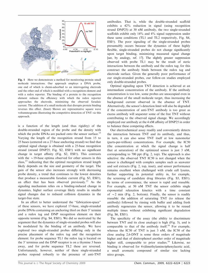
Fig. 1 Here we demonstrate a method for monitoring protein–small
molecule interactions. Our approach employs a DNA probe,
one end of which is chemi-adsorbed to an interrogating electrode
and the other end of which is modified with a recognition element and
with a redox reporter. The binding of a protein to the recognition
element reduces the efficiency with which the redox reporter
approaches the electrode, minimizing the observed faradaic
current. The addition of a small molecule that disrupts protein binding
reverses this effect. (Inset) Shown are representative square wave
voltammograms illustrating the competitive detection of TNT via this
approach.
This journal is �c The Royal Society of Chemistry 2009 Chem. Commun., 2009, 6222–6224 | 6223

Here we have demonstrated an electrochemical assay by
which small molecule–protein interactions are rapidly
and conveniently monitored even in crude, highly complex
sample matrices. The approach appears to be versatile,
requiring only the ability to conjugate an appropriate small
molecule recognition element to DNA in a manner that
does not significantly disrupt protein binding. Moreover,
as an electrochemical method our approach is easily scalable
and parallelizable18 for applications such as microtiter
plate-based assays.19 These attributes suggest our approach
is well suited for both massively parallel drug screening in the
laboratory and the detection of specific small molecules in
the field.
This work was supported by the NIH through grant
GM062958-01 and by the Institute for Collaborative
Biotechnologies through grant DAAD19-03-D-0004 from
the US Army Research Office. K. J. C. is supported by funds
from the California HIV/AIDS Research Program of the
University of California, Grant Number D07-SB-417.
Notes and references
1 L. Silverman, R. Campbell and J. R. Broach, Curr. Opin. Chem.Biol., 1998, 2, 397–403.
2 J. C. Spain, Annu. Rev. Microbiol., 1995, 49, 523–555.3 U. S. EPA, Washington, DC, 2006.4 U. Narang, P. R. Gauger and F. S. Ligler, Anal. Chem., 1997, 69,1961–1964.
5 G. P. Anderson, S. C. Moreira, P. T. Charles, I. L. Medintz,E. R. Goldman, M. Zeinali and C. R. Taitt, Anal. Chem., 2006, 78,2279–2285.
6 E. S. Bromage, T. Lackie, M. A. Unger, J. Ye and S. L. Kaattari,Biosens. Bioelectron., 2007, 22, 2532–2538.
7 E. R. Goldman, I. L. Medintz, J. L. Whitley, A. Hayhurst,A. R. Clapp, H. T. Uyeda, J. R. Deschamps, M. E. Lassmanand H. Mattoussi, J. Am. Chem. Soc., 2005, 127, 6744–6751.
8 J. C. Owicki, J. Biomol. Screening, 2000, 5, 297–306.9 T. Kawaguchi, D. R. Shankaran, S. J. Kim, K. V. Gobi,K. Matsumoto, K. Toko and N. Miura, Talanta, 2007, 72,554–560.
10 Y. Mizuta, T. Onodera, P. Singh, K. Matsumoto, N. Miura andK. Toko, Biosens. Bioelectron., 2008, 24, 191–197.
11 D. R. Shankaran, T. Kawaguchi, S. J. Kim, K. Matsumoto,K. Toko and N. Miura, Anal. Bioanal. Chem., 2006, 386,1313–1320.
12 D. R. Shankaran, K. Matsumoto, K. Toko and N. Miura, Sens.Actuators, B, 2006, 114, 71–79.
13 I. M. Ciumasu, P. M. Kramer, C. M. Weber, G. Kolb,D. Tiemann, S. Windisch, I. Frese and A. A. Kettrup, Biosens.Bioelectron., 2005, 21, 354–364.
14 P. R. Gauger, D. B. Holt, C. H. Patterson, P. T. Charles,L. Shriver-Lake and A. W. Kusterbeck, J. Hazard. Mater., 2001,83, 51–63.
15 K. J. Cash, F. Ricci and K. W. Plaxco, J. Am. Chem. Soc., 2009,131, 6955–6957.
16 A. Zeck, M. G. Weller and R. Niessner, Fresenius’ J. Anal. Chem.,1999, 364, 113–120.
17 F. Ricci, R. Y. Lai and K. W. Plaxco, Chem. Commun., 2007,3768–3770.
18 J. S. Swensen, Y. Xiao, B. S. Ferguson, A. A. Lubin, R. Y. Lai,A. J. Heeger, K. W. Plaxco and H. T. Soh, J. Am. Chem. Soc.,2009, 131, 4262–4266.
19 S. Piermarini, L. Micheli, N. H. S. Ammida, G. Palleschi andD. Moscone, Biosens. Bioelectron., 2007, 22, 1434–1440.
Fig. 2 (Top) As shown, the E-DNA scaffold readily detects protein–
small molecule interactions even in complex samples such as seawater
and crude soil extracts. The concentrations of half maximal inhibition
(IC50) observed in these complex sample matrices are within error of
the 3.1 � 0.5 nM observed in buffer. Data represent titrations
normalized to the same final signal gain. The inset shows the average
absolute signal gain for each condition.15 (Bottom) The assay is rapid;
shown are normalized sensor responses of three independently
fabricated sensors upon the addition of 50 nM TNT. The error bars
in these panels and the following figure represent the standard deviation
of three electrodes and are dominated by inter-electrode variability.
Fig. 3 Our approach is as specific as the protein–small molecule
interaction upon which it is based. For example, as expected,16 the
IC50 for 2,6-DNT is orders of magnitude poorer than that of TNT,
and the IC50 for dinitrophenol (DNPOL) is another order of magnitude
poorer still. No detectable response is observed for the non-nitroaromatic
control compound 4-(dimethylamino)phenylacetic acid (4DMAPA) even
at the highest concentrations we have employed.
6224 | Chem. Commun., 2009, 6222–6224 This journal is �c The Royal Society of Chemistry 2009