a fundamental system of cellular energy homeostasis regulated
TRANSCRIPT
A fundamental system of cellular energy homeostasisregulated by PGC-1�Lindsay M. Rohas, Julie St-Pierre*, Marc Uldry, Sibylle Jager, Christoph Handschin, and Bruce M. Spiegelman†
Dana–Farber Cancer Institute and Department of Cell Biology, Harvard Medical School, Boston, MA 02115
Contributed by Bruce M. Spiegelman, March 23, 2007 (sent for review February 23, 2007)
Maintenance of ATP levels is a critical feature of all cells. Mito-chondria are responsible for most ATP synthesis in eukaryotes. Weshow here that mammalian cells respond to a partial chemicaluncoupling of mitochondrial oxidative phosphorylation with adecrease in ATP levels, which recovers over several hours to controllevels. This recovery occurs through an increased expression of thetranscriptional coactivator peroxisome proliferator-activatedreceptor-coactivator 1� (PGC-1�) and mitochondrial genes. Cellsand animals lacking PGC-1� lose this compensatory mechanism andcannot defend their ATP levels or increase mitochondrial geneexpression in response to reduced oxidative phosphorylation. Theinduction of PGC-1� and its mitochondrial target genes is triggeredby a burst of intracellular calcium, which causes an increase incAMP-response-element-binding protein and transducer of regu-lated cAMP-response-element-binding proteins actions on thePGC-1� promoter. These data illustrate a fundamental transcrip-tional cycle that provides homeostatic control of cellular ATP. Inlight of this compensatory system that limits the toxicity of milduncoupling, the use of chemical uncoupling of mitochondria as ameans of treating obesity should be re-evaluated.
mitochondria � uncoupling
M itochondria are responsible for producing most of the ATPneeded for energy-requiring reactions in eukaryotic cells
(1). In the process of oxidative phosphorylation (OXPHOS), aproton gradient across the inner mitochondrial membrane iscoupled to the synthesis of ATP by the F1F0ATPase at complexV. Mitochondrial metabolism is dynamic and can be modulatedin response to external stimuli. For example, the �-adrenergicpathway is activated upon cold exposure in mice, which causesbrown adipose tissue to shift toward more uncoupled respiration(2–4). In uncoupled respiration, the protons pumped into theintermembrane space leak back into the mitochondrial matrix,bypassing ATP production and generating heat.
Mitochondria themselves can elicit intracellular signaling path-ways that alter nuclear gene expression, mitochondria number, andfunction (5, 6). This signaling process is referred to as retrogradesignaling. Retrograde signaling has been well studied in the buddingyeast, Saccharomyces cerevisiae. Yeast lacking mtDNA show in-creased expression of many genes important for the function ofmitochondria, such as citrate synthase, and several proteins havebeen identified in yeast that regulate retrograde signaling (7). Littleis known about the regulatory proteins involved in the mechanismsof retrograde signaling in higher eukaryotes. Similarly, how and towhat extent higher eukaryotic cells can defend their ATP levelsduring chronic challenges has not been extensively explored.
Peroxisome proliferator-activated receptor-coactivator 1�(PGC-1�) was first identified as a cold-inducible transcriptionalcoactivator of peroxisome proliferator-activated receptor � that canactivate a program of adaptive thermogenesis (8). PGC-1� hassince been shown to be a dominant regulator of mitochondrialfunction, biogenesis, and respiration in many tissues (9–13). Ectopicexpression of PGC-1� in white adipocytes increases cellular respi-ration and genes important for mitochondrial function, such asuncoupling protein 1, cytochrome c (Cyt c), and Cyt c oxidasesubunit II (COX II). Furthermore, increased expression of PGC-1�
increases mitochondrial volume density and cristae density (14).PGC-1� also can activate a program of fiber-type switching inskeletal muscle, including increased mitochondrial content and theexpression of myofibrillar proteins characteristic of type 1 and type2a muscle fibers (15). PGC-1�, the closest homolog of PGC-1�, hasalso been shown to regulate mitochondrial biogenesis and respira-tion in cells and transgenic animals (13, 16, 17).
2,4-Dinitrophenol (DNP), a chemical mitochondrial uncoupler,was widely used in the 1930s as a treatment for obesity (18–20).Mild doses were amazingly effective, often causing rapid loss ofadipose mass and body weight. While unregulated use led to reportsof toxicity and even death, it was not as lethal as might have beenexpected for a compound that disrupted OXPHOS. This toleranceindicates that cells can sustain mild chemical uncoupling, perhapsby using an energy compensatory mechanism. In the present study,we have investigated whether uncoupling of the mitochondrialmembrane potential could activate a compensatory pathway con-trolling ATP homeostasis. Indeed, chemical uncoupling increasesthe expression of PGC-1� and PGC-1�, as well as the expression ofseveral target mitochondrial genes. Studies using PGC-1� null cellsand animals indicate that PGC-1� is required for the induction ofmitochondrial gene expression, recovery of ATP levels, and cellsurvival. Together, these data illustrate a fundamental system ofenergy homeostasis whereby cells and tissues use PGC-1� torecover energy balance and also demonstrate that a powerfulcompensatory system exists that limits the toxicity of milduncoupling.
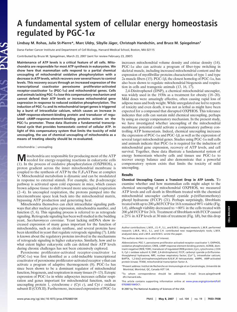
ResultsChemical Uncoupling Causes a Transient Drop in ATP Levels. Toexamine whether and how mammalian cells might adapt to thechemical uncoupling of mitochondrial OXPHOS, we measuredATP levels and cell death in fibroblasts treated with the chemicalmitochondrial uncoupler carbonyl cyanide p-(trifluromethoxy)phenyl hydrazone (FCCP) (21). Perhaps surprisingly, fibroblaststreated with up to 200 �M FCCP for 16 h remained 98% viable (Fig.1A), although viability decreased to �80% in the cells treated with200 �M FCCP for 24 h. Treatment of fibroblasts with FCCP causeda 25% in ATP levels at 30 min of treatment (Fig. 1B), but this drop
Author contributions: L.M.R., J.S.-P., S.J., and B.M.S. designed research; L.M.R. performedresearch; L.M.R., M.U., S.J., and C.H. contributed new reagents/analytic tools; L.M.R.analyzed data; and L.M.R. and B.M.S. wrote the paper.
The authors declare no conflict of interest.
Abbreviations: PGC-1, peroxisome proliferator-activated receptor-coactivator 1; OXPHOS,oxidative phosphorylation; CREB, cAMP-response-element-binding protein; ACREB, dom-inant-negative CREB; TORC, transducer of regulated CREB protein; Cyt c, cytochrome c; COXII, Cyt c oxidase subunit II; DNP, 2,4-dinitrophenol; FCCP, carbonyl cyanide p-(triflurome-thoxy)phenyl hydrazone; NRF, nuclear respiratory factor; [Ca2�]i, intracellular calcium;BAPTA, 1,2-bis(2-aminophenoxy)ethane-N,N,N�,N�-tetraacetate; AMPK, AMP-activatedprotein kinase; TFAM, mitochondrial transcription factor A.
*Present address: Institut de Recherche en Immunologie et en Cancerologie, Universite deMontreal, Montreal, QC, Canada H3T 1J4.
†To whom correspondence should be addressed. E-mail: bruce�[email protected].
This article contains supporting information online at www.pnas.org/cgi/content/full/0702683104/DC1.
© 2007 by The National Academy of Sciences of the USA
www.pnas.org�cgi�doi�10.1073�pnas.0702683104 PNAS � May 8, 2007 � vol. 104 � no. 19 � 7933–7938
CELL
BIO
LOG
Y
was transient, as ATP levels later recovered to those of theuntreated cells. We also examined the amount of lactate producedduring this experiment. Over 24 h, cells treated with FCCP secretedfour times more lactate than the untreated cells (Fig. 1C). Thisfinding suggests that glycolytic metabolism helps cells withstand adecrease in OXPHOS caused by chemical uncoupling.
We asked whether this mitochondrial uncoupling had any affecton mitochondrial density by using electron microscopy to quantifythe volume of mitochondria in cells treated with FCCP. Fibroblastswere exposed to 0 or 25 �M FCCP for 72 h, and mitochondrialvolume density was measured as described (14, 22). Fibroblaststreated with FCCP displayed a 1.8-fold higher mitochondrial vol-ume than control cells and higher levels of Cyt c protein [Fig. 1Dand supporting information (SI) Fig. 7], suggesting that chronicuncoupling induces a compensatory program that not only helps torecover ATP levels, but alters mitochondrial density well.
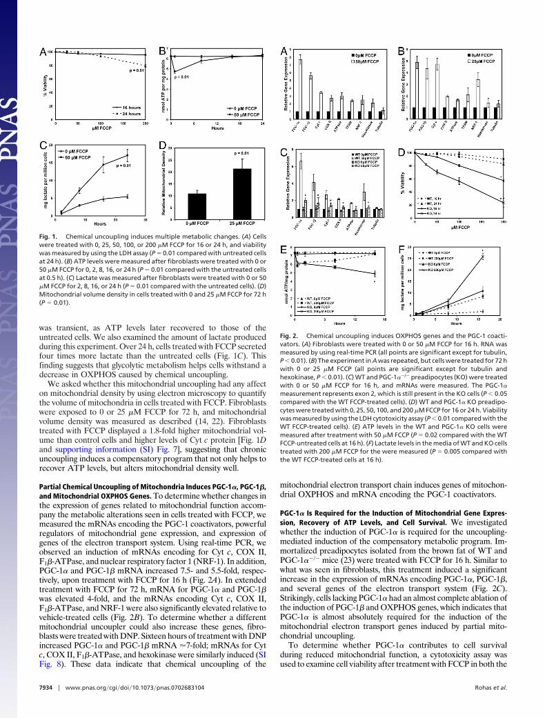
Partial Chemical Uncoupling of Mitochondria Induces PGC-1�, PGC-1�,and Mitochondrial OXPHOS Genes. To determine whether changes inthe expression of genes related to mitochondrial function accom-pany the metabolic alterations seen in cells treated with FCCP, wemeasured the mRNAs encoding the PGC-1 coactivators, powerfulregulators of mitochondrial gene expression, and expression ofgenes of the electron transport system. Using real-time PCR, weobserved an induction of mRNAs encoding for Cyt c, COX II,F1�-ATPase, and nuclear respiratory factor 1 (NRF-1). In addition,PGC-1� and PGC-1� mRNA increased 7.5- and 5.5-fold, respec-tively, upon treatment with FCCP for 16 h (Fig. 2A). In extendedtreatment with FCCP for 72 h, mRNA for PGC-1� and PGC-1�was elevated 4-fold, and the mRNAs encoding Cyt c, COX II,F1�-ATPase, and NRF-1 were also significantly elevated relative tovehicle-treated cells (Fig. 2B). To determine whether a differentmitochondrial uncoupler could also increase these genes, fibro-blasts were treated with DNP. Sixteen hours of treatment with DNPincreased PGC-1� and PGC-1� mRNA �7-fold; mRNAs for Cytc, COX II, F1�-ATPase, and hexokinase were similarly induced (SIFig. 8). These data indicate that chemical uncoupling of the
mitochondrial electron transport chain induces genes of mitochon-drial OXPHOS and mRNA encoding the PGC-1 coactivators.
PGC-1� Is Required for the Induction of Mitochondrial Gene Expres-sion, Recovery of ATP Levels, and Cell Survival. We investigatedwhether the induction of PGC-1� is required for the uncoupling-mediated induction of the compensatory metabolic program. Im-mortalized preadipocytes isolated from the brown fat of WT andPGC-1��/� mice (23) were treated with FCCP for 16 h. Similar towhat was seen in fibroblasts, this treatment induced a significantincrease in the expression of mRNAs encoding PGC-1�, PGC-1�,and several genes of the electron transport system (Fig. 2C).Strikingly, cells lacking PGC-1� had an almost complete ablation ofthe induction of PGC-1� and OXPHOS genes, which indicates thatPGC-1� is almost absolutely required for the induction of themitochondrial electron transport genes induced by partial mito-chondrial uncoupling.
To determine whether PGC-1� contributes to cell survivalduring reduced mitochondrial function, a cytotoxicity assay wasused to examine cell viability after treatment with FCCP in both the
Fig. 1. Chemical uncoupling induces multiple metabolic changes. (A) Cellswere treated with 0, 25, 50, 100, or 200 �M FCCP for 16 or 24 h, and viabilitywas measured by using the LDH assay (P � 0.01 compared with untreated cellsat 24 h). (B) ATP levels were measured after fibroblasts were treated with 0 or50 �M FCCP for 0, 2, 8, 16, or 24 h (P � 0.01 compared with the untreated cellsat 0.5 h). (C) Lactate was measured after fibroblasts were treated with 0 or 50�M FCCP for 2, 8, 16, or 24 h (P � 0.01 compared with the untreated cells). (D)Mitochondrial volume density in cells treated with 0 and 25 �M FCCP for 72 h(P � 0.01).
Fig. 2. Chemical uncoupling induces OXPHOS genes and the PGC-1 coacti-vators. (A) Fibroblasts were treated with 0 or 50 �M FCCP for 16 h. RNA wasmeasured by using real-time PCR (all points are significant except for tubulin,P � 0.01). (B) The experiment in A was repeated, but cells were treated for 72 hwith 0 or 25 �M FCCP (all points are significant except for tubulin andhexokinase, P � 0.01). (C) WT and PGC-1��/� preadipocytes (KO) were treatedwith 0 or 50 �M FCCP for 16 h, and mRNAs were measured. The PGC-1�
measurement represents exon 2, which is still present in the KO cells (P � 0.05compared with the WT FCCP-treated cells). (D) WT and PGC-1� KO preadipo-cytes were treated with 0, 25, 50, 100, and 200 �M FCCP for 16 or 24 h. Viabilitywas measured by using the LDH cytotoxicity assay (P � 0.01 compared with theWT FCCP-treated cells). (E) ATP levels in the WT and PGC-1� KO cells weremeasured after treatment with 50 �M FCCP (P � 0.02 compared with the WTFCCP-untreated cells at 16 h). (F) Lactate levels in the media of WT and KO cellstreated with 200 �M FCCP for the were measured (P � 0.005 compared withthe WT FCCP-treated cells at 16 h).
7934 � www.pnas.org�cgi�doi�10.1073�pnas.0702683104 Rohas et al.
WT and PGC-1� null cells. Whereas the WT cells showed very littledeath with up to 200 �M FCCP at either time point, the PGC-1�null cells showed a very substantial loss of viability (Fig. 2D). Only80% and 60% of the mutant cells survived 100 and 200 �M FCCP,respectively, after 16 h. After 24 h, only 30% of the mutant cellssurvived 200 �M FCCP compared with 85% of control cells. Thus,PGC-1� contributes substantially to the ability of these cells tosurvive partial uncoupling of the electron transport chain.
We next studied whether PGC-1� plays a role in the recovery ofthe ATP levels seen after treatment with FCCP (Fig. 1B). Both WTand PGC-1��/� cells displayed an initial 20% decrease in ATPlevels (Fig. 2E). After 16 h, the WT cells completely recovered theirATP levels to those of the untreated cells, whereas the PGC-1��/�
cells could not restore their ATP levels. By 16 h, the ATP levels inthe mutant cells were �68% of the ATP levels in the untreatedcells. In the same experiments, we measured lactate levels pro-duced. After 16 h, the FCCP-treated WT cells secreted approxi-mately three times more lactate than the untreated WT cells (Fig.2F). The untreated PGC-1��/� cells secreted significantly morelactate than both the treated and untreated WT cells, which wasalso true of the FCCP-treated PGC-1��/� cells. These experimentstogether indicate that the recovery of ATP levels and cell survivalseen after chronic treatment with FCCP requires PGC-1�.
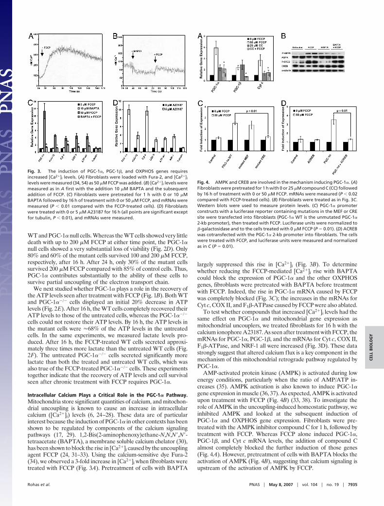
Intracellular Calcium Plays a Critical Role in the PGC-1� Pathway.Mitochondria store significant quantities of calcium, and mitochon-drial uncoupling is known to cause an increase in intracellularcalcium ([Ca2�]i) levels (6, 24–28). These data are of particularinterest because the induction of PGC-1� in other contexts has beenshown to be regulated by components of the calcium signalingpathways (17, 29). 1,2-Bis(2-aminophenoxy)ethane-N,N,N�,N�-tetraacetate (BAPTA), a membrane soluble calcium chelator (30),has been shown to block the rise in [Ca2�]i caused by the uncouplingagent FCCP (24, 31–33). Using the calcium-sensitive dye Fura-2(34), we observed a 3-fold increase in [Ca2�]i when fibroblasts weretreated with FCCP (Fig. 3A). Pretreatment of cells with BAPTA
largely suppressed this rise in [Ca2�]i (Fig. 3B). To determinewhether reducing the FCCP-mediated [Ca2�]i rise with BAPTAcould block the expression of PGC-1� and the other OXPHOSgenes, fibroblasts were pretreated with BAPTA before treatmentwith FCCP. Indeed, the rise in PGC-1� mRNA caused by FCCPwas completely blocked (Fig. 3C); the increases in the mRNAs forCyt c, COX II, and F1�-ATPase caused by FCCP were also ablated.
To test whether compounds that increased [Ca2�]i levels had thesame effect on PGC-1� and mitochondrial gene expression asmitochondrial uncouplers, we treated fibroblasts for 16 h with thecalcium ionophore A23187. As seen after treatment with FCCP, themRNAs for PGC-1�, PGC-1�, and the mRNAs for Cyt c, COX II,F1�-ATPase, and NRF-1 all were increased (Fig. 3D). These datastrongly suggest that altered calcium flux is a key component in themechanism of this mitochondrial retrograde pathway regulated byPGC-1�.
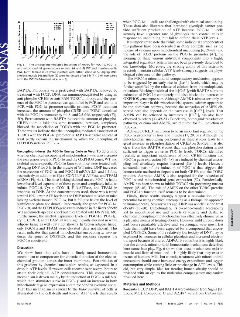
AMP-activated protein kinase (AMPK) is activated during lowenergy conditions, particularly when the ratio of AMP/ATP in-creases (35). AMPK activation is also known to induce PGC-1�gene expression in muscle (36, 37). As expected, AMPK is activatedupon treatment with FCCP (Fig. 4B) (33, 38). To investigate therole of AMPK in the uncoupling-induced homeostatic pathway, weinhibited AMPK and looked at the subsequent induction ofPGC-1� and OXPHOS gene expression. Fibroblasts were pre-treated with the AMPK inhibitor compound C for 1 h, followed bytreatment with FCCP. Whereas FCCP alone induced PGC-1�,PGC-1�, and Cyt c mRNA levels, the addition of compound Calmost completely blocked the further induction of those genes(Fig. 4A). However, pretreatment of cells with BAPTA blocks theactivation of AMPK (Fig. 4B), suggesting that calcium signaling isupstream of the activation of AMPK by FCCP.
Fig. 3. The induction of PGC-1�, PGC-1�, and OXPHOS genes requiresincreased [Ca2�]i levels. (A) Fibroblasts were loaded with Fura-2, and [Ca2�]i
levels were measured (34, 54) as 50 �M FCCP was added. (B) [Ca2�]i levels weremeasured as in A first with the addition 10 �M BAPTA and the subsequentaddition of FCCP. (C) Fibroblasts were pretreated for 1 h with 0 or 10 �MBAPTA followed by 16 h of treatment with 0 or 50 �M FCCP, and mRNAs weremeasured (P � 0.01 compared with the FCCP-treated cells). (D) Fibroblastswere treated with 0 or 5 �M A23187 for 16 h (all points are significant exceptfor tubulin, P � 0.01), and mRNAs were measured.
Fig. 4. AMPK and CREB are involved in the mechanism inducing PGC-1�. (A)Fibroblasts were pretreated for 1 h with 0 or 25 �M compound C (CC) followedby 16 h of treatment with 0 or 50 �M FCCP. mRNAs were measured (P � 0.02compared with FCCP-treated cells). (B) Fibroblasts were treated as in Fig. 3C.Western blots were used to measure protein levels. (C) PGC-1� promoterconstructs with a luciferase reporter containing mutations in the MEF or CREsite were transfected into fibroblasts (PGC-1� WT is the unmutated PGC-1�
2-kb promoter), then treated with FCCP. Luciferase units were normalized to�-galactosidase and to the cells treated with 0 �M FCCP (P � 0.01). (D) ACREBwas cotransfected with the PGC-1� 2-kb promoter into fibroblasts. The cellswere treated with FCCP, and luciferase units were measured and normalizedas in C (P � 0.01).
Rohas et al. PNAS � May 8, 2007 � vol. 104 � no. 19 � 7935
CELL
BIO
LOG
Y

Involvement of cAMP-Response-Element-Binding Protein (CREB) in theInduction of the PGC-1� Promoter. To elucidate the mechanism ofthis energy cycle further, we investigated the cis- and trans-actingfactors that play a role in the induction of the PGC-1� gene uponchemical uncoupling. Activity of a promoter segment containing 2kb of the 5� flanking region of the PGC-1� promoter driving aluciferase gene (29) was induced 3-fold upon treatment with FCCP(Fig. 4C). Transfections of the PGC-1� promoter harboring mu-tations in the MEF2 and CREB binding sites were analyzed.Promoter constructs with a mutation in the MEF2 binding siteshowed no difference in activity compared with the WT PGC-1�promoter (Fig. 4C). However, mutation of the CREB binding site(39) resulted in an almost complete ablation of the increase in thePGC-1� promoter activity in response to FCCP.
To investigate the role of the transcription factor CREB in theFCCP-mediated induction of the PGC-1� promoter, we examinedthe induction of the WT 2-kb PGC-1� promoter in the presence ofa dominant-negative version of the CREB protein (ACREB) (40).ACREB, a fusion of an acidic amphipathic domain and the leucinezipper dimerization domain of CREB, heterodimerizes with en-dogenous CREB and prevents it from binding to DNA. ACREBcompletely blocked the induction of the PGC-1� promoter ob-served upon treatment with 50 �M FCCP (Fig. 4D). TypicallyCREB protein requires activation by phosphorylation, often atSer-133. Fig. 4B shows that treatment with FCCP increases theregulatory Ser-133 phosphorylation of CREB. However, BAPTA,which blocks the induction of PGC-1� mRNA and OXPHOS geneexpression, does not block the phosphorylation of CREB. Thesedata together suggest a required role for CREB in the control of thePGC-1� promoter, but also suggest other regulated steps in thispathway must exist.
Involvement of Transducer of Regulated CREB Proteins (TORCs) in theRegulation of PGC-1�. We sought to identify other factors in thecalcium signaling pathway that were sensitive to BAPTA. Recently,
the TORC coactivators have emerged as important regulators ofCREB-dependent transcription, irrespective of the phosphoryla-tion state of CREB (41–48). Given this and their involvement incalcium signaling pathways (43, 44), the TORCs seemed to beplausible regulators in the pathway by which uncoupling inducesPGC-1�. To determine whether TORCs are involved in thePGC-1� energy cycle, fibroblasts were treated with FCCP. TORC1mRNA increased 1.6-fold, TORC2 mRNA increased 2.1-fold, andTORC3 mRNA increased 4.2-fold (Fig. 5A); pretreatment withBAPTA completely blocked the induction of all three TORCmRNAs. To determine whether TORCs play a role in the FCCP-mediated induction of the PGC-1� promoter, we measured theinduction of the WT 2-kb PGC-1� promoter in the presence of adominant-negative version of TORC (43). The dominant-negativeTORC protein (T1–44eGFP) is a fusion protein of the conservedCREB binding domain of TORC1 and eGFP. T1–44eGFP hasbeen shown to block the activation of CREB by TORC1, TORC2,and TORC3 (43). In the absence of T1–44eGFP, the PGC-1�promoter was induced 9.5-fold upon treatment with FCCP (Fig.5B). However, T1–44eGFP completely blocked the FCCP-mediated induction of the PGC-1� promoter, suggesting stronglythat TORCs are required for the induction of the PGC-1� promoterunder these conditions.
In a similar experiment, preadipocytes constitutively expressingT1–44eGFP were treated with FCCP for 16 h, and expression ofseveral mitochondrial genes was subsequently measured. Controlcells showed a 4.6-fold increase in PGC-1� mRNA and increases inPGC-1�, Cyt c, COX II, F1�-ATPase, and mitochondrial transcrip-tion factor A (TFAM) mRNAs (Fig. 5C). However, cells expressingT1–44eGFP had a blunted induction of mRNAs for all of thesegenes, again suggesting that TORCs are required for the PGC-1�energy cycle induced by uncoupling.
ChIP was used to measure the association of TORCs with theendogenous PGC-1� promoter in the presence of FCCP and
Fig. 5. TORC plays a critical role in the uncoupling-mediated induction of PGC-1� and mitochondrial genes. (A) Fibroblasts were treated as described in Fig.3C (P � 0.01 compared with the FCCP-treated samples), and mRNAs for TORC1, TORC2, and TORC3 were measured. (B) Fibroblasts were transfected with thePGC-1� 2-kb promoter, cotransfected with T1–44eGFP, and then treated with FCCP. Luciferase units were normalized to the cells treated with 0 �M FCCP (P �0.005). (C) Preadipocytes constitutively expressing T1–44eGFP or the eGFP control were treated with either 0 or 50 �M FCCP for 16 h, and mRNAs were measured(P � 0.01 compared with FCCP-treated control cells). (D) ChIP was used to measure phospho-CREB protein and TORC protein bound to the PGC-1� promoter upontreatment with FCCP. Fibroblasts were treated as described in Fig. 3C. Cells were cross-linked, and protein–DNA complexes were harvested by using phospho-CREBor PAN-TORC antibodies. The amount of PGC-1� promoter was then quantified by using PCR and real-time PCR with PGC-1�-specific primers (P � 0.02).
7936 � www.pnas.org�cgi�doi�10.1073�pnas.0702683104 Rohas et al.
BAPTA. Fibroblasts were pretreated with BAPTA, followed bytreatment with FCCP. DNA was immunoprecipitated by using ananti-phospho-CREB or anti-PAN TORC antibody, and the pres-ence of the PGC-1� promoter was quantified by PCR and real-timePCR with PGC-1� promoter-specific primers. FCCP treatmentincreased the amount of phospho-CREB and TORC associatedwith the PGC-1� promoter by �1.8- and 2.5-fold, respectively (Fig.5D). Pretreatment with BAPTA reduced the amount of phospho-CREB to �1.4-fold; this same treatment, however, completelyblocked the association of TORCs with the PGC-1� promoter.These results indicate that the uncoupling-mediated association ofTORCs with the PGC-1� promoter is BAPTA-sensitive and can atleast partly explain the mechanism by which the uncoupling ofOXPHOS induces PGC-1�.
Uncoupling Induces the PGC-1� Energy Cycle in Vivo. To determinewhether chemical uncoupling of mitochondria in vivo also increasesthe expression levels of PGC-1� and the OXPHOS genes, WT andskeletal muscle-specific PGC-1� knockout mice were treated with50 mg/kg DNP for 5 h. In the muscle of WT mice, DNP increasedthe expression of PGC-1� and PGC-1� mRNA 2.5- and 1.4-fold,respectively, in addition to Cyt c, COX II, F1�-ATPase, and TFAMmRNA (Fig. 6A). The mice lacking skeletal muscle PGC-1� had alower basal level expression of those genes and completely failed toinduce PGC-1�, Cyt c, COX II, F1�-ATPase, and TFAM inresponse to DNP. At the concentrations used, there was a trendtoward 10% lower ATP levels in the DNP-treated muscles in micelacking skeletal muscle PGC-1�, but it fell just below the level ofsignificance (data not shown). Importantly, the genes for PGC-1�,PGC-1�, and the OXPHOS genes were induced in the livers of bothWT and muscle-specific knockout mice treated with DNP (Fig. 6B).Furthermore, the mRNA expression levels of PGC-1�, PGC-1�,Cyt c, COX II, and TFAM all were significantly elevated in whiteadipose tissue as well (data not shown). In brown adipose tissue,only PGC-1� and TFAM were elevated (data not shown). Thisresult indicates that partial mitochondrial uncoupling in vivo in-duces the genes of OXPHOS, and this response requires thePGC-1� coactivator.
DiscussionWe show here that cells have a finely tuned homeostaticmechanism to compensate for chronic alteration of the electro-chemical gradient across the inner membrane. Perturbation ofthis gradient by chemical uncouplers results, as expected, in adrop in ATP levels. However, cells recover over several hours toattain their original ATP concentrations. This compensatorymechanism is driven mainly by the induction of PGC-1� mRNA,which then stimulates a rise in PGC-1� and an increase in bothmitochondrial gene expression and mitochondrial volume per se.That this mechanism is crucial to the basic survival of cells isillustrated by the cell death and loss of ATP levels that results
when PGC-1��/� cells are challenged with chemical uncoupling.These data also illustrate that increased glycolysis cannot pro-vide sufficient production of ATP because PGC-1��/� cellsactually have a greater rate of glycolysis than control cells inresponse to uncoupling, but fail to defend their ATP levels.
It is important to note that while some individual components ofthis pathway have been described in other contexts, such as therelease of calcium upon mitochondrial uncoupling (6, 24–28) andthe role of TORC proteins on the PGC-1� promoter (47), themerging of these various individual components into a highlyintegrated regulatory system has not been previously described toour knowledge. Moreover, the striking ability of this system toprecisely maintain cellular ATP levels strongly suggests the physi-ological relevance of this pathway.
The PGC-1� mitochondrial compensatory mechanism appearsto be triggered by an early rise in [Ca2�]i levels, which may befurther amplified by the release of calcium from the endoplasmicreticulum. Blocking this initial rise in [Ca2�]i with BAPTA stops theinduction of PGC-1� completely and also blocks the increases inOXPHOS gene expression. Although AMPK is almost certainly animportant player in this mitochondrial system, calcium appears tobe the dominant pathway, because the activation of AMPK ob-served here also depends on the early rise in [Ca2�]i levels. ThatAMPK can be activated by increases in [Ca2�]i has also beenobserved by others (33, 49–51). But clearly, both signal transductionpathways, calcium and AMPK, are required for full induction ofPGC-1�.
Activated CREB has proven to be an important regulator of thePGC-1� promoter in liver and muscle (17, 29, 39). Although themitochondrial uncoupling performed in these studies leads to agreat increase in phosphorylation of CREB on Ser-133, it is alsoclear from the BAPTA studies that this phosphorylation is notsufficient to trigger a rise in PGC-1� levels. TORCs, identifiedrecently as important modulators of both CREB function andPGC-1� gene expression (41–48), are induced by chemical uncou-pling and absolutely require increased [Ca2�]i levels. Hence, asubstantial part of the induction of PGC-1� that triggers thishomeostatic mechanism depends on both CREB and the TORCproteins. Activated AMPK is also required for the induction ofPGC-1� and mitochondrial gene expression (52). Paradoxically,AMPK has been shown to inhibit TORC2 via preventing nuclearimport (45, 46). The role of AMPK on the other TORC proteinsand PGC-1� function itself remains to be determined.
Finally, these results strongly suggest a re-evaluation of thepotential for using chemical uncoupling as a therapeutic approachto human obesity. Seventy years ago, DNP was widely used to treatobesity (18–20). Unfortunately, its over-the-counter accessibilityled to uncontrolled use and reports of toxicity and death, sochemical uncoupling of mitochondria was effectively eliminated asa plausible means of treating obesity. However, mild chronic doseshad been remarkably effective and, surprisingly, were much lesstoxic than might have been expected for a compound that uncou-pled OXPHOS. Some of the relatively low toxicity of DNP may beexplained by increases in cellular glycolysis and increased electrontransport because of altered ADP/ATP ratios, but it is highly likelythat the chronic mitochondrial homeostatic mechanisms describedhere come into play. Fig. 6 shows that these mechanisms exist inmuscle and liver of mice, and it is highly likely that they exist intissues of humans. Mild, but chronic, treatment with mitochondrialuncouplers should cause increased energy expenditure and oxygenconsumption while causing little or no change in ATP levels. Thisold, but very simple, idea for treating human obesity should berevisited with an eye to the molecular compensatory mechanismrevealed here.
Materials and MethodsReagents. FCCP, DNP, and BAPTA were obtained from Sigma (St.Louis, MO). Compound C and A23187 were from Calbiochem
Fig. 6. The uncoupling-mediated induction of mRNA for PGC-1�, PGC-1�,and mitochondrial genes occurs in vivo. (A and B) WT and muscle-specificPGC-1��/� female mice were injected with either saline or 50 mg/kg DNP.Skeletal muscle (A) and liver (B) were harvested after 5 h (P � 0.01 comparedwith the WT DNP-treated mice, n � 8).
Rohas et al. PNAS � May 8, 2007 � vol. 104 � no. 19 � 7937
CELL
BIO
LOG
Y
(San Diego, CA). Antibodies were from Cell Signaling (Danvers,MA) except for anti-Pan TORC (Calbiochem). The 2-kb PGC-1�promoter and promoter mutants have been described (29).ACREB was provided by Charles Vinson (40) (National Institutesof Health, Bethesda, MD). The dominant-negative TORC(T1–44eGFP) and control (eGFP) were provided by Novartis(Basel, Switzerland) (43). Immortalized preadipocytes fromPGC-1� WT and null mice (23, 53) and preadipocytes expressingT1–44eGFP were provided by Marc Uldry (Dana–Farber CancerInstitute).
Cell Culture, Transfection, and ChIP. 10T 1/2 fibroblasts were grownin DMEM (10% FBS). Immortalized preadipocytes from PGC-1�WT and �/� mice were cultured in DMEM (20% FBS). For thereporter gene assays, cells were transfected overnight with Super-Fect (Qiagen, Valencia, CA) and treated with FCCP for 24 h.Luciferase activity was normalized to �-galactosidase (Promega,Madison, WI), and then compared with the empty pGL3basicvector. PGL3basic vector, pSV vector, and eGFP served as thecontrols for the PGC-1� 2-kb promoter, ACREB, and T1–44eGFP,respectively. ChIP was performed with a ChIP-IT kit (Active Motif,Carlsbad, CA).
Analysis of Gene Expression. RNA was isolated by using TRIzol(Invitrogen, Carlsbad, CA) and measured by using iSCRIPT andSYBRGreen (Bio-Rad, Hercules, CA). mRNA levels were nor-malized to actin mRNA, and then relative mRNA levels weredetermined by using the ��Ct.
Electron Microscopy, Lactate, ATP, and Viability Measurements. ATPlevels were measured by using the ATP Determination Kit (In-vitrogen). Lactate in the media was quantified with Lactate Re-agent (Trinity Biotech, Bray, Ireland). Viability was measured byusing the LDH cytotoxicity kit (Roche, Indianapolis, IN). Electronmicroscopy was performed, and mitochondrial volume density wasmeasured as described (14, 22).
Calcium Measurements. Fura-2 ratios were used to calculate [Ca2�]as described (34, 54). For the measurements, EGTA was added tochelate the extracellular calcium. FCCP was then added followed bydigitonin and EGTA/Tris for calibration. When BAPTA was used,it was added before the addition of FCCP.
Animal Experiments. All animal experiments were performed ac-cording to a protocol approved by the Institutional Animal Careand Use Committee. Mice (see SI Text) were injected with 50 mg/kgDNP (90% saline, 10% DMSO solution) or 0 mg/kg DNP (90%saline, 10% DMSO solution). Mice were killed after 5 h.
Statistical Analysis. Results are expressed as �SD. Two-tailedStudent’s t tests were used to determine P values.
We thank Sherry Chin for assistance generating the PGC-1� muscle-specific �/� mice. This work was supported by National Institutes ofHealth Grants R01DK060837, NIDDK-DK54477, and DK61562 (toB.M.S). L.M.R. was supported by National Institutes of Health TrainingGrant/National Research Service Award 2-T32-GM07226-27.
1. Scheffler IE (1999) Mitochondria (Wiley-Liss, New York).2. Nicholls DG, Locke RM (1984) Physiol Rev 64:1–64.3. Lowell BB, Spiegelman BM (2000) Nature 404:652–660.4. Klingenberg M, Huang SG (1999) Biochim Biophys Acta 1415:271–296.5. Butow RA, Avadhani NG (2004) Mol Cell 14:1–15.6. Biswas G, Adebanjo OA, Freedman BD, Anandatheerthavarada HK, Vijayasar-
athy C, Zaidi M, Kotlikoff M, Avadhani NG (1999) EMBO J 18:522–533.7. Liu Z, Butow RA (2006) Annu Rev Genet 40:159–185.8. Puigserver P, Wu Z, Park CW, Graves R, Wright M, Spiegelman BM (1998) Cell
92:829–839.9. Kelly DP, Scarpulla RC (2004) Genes Dev 18:357–368.
10. Vega RB, Huss JM, Kelly DP (2000) Mol Cell Biol 20:1868–1876.11. Lehman JJ, Barger PM, Kovacs A, Saffitz JE, Medeiros DM, Kelly DP (2000)
J Clin Invest 106:847–856.12. Wu Z, Puigserver P, Andersson U, Zhang C, Adelmant G, Mootha V, Troy A,
Cinti S, Lowell B, Scarpulla RC, Spiegelman BM (1999) Cell 98:115–124.13. Lin J, Handschin C, Spiegelman BM (2005) Cell Metab 1:361–370.14. St-Pierre J, Lin J, Krauss S, Tarr PT, Yang R, Newgard CB, Spiegelman BM (2003)
J Biol Chem 278:26597–26603.15. Lin J, Wu H, Tarr PT, Zhang CY, Wu Z, Boss O, Michael LF, Puigserver P, Isotani
E, Olson EN, et al. (2002) Nature 418:797–801.16. Lin J, Puigserver P, Donovan J, Tarr P, Spiegelman BM (2002) J Biol Chem
277:1645–1648.17. Wu H, Kanatous SB, Thurmond FA, Gallardo T, Isotani E, Bassel-Duby R,
Williams RS (2002) Science 296:349–352.18. Parascandola J (1974) Mol Cell Biochem 5:69–77.19. Harper JA, Dickinson K, Brand MD (2001) Obes Rev 2:255–265.20. Cutting W, Mehrtens HG, Tainter ML (1933) J Am Med Assoc 101:193–195.21. Heytler PG, Prichard WW (1962) Biochem Biophys Res Commun 7:272–275.22. Weibel E (1979) Stereological Methods: Practical Methods for Biological Morphom-
etry (Academic, London).23. Uldry M, Yang W, St-Pierre J, Lin J, Seale P, Spiegelman BM (2006) Cell Metab
3:333–341.24. Luo Y, Bond JD, Ingram VM (1997) Proc Natl Acad Sci USA 94:9705–9710.25. Bernardi P, Paradisi V, Pozzan T, Azzone GF (1984) Biochemistry 23:1645–1651.26. Pozzan T, Bragadin M, Azzone GF (1977) Biochemistry 16:5618–5625.27. Sandoval ME (1980) Brain Res 181:357–367.28. Kessler RJ, Tyson CA, Green DE (1976) Proc Natl Acad Sci USA 73:3141–3145.29. Handschin C, Rhee J, Lin J, Tarr PT, Spiegelman BM (2003) Proc Natl Acad Sci
USA 100:7111–7116.30. Tsien RY (1980) Biochemistry 19:2396–2404.31. Yuan XJ, Sugiyama T, Goldman WF, Rubin LJ, Blaustein MP (1996) Am J Physiol
270:C321–C331.
32. Johnson JD, Chang JP (2005) Cell Calcium 37:573–581.33. Thors B, Halldorsson H, Thorgeirsson G (2004) FEBS Lett 573:175–180.34. Grynkiewicz G, Poenie M, Tsien RY (1985) J Biol Chem 260:3440–3450.35. Hardie DG, Carling D (1997) Eur J Biochem 246:259–273.36. Zong H, Ren JM, Young LH, Pypaert M, Mu J, Birnbaum MJ, Shulman GI (2002)
Proc Natl Acad Sci USA 99:15983–15987.37. Suwa M, Nakano H, Kumagai S (2003) J Appl Physiol 95:960–968.38. Hayashi T, Hirshman MF, Fujii N, Habinowski SA, Witters LA, Goodyear LJ
(2000) Diabetes 49:527–531.39. Herzig S, Long F, Jhala US, Hedrick S, Quinn R, Bauer A, Rudolph D, Schutz G,
Yoon C, Puigserver P, et al. (2001) Nature 413:179–183.40. Ahn S, Olive M, Aggarwal S, Krylov D, Ginty DD, Vinson C (1998) Mol Cell Biol
18:967–977.41. Conkright MD, Canettieri G, Screaton R, Guzman E, Miraglia L, Hogenesch JB,
Montminy M (2003) Mol Cell 12:413–423.42. Iourgenko V, Zhang W, Mickanin C, Daly I, Jiang C, Hexham JM, Orth AP,
Miraglia L, Meltzer J, Garza D, et al. (2003) Proc Natl Acad Sci USA 100:12147–12152.
43. Bittinger MA, McWhinnie E, Meltzer J, Iourgenko V, Latario B, Liu X, Chen CH,Song C, Garza D, Labow M (2004) Curr Biol 14:2156–2161.
44. Screaton RA, Conkright MD, Katoh Y, Best JL, Canettieri G, Jeffries S, GuzmanE, Niessen S, Yates JR, III, Takemori H, et al. (2004) Cell 119:61–74.
45. Koo SH, Flechner L, Qi L, Zhang X, Screaton RA, Jeffries S, Hedrick S, Xu W,Boussouar F, Brindle P, et al. (2005) Nature 437:1109–1111.
46. Shaw RJ, Lamia KA, Vasquez D, Koo SH, Bardeesy N, Depinho RA, MontminyM, Cantley LC (2005) Science 310:1642–1646.
47. Wu Z, Huang X, Feng Y, Handschin C, Feng Y, Gullicksen PS, Bare O, LabowM, Spiegelman B, Stevenson SC (2006) Proc Natl Acad Sci USA 103: 14379–14384.
48. Canettieri G, Koo SH, Berdeaux R, Heredia J, Hedrick S, Zhang X, Montminy M(2005) Cell Metab 2:331–338.
49. Hurley RL, Anderson KA, Franzone JM, Kemp BE, Means AR, Witters LA(2005) J Biol Chem 280:29060–29066.
50. Hawley SA, Pan DA, Mustard KJ, Ross L, Bain J, Edelman AM, Frenguelli BG,Hardie DG (2005) Cell Metab 2:9–19.
51. Woods A, Dickerson K, Heath R, Hong SP, Momcilovic M, Johnstone SR, CarlsonM, Carling D (2005) Cell Metab 2:21–33.
52. Kahn BB, Alquier T, Carling D, Hardie DG (2005) Cell Metab 1:15–25.53. Lin J, Wu PH, Tarr PT, Lindenberg KS, St-Pierre J, Zhang CY, Mootha VK, Jager
S, Vianna CR, Reznick RM, et al. (2004) Cell 119:121–135.54. Scorrano L, Oakes SA, Opferman JT, Cheng EH, Sorcinelli MD, Pozzan T,
Korsmeyer SJ (2003) Science 300:135–139.
7938 � www.pnas.org�cgi�doi�10.1073�pnas.0702683104 Rohas et al.