a functional dissociation between language and multiple
TRANSCRIPT

doi:10.1152/jn.00884.2013 112:1105-1118, 2014. First published 28 May 2014;J NeurophysiolIdan Blank, Nancy Kanwisher and Evelina FedorenkoBOLD signal fluctuationsmultiple-demand systems revealed in patterns of A functional dissociation between language and
You might find this additional info useful...
109 articles, 33 of which can be accessed free at:This article cites /content/112/5/1105.full.html#ref-list-1
including high resolution figures, can be found at:Updated information and services /content/112/5/1105.full.html
can be found at:Journal of Neurophysiologyabout Additional material and information http://www.the-aps.org/publications/jn
This information is current as of September 3, 2014.
American Physiological Society. ISSN: 0022-3077, ESSN: 1522-1598. Visit our website at http://www.the-aps.org/.(monthly) by the American Physiological Society, 9650 Rockville Pike, Bethesda MD 20814-3991. Copyright © 2014 by the
publishes original articles on the function of the nervous system. It is published 12 times a yearJournal of Neurophysiology
on Septem
ber 3, 2014D
ownloaded from
on Septem
ber 3, 2014D
ownloaded from

A functional dissociation between language and multiple-demand systemsrevealed in patterns of BOLD signal fluctuations
Idan Blank, Nancy Kanwisher, and Evelina FedorenkoBrain and Cognitive Sciences Department and McGovern Institute of Brain Research, Massachusetts Institute of Technology,Cambridge, Massachusetts
Submitted 13 December 2013; accepted in final form 27 May 2014
Blank I, Kanwisher N, Fedorenko E. A functional dissociationbetween language and multiple-demand systems revealed in patternsof BOLD signal fluctuations. J Neurophysiol 112: 1105–1118, 2014.First published May 28, 2014; doi:10.1152/jn.00884.2013.—What isthe relationship between language and other high-level cognitivefunctions? Neuroimaging studies have begun to illuminate this ques-tion, revealing that some brain regions are quite selectively engagedduring language processing, whereas other “multiple-demand” (MD)regions are broadly engaged by diverse cognitive tasks. Nonetheless,the functional dissociation between the language and MD systemsremains controversial. Here, we tackle this question with a synergisticcombination of functional MRI methods: we first define candidatelanguage-specific and MD regions in each subject individually (usingfunctional localizers) and then measure blood oxygen level-dependentsignal fluctuations in these regions during two naturalistic condi-tions (“rest” and story-comprehension). In both conditions, signalfluctuations strongly correlate among language regions as well asamong MD regions, but correlations across systems are weak ornegative. Moreover, data-driven clustering analyses based on theseinter-region correlations consistently recover two clusters corre-sponding to the language and MD systems. Thus although eachsystem forms an internally integrated whole, the two systemsdissociate sharply from each other. This independent recruitmentof the language and MD systems during cognitive processing isconsistent with the hypothesis that these two systems supportdistinct cognitive functions.
functional connectivity; language; multiple demand system
ALTHOUGH THE KEY BRAIN REGIONS engaged in language process-ing have been known since Broca (1861/2006) and Wernicke(1874/1969), debates continue on whether and to what extentthey overlap with regions engaged in other cognitive processes.Many neuroimaging studies have reported that brain regionsthat process language [particularly Broca’s (1861/2006) area]also engage in many nonlinguistic processes, including arith-metic (Dehaene et al. 1999; Stanescu-Cosson et al. 2000),music perception (Koelsch et al. 2002; Maess et al. 2001),working memory, and cognitive control (Blumstein 2009; Heinand Knight 2008; January et al. 2009; Kaan and Swaab 2002;Koechlin and Jubault 2006). Yet other studies have found thatregions activated during nonlinguistic tasks are distinct fromlanguage-processing regions (Fedorenko et al. 2011, 2012;Monti and Osherson 2012; Monti et al. 2009, 2012). Specifi-cally, many cognitively demanding tasks activate a set offrontal and parietal regions known as the “multiple-demand”(MD) system (Braver et al. 2003; Cole and Schneider 2007;
Dosenbach et al. 2008; Duncan 2010; Duncan and Owen 2000;Fedorenko et al. 2013; Miller and Cohen 2001), which does notoverlap with the classic fronto-temporal language system.Nonetheless, the dissociation between a putatively language-specific system and this domain-general MD system remainscontroversial (Blumstein and Amso 2013; Thompson-Schill etal. 2005).
To test for this dissociation, here, we compared the bloodoxygenation level-dependent (BOLD) signal time courses ofcandidate language and MD regions by synergistically com-bining two functional MRI (fMRI) methods: functional local-izers and functional correlations. First, we functionally local-ized candidate regions of interest (ROIs) in each subject(Fedorenko et al. 2010, 2013) using tasks that target linguisticprocessing (language localizer) and cognitive effort (MD lo-calizer). Next, subjects were scanned during a “rest” period orduring a story-comprehension task (conditions that are inde-pendent of and less constrained than the localizers). The timecourse of BOLD signal fluctuations during these two condi-tions was then extracted from each functionally defined region.Finally, we measured the pairwise correlations between timecourses of different regions (separately for each condition).This approach enabled us to answer three questions. 1) To whatextent do candidate language regions form a functionallyintegrated system (Cordes et al. 2000; Hampson et al. 2002;Newman et al. 2013; Turken and Dronkers 2011; Yue et al.2013), as indexed by high correlations among these regions? 2)To what extent do candidate MD regions similarly form anintegrated system (Dosenbach et al. 2007; Hampshire et al.2012; Seeley et al. 2007)? 3) Critically, how functionallydissociable are language and MD regions from each other, asindexed by weak (or negative) correlations between pairs ofregions straddling the two systems?
The current approach harnesses the complementarystrengths of functional localizers and functional correlations.First, the rest and story-comprehension conditions allow us tosample a broader, more naturalistic range of cognitive pro-cesses compared with task-based studies. Second, functionalcorrelations allow us to use not only hypothesis-driven meth-ods but also data-driven clustering to discover the relationshipbetween language and MD regions based on the covariation oftheir respective signal time courses. Third, we can straightfor-wardly interpret the emerging clusters in terms of their func-tionally characterized constituents, because our ROIs are func-tionally localized. Therefore, we do not have to rely on “re-verse inference” from stereotaxic coordinates (Poldrack 2006),which is inevitable when no functional localizers are used (Lee
Address for reprint requests and other correspondence: I. Blank, Dept. ofBrain and Cognitive Sciences, MIT, 43 Vassar St., 46-4141f, Cambridge, MA02139 (e-mail: [email protected]).
J Neurophysiol 112: 1105–1118, 2014.First published May 28, 2014; doi:10.1152/jn.00884.2013.
11050022-3077/14 Copyright © 2014 the American Physiological Societywww.jn.org
on Septem
ber 3, 2014D
ownloaded from

et al. 2012; Mantini et al. 2013; Tie et al. 2012; Yeo et al.2011).
MATERIALS AND METHODS
Subjects
Eighteen adult subjects (six men), aged 18–30, participated in aresting-state scan. Ten subjects (three men), aged 18–30, werescanned during a story-comprehension task (six of these also partic-ipated in the resting-state scan). All 22 subjects also completedindependent localizer runs that were used to define candidate languageand MD regions. Subjects were right handed and native speakers ofEnglish from Massachusetts Institute of Technology (MIT; Cam-bridge, MA) and the surrounding Cambridge community. All pro-vided informed consent and were paid for participating in the study.The protocol was approved by the Internal Review Board at MIT.
Functional Localization of Candidate Language and MD Regions
Data acquisition. Structural and functional data were collected ona whole-body 3 Tesla Siemens Trio scanner with a 32-channel headcoil at the Athinoula A. Martinos Imaging Center at the McGovernInstitute for Brain Research at MIT. T1-weighted structural imageswere collected in 176 sagittal slices [1 mm isotropic voxels; repetitiontime (TR) � 2,530 ms; echo time (TE) � 3.48 ms]. Functional BOLDdata were acquired using an echo planar imaging sequence with a flipangle of 90° and applying generalized autocalibrating partially paral-lel acquisition with an acceleration factor of two. Images werecollected in 31 near-axial slices, acquired in an interleaved order witha 10% distance factor [in-plane resolution: 2.1 � 2.1 mm; slicethickness: 4 mm; field of view: 200 mm in the phase encoding anteriorto posterior (A � P) direction; matrix size: 96 mm � 96 mm; TR:2,000 ms; TE: 30 ms]. Prospective acquisition correction (Thesen etal. 2000) was used to adjust the positions of the gradients based on thesubject’s head motion one TR back. The first 10 s of each run wasexcluded to allow for steady-state magnetization.
Design. Candidate language and MD ROIs were functionally de-fined. The language localizer has been demonstrated previously toidentify language-selective brain regions (Fedorenko et al. 2011)sensitive to high-level linguistic information, including syntax andlexical semantics (Fedorenko et al. 2012), and accordingly, contrastedreading of sentences with reading of sequences of pronounceablenonwords (presented one word/nonword at a time). After each sen-tence/nonword sequence, a probe word/nonword appeared, and sub-jects had to decide whether the probe item appeared in the precedingstimulus. Each of our 22 subjects completed two to four runs of thelocalizer, with Sentences and Nonwords blocks lasting either 24 s(four blocks/condition/run in a 336-s run, four subjects) or 18 s (eight
blocks/condition/run in a 378-s run, 12 subjects; or six blocks/condition/run in a 396-s run, six subjects). The order of Sentences andNonwords blocks was counterbalanced across runs and subjects. Thislocalizer (available from http://web.mit.edu/evelina9/www/funcloc/funcloc_localizers.html) is robust to changes in materials, task, andmodality of presentation (Fedorenko et al. 2010).
For the MD localizer, subjects performed a spatial working mem-ory task that we have found to activate the MD system broadly androbustly (Fedorenko et al. 2013). Subjects had to keep track of four(easy condition) or eight (hard condition) locations in a 3 � 4 grid(Fedorenko et al. 2011). In both conditions, subjects performed atwo-alternative, forced-choice task at the end of each trial to indicatethe set of locations that they just saw. The contrast Hard � Easytargets brain regions engaged in cognitively demanding tasks. Fe-dorenko et al. (2013) have shown that the regions activated by thistask are also activated by a wide range of other tasks contrasting adifficult vs. an easy condition. Each of our 22 subjects completed oneto three runs of this MD localizer, with Hard and Easy blocks lasting34 s (five blocks/condition/run in a 436-s run, 10 subjects), 32 s (sixblocks/condition/run in a 448-s run, 11 subjects), or 18 s (six blocks/condition/run in a 288-s run, one subject). The order of Hard and Easyblocks was counterbalanced across runs and subjects.
Spatial image preprocessing and localizer task analysis. MRI datawere spatially preprocessed using SPM5 (http://www.fil.ion.ucl.ac.uk/spm; Statistical Parametric Mapping). To reduce data transfor-mations, we performed all analyses in native, functional space. Eachsubject’s functional data were motion corrected and then smoothedwith a 4-mm full width at half-maximum Gaussian filter. In addition,the anatomical image of each subject was segmented into threeprobability maps, denoting areas of gray matter, white matter, andcerebrospinal fluid (CSF; see Functional Correlation Analysis), andthese maps were then coregistered to the native functional space. Theanatomical images were also further used for cortical surface recon-struction (see next section). The data for the localizer tasks weremodeled using a general linear model with a boxcar regressor con-volved with a canonical hemodynamic response function.
Definition of group-constrained, subject-specific fROIs. For eachsubject, functional ROIs (fROIs) were defined by combining twosources of information (Fedorenko et al. 2010; Julian et al. 2012): 1)the subject’s own activation map from the localizer runs and 2)group-level constraints (“masks”). The latter demarcated brain areaswithin which most or all individuals in prior studies showed activityfor the localizer contrasts (Table 1).
For the language fROIs, we used masks derived from a group-levelrepresentation of data for the Sentences � Nonwords contrast in anindependent group of subjects (Fedorenko et al. 2010) (masks avail-able for download at http://web.mit.edu/evelina9/www/funcloc/fun-cloc_parcels.html). Following Fedorenko et al. (2011), eight masks
Table 1. Functional regions of interest (fROIs)
Candidate Language Regions Candidate MD Regions
Left Hemisphere Right Hemisphere Left Hemisphere Right Hemisphere1 MidPostTemp 9 MidPostTemp 17 IFGop 26 IFGop2 PostTemp 10 PostTemp 18 MFG 27 MFG3 MidAntTemp 11 MidAntTemp 19 MFGOrb 28 MFGOrb4 AntTemp 12 AntTemp 20 ParInf 29 ParInf5 IFG 13 IFG 21 ParSup 30 ParSup6 IFGOrb 14 IFGOrb 22 ACC 31 ACC7 MFG 15 MFG 23 Insula 32 Insula8 AngG 16 AngG 24 SMA 33 SMA
25 PrecG 34 PrecG
MD, multiple-demand; MidPostTemp, middle-posterior temporal lobe; PostTemp, posterior temporal lobe; MidAntTemp, middle-anterior temporal lobe;AntTemp, anterior temporal lobe; IFG, inferior frontal gyrus; IFGOrb, orbital-IFG; MFG, middle frontal gyrus; AngG, angular gyrus; IFGop, opercular IFG;MFGOrb, orbital-MFG; ParInf, inferior parietal lobe; ParSup, superior parietal lobe; ACC, anterior cingulate cortex; Insula, insular cortex; SMA, supplementaryand presupplementary motor area; PrecG, precentral gyrus.
1106 LANGUAGE AND MD SYSTEMS FUNCTIONALLY DISSOCIATED
J Neurophysiol • doi:10.1152/jn.00884.2013 • www.jn.org
on Septem
ber 3, 2014D
ownloaded from

were used for defining fROIs in the left hemisphere, including regionsin the posterior temporal lobe (PostTemp) and middle-PostTemp,anterior and middle-anterior temporal lobe, inferior frontal gyrus(IFG) and orbital-IFG, middle frontal gyrus (MFG), and angular gyrus(AngG). These masks were mirror projected onto the right hemisphereto create eight homologous masks, resulting in a total of 16 languagemasks. As the masks (illustrated in Fig. 1) cover significant parts ofthe cortex, their mirrored version is likely to encompass the right-hemisphere homologues of the left-hemispheric, language-selectiveregions, despite possible hemispheric asymmetries in their preciseanatomical location.
The right-hemisphere homologues were included for two reasons.First, these regions appear to be activated during at least some aspectsof language processing, albeit usually not as strongly as the typical,left-lateralized language regions (Chiarello et al. 2003; Jung-Beeman2005). Second, given that left-hemisphere damage, but typically notright-hemisphere damage, leads to difficulties in language productionand comprehension (Damasio 1992; Geschwind 1970), we wanted toexamine hemispheric differences in functional correlations. For ex-ample, we wanted to see whether left-hemisphere language regionswould show stronger inter-region correlations, which might be ex-pected of a core language system (cf. the MD regions, which arestrongly bilateral and should thus show similarly strong inter-regioncorrelations in the left and right hemispheres).
For the MD fROIs, we used anatomical masks (Tzourio-Mazoyer etal. 2002) that included the main regions linked to MD activity in priorwork, following the methods of Fedorenko et al. (2013) [for a similarapproach, see Fedorenko et al. (2012)]. Eighteen masks were used,nine in each hemisphere, including regions in the opercular IFG,MFG, including its orbital part, insular cortex (Insula), precentralgyrus, supplementary and presupplementary motor area (SMA), infe-rior and superior parietal lobe, and anterior cingulate cortex (ACC).
These group-level masks, in the form of binary maps, were used toconstrain systematically the selection of subject-specific fROIs. Thusfor each subject, 16 candidate language fROIs were created byintersecting the subject’s unthresholded t-map for the Sentences �Nonwords contrast with the language masks. For each subject and foreach mask, the 10% of voxels with the highest t-values in theintersection image was then used to define a fROI (note that the voxelsincluded in the right-hemisphere fROIs were not constrained to bemirrored versions of their left-hemisphere counterparts but were onlyconstrained to land within a mirrored version of the broad masks).Similarly, 18 candidate MD fROIs were created for each subject byintersecting the subject’s unthresholded t-map for the Hard � Easycontrast with the MD masks, again selecting the 10% of voxels withthe highest t-values within each mask. Finally, we excluded a smallset of voxels that was contained in more than one fROI, due to smallspatial overlap between language and MD activation maps. Across
subjects, these excluded voxels comprised 1.67% (2.60) of our fROIs(for all subjects, 16 fROIs had no voxels excluded from them; each ofthe remaining 18 fROIs had �7% excluded voxels).
The definition of fROIs as the 10% of voxels with the highestt-values for a localizer contrast in a given mask balances the trade-offbetween: 1) choosing only voxels with a BOLD time course thatstrongly covaries with the localizer conditions (Sentences � Non-words or Hard � Easy) and 2) having a sufficient number of voxelsin the fROI. In addition, this procedure ensures that each fROI has aconstant size across subjects. However, we obtained similar results tothose reported below when fROIs were instead defined by intersectingthe language or MD masks with a thresholded t-map for the languageor MD contrasts, respectively (P � 0.001, uncorrected).
The language and MD masks used here were originally created inMontreal Neurological Institute (MNI) space. Therefore, before de-fining fROIs, the masks had to be projected onto each subject’snative functional space. This was done in two steps: first, combinedvolume and surface registration (Postelnicu et al. 2009) was usedto estimate the transformation of an MNI anatomical (T1) template to thenative anatomical space of each subject, and the resulting transfor-mation was applied to the masks. Second, affine coregistration wasused to project the masks from native anatomical space onto nativefunctional space. Only then were the masks intersected with subjects’t-maps from the functional localizers.
The localizer effects were highly reliable in all fROIs. Reliabilitywas tested via an n-fold, leave-one-out cross-validation across runs:for each subject, we defined fROIs based on all localizer runs but oneand then derived estimates of the localizer contrast effect for theleft-out run in these fROIs. The contrast effect estimates were aver-aged across all possible left-out runs and then tested for significanceacross subjects [false discovery rate (FDR) corrected for the numberof regions]. For all left-hemisphere candidate language regions,t(21) � 6.47, P � 10�5; for all right-hemisphere candidate languageregions, t(21) � 2.70, P � 0.007; and for all candidate MD fROIs,t(19) � 5.64, P � 10�4. Figure 1 shows the language and MD fROIsin the left hemisphere of three representative subjects, as well asprobability maps of fROI locations across all subjects.
Functional Correlation Analysis
Data acquisition. Functional data were collected using the sameparameters as for the functional localizers.
Design. In the resting-state condition, subjects were instructed toclose their eyes but to remain awake and let their mind wonder for 5min. In the story-comprehension condition, subjects listened to four tosix stories over the scanner-safe headphones (Sensimetrics, Malden,MA). Each story lasted between 4.5 and 6 min. Stories were con-structed from existing, publicly available texts (fairy tales, short
0 0.68
0 0.60
A
C
B
D
Fig. 1. Group-constrained, subject-specific functionalregions of interest (fROIs). A and C: probability mapsof the locations of fROIs across subjects, for thelanguage (red) and multiple-demand (MD; blue) sys-tems, respectively. Higher color saturation corre-sponds to a higher number of subjects having a sig-nificant activation in the relevant voxel. Apparentoverlap between fROIs is only at the group level, notthe individual subject level. B and D: candidate lan-guage fROIs (red) and candidate MD fROIs (blue) inthe left hemisphere of 3 representative subjects. In allsubfigures, dark gray lines demarcate the masks usedto constrain the location of fROIs (see Definition ofgroup-constrained, subject-specific fROIs). All sub-figures are in Montreal Neurological Institute spacefor illustration purposes only (fROI definition andfunctional correlation analyses were carried out in thenative functional space of each individual subject). Ap-parent overlap between different fROIs only results fromthe projection of fROIs onto the cortical surface.
1107LANGUAGE AND MD SYSTEMS FUNCTIONALLY DISSOCIATED
J Neurophysiol • doi:10.1152/jn.00884.2013 • www.jn.org
on Septem
ber 3, 2014D
ownloaded from

stories, and Wikipedia articles) but edited so as to include a variety oflinguistically interesting phenomena that do not occur with suffi-ciently high frequency in natural texts (e.g., infrequent words, nonlo-cal syntactic dependencies, unusual syntactic constructions, tempo-rary ambiguity, etc.; for examples, see Table 2). (The motivation forediting the stories had to do with the use of these materials in anotherproject aimed at understanding the processing of different kinds oflinguistic complexity.) The stories were then recorded by two nativeEnglish speakers (one man and one woman). After each story, sub-jects answered six comprehension questions, presented in a two-alternative, forced-choice format. For each subject, accuracy on thesequestions was significantly above chance, as indicated by the binomialtest [for all tests, P � 10�11, Bonferroni corrected for the number ofsubjects; mean accuracy across subjects: 83.09% (10)].
Spatial image preprocessing. Functional data were spatially pre-processed using the same procedure applied for the localizer runs.
Temporal preprocessing. Temporal preprocessing was carried outusing the CONN toolbox (Whitfield-Gabrieli and Nieto-Castanon2012) with default parameters, unless specified otherwise. First, noiseintroduced by signal fluctuations originating from non-neuronalsources (e.g., respiratory and cardiac activity) was removed. To thisend, the first five temporal principal components of the BOLD signaltime course extracted from the white matter were regressed out ofeach voxel’s time course; signal originating in the CSF was similarlyregressed out. White matter and CSF voxels were identified based onsegmentation of the anatomical image (Behzadi et al. 2007). The firstsix principal components of the six motion parameters estimatedduring offline motion correction were also regressed out, as well astheir first time derivative. Second, the residual signal was bandpassfiltered (0.008–0.09 Hz) to preserve only low-frequency signal fluc-tuations (Cordes et al. 2001).
Data analysis. Analysis of functional correlations was carried outseparately for each of the two experimental conditions (resting-stateand story-comprehension). For each subject, we averaged the BOLDsignal time course across all voxels in each fROI. For each pair offROIs, Pearson’s moment correlation coefficient was then computedbetween their respective time courses. These correlations were Fishertransformed to improve normality, and three one-way, repeated-measures ANOVAs were then performed on the data to assesswhether different regions showed different patterns of functionalcorrelations. First, we compared the average correlation within thecandidate language system (i.e., the average of all 120 pairwisecorrelations among the 16 language fROIs), the average correlationwithin the candidate MD system (i.e., the average of all 153 pairwise
correlations among the 18 MD fROIs), and the average correlationacross the two systems (i.e., the average of all 288 pairwise correla-tions between a language fROI and a MD fROI). Second, for thelanguage system, we compared the mean correlation within the lefthemisphere (averaging across all 28 pairwise correlations among theeight language fROIs in the left hemisphere), the analogous meancorrelation within the right hemisphere, and the mean correlationacross the two hemispheres (averaging across 64 pairwise correla-tions). Third, the same within-hemisphere vs. across-hemispherescomparison was carried out for the MD system.
For visualization purposes, we also created two group-level matri-ces of fROI-to-fROI correlations, one for each experimental condi-tion. Specifically, the Fisher-transformed correlation between eachpair of fROIs was averaged across subjects (the Fisher transformdecreases the bias in averaging) (Silver and Dunlap 1987), and theresulting average correlations were then inverse Fisher transformed.The two group-level fROI-to-fROI correlation matrices are presentedin Fig. 2. However, to ensure that the patterns of functional correla-tions reported here were observed consistently across individualsubjects, the majority of our analyses did not use these averagecorrelation matrices but was instead performed within subjects (e.g.,the repeated-measures ANOVAs described above). Only the hierar-chical clustering analysis (see below) relied on group-level averagecorrelations.
Controlling for the effects of head motion. Head-motion artifactshave been reported to affect functional correlations (Power et al. 2012;Satterthwaite et al. 2012; Van Dijk et al. 2010). To ensure that suchartifacts could not account for the results reported here, we performedtwo control analyses. First, for each experimental run of each subject,time points with excessive head motion (“motion spikes”) wereidentified using the Artifact Detection Toolbox implemented in Mat-lab (available for download at http://www.nitrc.org/projects/artifact_detect/). Each motion spike was then included as a regressor duringtemporal preprocessing (see above), thus removing the effects of thesetime points on our residual BOLD time courses (Lemieux et al. 2007;Satterthwaite et al. 2013). This control analysis and our originalanalysis (without “spike regression”) resulted in qualitatively similarpatterns of functional correlations. To minimize data manipulations,we report the results of our original analysis.
Second, we tested whether individual differences in estimated headmotion could explain individual differences in functional correlationpatterns. First, each subject’s six motion parameters were collapsed toa single value, mean relative displacement (MRD) (Jenkinson et al.2002). Then, we computed the Pearson correlation across subjects,between MRD, and each of three measures described above: 1) themean functional correlation within the language system, 2) the meanfunctional correlation within the MD system, and 3) the mean func-tional correlation across the two systems. None of these measures wascorrelated significantly with MRD (FDR corrected) in either theresting-state or the story-comprehension condition (the correlationsthat were significant before FDR correction were opposite in directionto our reported effects). On average, individual differences in headmotion explained 5% of the individual differences in functionalcorrelation patterns. We then repeated this analysis using the pointbiserial correlation instead of the Pearson correlation by splitting oursample into a “high MRD” half and a “low MRD” half. Whereas thetwo halves differed significantly in MRD, they did not differ in theirfunctional correlation patterns. We therefore conclude that our resultsreported below cannot result from a head-motion artifact.
Clustering Analyses
k-Means. To reveal the dominant patterns of functional correlationsacross our fROIs in a relatively data-driven fashion, we submitted,separately for each subject, the average BOLD signal time coursesfrom candidate language and MD fROIs to the k-means clusteringalgorithm from Matlab. To ensure that the choice of k did not impose
Table 2. Linguistic materials used in the story-comprehensiontask
Examples
Infrequent words “Autosomal”“Brunt”“Conjectured”
Nonlocal syntacticdependencies
“The kindly Lord of the Manor who the people hadoften asked for help. . .”
“The severity of the problem the people faced. . .”“The water snail that she had discovered a couple of
days ago. . .”Unusual syntactic
constructions“A source of great trouble to the local folk the boar
was. . .”“It was the first huntsman who was. . .”“Into vapor the water drops that danced in the ocean
had been changed. . .”Temporary
ambiguity“The huntsman questioned by the Lord. . .”“The matron understood my idea was something that
I was excited about. . .”“Abby’s mom denied Abby’s version of the story
was true. . .”
1108 LANGUAGE AND MD SYSTEMS FUNCTIONALLY DISSOCIATED
J Neurophysiol • doi:10.1152/jn.00884.2013 • www.jn.org
on Septem
ber 3, 2014D
ownloaded from

on the data an implicitly hypothesized division into language and MDsystems, separate analyses were run with values of k ranging from twoto six clusters. The pairwise distance measure used for clustering wasdefined as one minus the correlation between different time courses.To choose the initial cluster centroids, k time courses out of a subject’sdata were sampled randomly, and this procedure was repeated 50times to generate multiple clustering solutions. To pool the resultingdata, we then computed, for each pair of fROIs, the probability(percentage of solutions across random initializations and subjects)that the two fROIs would both be assigned to the same cluster. Suchpooling provides a straightforward way to collapse results acrosssubjects with different cluster solutions or cluster numbering (i.e.,order; such pooling is also known as “consensus clustering”) [seeBassett et al. (2013); Lancichinetti and Fortunato (2012)].
We assessed the significance of our clustering results with apermutation test. For each subject, we created a surrogate BOLDsignal time course for each fROI by phase-shuffling its original timecourse (i.e., reassigning the phases of different frequencies uniformlyat random with replacement). The 34 surrogate time courses of eachsubject were then clustered, and the clustering solutions were pooledacross subjects, using the same procedures described above. Werepeated this permutation procedure 1,000 times, generating for eachpair of fROIs a null distribution of the probability that they would beassigned to the same cluster. The true probability, based on clusteringthe real data, was compared against this distribution to produce atwo-tailed P value. Multiple comparisons were FDR corrected (Ben-
jamini and Yekutieli 2001) separately for each fROI (each pair of the34 fROIs was assigned a P value, so each fROI had 33 P valuesassociated with it). This test yielded results similar to those obtainedwhen we shuffled the original time courses across fROIs instead ofgenerating surrogate time courses.
In the previous analysis, the clustering algorithm was data driven inthe sense that it was not provided with information about which fROIswere candidate language regions and which were candidate MDregions. Nonetheless, the analysis was still constrained to treat eachfROI as a distinct entity, as the clustering was run on time courses thatwere averaged across all voxels within each fROI. To relax thisconstraint partially, our next analysis clustered the BOLD signal timecourses of individual voxels across all fROIs. As in the previousanalysis, 50 clustering solutions were generated for each subject,where randomly sampled time courses served as initial cluster cen-troids. For each clustering solution, we then performed the followingcomputation: first, we examined each set of voxels, originating fromwithin a single fROI, to determine its “dominant cluster” (i.e., thecluster that had the largest number of voxels in that fROI assigned toit). Then, for each pair of fROIs, we computed the percentage ofvoxels in the first fROI assigned to the cluster that was dominant inthe second fROI (this resulted in two measures, depending on whichfROI was “first” and which was “second”). This procedure provided,for each pair of fROIs, a voxel-wise measure of cluster similarity,which was then averaged across random initializations and subjects.To test the significance of this cluster similarity measure for each pair
A Rest: all correlations
B Rest: significant correlations
C Story comprehension: all correlations
D Story comprehension: significant correlations
corr
elat
ion
1
0.8
0.6
0.2
0.4
-0.6
-0.4
-0.2
0
-0.8
-1n.s.
LanguageLH
MDRH
MDLH
LanguageRH
Fig. 2. Matrices of fROI-to-fROI functional correlations, for (A) the resting-state condition and (C) the story-comprehension condition. Matrices in B and Dpresent the same data as A and C, respectively, but show only significant correlations [� � 0.05, false discovery rate (FDR) corrected]. Nonsignificant correlationsare colored in black. The order of fROIs across rows (and columns) follows Table 1, where regions are sorted by system (language, then MD). Within eachsystem, fROIs are sorted by hemisphere [left hemisphere (LH), then right hemisphere (RH)]. Thick, white lines separate these subsets of fROIs.
1109LANGUAGE AND MD SYSTEMS FUNCTIONALLY DISSOCIATED
J Neurophysiol • doi:10.1152/jn.00884.2013 • www.jn.org
on Septem
ber 3, 2014D
ownloaded from

of fROIs, we applied a phase-shuffling permutation test following thesame procedure as described above.
This second analysis used data from single voxels rather thanfROIs, yet the similarity measure that we computed for pooling theresults across subjects still referred to the original grouping of voxelsinto fROIs. However, this measure was only computed after voxel-wise clustering had taken place. We chose this measure because ourdata did not allow for comparing single voxels across subjects: asfROIs were defined in a subject-specific manner, voxels falling withina fROI for one subject might not have fallen within any fROI foranother subject, thus entering the clustering analysis for the formersubject but not the latter.
Hierarchical clustering. Hierarchical clustering is an algorithm thatcreates a binary tree structure connecting elements in a set, such thatthe length of branches on the tree approximates the distances amongthe elements, as provided by the user (Hartigan 1975). The clusteringtogether of elements, whose connecting path on the tree is shorter thana chosen length, therefore creates a partition of the element set withoutprespecifying the number of resulting clusters (in contrast to k-means). We performed hierarchical clustering on our fROIs, provid-ing the group-level fROI-to-fROI correlation matrix as input so thatthe distance between two fROIs was defined as one minus theircorrelation. Clustering was based on average linkage so that twoclusters were merged into a bigger cluster based on the mean distancebetween their respective members.
The optimal partition of fROIs, based on the resulting tree, wasidentified via a measure of modularity (Newman and Girvan 2004).First, by gradually decreasing the path length used as a criterion forclustering fROIs, we generated the set of all possible partitionslicensed by our hierarchical clustering solution (the longest pathlength generates a single cluster consisting of all 34 fROIs; theshortest path length generates 34 singleton clusters). Then, for eachpartition, we computed a reformulated modularity measure that isappropriate for detecting clusters in correlated data (Gómez et al.2009). High modularity values indicate clustering solutions, wherewithin each cluster, the positive functional correlations are stronger(and the negative functional correlations are weaker) compared withwhat is expected under a null model. The null model is a randomfROI-to-fROI correlation matrix that preserves, for each fROI, thesum of its positive correlations and the sum of its negative correlationswith the other fROIs.
RESULTS
Functional Correlation Analysis: Comparing Systems andHemispheres
Comparison of the language and MD systems. Figure 2presents, for the resting-state and story-comprehension condi-tions, matrices of pairwise correlations between candidatelanguage and MD fROIs computed on the time courses ofBOLD signal fluctuations. A clear partition of the fROIs isvisually evident before any statistical analysis: most pairs oflanguage fROIs are strongly and positively correlated witheach other (cf. top-left quadrant of the correlation matrices),and most pairs of MD ROIs are also strongly and positivelycorrelated with each other (cf. bottom-right quadrant of thecorrelation matrices); but correlations of most pairs consistingof a language fROI and a MD fROI are noticeably weaker (cf.bottom-left and top-right quadrants of the correlation matri-ces). Moreover, during story comprehension, there are signif-icant, negative correlations between candidate left-hemispherelanguage fROIs and right-hemisphere MD fROIs. These resultsindicate a functional architecture comprised of two systems,one consisting of language regions and the other consisting of
MD regions. BOLD signal fluctuations within each system arehighly synchronized, but the two systems are functionallydissociated.
To test quantitatively for the language-MD functional dis-sociation, we compared the average pairwise correlation withinthe language system (across all fROI pairs) with the averagepairwise correlation within the MD system and the averagepairwise correlation across the two systems (Fig. 3; averages werecomputed based on Fisher-transformed correlations; see MATERIALS
AND METHODS). Specifically, a one-way, repeated-measuresANOVA was carried out to compare functional correlationsamong these three levels (“within language,” “within MD,”and “across systems”). Consistent with our qualitative obser-vations, a highly robust effect was revealed in both conditions[resting-state: F(2,34) � 62.84, P � 10�11; story-comprehen-sion: F(2,18) � 78.56, P � 10�8]. In the resting-state condition,
A Rest
******
** *****
*****
Lang
uage
MDLa
ngua
ge−
MD
Between Systems
LH RH LH−RH
Within Language
LH RH LH−RH
Within MD
aver
age
corr
elat
ion
0.90.8
0.30.40.50.60.7
-0.10
0.10.2
0.90.8
0.30.40.50.60.7
-0.10
0.10.2
0.90.8
0.30.40.50.60.7
-0.10
0.10.2
B Story Comprehension
* ******
******
***
aver
age
corr
elat
ion
0.90.8
0.30.40.50.60.7
-0.10
0.10.2
0.90.8
0.30.40.50.60.7
-0.10
0.10.2
0.90.8
0.30.40.50.60.7
-0.10
0.10.2
Lang
uage
MDLa
ngua
ge−
MD
Between Systems
LH RH LH−RH
Within Language
LH RH LH−RH
Within MD
Fig. 3. Comparisons of average correlations within and across systems andhemispheres for (A) the resting-state condition and (B) the story-comprehen-sion condition. Three repeated-measures comparisons are presented. Left:comparison of the average pair-wise correlation within the language system(i.e., across all language fROI pairs), the average correlation within the MDsystem (i.e., across all MD fROI pairs), and the average correlation betweenthe 2 systems (i.e., across all pairs of a language fROI and a MD fROI).Middle: comparison of the average pairwise correlation within the LH, withinthe RH, and between hemispheres in the language system. Right: comparisonof the average pairwise correlation within the LH, within the RH, and betweenhemispheres in the MD system. Error bars show SDs across subjects; *P �0.05, **P � 0.01, ***P � 0.001 (Bonferroni corrected for multiple compar-isons).
1110 LANGUAGE AND MD SYSTEMS FUNCTIONALLY DISSOCIATED
J Neurophysiol • doi:10.1152/jn.00884.2013 • www.jn.org
on Septem
ber 3, 2014D
ownloaded from

post hoc pairwise comparisons (Bonferroni corrected, for all thatfollow) showed that the average correlation across the two sys-tems (r � 0.03, SD 0.11 across subjects) was weaker than theaverage correlation within the language system (r � 0.38, SD0.13) and within the MD system [r � 0.41, SD 0.09; for both tests,t(17) � 8.47, P � 10�6]. Similarly, in the story-comprehensioncondition, the average correlation across the two systems (r ��0.03, SD 0.10) was weaker than the average correlation withinthe MD system (r � 0.37, SD 0.11), which was, in turn, weakerthan the average correlation within the language system [r � 0.49,SD 0.08; for both tests, t(9) � 3.68, P � 0.016].
Within-system and between-system correlations were alsocompared while controlling for the effect of anatomical dis-tance among fROIs. To this end, we computed the Euclideandistances between each pair of ipsilateral fROIs, based oneither their respective center-of-mass coordinates or their pointof maximal proximity. Then, for each fROI, its distances fromall other ipsilateral fROIs were regressed out from the corre-sponding functional correlations (separate analyses were per-formed for the two distance measures). For each fROI, we thentested whether its residual correlations with other fROIs thatbelonged to its own system were stronger than its residualcorrelations with fROIs that belonged to the other system. Wefound that correlations within each system remained strongerthan correlations across the two systems. This difference incorrelation strength reached significance for all fROIs (Bon-ferroni corrected for multiple comparisons) except for twolanguage fROIs in the right hemisphere: the AngG (in both theresting-state and story-comprehension conditions) and theMFG (in the resting-state condition).
Comparison of the left and right hemispheres. We nextcompared, for each of the language and MD systems, theaverage pairwise correlation within the left hemisphere withthe average pairwise correlation within the right hemisphereand the average pairwise correlation across the two hemi-spheres (Fig. 3). Specifically, a one-way, repeated-measuresANOVA was carried out to compare functional correlationsamong these three levels (“within right hemisphere,” “withinleft hemisphere,” and “across hemispheres”).
For the language system, a significant hemispheric differencewas revealed in both the resting-state [F(2,34) � 28.97, P � 10�7]and the story-comprehension [F(1.11, 9.99) � 63.95, P � 10�5,Greenhouse-Geisser corrected for nonsphericity] conditions. Inthe resting-state condition, post hoc pairwise comparisons showedthat the average correlation in the left hemisphere (r � 0.50, SD0.14) was stronger than the average correlation in the righthemisphere (r � 0.40, SD 0.13), which was, in turn, higher thanthe average correlation across hemispheres [r � 0.31, SD 0.15; forall tests, t(17) � 3.61, P � 0.007]. In the story-comprehensioncondition, post hoc pairwise comparisons showed that the averagecorrelation within the left hemisphere (r � 0.66, SD 0.10) wasstronger than the average correlation in the right hemisphere (r �0.42, SD 0.11), as well as across hemispheres [r � 0.43, SD 0.10;for both tests, t(9) � 7.54, P � 0.001], but the latter two did notdiffer significantly.
In the MD system, significant hemispheric effects were alsorevealed in the resting-state [F(1.22 20.69) � 8.19, P � 0.007,Greenhouse-Geisser corrected] and story-comprehension[F(2,18) � 13.51, P � 0.001] conditions. In the resting-statecondition, post hoc pairwise comparisons showed that theaverage correlation across hemispheres (r � 0.37, SD 0.10)
was weaker than the average correlation within the left hemi-sphere (r � 0.44, SD 0.14) and within the right hemisphere[r � 0.46, SD 0.12; for both tests, t(17) � 4.27, P � 0.002].Similar results were found for the story-comprehension condi-tion [across hemispheres: r � 0.33, SD 0.13; left hemisphere:r � 0.42, SD 0.12; right hemisphere: r � 0.43, SD 0.12; forboth tests, t(9) � 3.88, P � 0.012].
k-Means
Clustering fROIs. We clustered fROIs based on the correla-tions among their respective average BOLD signal timecourses, separately for each subject. For both the resting-stateand the story-comprehension conditions, clustering the fROIsinto k � 2 clusters revealed a clear partition between thelanguage system and the MD system (Fig. 4A). Across subjectsin the resting-state condition, an average of 14.24 (1.44) fROIsout of the 16 candidate language fROIs, or 89% (0.09), wasgrouped into one cluster, whereas an average of 16.39 (1.32)fROIs out of the 18 candidate MD fROIs, or 91% (0.07), wasgrouped into a different cluster. Similarly, across subjects inthe story-comprehension condition, an average of 14.59 (1.00)fROIs out of the 16 candidate language fROIs, or 91% (0.06),was grouped into one cluster, whereas an average of 16.49(1.80) fROIs out of the 18 candidate MD fROIs, or 92% (0.1),was grouped into a different cluster. A notably inconclusiveclustering pattern was only observed for two candidate, right-hemisphere, homologue-language fROIs, namely the rightAngG and MFG. Across subjects in the resting-state condition,these two regions were assigned to the language-dominantcluster only on 57.2% (47) and 51.8% (48) of the clusteringsolutions, respectively. Across subjects in the story-compre-hension condition, these two regions were assigned to thelanguage-dominant cluster only on 63.5% (28) and 50.5% (34)of the clustering solutions, respectively. Importantly, the sep-aration between language and MD systems did not result fromconstraining the algorithm to generate exactly two clusters; asimilar pattern was obtained for values of k ranging from threeto six clusters, where candidate language fROIs were stillclustered with each other more often than with MD fROIs andvice versa (Fig. 5).
The partition of fROIs into a language cluster and a MDcluster was not expected to occur at random, as indicated by apermutation test using surrogate BOLD time courses (createdvia phase-shuffling of the original data). Out of the 91 possiblepairs of 14 language fROIs (excluding the right AngG andMFG), 86 pairs (94.5%) in the resting-state condition and 91pairs (100%) in the story-comprehension condition werejointly clustered significantly more often than expected bychance. Similarly, out of the 153 possible pairs of 18 MDfROIs, 137 pairs (89.5%) in the resting-state condition and 153pairs (100%) in the story-comprehension condition werejointly clustered significantly more often than expected bychance. Conversely, out of 288 possible pairs consisting of alanguage fROI and a MD fROI, 285 pairs (99%) in theresting-state condition and 288 pairs (100%) in the story-comprehension condition were jointly clustered less often thanexpected by chance.
Clustering individual voxels. When BOLD time coursesfrom all individual voxels within our fROIs were clustered intok � 2 clusters, a “language-dominant” cluster and a “MD-dominant” cluster again emerged. Namely, a high percentage
1111LANGUAGE AND MD SYSTEMS FUNCTIONALLY DISSOCIATED
J Neurophysiol • doi:10.1152/jn.00884.2013 • www.jn.org
on Septem
ber 3, 2014D
ownloaded from

LanguageLH
MDRH
MDLH
LanguageRH
A Rest: fROI-wise K-Means
B Rest: voxel-wise K-Means
D Story comprehension: fROI-wise K-Means
E Story comprehension: voxel-wise K-Means
C Rest: voxel-wise K-Means F Story comprehension: voxel-wise K-Means
% v
oxel
s as
sign
ed
0
20
40
60
80
100
cluster 1 cluster 2 0
20
40
60
80
100
cluster 1 cluster 2
Language voxels − LHLanguage voxels − RH
MD voxels − LHMD voxels − RH
sim
ilarit
y (%
)
100
80
0
20
40
60
n.s.
Fig. 4. k-Means clustering results for the resting-state (left) and story-comprehension (right) conditions, with k � 2 clusters. A and D: the average bloodoxygenation level-dependent (BOLD) signal time course of each fROI was extracted, and the resulting time courses were clustered. In the fROI-to-ROI similaritymatrices plotted here, the color of an entry (i,j) for a given pair of fROIs represents the probability (percentage of clustering solutions across subjects andinitializations) that the 2 fROIs would both be assigned to the same cluster. B and E: BOLD signal time courses of all voxels falling within our fROIs wereclustered. For each fROI, its “dominant cluster” was then defined as the cluster to which most of the voxels originating within that fROI were assigned. In thefROI-to-ROI similarity matrices plotted here, the color of an entry (i,j) for a given pair of fROIs represents the percentage of voxels in fROI j that was assignedto the dominant cluster of fROI i (note that this is not symmetrical). Percentages are averaged across subjects and initializations. In all matrices (A–D), onlysignificant entries are shown (as assessed with a permutation test, based on phase-shuffling of the original BOLD time courses; � � 0.05, FDR corrected).Nonsignificant entries are colored in black. The order of fROIs across rows (and columns) follows Table 1, where regions are sorted by system (language, thenMD). Within each system, fROIs are sorted by hemisphere (LH, then RH). Thick, white lines separate these subsets of fROIs. C and F: same data as in B andE, respectively. The proportion of “language voxels” and “MD voxels” from each hemisphere that were assigned to each cluster is presented (across the 2 clusters,bars of the same color add to 100%). Error bars show SDs across subjects.
1112 LANGUAGE AND MD SYSTEMS FUNCTIONALLY DISSOCIATED
J Neurophysiol • doi:10.1152/jn.00884.2013 • www.jn.org
on Septem
ber 3, 2014D
ownloaded from

of voxels originating within language fROIs was assigned toone cluster [resting-state: 76.59% (9.11); story-comprehen-sion: 83.76% (6.44); averaged across subjects], but this samecluster consisted of much fewer voxels originating within MDfROIs [resting state: 19.66% (7.67); story comprehension:19.38% (9.14)]. This cluster was hence language dominant, withthe other cluster showing the opposite, MD-dominant pattern. Thedifference between the percentage of language voxels assigned toa cluster and the percentage of MD voxels assigned to the samecluster was significant [resting-state: t(17) � 16.61, P � 10�11;story-comprehension: t(9) � 15.28, P � 10�7; Fig. 4, C and F].
When we compared, for every given pair of fROIs, theassignments of their constituent voxels into the two clusters,we observed a clustering pattern similar to that reported forfROI-wise clustering. Namely, in both the resting-state and the
story-comprehension conditions, a partition between the lan-guage and MD systems again emerged (Fig. 4, B and E). Thusthe majority of voxels originating in language fROIs wereassigned to the same cluster, whereas the majority of voxelsoriginating in MD fROIs were assigned to the other cluster.The least conclusive clustering pattern was again observed forthe right AngG and MFG (candidate, right-homologue lan-guage fROIs). Only about one-half of the voxels originating inthese fROIs was assigned to the language-dominant cluster,whereas the other half was assigned to the MD-dominantcluster. Out of the 91 possible pairs of the remaining 14language fROIs, 86 pairs (94.5%) in the resting-state conditionand 91 pairs (100%) in the story-comprehension condition hadtheir voxels jointly clustered significantly more often thanexpected by chance. Similarly, out of the 153 possible pairs of
similarity (%)
100800 20 40 60
K = 3 clusters
A Rest: fROI-wise K-Means
B Rest: voxel-wise K-Means
LanguageLH
MDRH
MDLH
LanguageRH
C Story comprehension: fROI-wise K-Means
D Story comprehension: voxel-wise K-Means
K = 4 clusters
A Rest: fROI-wise K-Means
B Rest: voxel-wise K-Means
LanguageLH
MDRH
MDLH
LanguageRH
C Story comprehension: fROI-wise K-Means
D Story comprehension: voxel-wise K-Means
K = 6 clusters
A Rest: fROI-wise K-Means
B Rest: voxel-wise K-Means
LanguageLH
MDRH
MDLH
LanguageRH
C Story comprehension: fROI-wise K-Means
D Story comprehension: voxel-wise K-Means
K = 5 clusters
A Rest: fROI-wise K-Means
B Rest: voxel-wise K-Means
LanguageLH
MDRH
MDLH
LanguageRH
C Story comprehension: fROI-wise K-Means
B Story comprehension: voxel-wise K-Means
Fig. 5. k-Means clustering results of functional correlation data as a function of k. Conventions are the same as in Fig. 4.
1113LANGUAGE AND MD SYSTEMS FUNCTIONALLY DISSOCIATED
J Neurophysiol • doi:10.1152/jn.00884.2013 • www.jn.org
on Septem
ber 3, 2014D
ownloaded from
the 18 MD fROIs, 140 pairs (91.5%) in the resting-statecondition and 153 pairs (100%) in the story-comprehensioncondition had their voxels jointly clustered significantly moreoften than expected by chance. Conversely, out of the 288possible pairs consisting of a language fROI and a MD fROI,286 pairs (99.3%) in the resting-state condition and 287 pairs(99.7%) in the story-comprehension condition had their voxelsjointly clustered less often than expected by chance. As was thecase for fROI-wise clustering, the general patterns of voxel-wise clustering also did not depend on the choice of k (Fig. 5).
Hierarchical Clustering
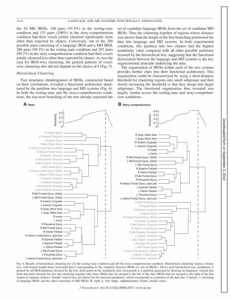
Tree structures (dendrograms) of fROIs, constructed basedon their correlations, revealed a functional architecture domi-nated by the partition into language and MD systems (Fig. 6).In both the resting-state and the story-comprehension condi-tions, the top-most branching of the tree already separated the
set of candidate language fROIs from the set of candidate MDfROIs. Thus the clustering together of regions whose distancewas shorter than the height of the first branching partitioned thedata into language and MD systems. In both experimentalconditions, this partition into two clusters had the highestmodularity value compared with all other possible partitionslicensed by the hierarchical tree, suggesting that the functionaldissociation between the language and MD systems is the keyorganizational principle underlying the data.
The organization of fROIs within each of the two systemsprovides further clues into their functional architecture. Thisorganization could be characterized by using a short-distancethreshold for clustering regions into small subgroups and thenslowly increasing the threshold so that they merge into largersubgroups. The functional organization thus revealed waslargely similar across the resting-state and story-comprehen-sion conditions.
B Story comprehension:A Rest:
L Inferior Frontal Gyrus, opercularL Precentral Gyrus
L Mid Frontal GyrusL Inferior Parietal
L Superior ParietalR Superior Parietal
R Inferior Frontal Gyrus, opercularR Inferior Parietal
R Mid Frontal GyrusR Precentral Gyrus
L InsulaR Insula
L Supp. Motor AreaR Supp. Motor AreaL Anterior CingulateR Anterior Cingulate
L Mid Frontal Gyrus, OrbitalR Mid Frontal Gyrus, Orbital
L Mid Posterior TemporalL Mid Anterior Temporal
L Anterior TemporalL Posterior Temporal
L Inferior Frontal GyrusL Inferior Frontal Gyrus, Orbital
L Mid Frontal GyrusR Mid Posterior TemporalR Mid Anterior Temporal
R Anterior TemporalR Inferior Frontal Gyrus
R Inferior Frontal Gyrus, OrbitalR Mid Frontal Gyrus
L Angular GyrusR Posterior Temporal
R Angular Gyrus
0
0.1
0.2
0.3
Mod
ular
ity
L Mid Posterior TemporalL Inferior Frontal Gyrus
L Inferior Frontal Gyrus, OrbitalL Mid Frontal Gyrus
L Posterior TemporalL Mid Anterior Temporal
L Anterior TemporalR Mid Posterior Temporal
R Inferior Frontal GyrusR Mid Anterior Temporal
R Anterior TemporalR Inferior Frontal Gyrus, Orbital
L Angular GyrusR Posterior Temporal
R Angular GyrusR Mid Frontal Gyrus
L Inferior Frontal Gyrus, opercularL Precentral Gyrus
L Inferior ParietalL Superior Parietal
R Inferior Frontal Gyrus, opercularR Precentral Gyrus
R Mid Frontal GyrusR Inferior Parietal
R Superior ParietalL Mid Frontal Gyrus
L Mid Frontal Gyrus, OrbitalR Mid Frontal Gyrus, Orbital
L InsulaR Insula
L Anterior CingulateR Anterior CingulateL Supp. Motor AreaR Supp. Motor Area
0.10.20.30.4
Mod
ular
ity
0
Fig. 6. Results of hierarchical clustering for (A) the resting-state condition and (B) the story-comprehension condition. Hierarchical clustering creates a binarytree, with branch length (here, horizontal lines) corresponding to the similarity between fROIs (or sets of fROIs). Above each hierarchical tree, modularity isplotted for all fROI partitions licensed by the tree. Each point on the modularity plot corresponds to a partition generated by drawing an imaginary vertical linefrom that point through the tree and clustering together only those fROIs that are merged to the left of this line (fROIs that are merged to the right of the lineremain in separate clusters). Sample vertical lines are drawn for the maximal modularity, which corresponds to a partition of the data into 2 clusters, 1 consistingof language fROIs and the other consisting of MD fROIs. R, right; L, left; Supp., supplementary; Insula, insular cortex.
1114 LANGUAGE AND MD SYSTEMS FUNCTIONALLY DISSOCIATED
J Neurophysiol • doi:10.1152/jn.00884.2013 • www.jn.org
on Septem
ber 3, 2014D
ownloaded from

In the language system, especially during rest, clusteringwas dominated by hemisphere, and within each hemisphere,regions were clustered according to their lobe. Namely, left-temporal regions were clustered together and only then mergedwith left-frontal regions. Next, these left-hemisphere regionswere merged with right-hemisphere regions, which themselvesalso formed temporal and frontal subsets. The right AngG andMFG were among the last to merge with the rest of thelanguage system (consistent with our k-means results), alongwith the left AngG and right PostTemp.
In the MD system, clustering was sometimes dominated byhemisphere and sometimes by interhemispheric homology.Namely, most frontal and parietal regions tended to merge witheach other ipsilaterally before merging across hemispheres.However, the Insula, SMA, ACC, and MFG were each firstclustered with their contralateral homologue and only thenmerged with each other and with the former “fronto-parietal”subset.
DISCUSSION
The findings reported here demonstrate that fMRI BOLDsignal fluctuations are strongly correlated among differentbrain regions of the language system, as well as among differ-ent regions of the MD system, but correlations across these twosystems are weak or negative. These results are robust, gener-alizing across two conditions: rest, where signal fluctuationsare not driven by an external task, and story comprehension,where signal fluctuations are task evoked; and they arise insimilar form from both hypothesis-driven and data-driven anal-yses. These data provide powerful, new evidence that each ofthese systems forms a cohesive, integrated whole, yet the twosystems are functionally dissociated from each other.
Previous studies that used data-driven clustering of voxelsacross the brain, based on resting-state functional data, pro-vided the groundwork for the present study and even revealedsystems that approximately resemble the language and MDsystems investigated here (Lee et al. 2012; Mantini et al. 2013;Tie et al. 2012; Yeo et al. 2011). However, the only way to linkthe clusters that emerged in those studies to the wealth ofknowledge about the functions of different brain regions isthrough reverse inference, based on stereotaxic coordinates(Poldrack 2006). Such anatomy-based inferences are particu-larly challenging for the language and MD systems, becausethe mapping between stereotaxic coordinates and functionalregions is degraded by both the high variability across subjectsin the anatomical locations of each region (Amunts et al. 1999;Frost and Goebel 2012; Juch et al. 2005; Paus et al. 1996;Tahmasebi et al. 2012; Tomaiuolo et al. 1999) and the frequentproximity of language and MD regions (Fedorenko et al.2012). Here, we circumvented these problems by performingour clustering analyses on regions (and voxels) that weredefined functionally within each subject, thereby allowing adirect interpretation of the resulting clusters in terms of specificfunctional hypotheses.
The combination of a subject-specific, functional localiza-tion approach with an analysis of functional correlations hasbeen applied previously to the ventral visual pathway (Turk-Browne et al. 2010; Zhen et al. 2013; Zhu et al. 2011) and otherregions (Harmelech et al. 2013; Heinzle et al. 2012). In fact,this method was used by the first paper to report resting-state
functional correlations (Biswal et al. 1995). However, no priorstudy has used this method to study the functional relationshipbetween the language and MD systems. Specifically, whereasa few prior functional correlation studies did use functionallocalizers for defining either language (Makuuchi and Fried-erici 2013; Newman et al. 2013) or MD (Dosenbach et al.2007; Seeley et al. 2007) regions, most of these studies usedgroup-level analyses of the localizer data (or coordinates fromprior studies, cf. Turken and Dronkers 2011), again with thepotential shortcomings described above. The few studies thatdid define candidate language regions in individual subjects(Hampson et al. 2002; Morgan et al. 2009) have focused onsmall subsets of the language network rather than providingwide coverage of ROIs.
The current study therefore provides new support for thehypothesis that the language and MD systems are dissociablefrom each other and are recruited for distinct cognitive pro-cesses. Our findings complement prior evidence from neuro-imaging studies that used standard functional contrasts (Fe-dorenko et al. 2011, 2012; Monti and Osherson 2012; Monti etal. 2009, 2012), as well as evidence from double dissociationsin the patient literature (Apperly et al. 2006; Bek et al. 2010;Broca 1861/2006; Butterworth 2000; Happé et al. 1999;Klessinger et al. 2007; Luria et al. 1965; Peretz and Coltheart2003; Varley et al. 2005; Varley and Siegal 2000; Wernicke1874/1969).
Remaining Questions
A crucial question for further investigation concerns thefine-grained functional organization within the language sys-tem and within the MD system. Although each system is highlyintegrated, as indexed by the strong correlations among itsconstituent regions found here, further functional subdivisionswithin each system are likely. Indeed, our clustering resultsalready capture some possible subdivisions within each sys-tem, and some of these appear to correspond to those suggestedin prior studies. Namely, within the MD system, our hierarchi-cal clustering analyses revealed two subsets that may corre-spond to the previously identified fronto-parietal and “cingulo-opercular” networks (Dosenbach et al. 2006, 2007; Koechlin etal. 1999; Mantini et al. 2013; Nomura et al. 2010; Power et al.2011). These two networks are hypothesized to be differentlyrecruited to control task-relevant, cognitive strategies [for areview, see Power and Petersen (2013)]. Within the languagesystem, our hierarchical clustering revealed frontal and tempo-ral subsets in each hemisphere, consistent with previous resultsfrom both aphasic patients (Geschwind 1970; Gorno-Tempiniet al. 2004) and fMRI studies (Hagoort 2003, 2005; Snijders etal. 2009; Tie et al. 2012). The current approach of combiningfunctional correlation measures with functional localizers thattarget specific cognitive functions is likely to prove powerful infurther elucidating these hypothesized subdivisions within thelanguage and MD systems.
A second unanswered question concerns the neurobiologicalsignificance of functional correlations across brain regions. Ithas been suggested that these correlations may, in part, reflect:1) anatomical connectivity (direct and indirect) and/or 2) his-tory of coactivation (Deco and Corbetta 2011; Deco et al. 2010,2013) [for reviews and additional accounts, see He et al.(2008); Keller et al. (2011); Matsui et al. (2011); Schölvinck et
1115LANGUAGE AND MD SYSTEMS FUNCTIONALLY DISSOCIATED
J Neurophysiol • doi:10.1152/jn.00884.2013 • www.jn.org
on Septem
ber 3, 2014D
ownloaded from

al. (2010); Shmuel and Leopold (2008)]. The extent to whichthe patterns of correlations reported here correspond to directanatomical connections therefore remains to be discovered.Although evidence from diffusion imaging is generally con-sistent with resting functional correlation measures (Hermund-stad et al. 2013), neither is a perfect measure of structuralconnectivity (Ugurbil et al. 2013), posing substantial chal-lenges for a definitive answer to this question. Moreover, somefunctional correlations are critically task dependent (Hermund-stad et al. 2013). Thus although the functional dissociationbetween the language and MD systems generalized acrossresting state and story comprehension, it is possible that thisdissociation would be modulated under other cognitive states.
Third, although our data indicate that the language andMD regions are independently recruited during cognitiveprocessing, this conclusion need not imply that the twosystems can never be engaged simultaneously. Indeed, manyprevious fMRI studies have reported activations in MDregions during some language-processing tasks, especiallywhen such processing is effortful (January et al. 2009;Kuperberg et al. 2003; McMillan et al. 2012, 2013; Meltzeret al. 2010; Nieuwland et al. 2012; Novais-Santos et al.2007; Rodd et al. 2005; Wild et al. 2012). These findingssuggest that the domain-general, cognitive-control mecha-nisms associated with the MD system may play a role inlanguage processing [E. Fedorenko, unpublished observa-tions; Fedorenko and Thompson-Schill (2014)] and hence,that the MD and language systems may coactivate in somecircumstances. Interactions between these two systems,however, may be more pronounced on a fast millisecond-level time scale and therefore, may not be detectable in theBOLD signal fluctuations measured here, given the lowtemporal resolution of this signal. Thus an important ques-tion for future research concerns the frequency, nature, andfunctional importance of interactions and coactivations ofthe language and MD systems.
Conclusions
Our results support a functional dissociation between thelanguage and MD systems: each system is strongly correlatedwithin itself, but pairs of regions straddling the two systemsshow weak (or negative) correlations. The robustness of thisdissociation across conditions and analyses suggests that itreflects a deep principle of the functional organization of thehuman brain. Thus the current data help resolve the contro-versy in the prior neuroimaging literature (Blumstein andAmso 2013; Thompson-Schill et al. 2005) in favor of thehypothesis that at least some of the neural mechanisms used forhigh-level language processing are distinct from those thatsupport other cognitive functions.
ACKNOWLEDGMENTS
The authors thank Susan Whitfield-Gabrieli for valuable advice on connec-tivity analysis in the CONN toolbox and Terri Lynn Scott for generating fROIsin native functional space. The authors are also grateful to Anastasia Vish-nevetsky, Steve Piantadosi, and Ted Gibson for help in constructing andrecording the materials for the story-comprehension task and to Eyal Dechterand Alex Kell for help with the script for the stories experiment. For technicalsupport during scanning, the authors thank Christina Triantafyllou, SteveShannon, and Sheeba Arnold. We also acknowledge the Athinoula A. MartinosImaging Center at the McGovern Institute for Brain Research, MIT.
GRANTS
Support for this research was provided by the following funding sources:grant EY13455 to N. Kanwisher, a grant from the Ellison Medical Foundationto N. Kanwisher, a seed grant from the Simons Center for the Social Brain toN. Kanwisher, and Eunice Kennedy Shriver National Institute of Child Healthand Human Development Award K99 HD-057522 to E. Fedorenko.
DISCLOSURES
No conflicts of interest, financial or otherwise, are declared by the authors.
AUTHOR CONTRIBUTIONS
Author contributions: N.K. and E.F. conception and design of research; E.F.performed experiments; I.B. and E.F. analyzed data; I.B., N.K., and E.F.interpreted results of experiments; I.B. prepared figures; I.B. drafted manu-script; I.B., N.K., and E.F. edited and revised manuscript; I.B., N.K., and E.F.approved final version of manuscript.
REFERENCES
Amunts K, Schleicher A, Bürgel U, Mohlberg H, Uylings H, Zilles K.Broca’s region revisited: cytoarchitecture and intersubject variability. JComp Neurol 412: 319–341, 1999.
Apperly IA, Samson D, Carroll N, Hussain S, Humphreys G. Intact first-and second-order false belief reasoning in a patient with severely impairedgrammar. Soc Neurosci 1: 334–348, 2006.
Bassett DS, Porter MA, Wymbs NF, Grafton ST, Carlson JM, Mucha PJ.Robust detection of dynamic community structure in networks. Chaos 23:013142, 2013.
Behzadi Y, Restom K, Liau J, Liu TT. A component based noise correctionmethod (CompCor) for BOLD and perfusion based fMRI. Neuroimage 37:90–101, 2007.
Bek J, Blades M, Siegal M, Varley RA. Language and spatial reorientation:evidence from severe aphasia. J Exp Psychol 36: 646, 2010.
Benjamini Y, Yekutieli D. The control of the false discovery rate in multipletesting under dependency. Ann Statist 29: 1165–1188, 2001.
Biswal B, Zerrin Yetkin F, Haughton VM, Hyde JS. Functional connectiv-ity in the motor cortex of resting human brain using echo-planar MRI. MagnReson Med 34: 537–541, 1995.
Blumstein SE. Reflections on the cognitive neuroscience of language. In: TheCognitive Neurosciences, edited by Gazzaniga M. Cambridge, MA: MITPress, 2009, p. 1235–1240.
Blumstein SE, Amso D. Dynamic functional organization of language: in-sights from functional neuroimaging. Perspect Psychol Sci 8: 44–48, 2013.
Braver TS, Reynolds JR, Donaldson DI. Neural mechanisms of transient andsustained cognitive control during task switching. Neuron 39: 713–726,2003.
Broca P. Comments regarding the seat of the faculty of spoken language,followed by an observation of aphemia (loss of speech). In: Broca’s Region,edited by Grodzinsky Y and Amunts K. New York: Oxford UniversityPress, 1861/2006, p. 291–304.
Butterworth B. The Mathematical Brain. London: Macmillan, 2000.Chiarello C. Parallel systems for processing language: hemispheric comple-
mentarity in the normal brain. In: Mind, Brain, and Language: Multidisci-plinary Perspectives, edited by Banich M and Mack M. Oxon, UK: Psy-chology, 2003, p. 229–247.
Cole MW, Schneider W. The cognitive control network: integrated corticalregions with dissociable functions. Neuroimage 37: 343–360, 2007.
Cordes D, Haughton VM, Arfanakis K, Carew JD, Turski PA, Moritz CH,Quigley MA, Meyerand ME. Frequencies contributing to functional con-nectivity in the cerebral cortex in “resting-state” data. Am J Neuroradiol 22:1326–1333, 2001.
Cordes D, Haughton VM, Arfanakis K, Wendt GJ, Turski PA, MoritzCH, Quigley MA, Meyerand ME. Mapping functionally related regions ofbrain with functional connectivity MR imaging. Am J Neuroradiol 21:1636–1644, 2000.
Damasio AR. Aphasia. N Engl J Med 326: 531–539, 1992.Deco G, Corbetta M. The dynamical balance of the brain at rest. Neurosci-
entist 17: 107–123, 2011.Deco G, Jirsa VK, Mcintosh AR. Emerging concepts for the dynamical
organization of resting-state activity in the brain. Nat Rev Neurosci 12:43–56, 2010.
1116 LANGUAGE AND MD SYSTEMS FUNCTIONALLY DISSOCIATED
J Neurophysiol • doi:10.1152/jn.00884.2013 • www.jn.org
on Septem
ber 3, 2014D
ownloaded from

Deco G, Jirsa VK, Mcintosh AR. Resting brains never rest: computationalinsights into potential cognitive architectures. Trends Neurosci 36: 268–274, 2013.
Dehaene S, Spelke E, Pinel P, Stanescu R, Tsivkin S. Sources of mathe-matical thinking: behavioral and brain-imaging evidence. Science 284:970–974, 1999.
Dosenbach NU, Fair DA, Cohen AL, Schlaggar BL, Petersen SE. Adual-networks architecture of top-down control. Trends Cogn Sci 12: 99–105, 2008.
Dosenbach NU, Fair DA, Miezin FM, Cohen AL, Wenger KK, DosenbachRA, Fox MD, Snyder AZ, Vincent JL, Raichle ME. Distinct brainnetworks for adaptive and stable task control in humans. Proc Natl Acad SciUSA 104: 11073–11078, 2007.
Dosenbach NU, Visscher KM, Palmer ED, Miezin FM, Wenger KK, KangHC, Burgund ED, Grimes AL, Schlaggar BL, Petersen SE. A coresystem for the implementation of task sets. Neuron 50: 799–812, 2006.
Duncan J. The multiple-demand (MD) system of the primate brain: mentalprograms for intelligent behaviour. Trends Cogn Sci 14: 172–179, 2010.
Duncan J, Owen AM. Common regions of the human frontal lobe recruitedby diverse cognitive demands. Trends Neurosci 23: 475–483, 2000.
Fedorenko E, Behr MK, Kanwisher N. Functional specificity for high-levellinguistic processing in the human brain. Proc Natl Acad Sci USA 108:16428–16433, 2011.
Fedorenko E, Duncan J, Kanwisher N. Broad domain generality in focalregions of frontal and parietal cortex. Proc Natl Acad Sci USA 110:16616–16621, 2013.
Fedorenko E, Duncan J, Kanwisher N. Language-selective and domain-general regions lie side by side within Broca’s area. Curr Biol 22: 2059–2062, 2012.
Fedorenko E, Hsieh PJ, Nieto-Castañón A, Whitfield-Gabrieli S, Kan-wisher N. New method for fMRI investigations of language: defining ROIsfunctionally in individual subjects. J Neurophysiol 104: 1177–1194, 2010.
Fedorenko E, Nieto-Castanon A, Kanwisher N. Lexical and syntacticrepresentations in the brain: an fMRI investigation with multi-voxel patternanalyses. Neuropsychologia 50: 499–513, 2012.
Fedorenko E, Thompson-Schill SL. Reworking the language network.Trends Cogn Sci 18: 120–126, 2014.
Frost MA, Goebel R. Measuring structural-functional correspondence: spatialvariability of specialised brain regions after macro-anatomical alignment.Neuroimage 59: 1369–1381, 2012.
Geschwind N. The organization of language and the brain. Science 170:940–944, 1970.
Gómez S, Jensen P, Arenas A. Analysis of community structure in networksof correlated data. Phys Rev E Stat Nonlin Soft Matter Phys 80: 016114,2009.
Gorno-Tempini ML, Dronkers NF, Rankin KP, Ogar JM, PhengrasamyL, Rosen HJ, Johnson JK, Weiner MW, Miller BL. Cognition andanatomy in three variants of primary progressive aphasia. Ann Neurol 55:335–346, 2004.
Hagoort P. How the brain solves the binding problem for language: aneurocomputational model of syntactic processing. Neuroimage 20: S18–S29, 2003.
Hagoort P. On Broca, brain, and binding: a new framework. Trends Cogn Sci9: 416–423, 2005.
Hampshire A, Highfield RR, Parkin BL, Owen AM. Fractionating humanintelligence. Neuron 76: 1225–1237, 2012.
Hampson M, Peterson BS, Skudlarski P, Gatenby JC, Gore JC. Detectionof functional connectivity using temporal correlations in MR images. HumBrain Mapp 15: 247–262, 2002.
Happé F, Brownell H, Winner E. Acquired ‘theory of mind’ impairmentsfollowing stroke. Cognition 70: 211–240, 1999.
Harmelech T, Preminger S, Wertman E, Malach R. The day-after effect:long term, Hebbian-like restructuring of resting-state fMRI patterns inducedby a single epoch of cortical activation. J Neurosci 33: 9488–9497, 2013.
Hartigan JA. Clustering Algorithms. New York: John Wiley & Sons, 1975.He BJ, Snyder AZ, Zempel JM, Smyth MD, Raichle ME. Electrophysio-
logical correlates of the brain’s intrinsic large-scale functional architecture.Proc Natl Acad Sci USA 105: 16039–16044, 2008.
Hein G, Knight RT. Superior temporal sulcus—it’s my area: or is it? J CognNeurosci 20: 2125–2136, 2008.
Heinzle J, Wenzel MA, Haynes JD. Visuomotor functional network topologypredicts upcoming tasks. J Neurosci 32: 9960–9968, 2012.
Hermundstad AM, Bassett DS, Brown KS, Aminoff EM, Clewett D,Freeman S, Frithsen A, Johnson A, Tipper CM, Miller MB. Structural
foundations of resting-state and task-based functional connectivity in thehuman brain. Proc Natl Acad Sci USA 110: 6169–6174, 2013.
January D, Trueswell JC, Thompson-Schill SL. Co-localization of stroopand syntactic ambiguity resolution in Broca’s area: implications for theneural basis of sentence processing. J Cogn Neurosci 21: 2434–2444, 2009.
Jenkinson M, Bannister P, Brady M, Smith S. Improved optimization forthe robust and accurate linear registration and motion correction of brainimages. Neuroimage 17: 825–841, 2002.
Juch H, Zimine I, Seghier ML, Lazeyras F, Fasel JH. Anatomical variabil-ity of the lateral frontal lobe surface: implication for intersubject variabilityin language neuroimaging. Neuroimage 24: 504–514, 2005.
Julian J, Fedorenko E, Webster J, Kanwisher N. An algorithmic method forfunctionally defining regions of interest in the ventral visual pathway.Neuroimage 60: 2357–2364, 2012.
Jung-Beeman M. Bilateral brain processes for comprehending natural lan-guage. Trends Cogn Sci 9: 512–518, 2005.
Kaan E, Swaab TY. The brain circuitry of syntactic comprehension. TrendsCogn Sci 6: 350–356, 2002.
Keller CJ, Bickel S, Entz L, Ulbert I, Milham MP, Kelly C, Mehta AD.Intrinsic functional architecture predicts electrically evoked responses in thehuman brain. Proc Natl Acad Sci USA 108: 10308–10313, 2011.
Klessinger N, Szczerbinski M, Varley RA. Algebra in a man with severeaphasia. Neuropsychologia 45: 1642–1648, 2007.
Koechlin E, Basso G, Pietrini P, Panzer S, Grafman J. The role of theanterior prefrontal cortex in human cognition. Nature 399: 148–151, 1999.
Koechlin E, Jubault T. Broca’s area and the hierarchical organization ofhuman behavior. Neuron 50: 963–974, 2006.
Koelsch S, Gunter TC, Zysset S, Lohmann G, Friederici AD. Bach speaks:a cortical “language-network” serves the processing of music. Neuroimage17: 956–966, 2002.
Kuperberg GR, Holcomb PJ, Sitnikova T, Greve D, Dale AM, Caplan D.Distinct patterns of neural modulation during the processing of conceptualand syntactic anomalies. J Cogn Neurosci 15: 272–293, 2003.
Lancichinetti A, Fortunato S. Consensus clustering in complex networks. SciRep 2: 336, 2012.
Lee MH, Hacker CD, Snyder AZ, Corbetta M, Zhang D, Leuthardt EC,Shimony JS. Clustering of resting state networks. PloS One 7: e40370,2012.
Lemieux L, Salek-Haddadi A, Lund TE, Laufs H, Carmichael D. Model-ling large motion events in fMRI studies of patients with epilepsy. MagnReson Imaging 25: 894–901, 2007.
Luria AR, Tsvetkova LS, Futer D. Aphasia in a composer. J Neurol Sci 2:288–292, 1965.
Maess B, Koelsch S, Gunter TC, Friederici AD. Musical syntax is processedin Broca’s area: an MEG study. Nat Neurosci 4: 540–545, 2001.
Makuuchi M, Friederici AD. Hierarchical functional connectivity betweenthe core language system and the working memory system. Cortex 49:2416–2423, 2013.
Mantini D, Corbetta M, Romani GL, Orban GA, Vanduffel W. Evolution-arily novel functional networks in the human Brain? J Neurosci 33: 3259–3275, 2013.
Matsui T, Tamura K, Koyano KW, Takeuchi D, Adachi Y, Osada T,Miyashita Y. Direct comparison of spontaneous functional connectivity andeffective connectivity measured by intracortical microstimulation: an fMRIstudy in macaque monkeys. Cereb Cortex 21: 2348–2356, 2011.
McMillan CT, Clark R, Gunawardena D, Ryant N, Grossman M. fMRIevidence for strategic decision-making during resolution of pronoun refer-ence. Neuropsychologia 50: 674–687, 2012.
McMillan CT, Coleman D, Clark R, Liang TW, Gross RG, Grossman M.Converging evidence for the processing costs associated with ambiguousquantifier comprehension. Front Psychol 4: 153, 2013.
Meltzer JA, Mcardle JJ, Schafer RJ, Braun AR. Neural aspects of sentencecomprehension: syntactic complexity, reversibility, and reanalysis. CerebCortex 20: 1853–1864, 2010.
Miller EK, Cohen JD. An integrative theory of prefrontal cortex function.Annu Rev Neurosci 24: 167–202, 2001.
Monti MM, Osherson DN. Logic, language and the brain. Brain Res 1428:33–42, 2012.
Monti MM, Parsons LM, Osherson DN. The boundaries of language andthought in deductive inference. Proc Natl Acad Sci USA 106: 12554–12559,2009.
Monti MM, Parsons LM, Osherson DN. Thought beyond language neuraldissociation of algebra and natural language. Psychol Sci 23: 914–922,2012.
1117LANGUAGE AND MD SYSTEMS FUNCTIONALLY DISSOCIATED
J Neurophysiol • doi:10.1152/jn.00884.2013 • www.jn.org
on Septem
ber 3, 2014D
ownloaded from

Morgan VL, Mishra A, Newton AT, Gore JC, Ding Z. Integrating func-tional and diffusion magnetic resonance imaging for analysis of structure-function relationship in the human language network. PloS One 4: e6660,2009.
Newman AJ, Kenny S, Saint-Aubin J, Klein RM. Can skilled readersperform a second task in parallel? A functional connectivity MRI study.Brain Lang 124: 84–95, 2013.
Newman ME, Girvan M. Finding and evaluating community structure innetworks. Phys Review E Stat Nonlin Soft Matter Phys 69: 026113, 2004.
Nieuwland MS, Martin AE, Carreiras M. Brain regions that process case:evidence from basque. Hum Brain Mapp 33: 2509–2520, 2012.
Nomura EM, Gratton C, Visser RM, Kayser A, Perez F, D’esposito M.Double dissociation of two cognitive control networks in patients with focalbrain lesions. Proc Natl Acad Sci USA 107: 12017–12022, 2010.
Novais-Santos S, Gee J, Shah M, Troiani V, Work M, Grossman M.Resolving sentence ambiguity with planning and working memory re-sources: evidence from fMRI. Neuroimage 37: 361–378, 2007.
Paus T, Tomaiuolo F, Otaky N, MacDonald D, Petrides M, Atlas J, MorrisR, Evans AC. Human cingulate and paracingulate sulci: pattern, variability,asymmetry, and probabilistic map. Cereb Cortex 6: 207–214, 1996.
Peretz I, Coltheart M. Modularity of music processing. Nat Neurosci 6:688–691, 2003.
Poldrack RA. Can cognitive processes be inferred from neuroimaging data?Trends Cogn Sci 10: 59–63, 2006.
Postelnicu G, Zollei L, Fischl B. Combined volumetric and surface registra-tion. IEEE Trans Med Imaging 28: 508–522, 2009.
Power JD, Barnes KA, Snyder AZ, Schlaggar BL, Petersen SE. Spuriousbut systematic correlations in functional connectivity MRI networks arisefrom subject motion. Neuroimage 59: 2142–2154, 2012.
Power JD, Cohen AL, Nelson SM, Wig GS, Barnes KA, Church JA, VogelAC, Laumann TO, Miezin FM, Schlaggar BL. Functional networkorganization of the human brain. Neuron 72: 665–678, 2011.
Power JD, Petersen SE. Control-related systems in the human brain. CurrOpin Neurobiol 23: 223–228, 2013.
Rodd JM, Davis MH, Johnsrude IS. The neural mechanisms of speechcomprehension: fMRI studies of semantic ambiguity. Cereb Cortex 15:1261–1269, 2005.
Satterthwaite TD, Elliott MA, Gerraty RT, Ruparel K, Loughead J,Calkins ME, Eickhoff SB, Hakonarson H, Gur RC, Gur RE. Animproved framework for confound regression and filtering for control ofmotion artifact in the preprocessing of resting-state functional connectivitydata. Neuroimage 64: 240–256, 2013.
Satterthwaite TD, Wolf DH, Loughead J, Ruparel K, Elliott MA, Hako-narson H, Gur RC, Gur RE. Impact of in-scanner head motion on multiplemeasures of functional connectivity: relevance for studies of neurodevelop-ment in youth. Neuroimage 60: 623–632, 2012.
Schölvinck ML, Maier A, Frank QY, Duyn JH, Leopold DA. Neural basisof global resting-state fMRI activity. Proc Natl Acad Sci USA 107: 10238–10243, 2010.
Seeley WW, Menon V, Schatzberg AF, Keller J, Glover GH, Kenna H,Reiss AL, Greicius MD. Dissociable intrinsic connectivity networks forsalience processing and executive control. J Neurosci 27: 2349–2356, 2007.
Shmuel A, Leopold DA. Neuronal correlates of spontaneous fluctuations infMRI signals in monkey visual cortex: implications for functional connec-tivity at rest. Hum Brain Mapp 29: 751–761, 2008.
Silver NC, Dunlap WP. Averaging correlation coefficients: should Fisher’s ztransformation be used? J Appl Psychol 72: 146, 1987.
Snijders TM, Vosse T, Kempen G, Van Berkum JJ, Petersson KM,Hagoort P. Retrieval and unification of syntactic structure in sentencecomprehension: an fMRI study using word-category ambiguity. CerebCortex 19: 1493–1503, 2009.
Stanescu-Cosson R, Pinel P, Van De Moortele PF, Le Bihan D, Cohen L,Dehaene S. Understanding dissociations in dyscalculia A brain imagingstudy of the impact of number size on the cerebral networks for exact andapproximate calculation. Brain 123: 2240–2255, 2000.
Tahmasebi AM, Davis MH, Wild CJ, Rodd JM, Hakyemez H, Abolmae-sumi P, Johnsrude IS. Is the link between anatomical structure andfunction equally strong at all cognitive levels of processing? Cereb Cortex22: 1593–1603, 2012.
Thesen S, Heid O, Mueller E, Schad LR. Prospective acquisition correctionfor head motion with image-based tracking for real-time fMRI. Magn ResonMed 44: 457–465, 2000.
Thompson-Schill SL, Bedny M, Goldberg RF. The frontal lobes and theregulation of mental activity. Curr Opin Neurobiol 15: 219–224, 2005.
Tie Y, Rigolo L, Norton IH, Huang RY, Wu W, Orringer D, MukundanS, Golby AJ. Defining language networks from resting-state fMRI forsurgical planning—a feasibility study. Hum Brain Mapp 35: 1018–1030,2014.
Tomaiuolo F, MacDonald J, Caramanos Z, Posner G, Chiavaras M, EvansAC, Petrides M. Morphology, morphometry and probability mapping of thepars opercularis of the inferior frontal gyrus: an in vivo MRI analysis. EurJ Neurosci 11: 3033–3046, 1999.
Turk-Browne NB, Norman-Haignere SV, McCarthy G. Face-specific rest-ing functional connectivity between the fusiform gyrus and posterior supe-rior temporal sulcus. Front Hum Neurosci 4: 176, 2010.
Turken U, Dronkers NF. The neural architecture of the language compre-hension network: converging evidence from lesion and connectivity analy-ses. Front Syst Neurosci 5: 1, 2011.
Tzourio-Mazoyer N, Landeau B, Papathanassiou D, Crivello F, Etard O,Delcroix N, Mazoyer B, Joliot M. Automated anatomical labeling ofactivations in SPM using a macroscopic anatomical parcellation of the MNIMRI single-subject brain. Neuroimage 15: 273–289, 2002.
Ugurbil K, Xu J, Auerbach EJ, Moeller S, Vu A, Duarte-Carvajalino JM,Lenglet C, Wu X, Schmitter S, Van De Moortele PF, Strupp J, SapiroG, De Martino F, Wang D, Harel N, Garwood M, Chen L, Feinberg DA,Smith SM, Miller KL, Sotiropoulos SN, Jbabdi S, Andersson JL,Behrens TE, Glasser MF, Van Essen DC, Yacoub E, WU-Minn HCPConsortium. Pushing spatial and temporal resolution for functional anddiffusion MRI in the Human Connectome Project. Neuroimage 80: 80–104,2013.
Van Dijk KR, Hedden T, Venkataraman A, Evans KC, Lazar SW,Buckner RL. Intrinsic functional connectivity as a tool for human connec-tomics: theory, properties, and optimization. J Neurophysiol 103: 297, 2010.
Varley RA, Klessinger NJ, Romanowski CA, Siegal M. Agrammatic butnumerate. Proc Natl Acad Sci USA 102: 3519–3524, 2005.
Varley RA, Siegal M. Evidence for cognition without grammar from causalreasoning and ‘theory of mind’ in an agrammatic aphasic patient. Curr Biol10: 723–726, 2000.
Wernicke C. The symptom complex of aphasia: a psychological study on ananatomical basis. In: Boston Studies in the Philosophy of Science, edited byCohen RS and Wartofsky MW. Dordrecht, Netherlands: D. Reidel, 1874/1969, p. 34–97.
Whitfield-Gabrieli S, Nieto-Castanon A. Conn: a functional connectivitytoolbox for correlated and anticorrelated brain networks. Brain Connect 2:125–141, 2012.
Wild CJ, Yusuf A, Wilson DE, Peelle JE, Davis MH, Johnsrude IS.Effortful listening: the processing of degraded speech depends critically onattention. J Neurosci 32: 14010–14021, 2012.
Yeo BT, Krienen FM, Sepulcre J, Sabuncu MR, Lashkari D, HollinsheadM, Roffman JL, Smoller JW, Zöllei L, Polimeni JR. The organization ofthe human cerebral cortex estimated by intrinsic functional connectivity. JNeurophysiol 106: 1125–1165, 2011.
Yue Q, Zhang L, Xu G, Shu H, Li P. Task-modulated activation andfunctional connectivity of the temporal and frontal areas during speechcomprehension. Neuroscience 237: 87–95, 2013.
Zhen Z, Fang H, Liu J. The hierarchical brain network for face recognition.PloS One 8: e59886, 2013.
Zhu Q, Zhang J, Luo YL, Dilks DD, Liu J. Resting-state neural activityacross face-selective cortical regions is behaviorally relevant. J Neurosci 31:10323–10330, 2011.
1118 LANGUAGE AND MD SYSTEMS FUNCTIONALLY DISSOCIATED
J Neurophysiol • doi:10.1152/jn.00884.2013 • www.jn.org
on Septem
ber 3, 2014D
ownloaded from