a fluorescent molecular imaging probe with selectivity for
TRANSCRIPT

ChemicalScience
EDGE ARTICLE
Ope
n A
cces
s A
rtic
le. P
ublis
hed
on 2
1 A
pril
2020
. Dow
nloa
ded
on 1
0/5/
2021
1:3
7:34
AM
. T
his
artic
le is
lice
nsed
und
er a
Cre
ativ
e C
omm
ons
Attr
ibut
ion-
Non
Com
mer
cial
3.0
Unp
orte
d L
icen
ce.
View Article OnlineView Journal | View Issue
A fluorescent mo
aMolecular Imaging Chemistry Laboratory, W
of Clinical Neurosciences, University of Cam
medschl.cam.ac.ukbJohn van Geest Centre for Brain Repair
University of Cambridge, Cambridge, UKcTTK-NAP B – Drug Discovery Research Grou
of Organic Chemistry, Research Center for NdDepartment of Chemistry, University of CameUK Dementia Research Institute, University
† Electronic supplementary informa10.1039/c9sc05620c
‡ These authors contributed equally.
Cite this: Chem. Sci., 2020, 11, 4773
All publication charges for this articlehave been paid for by the Royal Societyof Chemistry
Received 6th November 2019Accepted 19th April 2020
DOI: 10.1039/c9sc05620c
rsc.li/chemical-science
This journal is © The Royal Society o
lecular imaging probe withselectivity for soluble tau aggregated protein†
Yanyan Zhao, ‡a Ole Tietz, ‡a Wei-Li Kuan, b Abdul K. Haji-Dheere,a
Stephen Thompson, a Benjamin Vallin,b Elisabetta Ronchi,a Gergely Toth,c
David Klenermande and Franklin I. Aigbirhio *a
Soluble forms of aggregated tau misfolded protein, generally termed oligomers, are considered to be the
most toxic species of the different assembly states that are the pathological components of
neurodegenerative disorders. Therefore, a critical biomedical need exists for imaging probes that can
identify and quantify them. We have designed and synthesized a novel fluorescent probe, pTP-TFE for
which binding and selectivity profiles towards aggregated tau and Ab proteins were assessed. Our results
have shown pTP-TFE to be selective for early forms of soluble tau aggregates, with high affinity of
dissociation constants (Kd) ¼ 66 nM, and tenfold selectivity over mature tau fibrils. Furthermore, we
found that pTP-TFE is selective for tau over Ab aggregates and had good cell permeability. This
selectivity of pTP-TFE towards early forms of aggregated tau protein ex vivo was also supported with
studies on human brain tissue containing tau and Ab pathology. To the best of our knowledge, this is the
first fluorescent molecule to be reported to have this form of selectivity profile, which suggests that
pTP-TFE is a unique probe candidate for imaging-based detection of early stages of Alzheimer's disease
and other tauopathies.
Introduction
The accumulation of misfolded protein aggregates is a patho-logical hallmark of neurodegenerative diseases.1 In the case ofAlzheimer's disease (AD), the most prevalent pathology consistsof extracellular b-amyloid protein (Ab) plaques and intracellulartau protein-containing neurobrillary tangles.2 Protein aggre-gates have high beta-sheet structural characteristic,3 which ledto the identication of small molecules with specic bindingaffinity for them,4 several of which have been used successfullyfor in vivo imaging with positron emission tomography (PET).5
However, it is becoming increasingly apparent that proteinsassociated with neurodegenerative diseases can adopt differentassembly states, which can impact disease onset and theirprogression to different extent.6 Research on tau protein has
olfson Brain Imaging Centre, Department
bridge, Cambridge, UK. E-mail: a20@
, Department of Clinical Neuroscience,
p – Neurodegenerative Diseases, Institute
atural Sciences, Budapest, Hungary
bridge, Lenseld Road, Cambridge, UK
of Cambridge, Cambridge, UK
tion (ESI) available. See DOI:
f Chemistry 2020
shown that the earlier forming smaller soluble aggregates,generally termed oligomers, are the most toxic species and theirin vivo levels are likely to be more correlated with diseaseprogression than the levels of mature brils.7 Given that earlydiagnosis of neurodegenerative disease has become an impor-tant objective, a critical need exists for the development of novelimaging probes that can identify and quantify soluble aggre-gated protein species in vivo. Moreover, such probes couldgreatly enhance our understanding of disease progression andbe used for assessing the efficacy of new therapeutic candidates.
Efforts to design a facile imaging agent that can be used forinvestigation of soluble protein aggregates have thus farfocused on Ab-protein, leading to the development of a mono-clonal antibody based probe ([125I]8D3-F(ab0)2-h158)8 anda modest number of small molecules, most notably BD-Oligobased on the boron-dipyrromethene (BODIPY) uorescencestructure9 and AN-SP based on a spiropyran scaffold.10 Inaddition, luminescent conjugated oligothiophenes (LCOs) havebeen shown to bind to soluble aggregates of both Ab and tauprotein.11 Despite the success in recent years of developing PETprobes for imaging tau neurobrillary tangles12 presently thereare no probes that have selective binding toward tau solubleaggregated species. With tau protein pathology being a constit-uent feature of several neurodegenerative disorders, collectivelytermed tauopathies, the development of such probes, repre-sents a major biomedical need.
Chem. Sci., 2020, 11, 4773–4778 | 4773
Chemical Science Edge Article
Ope
n A
cces
s A
rtic
le. P
ublis
hed
on 2
1 A
pril
2020
. Dow
nloa
ded
on 1
0/5/
2021
1:3
7:34
AM
. T
his
artic
le is
lice
nsed
und
er a
Cre
ativ
e C
omm
ons
Attr
ibut
ion-
Non
Com
mer
cial
3.0
Unp
orte
d L
icen
ce.
View Article Online
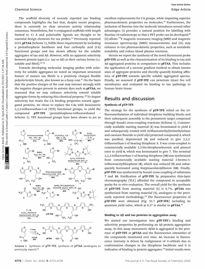
The scaffold diversity of recently reported tau bindingcompounds highlights the fact that, despite recent progress,there is currently no clear structure activity relationshipconsensus. Nonetheless, at p-conjugated scaffolds with lengthlimited to 13 �A and polarizable ligands are thought to beessential design elements for tau probes.13 Previously reportedLCO, pFTAA (Scheme 1), fulls these requirements by includinga pentathiophene backbone and four carboxylic acid (CA)functional groups and has shown affinity for the solubleaggregates of tau and Ab. However, with no apparent selectivitybetween protein types (i.e. tau or Ab) or their various forms (i.e.soluble and bril).14–16
Towards developing molecular imaging probes with selec-tivity for soluble aggregates we noted an important structuralfeature of mature tau brils is a positively charged exiblepolyelectrolyte brush, also known as a fuzzy coat.17 On the basisthat the positive charges of the coat may interact strongly withthe negative charges present in anionic dyes such as pFTAA, wereasoned that we may enhance selectivity toward solubleaggregate forms by reducing this chemical property.18 To impartselectivity but retain the CA binding properties toward aggre-gated proteins, we chose to replace the CAs with bioisosteric2,2,2-triuoroethan-1-ol (TFE) functional groups, to yield thecompound pTP-TFE (pentathiophene-triuoroethanol –
Scheme 1). TFE functional groups have been shown to act as
Scheme 1 Synthesis of pTP-TFE; synthesis of pFTAA (analogues topreviously report).14
4774 | Chem. Sci., 2020, 11, 4773–4778
excellent replacements for CA groups, while imparting superiorpharmacokinetic properties on molecules.19 Furthermore, theinclusion of uorine into the molecule introduces several otheradvantages; (i) provides a natural position for labelling withuorine-18 radioisotope so that a PET probe can be developed;20
(ii) allows 19F magnetic resonance imaging (MRI) and magneticresonance spectroscopy (MRS) measurements; and (iii) mayenhance in vivo pharmacokinetic properties, such as metabolicinstability and reduce blood plasma retention.
Herein we report the synthesis of the novel uorescent probepTP-TFE as well as the characterization of its binding to tau andAb aggregated proteins in comparison to pFTAA. This includesthe application of a sucrose gradient method to obtain knownsizes of aggregate proteins and thereby quantify binding affin-ities of pTP-TFE towards specic soluble aggregated species.Finally, we assessed if pTP-TFE can penetrate intact live cellmembranes and evaluated its binding to tau pathology inhuman brain tissue.
Results and discussionSynthesis of pTP-TFE
The strategy for the synthesis of pTP-TFE relied on the tri-uoromethylation of individual thiophene building blocks andtheir subsequent assembly to the pentameric target compoundthrough Suzuki cross-coupling reactions (Scheme 1). Commer-cially available starting material (1) was brominated to yield 2and subsequently treated with (triuoromethyl)trimethylsilaneand caesium uoride to yield silyl protected compound 3, whichwas puried, deprotected (4) and reduced to give 2,2,2-triuoroethan-1-ol bearing thiophene 5. 5 was cross-coupled tocommercially available 2,5-bis-thiopheneboronic acid pinacolester to yield 6, which was brominated to give 7. The terminal2,2,2-triuoroethan-1-ol bearing thiophene (10) was synthesizedfrom commercially available starting material 2-bromo-5-triuoroacetylthiophene (8), which was reduced (9) and subse-quently boronated using bis(pinacolato)diboron (10). Finally,pTP-TFE was synthesized by Suzuki cross-coupling of substrates7 and 10. Purication of pTP-TFE by preparative thin-layerchromatography (TLC) afforded the compound in acceptablepurity for in vitro evaluation. The overall yield for the synthesisof pTP-TFE from starting material (1) is 0.7%. pFTAA wassynthesized from starting material (1), analogues to the previ-ously reported methodology.14 The uorescence properties ofpTP-TFE were obtained (Fig. S1.† pTP-TFE) including itsquantum yield value, which at 0.27 is similar to pFTAA.14
Binding to Ab and tau proteins in aggregation assay
We started our investigation into pTP-TFE's binding andselectivity properties by performing an Ab protein aggregationassay. In this assay monomeric Ab40 is aggregated in the pres-ence of pTP-TFE or pFTAA and the uorescence intensities ofthe compounds monitored over time. An increase in uores-cence intensity is driven by realignment of p-orbitals due toconformation changes in the thiophene backbone and it isindicative of binding to protein aggregates.14 Initial results were
This journal is © The Royal Society of Chemistry 2020

Fig. 2 (A) Normalized fluorescence intensities of pTP-TFE (n ¼ 3),pFTAA (n ¼ 3) and ThT (n ¼ 3) in an aggregation assay starting with taumonomers. (B–D) Representative TEM image of tau aggregates 24 h(B), 96 h (C) and 240 h (D) (white bar ¼ 200 nm). (E and F) Bindingaffinity of pTP-TFE to tau aggregates collected at 96 hours (E) and 240hours (F) of the aggregation assay.
Fig. 1 (A) Normalized fluorescence intensities of pTP-TFE, pFTAA andThT in an Ab aggregation assay starting with Ab40 monomers (n ¼ 3).(B–D) Representative TEM image of Ab aggregates 90min (B), 200min(C) and 700 min (D) (white bar ¼ 200 nm). (E and F) Fluorescenceintensity based binding curve of pTP-TFE (E) and pFTAA (F) with Abaggregates collected at 90 min (red), 200 min (blue) and 24 hours(black) of the aggregation assay.
Edge Article Chemical Science
Ope
n A
cces
s A
rtic
le. P
ublis
hed
on 2
1 A
pril
2020
. Dow
nloa
ded
on 1
0/5/
2021
1:3
7:34
AM
. T
his
artic
le is
lice
nsed
und
er a
Cre
ativ
e C
omm
ons
Attr
ibut
ion-
Non
Com
mer
cial
3.0
Unp
orte
d L
icen
ce.
View Article Online
encouraging as pTP-TFE showed an earlier response to Ab40aggregates in comparison to pFTAA (Fig. 1A). Transmissionelectron microscopy (TEM) images of Ab40 aggregates at 90 minconrm the presence of small aggregate forms (Fig. 1B) andmature brils at later time points (700 min – Fig. 1D). Toquantitatively assess the binding affinity of pTP-TFE and pFTAAto different types of aggregates, Ab40 protein was collected froman aggregation assay (not containing compounds) at timepoints of 90 min, 200 min and 24 h. These aggregates weretreated with different concentrations of the probes to determinebinding constants (Kd) (Fig. 1E and F) by measuring theiruorescence intensity. These experiments conrmed that pTP-TFE displays a higher binding affinity to the earlier species (90min) of aggregates (Kd ¼ 7.58 mM), compared to pFTAA whichshowed no binding affinity for aggregates at this stage ofmaturation. Inversely, pFTAA showed stronger binding affinityto later aggregates (Kd ¼ 0.93 mM {200 min}, Kd ¼ 0.65 mM {24h}) compared to pTP-TFE (Kd¼ 3.27 mM {200min}, Kd¼ 3.79 mM{24 h}). In addition, the binding prole of pTP-TFE to a mixtureof Ab40 : Ab42 (9 : 1 ratio), which is considered a more accuraterepresentation of human cerebrospinal uid Ab aggregates invivo, was also determined. On the basis of the analysis of uo-rescence intensities during the assays (Fig. S6A†) pTP-TFEinteracted with Ab aggregates at an earlier time compared topFTAA and ThT. Thus, we observe a similar relative interactionprole as with just Ab40 (Fig. 1A). The binding affinities of pTP-TFE to Ab40 : Ab42 (9 : 1) mixture studied were Kd ¼ 2.1 mM at200 min and Kd ¼ 0.75 mM at 24 h, therefore a higher bindingaffinity than just with Ab40. This is consistent with higheraffinity binding to Ab42 as previously observed.21
This journal is © The Royal Society of Chemistry 2020
We then investigated the binding prole of pTP-TFE to tauprotein. To this end, tau monomers were subjected to bril-forming conditions using heparin and aliquots removed at 1,5, 24, 48, 80, 96, 168 and 240 h. Treatment of these aliquots withpTP-TFE showed a gradual increase of uorescence intensity,which reached a maximum aer 96 h and declined thereaeruntil the end point of the experiment (Fig. 2A). Experimentswith pFTAA showed a curve of similar shape and characteristictrajectory.16 TEM images of tau aggregates at different timepoints conrmed that initially small tau aggregates weredetectable as early as 24 h (Fig. 2B), which became moreapparent at 96 h (Fig. 2C) and then mature brils were found inthe 240 h samples (Fig. 2D). These results suggest preferentialbinding of pTP-TFE to early soluble tau aggregates in compar-ison to pFTAA.
Interestingly, we observed a shi in the emission spectrumof pTP-TFE over the course of the experiment. Emission spectrarecorded with tau aggregates at 24, 96 and 240 h show distinctshapes (Fig. S4A†). An analysis of the ratio of normalized uo-rescence intensities at 495 nm and 560 nm revealed a gradualshi towards the blue until 96 h (ratio¼ 2.2� 0.20), followed bya shi back towards the red (ratio ¼ 1.5 � 0.74) (Fig. S4B†).These results suggest the pTP-TFE can bind to distinct oligo-mers of tau that are generated along the aggregation pathway ofthe protein. No emission spectrum shis were observed inexperiments with pFTAA (Fig. S5B†).
The binding affinity for pTP-TFE towards aggregatescollected from the aggregation assay (not containingcompounds) at 96 h and 240 h were determined by treatmentwith different concentrations of probe (Fig. 2E and F). pTP-TFEdisplayed exceptional affinity for small aggregates (96 h) with
Chem. Sci., 2020, 11, 4773–4778 | 4775
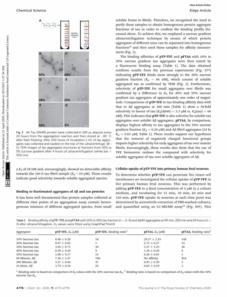
Fig. 3 (A) Tau (0N4R) protein were collected in 100 mL aliquots every24 hours from the aggregation reaction and then stored at �80 �Cafter flash freezing. After 240 hours of incubation 1 mL of tau aggre-gates was collected and loaded on the top of the ultracentrifuge. (B–G) TEM images of tau aggregates structures at fractions from 50% to5% sucrose gradient after 4 hours of ultracentrifugation (white bar ¼500 nm).
Chemical Science Edge Article
Ope
n A
cces
s A
rtic
le. P
ublis
hed
on 2
1 A
pril
2020
. Dow
nloa
ded
on 1
0/5/
2021
1:3
7:34
AM
. T
his
artic
le is
lice
nsed
und
er a
Cre
ativ
e C
omm
ons
Attr
ibut
ion-
Non
Com
mer
cial
3.0
Unp
orte
d L
icen
ce.
View Article Online
a Kd of 38 nM and, encouragingly, showed no detectable affinitytowards the 240 h tau bril sample (Kd < 10 mM). These resultsindicate good selectivity towards soluble aggregated species.
Binding to fractionated aggregates of Ab and tau proteins
It has been well documented that protein samples collected atdifferent time points of an aggregation assay contain hetero-geneous mixtures of different aggregated species, from small
Table 1 Binding affinity of pTP-TFE and pFTAAwith 10% to 50% tau fractio3) after ultracentrifugation, Kd values were fitted using GraphPad Prism5
Aggregates pTP-TFE, Kd (mM) pTP-TFE, bind
10% Sucrose tau 0.96 � 0.20 1420% Sucrose tau 0.07 � 0.03 130% Sucrose tau 3.04 � 0.71 4940% Sucrose tau 0.59 � 0.20 950% Sucrose tau 1.08 � 0.17 1590 Minute, Ab 7.58 � 2.47 108200 Minute, Ab 3.27 � 0.64 4724 Hour, Ab 3.79 � 0.34 54
a Binding ratio is based on comparison of Kd values with the 20% sucrosesucrose tau Kd.
4776 | Chem. Sci., 2020, 11, 4773–4778
soluble forms to brils. Therefore, we recognized the need topurify these samples to obtain homogenous protein aggregatefractions of tau in order to conrm the binding prole dis-cussed above. To achieve this, we employed a sucrose gradientultracentrifugation technique by means of which proteinaggregates of different sizes can be separated into homogenousfractions22 and then used these samples for affinity measure-ment (Fig. 3).
The binding affinities of pTP-TFE and pFTAA with 10% to50% sucrose gradient tau aggregates were then tested bya uorescent binding assay (Table 1). The data obtainedconrms results from the previous experiments (Fig. S7†)indicating pTP-TFE binds most strongly to the 20% sucrosegradient fraction (Kd ¼ 66 nM), which consist of solubleaggregated tau as conrmed by TEM (Fig. 3). Furthermore,selectivity of pTP-TFE for small aggregates over brils wasconrmed by a difference in Kd for 40% and 50% sucrosegradient tau aggregates of approximately one order of magni-tude. Comparisons of pTP-TFE to tau binding affinity data withthat to Ab aggregates at 200 min (Table 1) show a 50-foldselectivity in favour of tau (Kd(Ab40) ¼ 3.3 mM vs. Kd(tau) ¼ 66nM). This indicates that pTP-TFE is also selective for soluble tauaggregates over soluble Ab aggregates. pFTAA, by comparison,displays highest affinity to tau aggregates in the 50% sucrosegradient fraction (Kd ¼ 0.20 mM) and Ab bril aggregates (24 h;Kd ¼ 0.65 mM; Table 1). These results support our hypothesisthat the removal of negatively charged functional groupsimparts higher selectivity for early aggregates of tau over maturebrils. Encouragingly, these results also show that the use ofTFE bioisostere endows the compound with selectivity forsoluble aggregates of tau over soluble aggregates of Ab.
Cellular uptake of pTP-TFE into primary human fetal neurons
To determine whether pTP-TFE can penetrate live intact cellmembranes we investigated the cellular uptake of pTP-TFE inlive primary human fetal neurons. This was performed byadding pTP-TFE to a nal concentration of 4 mM in a culturemedium, and incubating for 15 min, 30 min, 60 min and120 min. pTP-TFE uptake in neurons at each time point wasdetermined by acetonitrile extraction of PBS-washed cultures,and quantied using an LC-MS/MS assay23 (Fig. S9†). This
n (n¼ 3–6) and Ab40 aggregates at 90min, 200min and 24 hours (n¼
ing ratioa pFTAA, Kd (mM) pFTAA, binding ratiob
29.57 � 2.10 1482.75 � 0.57 145.27 � 1.25 261.29 � 0.20 60.20 � 0.01 1No affinity N/A0.93 � 0.30 50.65 � 0.19 3
tau Kd.b Binding ratio is based on comparison of Kd values with the 50%
This journal is © The Royal Society of Chemistry 2020

Fig. 4 Kinetics of pTP-TFE uptake in primary human fetal neuronsquantified by measuring the concentration of pTP-TFE in live neuronsusing LC-MS/MS; data presented as % of total administered amount. Yvalues calculated from Table S1† (n ¼ 3).
Edge Article Chemical Science
Ope
n A
cces
s A
rtic
le. P
ublis
hed
on 2
1 A
pril
2020
. Dow
nloa
ded
on 1
0/5/
2021
1:3
7:34
AM
. T
his
artic
le is
lice
nsed
und
er a
Cre
ativ
e C
omm
ons
Attr
ibut
ion-
Non
Com
mer
cial
3.0
Unp
orte
d L
icen
ce.
View Article Online
showed 5% of the total applied pTP-TFE in the neuronal cellswithin 30 min and 10% in 2 hours (Fig. 4 and Table S1†),which indicates the compound can cross the cell membranewith rapid uptake. The quantity and kinetics of this uptake arecomparable to other compounds, including established PETprobes in oncology24 and agents delivered into neuronalcells.25
Ex vivo imaging of early tau aggregates in AD and PSP humanbrain slices
Finally, we investigated whether the selectivity demonstrated inin vitro assays could be replicated in ex vivo human brain tissue.To this end we evaluated the binding of pTP-TFE to taupathology in human brain tissue of progressive supranuclearpalsy (PSP), a pure tauopathy, and Alzheimer's disease, whichcontains both Ab and tau pathology. Low-magnication epi-uorescence microscopy showed that pTP-TFE signals can befound colocalizing with hyperphosphorylated, AT8-positive taupathology in both PSP and AD brains (Fig. 5A). Closer inspectionusing confocal microscopy revealed that pTP-TFE signals werepredominantly observed within the cell soma, co-localizing withAT8-positive tau pathology, although the presence of pTP-TFE
Fig. 5 pTP-TFE staining with human PSP (n ¼ 5) and AD brain slides (n¼ 4). (A) Epifluorescence images of pTP-TFE in AD and PSP humanbrain slides with scale bar ¼ 50 mm. (B) Confocal images of pTP-TFE inAD and PSP with scale bar ¼ 10 mm. The arrows indicate an axonal/dendritic compartment.
This journal is © The Royal Society of Chemistry 2020
signals could also be found in the axonal/dendritic compart-ments (Fig. 5B, arrows). Conversely, co-localization with taubril antibody AT100 is less well established; AT100-positiveimmunoreactivity has been shown to selectively label lamen-tous tau pathology,26 this substantiates our in vitro binding dataand suggests that pTP-TFE demonstrates preferential reactivityto early stage soluble tau aggregates.
Conclusions
We have designed and synthesized a novel uorescent probe,pTP-TFE for which binding and selectivity proles towardsaggregated tau and Ab proteins were assessed. This includeduse of a sucrose gradient ultracentrifugation method for moreaccurate quantication of binding affinities to specic size tauaggregates. Our results show pTP-TFE to be selective for solubletau aggregates, with a high affinity of Kd ¼ 66 nM, and ten-foldselectivity over mature brils. Furthermore, we found that pTP-TFE is tau selective over Ab, the other major misfolded proteinaggregate of neurodegenerative disorders. To the best of ourknowledge, pTP-TFE is the rst uorescent molecule to havethis form of selectivity for soluble tau aggregates over tau brils.In addition, we established that pTP-TFE could penetrate intactlive cell membranes rapidly, while its selectivity towards earlyforms of aggregated tau protein was also supported by studieson human brain tissue containing tau and Ab pathology.
The high affinity and selectivity for early soluble aggregatesof tau and its cell permeability make pTP-TFE a best in classmolecular tool for the study of tauopathy development andprogression as well as a proof of concept chemotype for thedevelopment of small molecule therapeutics, with enhancedaffinity and selectivity. We aim to utilize the potential displayedby pTP-TFE in vitro and ex vivo through in vivo optical, uorine-19 MRI/MRS and uorine-18 PET imaging modalities in thefuture.
Conflicts of interest
There are no conicts to declare.
Acknowledgements
The authors thank Dr Gabriele Schierle and Dr Na Yu (ChemicalEngineering Department, University of Cambridge), Dr KarinMuller (Cambridge Advanced Imaging Center) for technicalsupport, Prof James Rowe (Department of Clinical Neurosci-ences, University of Cambridge), Dr David Williamson andCambridge Brain Bank for the post-mortem brain samples,EPSRC Mass Spectrometry Service (University of Swansea).Cambridge Brain Bank is supported by the NIHR CambridgeBiomedical Research Centre. This study was funded by theNational Institute for Health Research (NIHR) CambridgeBiomedical Research Centre: Dementia and Neurodegenerationtheme, Medical Research Council grant (MR/K02308X/1), theEngineering and Physical Sciences Research Council (ST, EP/P008224/1), Royal Society (DK), and Amgen Foundation Schol-arship (ER). WLK and BV are supported by the Medical Research
Chem. Sci., 2020, 11, 4773–4778 | 4777
Chemical Science Edge Article
Ope
n A
cces
s A
rtic
le. P
ublis
hed
on 2
1 A
pril
2020
. Dow
nloa
ded
on 1
0/5/
2021
1:3
7:34
AM
. T
his
artic
le is
lice
nsed
und
er a
Cre
ativ
e C
omm
ons
Attr
ibut
ion-
Non
Com
mer
cial
3.0
Unp
orte
d L
icen
ce.
View Article Online
Council (MR/S005528/1). GT was supported by the HungarianBrain Research Program (2017-1.2.1-NKP-2017-00002).
Notes and references
1 F. Chiti and C. M. Dobson, Annu. Rev. Biochem., 2006, 7, 333.2 C. Ballatore, V. M. Lee and J. Q. Trojanowski, Nat. Rev.Neurosci., 2007, 8, 663; L. M. Ittner and J. Gotz, Nat. Rev.Neurosci., 2011, 12, 67; B. Ghetti, A. L. Oblak, B. F. Boeve,K. A. Johnson, B. C. Dickerson and M. Goedert,Neuropathol. Appl. Neurobiol., 2015, 41, 24.
3 E. D. Eanes and G. G. Glenner, J. Histochem. Cytochem., 1968,16, 673; A. W. Fitzpatrick, B. Falcon, S. He, A. G. Murzin,G. Murshudov, H. J. Garringer, R. A. Crowther, B. Ghetti,M. Goedert and S. H. Scheres, Nature, 2017, 547, 185.
4 M. R. Jones, E. Mathieu, C. Dyrager, S. Faissner,Z. Vaillancourt, K. J. Korshavn, M. H. Lim,A. Ramamoorthy, V. W. Yong, S. Tsutsui and P. K. Stys,Chem. Sci., 2017, 8, 5636; Y. Li, D. Xu, A. Sun, S. L. Ho,C. Y. Poon, H. N. Chan, O. T. Ng, K. K. Yung, H. Yan,H. W. Li and M. S. Wong, Chem. Sci., 2017, 8, 8279;K. P. Nilsson, FEBS Lett., 2009, 583, 2593.
5 Y. Liu, Y. Yang, M. Sun, M. Cui, Y. Fu, Y. Lin, Z. Li and L. Nie,Chem. Sci., 2017, 8, 2710; A. Nordberg, J. O. Rinne, A. Kadirand B. Langstrom, Nat. Rev. Neurol., 2010, 6, 78.
6 D. Eisenberg and M. Jucker, Cell, 2012, 148, 1188; J. L. Guo,D. J. Covell, J. P. Daniels, M. Iba, A. Stieber, B. Zhang,D. M. Riddle, L. K. Kwong, Y. Xu, J. Q. Trojanowski andV. M. Lee, Cell, 2013, 154, 103.
7 S. Maeda, N. Sahara, Y. Saito, S. Murayama, A. Ikai andA. Takashima, Neurosci. Res., 2006, 54, 197; M. Goedert,Alzheimers Disease & Dementia, 2016, 12, 1040.
8 D. Sehlin, X. T. Fang, L. Cato, G. Antoni, L. Lannfelt andS. Syvanen, Nat. Commun., 2016, 7, 10759.
9 C. L. Teoh, D. Su, S. Sahu, S. W. Yun, E. Drummond, F. Prelli,S. Lim, S. Cho, S. Ham, T. Wisniewski and Y. T. Chang, J. Am.Chem. Soc., 2015, 137, 13503; L. P. Jameson and S. V. Dzyuba,Bioorg. Med. Chem. Lett., 2013, 23, 1732; S. Lim, M.M. Haque,D. Su, D. Kim, J. S. Lee, Y. T. Chang and Y. K. Kim, Chem.Commun., 2017, 53, 1607.
10 G. Lv, A. Sun, P. Wei, N. Zhang, H. Lan and T. Yi, Chem.Commun., 2016, 52, 8865.
11 T. Klingstedt, H. Shirani, K. A. Aslund, N. J. Cairns,C. J. Sigurdson, M. Goedert and K. P. Nilsson, Chem.–Eur.J., 2015, 19, 10179.
12 V. L. Villemagne, V. Dore, S. C. Burnham, C. L. Masters andC. C. Rowe, Nat. Rev. Neurol., 2018, 14, 225; M. Goedert,Y. Yamaguchi, S. K. Mishra, M. Higuchi and N. Sahara,Frontiers in Neurology, 2018, 15, 70.
4778 | Chem. Sci., 2020, 11, 4773–4778
13 P. Verwilst, H. S. Kim, S. Kim, C. Kang and J. S. Kim, Chem.Soc. Rev., 2018, 47, 2249.
14 A. Aslund, C. J. Sigurdson, T. Klingstedt, S. Grathwohl,T. Bolmont, D. L. Dickstein, E. Glimsdal, S. Prokop,M. Lindgren, P. Konradsson and D. M. Holtzman, ACSChem. Biol., 2009, 4, 673.
15 T. Klingstedt and K. P. Nilsson, Biochem. Soc. Trans., 2012,40, 704.
16 T. Klingstedt, H. Shirani, J. Mahler, B. M. Wegenast-Braun,S. Nystrom, M. Goedert, M. Jucker and K. P. Nilsson,Chem.–Eur. J., 2015, 21, 9072.
17 J. E. Gerson and R. Kayed, Frontiers in Neurology, 2013, 17,93; L. D. Leon, I. Karla, P. Garcıa-Gutierrez, I. N. Serratos,M. Palomera-Cardenas, M. D. Figueroa-Corona, V. Campos-Pena and M. A. Meraz-Rıos, J. Alzheimer's Dis., 2013, 35, 319.
18 C. A. Lasagna-Reeves, D. L. Castillo-Carranza, U. Sengupta,M. J. Guerrero-Munoz, T. Kiritoshi, V. Neugebauer,G. R. Jackson and R. Kayed, Sci. Rep., 2012, 2, 700.
19 C. Ballatore, D. M. Huryn and A. B. Smith, ChemMedChem,2013, 8, 385; Y. Ducharme, M. Blouin, M. C. Carriere,A. Chateauneuf, B. Cote, D. Denis, R. Frenette, G. Greig,S. Kargman, S. Lamontagne and E. Martins, Bioorg. Med.Chem. Lett., 2005, 15, 1155.
20 P. Nordeman, L. B. Johansson, M. Back, S. Estrada, H. Hall,D. Sjolander, G. T. Westermark, P. Westermark, L. Nilsson,P. Hammarstrom and K. P. Nilsson, ACS Med. Chem. Lett.,2016, 7, 368.
21 G. Yamin and D. B. Teplow, J. Neurochem., 2017, 140, 210;A. Jan, O. Gokce, R. Luthi-Carter and H. A. Lashuel, J. Biol.Chem., 2008, 283, 28176.
22 S. Maeda, N. Sahara, Y. Saito, M. Murayama, Y. Yoshiike,H. Kim, T. Miyasaka, S. Murayama, A. Ikai andA. Takashima, Biochemistry, 2007, 46, 3856.
23 J. Bhat, A. Narayan, J. Venkatraman and M. Chatterji, J.Microbiol. Methods, 2013, 94, 152; K. H. Richards,N. Schanze, R. Monk, E. Rijntjes, D. Rathmann andJ. A. Kohrle, PLoS One, 2017, 12, 1.
24 M. G. MacAskill, A. S. Tavares, J. Wu, C. Lucatelli,J. C. Mountford, A. H. Baker, D. E. Newby andP. W. Hadoke, Sci. Rep., 2017, 7, 44233.
25 L. Hasadsri, J. Kreuter, H. Hattori, T. Iwasaki andJ. M. George, J. Biol. Chem., 2009, 284, 6972.
26 F. Clavaguera, H. Akatsu, G. Fraser, R. A. Crowther, S. Frank,J. Hench, A. Probst, D. T. Winkler, J. Reichwald,M. Staufenbiel, B. Ghetti, M. Goedert and M. Tolnay, Proc.Natl. Acad. Sci. U. S. A., 2013, 110, 9535.
This journal is © The Royal Society of Chemistry 2020