a fluometric determination of histamine upon the
TRANSCRIPT

788 THE JOURNAL OF ANTIBIOTICS OCT. 1974
A FLUOMETRIC DETERMINATION OF HISTAMINE UPON
THE ADMINISTRATION OF MACROLIDE ANTIBIOTICS
SHIGEO YAMADA and KAZUO WAKABAYASHI
Department of Pharmacology, School of Pharmaceutical Sciences, Showa University, Shinagawa-ku, Tokyo, Japan
(Received for publication January 25, 1974)
The administration of the macrolide antibiotics, oleandomycin, spiramycin and
erythromycin, caused marked depressor effect and increased histamine content in
the blood. Histamine in the blood was measured by colorimetry, fluorometry and
bioassay. As method of measurement of minute quantities of histamine, fluoro-
metry and bioassay were superior, and the technic of fluorometry was most simple.
The intravenous injection of the macrolide antibiotics such as spiramycin (30 mg/kg), ole-
andomycin (50 mg/kg), and erythromycin (30 mg/kg) in cats and dogs resulted in marked dec-
lines of blood pressure in our previous experiments1). The mechanism of action was shown to be due to the in vivo release of histamine (H) by a biological method.
Of the methods of bioassay for H, cat blood pressure2) and isolated guinea pig intestine3)
have been utilized. These methods are subject to spontaneous errors. The influence of in-
dividual and sexual differences of animals, as well as skill in the experimental technic is re-
cognized. As a consequence, many experiments are required and the results have to be
analyzed statistically. According to YAMADA4) in the current concept of the presence of H releasers, the biological
assay requires both the blood pressure method and the guinea-pig intestine method. In the
present study H in the blood was determined chemically and by bioassay at the time of the blood pressure fall following the administration of macrolide antibiotics.
Experimental Materials
Experimental drugs Erythromycin (EM, Dainihon Pharmaceutical Co.), oleandomycin (OM, Yamanouchi Phar-maceutical Co.), and spiramycin (SPM, Kyowa Hakko Kogyo Co.) were used.
Method of blood pressure measurement Healthy dogs weighing 7- 14 kg were anesthetized with sodium pentobarbital, 30 mg/kg . The femoral artery was exposed and an arterial cannula was inserted. A mercury mano-meter was connected to the cannula for the blood pressure measurement. Drugs were admini-stered via a venous cannula inserted into the femoral vein. Chemical determination of H For the chemical determination of H, colorimetric and fluorometric methods were
employed. A comparison was made between these two methods. (1) Colorimetric determination: Fig 1 shows the procedure of the colorimetric deter-
mination of H in the blood. Fifteen ml of 5 % sodium nitrite was added to 15 ml of 0.9 % sulfanylic acid and mixed. After a few minutes, 6 ml of sodium nitrite was further added and mixed. After another 5 minutes, water was added to 2 ml of the sample or standard H solution and mixed. After 30 minutes, 10 ml of 1 % sodium carbonate was added and mixed. Three ml of the mixture was placed in a cuvette and the colorimetry was carried out at 420 mp wave length with a photoelectric colorimeter (Hitachi EPW-4).

789VOL. XXVII NO. 10 THE JOURNAL OF ANTIBIOTICS
(2) Fluorometry: According to the method of SHORE et al.5), 5 ml of blood was deproteinized with perchloric acid, followed by the extraction of H with an alkaline butanol solution. Finally H was transferred to hydrochloric acid solution. The addition of
phthalaldehyde to this solution yielded the fluorescence. This was measured in a Beckmann fluorometer (Fig. 2).
Experimental Results
1. Colorimetry of Histamine
H was extracted from the blood and de-
gree of recovery was measured with paper chromatography, column chromatography and
an ion-exchange resin.
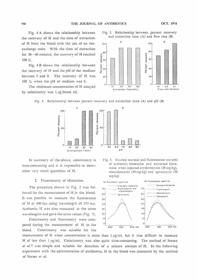
Fig. 3 A shows the relationship between
the recovery of H and the time of extraction with the use of paper chromatography which is
developed by a medium composed of ethyl acetate, ethyl alcohol and distilled water for 24 hours.
When the time was 10 minutes for the extraction of H from the blood, the recovery of H
was about 60 %. Even if the time of extraction was prolonged to more than 20 minutes, the
maximum recovery did not exceed 92.5 %.
Fig. 3 B shows that relationship between the recovery of H and the elution rate of
0.1, 0.5 and 1.0 ml/min. from the ion exchange resin column. One hundred % of the H was
recovered at an elution rate of 0.1 ml/min.
Fig. 1. Procedure for histamine determination
of diazo-method
A solution 15 ml+B solution 15 ml
mix after 5 min.
add B solution 6 ml
mix after 5 min. add water
make total volume 50 ml (A- B solution) sample 2 ml+A.B solution 4 ml
mix after 30 min.
add C solution 10 ml
mix
transfer 3 ml to a cuvette
A solution: 0.9% sulfanylic acid B solution: 5% sodium nitrite C solution: 1% sodium carbonate
Fig. 2. Procedure for extraction and determination of histamine in blood
Oxalated blood 5 ml+Water 4.5 ml+Conc. perchloric acid 0.5 ml
shake 10 min. centrifuge 10 min. (2,500 r.p.m.)
Supernate 4 ml+5 N NaOH 0.5 ml+n-Butanol 10 ml
shake 5 min. centrifuge 10 min.
Organic phase+Salt-saturated 0.1 N NaOH 5 ml
shake 1 min. centrifuge 10 min.
Organic phase 8 Ml +0.1 N HCl 5 ml+n-Heptane 15 ml
shake 1 min. centrifuge 10 min.
Aqueous phase 2 m1+1 N NaOH 0.4 ml
mix
+1 % OPT 0.1 ml
mix after 4 min.
+3 N HCl
mix
Sample
790 THE JOURNAL OF ANTIBIOTICS OCT. 1974
Fig. 4 A shows the relationship between
the recovery of H and the time of extraction
of H from the blood with the use of an ion-
exchange resin. With the time of extraction
for 30-40 minutes, the recovery of H reached
100 %.
Fig. 4 B shows the relationship between
the recovery of H and the pH of the medium
between 5 and 9. The recovery of H was
100 % when the pH of medium was 6.
The minimum concentration of H assayed
by colorimetry was 1 pg/blood ml.
In summary of the above, colorimetry is
time-consuming and it is impossible to deter-
mine very small quantities of H.
2. Fluorometry of Histamine
The procedure shown in Fig. 2 was fol-
lowed for the measurement of H in the blood.
It was possible to measure the fluorescence
of H at 440 mft using wavelength of 355 my.
Authentic H was also measured at the same
wavelengths and gave the same values (Fig. 5).
Colorimetry and fluorometry were com-
pared during the measurement of H in the blood. Colorimetry was suitable for the
measurement of H when concentration is more than 1 ug/ml, but it was difficult to measure
H of less than 1 leg/ml. Colorimetry was also quite time-consuming. The method of SHORE
et al.5) was simple and suitable for detection of a minute amount of H. In the following
experiment with the administration of antibiotics, H in the blood was measured by the method
of SHORE et al.
Fig. 3. Relationship between percent recovery
and extraction time (A) and flow rate (B)
A100
80
60
40
20
010 20 30 60
Extraction time (min.)
B
100
80
60
40
20
01.0 0.5 0.1
Flow rats (ml/min.)
Fig. 4. Relationship between percent recovery and extraction time (A) and pH (B)
A
100
50
0
10 20 30 40
Extraction (min.)
B
too
50
05.0 6.0 7.0 8.0 9 0
pH
Fig. 5. Excited wave(a) and fluorescence wave(b) of authentic histamine and extracted hista-
mine when injected erythromycin (30 mg/kg), oleandomycin (50 mg/kg) and spiramycin (30 mg/kg)
(a) Excitation spectrum
70
60
50
40
30
20
10
0
300 350 400 nm
Standard histamine
Erythromycin and ole on do myc in
Spira mycin
(b) Fluorescence spectrum
70
60
50
40
30
20
10
0
400 450 500 nm
Standard histamine
Erythromycin
Oleondomycin
Spiramycin

791VOL. XXVII NO. 10 THE JOURNAL OF ANTIBIOTICS
3. H in the Blood after the Administration of Macrolide Antibiotics
In Fig. 6 the hypotensive effects of 50 mg/kg of oleandomycin, 30 mg/kg of spiramycin
and 30 mg/kg of erythromycin in dogs are shown. At various intervals before and after the
injection 5 ml of blood was withdrawn for H determination. One minute after the admini-
stration of 50 mg/kg of oleandomycin, the blood pressure gave a maximum fall of 69 % fol-
lowed by recovery after 55 minutes. H in the blood at this time was 0.130±0.040 (S. E.)
pg'ml, representing about a 7-fold increase over the control H value before the administration of oleandomycin (Blood pressure 136 mmHg) of 0.019±0.011 pg/ml. When 30 mg/kg of spira-
mycin was administered, H in the blood increased about 16-fold, while administration of 30
mg/kg of erythromycin led to an approximate 7-fold increase. H in the blood at the time of
the maximum fall in blood pressure is shown in Table 1. This was compared with the value
obtained by bioassay.
4. Comparison between Fluorometry and Bioassay
In the previous report, H in the blood at the time of a fall in the blood pressure in re-
sponse to macrolide series antibiotics was measured by bioassay.
When a small amount of H was measured, the measurable range of H was not much
different between fluorometric assay and bioassay. From the present result fluorometric assay
Fig. 6. Effects of three macrolide antibiotics, oleandomycin (50 mg/kg), spiramycin (30 mg/kg) and erythromycin (30 mg/kg) on the blood pressure in dogs
OLEANDOMYCLN
50mg/kg1 min.,
b SPIRAMYCIN
30mg/kg,1 min.,
ERYTHROMYCIN
30gm/kg1_ min.,
Table 1. Comparison of blood histamine content assayed by fluorometry and bioassay.
Drugs
Oleandomycin
50 mg/kg i.v.
Spiramycin
30 mg/kg i.v.
Erythromycin
30 mg/kg i.v.
Fluorometry (pg/ml)
Control (normal)
0.019 ±0.011
(140)
0.012 ±0.006
(136)
0.028 ±0.017
(138)
Maximum falling of blood
pressure
0.130 ±0.04
(78)
0.193 ±0.035
(44)
0.185 0.021
(30)
Falling of blood
pressure
0.120 ±0.033
(98)
0.045 ±0.041
(94)
0.850 ±0.022
(88)
Recovery
0.029 ±0.009
(140)
0.013 ±0.008
(136)
0.010 ±0.027
(138)
Bioassay (pg/ml)
Control (normal)
0.020 ±0.011
0.018 ±0.002
0.019 +0.002
Maximum falling of
blood
pressure
0.260 ±0.040
0.183 ±0.024
0.136 +0.046
Falling of blood
pressure
0.077 ±0.012
0.051 ±0.016
0.053 +0.005
Recovery
0.022 ±0.005
0.014 ±0.005
0.027 +0.014
Blood pressure are indicated in parentheses. Each value is the mean of four experiments and
standard error. These are the same samples.

792 THE JOURNAL OF ANTIBIOTICS OCT. 1974
appeared to be easy to handle and required only a short time.
Discussion
SHORE et al.5) introduced the method of fluorometry for microquantities of H contained in
various tissues of the bodies of animals and this method is widely used. In the present study,
the authors measured the release of H by macrolide antibiotics as in the previous report by fluorometry.
For the method of colorimetry, paper chromatography was used to extract H. When the time of extraction was 10 minutes the recovery of H was 61.5 %. Even if the time of ext-
raction was prolonged the maximum recovery only reached 90 %. This was because a com-
plete separation of H from the paper did not occur. When column chromatography was used and the speed of elution from the column was less than 0.1 ml/min., the recovery of H was excellent, and a pH of 5-6 appeared to be adequate. However, this procedure took a long time. When H was extracted with an ion-exchange resin, 30 minutes of extraction suf-ficed. Fluorometry was quite simple and excellent for quantification of H in the blood. As
was pointed out by SHORE et a1.5), this method appeared to be adequate for the measurement of H contained in an organ.
H releasers had been cited from ancient times. Among the antibiotics, the polymyxin series antibiotics6) were known as histamine releasers. This was demonstrated by a biological
measurement of H in the blood. Many difficulties were encountered in the measurement of minute amounts of H by colori-
metry, because of the ready tendencies of histamine, tyrosine and indole to become diazo compounds. These antibiotics did not contain H according to bioassay methods.
Conclusion
When blood pressure falls in response to the administration of macrolide antibiotics, ole-
andomycin, spiramycin and erythromycin, H in the blood was measured by a fluorometric
assay and also by colorimetry. The following conclusions were drawn:
1. The H content in blood increased 7-16-fold in response to the administration of
oleandomycin, spiramycin and erythromycin.
2. Colorimetry, fluorometry and bioassay were compared for their measurement of
minute quantities of H; fluorometry and bioassay were superior, and the technic of fluoro-
metry was most simple.
References
1) WAKABAYASHI, K. & S. YAMADA: Effects of several macrolide antibiotics on blood pressure of dogs. Jap. J. Pharmacol. 22: 799-807, 1972 2) BEST, C. H. & E. W. MCHENRY: Histamine. Physiol. Rev. 11 : 371--477, 1931 3) GUGGENHEIM, M. & W. LOFFLER: Biologischer Nachweis proteinogener Amine in Organex-
trakten and Kbrperflussigkeiten. Biochem. Zts. 72 : 303-324, 1916 4) YAMADA, S.; Y. TOYOSHIMA, T. KOEDA, T. MATSUMOTO & K. MATSUO: A method of biological
test for histamine-releasing antibiotics. Jap. J. Antibiotics 22 : 8-13, 1969 5) SHORE, P. A.; A. BURKHALTER & V. H. COHN, Jr.: A method for the fuoronletric assay of histamine in tissues. J. Pharmacol. Exptl. Therap. 127 : 182-186, 1959 6) BUSHBY, S. R. M. & A. F. GREEN: The release of histamine by polymyxin B and polvmyxin E. Brit. J. Pharmacol. & Chemoth. 10 : 215-219, 1955.