a dual phosphorylation switch controls 14-3-3-dependent cell … · 2016-02-09 · research article...
TRANSCRIPT

RESEARCH ARTICLE
A dual phosphorylation switch controls 14-3-3-dependent cellsurface expression of TASK-1Markus Kilisch1,*, Olga Lytovchenko1,*, Eric C. Arakel1, Daniela Bertinetti2 and Blanche Schwappach1,3,‡
ABSTRACTThe transport of the K+ channels TASK-1 and TASK-3 (also known asKCNK3 and KCNK9, respectively) to the cell surface is controlled bythe binding of 14-3-3 proteins to a trafficking control region at theextremeC-terminus of the channels. The current model proposes thatphosphorylation-dependent binding of 14-3-3 sterically masks aCOPI-binding motif. However, the direct effects of phosphorylation onCOPI binding and on the binding parameters of 14-3-3 isoforms arestill unknown. We find that phosphorylation of the trafficking controlregion prevents COPI binding even in the absence of 14-3-3, and wepresent a quantitative analysis of the binding of all human 14-3-3isoforms to the trafficking control regions of TASK-1 and TASK-3.Surprisingly, the affinities of 14-3-3 proteins for TASK-1 are twoorders of magnitude lower than for TASK-3. Furthermore, we find thatphosphorylation of a second serine residue in the C-terminus ofTASK-1 inhibits 14-3-3 binding. Thus, phosphorylation of thetrafficking control region can stimulate or inhibit transport of TASK-1to the cell surface depending on the target serine residue. Ourfindings indicate that control of TASK-1 trafficking by COPI, kinases,phosphatases and 14-3-3 proteins is highly dynamic.
KEY WORDS: 14-3-3 protein, Endoplasmic reticulum, Golgi,Membrane trafficking, Phosphorylation, Two-pore-domain K+
channel, COPI, Protein kinase A, TASK-1
INTRODUCTIONTwo-pore-domain K+ (K2P) channels are dimeric membraneproteins that function at the cell surface and contribute to theelectrical properties of many different cell types (Renigunta et al.,2015). It is well established that two members of the K2P-channelfamily – i.e. TASK-1 and TASK-3 (also known as KCNK3 andKCNK9, respectively) – require phosphorylation-dependent high-affinity binding of 14-3-3 proteins to their distal C-terminus in orderto reach the cell surface (O’Kelly et al., 2002; Rajan et al., 2002;Smith et al., 2011; Kilisch et al., 2015). 14-3-3 proteins are smalladaptor proteins with no specific enzyme activity; they canmodulate the function of many ‘client’ proteins. The distal C-terminus of both channels functions as a trafficking control region; itcontains a binding motif for the COPI vesicle coat and anoverlapping mode III 14-3-3-binding motif [R-x-x-pS/pT-x-
COOH, where ‘p’ denotes a phosphorylated residue and ‘x’ anyamino acid (Coblitz et al., 2005)]. In the classic model of TASKtrafficking, interaction with COPI retains TASK channels inthe early secretory pathway until phosphorylation of the distalC-terminus allows binding of 14-3-3, which sterically preventsCOPI from accessing the trafficking control region. Consequently,the phosphorylated channel exits the Golgi and is expressed at thecell surface. Based on studies using in vitro phosphorylation, onmutagenesis or application of specific inhibitors, protein kinase A(cAMP-dependent protein kinase, PKA) has emerged as the kinasethat is most likely to be responsible for phosphorylating TASK C-termini (Mant et al., 2011). However, the direct effect ofphosphorylation on COPI binding has not yet been determined,and the protein–protein interactions involved in TASK traffickingcontrol have not been assessed quantitatively. Most studies treat theseven 14-3-3 proteins (encoded by seven distinct genes butcommonly termed isoforms in the field) as one entity, and little isknown about the significance of individual 14-3-3 proteins (denotedwith Greek letters as β, γ, η, ε, σ, τ and ζ) in modulating the functionof specific 14-3-3 clients.
The observation that the intracellular trafficking of TASKchannels depends on phosphorylation and interaction with 14-3-3proteins suggests that the surface expression of TASK channelsmight be regulated by protein kinases and phosphatases. This aspectis poorly understood because studies in heterologous systems havefocused on the fundamental prerequisites for cell surface expressionrather than on its modulation by signal transduction. However, thecell surface expression of many channels is highly regulated underdifferent physiological conditions. We have recently shown that theCOPI-binding motif, which prevents the cell surface expression ofATP-sensitive K+ channels, can be phosphorylated and thusinactivated upon β-adrenergic stimulation in cardiac myocytes(Arakel et al., 2014). Here, we have used an array of wild-type andmutated TASK distal C-termini, and all seven mammalian 14-3-3proteins to systematically and quantitatively delineate the molecularevents that determine the role of the TASK trafficking control regionin regulated cell surface expression.
RESULTSAffinity of 14-3-3 for the TASK-1 C-terminus is substantiallylower than that for the TASK-3 C-terminusCurrent insight into COPI and 14-3-3 binding to the traffickingcontrol region of the TASK C-terminus is qualitative (O’Kellyet al., 2002; Rajan et al., 2002; O’Kelly and Goldstein, 2008;Zuzarte et al., 2009). The equilibrium dissociation constant (Kd)for the binding of 14-3-3σ to a hexapeptide corresponding to theC-terminus of TASK-3 that included phosphorylated residueS373 has been determined previously (4.1±0.8 µM; Anderset al., 2013), but it is not known how the parameters of 14-3-3binding compare between TASK-1 and TASK-3, or between thedifferent 14-3-3 isoforms. To determine them systematically, weReceived 7 September 2015; Accepted 29 December 2015
1Department of Molecular Biology, Universitatsmedizin Gottingen,Humboldtallee 23, Gottingen 37073, Germany. 2Department of Biochemistry,University of Kassel, Kassel 34132, Germany. 3Max-Planck Institute forBiophysical Chemistry, Gottingen 37077, Germany.*These authors contributed equally to this work
‡Author for correspondence ([email protected])
This is an Open Access article distributed under the terms of the Creative Commons AttributionLicense (http://creativecommons.org/licenses/by/3.0), which permits unrestricted use,distribution and reproduction in any medium provided that the original work is properly attributed.
831
© 2016. Published by The Company of Biologists Ltd | Journal of Cell Science (2016) 129, 831-842 doi:10.1242/jcs.180182
Journal
ofCe
llScience

chose fluorescence polarization titration – a solution-basedmethod suited to measuring the binding affinity between one14-3-3 ligand-binding groove and the TASK-derived peptide(Fig. 1A,B). The use of synthetic fluorescein-labeled peptides isparticularly useful for studying the C-terminus of TASK-1because the phosphorylation status of the two serine residues(S392 and S393) present in its sequence can be controlledindividually. All seven human 14-3-3 isoforms were affinitypurified as fusions to the maltose binding protein (MBP). Aftercleavage of the MBP tag, dimeric 14-3-3 proteins were purifiedby using size exclusion chromatography (Fig. S1).TASK-1 or TASK-3 C-terminal fluorescent peptides (10 nM),
comprising the last 15 amino acids of the respective C-termini, weretitrated with concentrations of the 14-3-3 proteins ranging from1 nM to 120 µM (Fig. 2), and the equilibrium dissociation constantsof 14-3-3 proteins were calculated for TASK-1 phosphorylated atS393 (TASK-1 pS393; the conserved serine residue of the mode III14-3-3-binding motif ) and for TASK-3 phosphorylated at S373(TASK-3 pS373) (Table 1). They ranged from 7.5±0.14 µM (mean±s.e.m.; 14-3-3β) to 49.4±2 µM (14-3-3τ) for TASK-1, whereasdissociation constants for TASK-3 ranged from 110±10 nM (14-3-3η) to 3.6± 0.44 µM (14-3-3σ). The latter value matches the resultdetermined previously by Anders et al. (2013) for the TASK-3 C-terminal hexapeptide using isothermal titration calorimetry. Thedissociation constants of the seven mammalian 14-3-3 isoforms forthe same TASK C-terminal peptide differed up to sevenfold forTASK-1-derived peptides and up to 30-fold for TASK-3-derivedpeptides. For both TASK C-termini, 14-3-3γ and 14-3-3η boundwith high affinity, whereas 14-3-3σ bound with low affinity. Nobinding of any 14-3-3 isoform to any TASK C-terminal peptide wasobserved when the peptides were not phosphorylated.In addition to fluorescence polarization, we used surface
plasmon resonance (SPR) as a second, complementary approachto quantify the binding parameters for the TASK-3 C-terminus(Fig. 3, Table 1). A glutathione S-transferase (GST)-taggedTASK-3 C-terminal 15 residues fusion protein (GST–TASK-3)was expressed in and purified from Escherichia coli (and hence wasnot phosphorylated). We developed an on-chip phosphorylationprotocol (Fig. 3A) where we first captured the GST fusion proteins
on the chip surface with an antibody against GST (which wascrosslinked at a high density to the metal surface) and thenphosphorylated GST–TASK-3 with recombinantly expressed PKAcatalytic subunit (also known as PRKACA; Knape et al., 2015). Weverified efficient phosphorylation of the fusion proteins byexamining the binding parameters of a phospho-specific antibodythat recognizes a phosphorylated PKA target motif to the PKA-treated TASK-3 C-terminus (Fig. 3B). We observed high-affinitybinding with an equilibrium dissociation constant of 4.5±0.6 nM.Consistent with the results of fluorescence polarization titration,no binding of 14-3-3 proteins was observed prior to on-chipphosphorylation, whereas all 14-3-3 isoforms did bind uponphosphorylation of the TASK-3 C-terminus (Fig. 3C) withequilibrium dissociation constants (Fig. 3D, Table 1) between45±9 nM (14-3-3γ) to 742±29 nM (14-3-3σ). Depending onthe 14-3-3 isoform, these values were very similar (14-3-3η and14-3-3τ) or differed two- (14-3-3β) to fivefold (14-3-3ε, 14-3-3ζand 14-3-3σ) from the values determined by fluorescencepolarization (Fig. 2, Table 1). Importantly, 14-3-3γ and 14-3-3ηdisplayed the same high-affinity binding as that observed in thefluorescence polarization assay. We conclude that the two methodsyield comparable parameters for 14-3-3 binding to the TASK-3pS373 C-terminal peptide. The differences in absolute values mightbe due to the fact that the presentation of the phosphorylatedC-termini is different in the two methods (soluble peptide comparedto immobilized GST fusion protein).
Deletion of the C-terminal valine residue in the TASK-3C-terminus does not abolish 14-3-3 bindingBased on yeast two-hybrid and qualitative pull-down assays, it hasbeen proposed that deleting the C-terminal valine residue of themode III 14-3-3-binding motif abolishes 14-3-3 binding (O’Kellyet al., 2002; Rajan et al., 2002). To test this hypothesis, we measuredthe affinity of all seven 14-3-3 isoforms for the TASK-3 C-terminalpeptide containing phosphorylated S373, but lacking the C-terminalvaline (V374), by fluorescence polarization titration (Fig. 2,Table 1). Indeed, the interaction could no longer be measured for14-3-3σ, but the affinities remained in the low micromolar range for14-3-3β, 14-3-3γ, 14-3-3ζ, 14-3-3η and 14-3-3τ. The C-terminal
Fig. 1. Peptides and proteins used in this study. The last 15 amino acids of human TASK-1 and TASK-3 C-termini contain a minimal trafficking control regioncomprising a COPI recognition motif (green), a PKA target site (red) and a mode III 14-3-3-binding motif (-K-R-R-K/S-pS-V-COOH, where ‘p’ denotes aphosphorylated residue). Note the presence of a serine residue (S392) preceding the conserved serine residue of the mode III 14-3-3-binding motif in TASK-1(S393) but not TASK-3 (S373). Fluorescein-labeled peptides comprising the TASK C-termini were used for the determination of equilibrium binding constants byusing fluorescence polarization.
832
RESEARCH ARTICLE Journal of Cell Science (2016) 129, 831-842 doi:10.1242/jcs.180182
Journal
ofCe
llScience
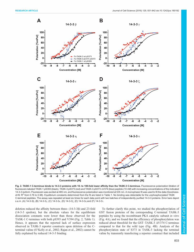
deletion reduced the affinity between three- (14-3-3β) and 23-fold(14-3-3 epsilon), but the absolute values for the equilibriumdissociation constants were lower than those observed for theTASK-1 C-terminus with both pS393 and V394 (Fig. 2, Table 1).Hence, it appears that the reported lack of surface expressionobserved in TASK-3 reporter constructs upon deletion of the C-terminal valine (O’Kelly et al., 2002; Rajan et al., 2002) cannot befully explained by reduced 14-3-3 binding.
To further clarify this point, we studied the phosphorylation ofGST fusion proteins of the corresponding C-terminal TASK-3peptides by using the recombinant PKA catalytic subunit in vitro(Fig. 4A), and we found that the efficiency of phosphorylation wasreduced about threefold for the GST–TASK-3 ΔV374 C-terminuscompared to that for the wild type (Fig. 4B). Analysis of thephosphorylation state of S373 in TASK-3 lacking the terminalvaline by transiently transfecting a reporter construct that included
Fig. 2. TASK-1 C-terminus binds to 14-3-3 proteins with 10- to 100-fold lower affinity than the TASK-3 C-terminus. Fluorescence polarization titration offluorescein-labeled TASK-1 pS393 (black), TASK-3 pS373 (red) and TASK-3 pS373 ΔV374 (blue) peptide (10 nM) with increasing concentrations of the indicated14-3-3 isoform. Fluorescein was excited at 485 nm, and fluorescence polarization wasmonitored at 535 nm. Amonophasic fit was used to fit the data (Goodness-of-fit: R2 from 0.76 to 0.98). Equilibrium constants determined from the fit are listed in Table 1. No binding was detectable for the unphosphorylated TASKC-terminal peptides. The assay was repeated at least six times for each data point with two batches of independently purified 14-3-3 proteins. Error bars depicts.e.m. (A) 14-3-3β, (B) 14-3-3γ, (C) 14-3-3ε, (D) 14-3-3ζ, (E) 14-3-3η and (F) 14-3-3τ.
833
RESEARCH ARTICLE Journal of Cell Science (2016) 129, 831-842 doi:10.1242/jcs.180182
Journal
ofCe
llScience

the extracellular domain of CD8 (CD8–CFP–TASK-3 ΔV374) intoCOS-7 cells and subsequent SDS-PAGE analysis with gelscontaining a phospho-chelating agent (Phostag) also revealed thatthe efficiency of S372 phosphorylation was decreased in theabsence of the C-terminal valine. The reporter proteins exposingthe wild-type TASK-3 C-terminal 15 residues or a mutant, in which
the COPI-dependent ER retention–retrieval motif (KRR) wasdisrupted (K369A), migrated slower than the S373A mutant,implying that both the wild-type TASK-3 and the K369A mutantwere phosphorylated. Consistent with this interpretation, treatmentwith phosphatase altered the migratory behavior of the reporterproteins to match that of the S373A mutant. In contrast to the wild
Table 1. Binding affinities of 14-3-3 proteins to TASK-1 and TASK-3 C-terminal peptides determined by fluorescence polarization and SPR
Fluorescence polarizationSPR
Kd [nM] Kd [nM]
14-3-3 isoform TASK-1 pS393 TASK-3 pS373 TASK-3 pS373 ΔV374 GST–TASK-3-CT15
β 7500±140 370±40 1200±100 181±21γ 9800±640 150±20 1100±140 45±9ε 29,000±1100 800±80 18,000±4800 150±6ζ 23,000±1400 1000±60 3300±300 221±35η 9200±110 110±10 1600±140 115±28τ 49,400±2000 220±20 2400±100 246±19σ 36,000±2900 3600±400 n.b.±n.b. 742±29
Equilibrium dissociation constants (Kd) are displayed in [nM]. Error is s.e.m. n.b., no binding detectable.
Fig. 3. Affinities of the seven mammalian 14-3-3 isoforms for the phosphorylated TASK-3C-terminus obtained by surface plasmonresonance are consistent with valuesobtained using fluorescence polarization.(A) Schematic illustration of the surface plasmonresonance (SPR) set-up used to monitor 14-3-3binding to immobilized TASK-3 C-terminifused to GST and phosphorylated on thecarboxymethyldextran sensor chip. (B) Anantibody against PKA-phosphorylated substrateswas used to confirm on-chip phosphorylation ofthe TASK-3 C-termini. (C) Representative familyof SPR sensograms reflecting the association anddissociation phases of 14-3-3β and 14-3-3σbinding to immobilized phosphorylated TASK-3C-termini. No binding was observed beforephosphorylation of the immobilized GST fusionprotein. (D) Dose-response curve for therespective pairs of GST–TASK-3 C-terminalfusion proteins and the individual 14-3-3 proteinswith SPR analysis. Data were analyzed with asigmoidal dose-response curve fit (Goodness-of-fit: R2 from 0.96 to 0.99). Equilibrium constantsdetermined from the fit are listed in Table 1. Theassay was repeated at least six times for eachdata point with two batches of independentlypurified 14-3-3 proteins. Error bars depict s.e.m.
834
RESEARCH ARTICLE Journal of Cell Science (2016) 129, 831-842 doi:10.1242/jcs.180182
Journal
ofCe
llScience

type and to the K369A variant, we observed substantial amounts ofunphosphorylated CD8–CFP–TASK-3 ΔV374 reporter proteinprior to phosphatase treatment. These findings suggest thattruncation of the mode III 14-3-3-binding motif affects theefficiency of phosphorylation by PKA in vivo and might alsoimprove access for cellular phosphatases.To assess the functional consequences of phosphorylation on cell
surface expression of the reporter protein, we employed flowcytometry (Fig. 4C). In this assay, transfected cells were identifiedusing the CFP fluorescence signal, and the relative cell surfaceexpression was determined by staining an extracellular epitope ofCD8 with a specific antibody. We determined the relative cell
surface expression of wild-type CD8–CFP–TASK-3 as well as thatof the ΔV374 and S373Amutants, and we found that the C-terminaltruncation variant displayed moderately higher cell surfaceexpression levels than the non-phosphorylatable S373A mutant.We also assessed the steady-state localization of the bulk of theCD8–CFP fusion proteins with indirect immunostaining (Fig. 4D;Fig. S2) and observed a very similar pattern for both, CD8–CFP–TASK-3 ΔV374 and CD8–CFP–TASK-3 S373A. As reportedpreviously (Zuzarte et al., 2009), proteins unable to recruit 14-3-3,which is required to counteract the COPI-binding motif present inthe C-terminus of TASK proteins, accumulated in a punctateperinuclear COPI-positive structure (Fig. S2) in the vicinity of a
Fig. 4. Reduced 14-3-3 bindingaffinity does not explain the lackof cell surface transport of theTASK-3 ΔV mutant. (A) In vitrophosphorylation of recombinant GST–TASK-3 (15 C-terminal residues) wild-type (WT), GST–TASK-3 ΔV374 andGST–TASK-3 S373A fusion proteinswith the PKA catalytic subunit.Samples were resolved on an SDS-PAGE gel containing 75 µM Phostagreagent and 75 µM MnCl2, whichretards the migration of proteins bychelating phosphorylated amino acidside chains. The Coomassie-stainedgel is representative of six independentexperiments. TASK-3-P,phosphorylated TASK-3. (B) Bardiagram of the relative amount ofphosphorylated TASK-3 traffickingcontrol region constructs shownin A. n=6, error bars depict s.e.m.(C) In vivo phosphorylation status ofthe indicated CD8–CFP reporterproteins, as reflected by migration inPhostag SDS-PAGE gels. λPPase, λphosphatase. The blot isrepresentative of 11 independenttransfections. (D) Flow cytometryassay to determine the effect of theindicated TASK C-termini on the cellsurface expression of a CD8–CFPreporter protein. Cells were transfectedwith the same series of constructs inthree independent experiments. Foreach experiment, 10,000 cells perconstruct were analyzed. The mean ofthe CD8 signal was expressed as aratio with the mean of the CFP signal inCFP-positive cells and normalized tothat of the wild-type TASK-3 construct.Error bars depict s.e.m. (E) Subcellularlocalization of CD8–CFP–TASK-3ΔV374 is indistinguishable from that ofCD8–CFP–TASK-3 S373A.Constructs were transfected in threeindependent experiments and ca. 100cells per transfection were analyzed byindirect immunostaining against theindicated antigens.
835
RESEARCH ARTICLE Journal of Cell Science (2016) 129, 831-842 doi:10.1242/jcs.180182
Journal
ofCe
llScience
marker of the Golgi compartment (GM130, also known asGOLGA2; Fig. 4D). We conclude that in the mutant TASK-3ΔV374, less of the CD8–CFP reporter fusion protein isphosphorylated and that the phosphorylated form binds to 14-3-3proteins with lower affinity than wild-type TASK-3 but as stronglyas TASK-1 pS393. The simplest interpretation of these findings isthat strongly reduced cell surface expression of C-terminallytruncated TASK-3 is attributable to the combination of inefficientphosphorylation and a moderately reduced 14-3-3 binding affinity.
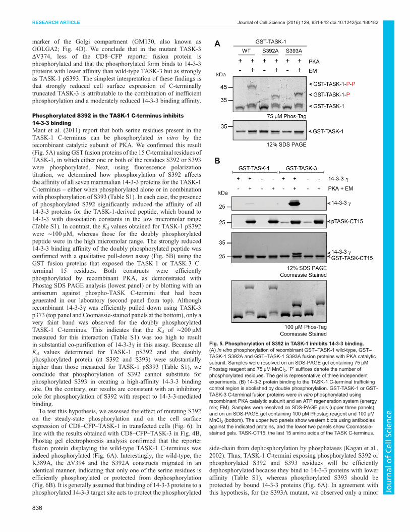
Phosphorylated S392 in the TASK-1 C-terminus inhibits14-3-3 bindingMant et al. (2011) report that both serine residues present in theTASK-1 C-terminus can be phosphorylated in vitro by therecombinant catalytic subunit of PKA. We confirmed this result(Fig. 5A) using GST fusion proteins of the 15 C-terminal residues ofTASK-1, in which either one or both of the residues S392 or S393were phosphorylated. Next, using fluorescence polarizationtitration, we determined how phosphorylation of S392 affectsthe affinity of all seven mammalian 14-3-3 proteins for the TASK-1C-terminus – either when phosphorylated alone or in combinationwith phosphorylation of S393 (Table S1). In each case, the presenceof phosphorylated S392 significantly reduced the affinity of all14-3-3 proteins for the TASK-1-derived peptide, which bound to14-3-3 with dissociation constants in the low micromolar range(Table S1). In contrast, the Kd values obtained for TASK-1 pS392were ∼100 µM, whereas those for the doubly phosphorylatedpeptide were in the high micromolar range. The strongly reduced14-3-3 binding affinity of the doubly phosphorylated peptide wasconfirmed with a qualitative pull-down assay (Fig. 5B) using theGST fusion proteins that exposed the TASK-1 or TASK-3 C-terminal 15 residues. Both constructs were efficientlyphosphorylated by recombinant PKA, as demonstrated withPhostag SDS PAGE analysis (lowest panel) or by blotting with anantiserum against phospho-TASK C-termini that had beengenerated in our laboratory (second panel from top). Althoughrecombinant 14-3-3γ was efficiently pulled down using TASK-3p373 (top panel and Coomassie-stained panels at the bottom), only avery faint band was observed for the doubly phosphorylatedTASK-1 C-terminus. This indicates that the Kd of ∼200 µMmeasured for this interaction (Table S1) was too high to resultin substantial co-purification of 14-3-3γ in this assay. Because allKd values determined for TASK-1 pS392 and the doublyphosphorylated protein (at S392 and S393) were substantiallyhigher than those measured for TASK-1 pS393 (Table S1), weconclude that phosphorylation of S392 cannot substitute forphosphorylated S393 in creating a high-affinity 14-3-3 bindingsite. On the contrary, our results are consistent with an inhibitoryrole for phosphorylation of S392 with respect to 14-3-3-mediatedbinding.To test this hypothesis, we assessed the effect of mutating S392
on the steady-state phosphorylation and on the cell surfaceexpression of CD8–CFP–TASK-1 in transfected cells (Fig. 6). Inline with the results obtained with CD8–CFP–TASK-3 in Fig. 4B,Phostag gel electrophoresis analysis confirmed that the reporterfusion protein displaying the wild-type TASK-1 C-terminus wasindeed phosphorylated (Fig. 6A). Interestingly, the wild-type, theK389A, the ΔV394 and the S392A constructs migrated in anidentical manner, indicating that only one of the serine residues isefficiently phosphorylated or protected from dephosphorylation(Fig. 6B). It is generally assumed that binding of 14-3-3 proteins to aphosphorylated 14-3-3 target site acts to protect the phosphorylated
side-chain from dephosphorylation by phosphatases (Kagan et al.,2002). Thus, TASK-1 C-termini exposing phosphorylated S392 orphosphorylated S392 and S393 residues will be efficientlydephosphorylated because they bind to 14-3-3 proteins with loweraffinity (Table S1), whereas phosphorylated S393 should beprotected by bound 14-3-3 proteins (Fig. 6A). In agreement withthis hypothesis, for the S393A mutant, we observed only a minor
Fig. 5. Phosphorylation of S392 in TASK-1 inhibits 14-3-3 binding.(A) In vitro phosphorylation of recombinant GST–TASK-1 wild-type, GST–TASK-1 S392A and GST–TASK-1 S393A fusion proteins with PKA catalyticsubunit. Samples were resolved on an SDS-PAGE gel containing 75 µMPhostag reagent and 75 µM MnCl2. ‘P’ suffixes denote the number ofphosphorylated residues. The gel is representative of three independentexperiments. (B) 14-3-3 protein binding to the TASK-1 C-terminal traffickingcontrol region is abolished by double phosphorylation. GST-TASK-1 or GST-TASK-3 C-terminal fusion proteins were in vitro phosphorylated usingrecombinant PKA catalytic subunit and an ATP regeneration system (energymix; EM). Samples were resolved on SDS-PAGE gels (upper three panels)and on an SDS-PAGE gel containing 100 µM Phostag reagent and 100 µMMnCl2 (bottom). The upper two panels show western blots using antibodiesagainst the indicated proteins, and the lower two panels show Coomassie-stained gels. TASK-CT15, the last 15 amino acids of the TASK C-terminus.
836
RESEARCH ARTICLE Journal of Cell Science (2016) 129, 831-842 doi:10.1242/jcs.180182
Journal
ofCe
llScience

band reflecting the phosphorylated species. We conclude that S392can be phosphorylated by the catalytic subunit of PKA (Fig. 5), butour results suggest that either phosphorylation is inefficient or thatthis residue is efficiently dephosphorylated in COS-7 cells.
S392 reduces the cell surface expression of CD8–CFP–TASK-1To test the functional consequences of S392 phosphorylation intransfected cells, we determined the relative cell surface expressionof CD8–CFP–TASK-1 S392A using flow cytometry (Fig. 6B).Surprisingly, the surface expression of this construct was more thantwofold higher than that of ‘wild-type’ CD8–CFP–TASK-1. Incontrast, CD8–CFP–TASK-1 S393A did not reach the cell surface,most probably owing to its inability to bind to 14-3-3 proteins. Thisfinding is consistent with the results obtained with the CD8–CFP–TASK-3 S373A mutant (Fig. 4C).It has been reported previously that residue K389 in the
trafficking control region is a crucial residue of the ERlocalization motif of TASK-1 (Zuzarte et al., 2009). Mutating thelysine residue to alanine induced the same twofold increase in CD8–CFP–TASK-1 surface expression as the S392A mutation. Theseobservations are consistent with the notion that transientphosphorylation of S392 is sufficient to reduce the efficacy bywhich 14-3-3 proteins can remove the cargo protein from the part ofthe secretory pathway where COPI operates. In the absence of a
functional COPI-interaction motif (Zuzarte et al., 2009), 14-3-3protein binding to the TASK C-terminus is no longer required forcell surface expression. Hence, the corresponding construct K389Areaches the cell surface efficiently. For TASK-3, which displayshigh-affinity 14-3-3 binding and no second inhibitory serine, thecorresponding K369A mutant is expressed at levels at the cellsurface that are nearly equal to those of the TASK-3 wild-typeconstruct (Fig. 4C). Hence, in the case of TASK-3, 14-3-3efficiently displaces COPI. The reduced cell surface expression ofthe TASK-1 wild-type construct with respect to both the TASK-1K389A and S392A constructs is consistent with the notion that bothCOPI and 14-3-3 proteins can access the trafficking control regionin the steady state.
Phosphorylation of TASK C-termini abolishes COPI bindingAlthough it is well established that phosphorylation of the mode IIIbinding motif is a prerequisite for 14-3-3 binding to TASKC-termini, the effect of phosphorylation on COPI binding isunknown. Functionally, the COPI-binding motif was mapped in thecontext of TASK ΔV constructs because retention of theseconstructs from the cell surface reflects COPI activity. Pull-downassays to directly assess the physical interaction were performedwith cellular lysates that also contained kinases, phosphatases and14-3-3 proteins (O’Kelly et al., 2002; O’Kelly and Goldstein, 2008;Zuzarte et al., 2009). In these experiments, detection of the β-COP
Fig. 6. Phosphorylation of S392 in TASK-1 reduces cell surface expression. (A) In vivo phosphorylation status of the CD8 reporter protein as reflected bymigration in Phostag SDS-PAGE gels. The blot is representative of seven independent transfections. TASK-1-P, phosphorylated TASK-1. (B) Flow cytometryassay to determine the effect of replacing either S392 or S393 in the TASK-1 C-terminus on the cell surface expression of the indicated CD8–CFP reporterproteins. Cells were transfected with the same series of constructs in three independent experiments. For each experiment, 10,000 cells per construct wereanalyzed. The mean of the CD8 signal is expressed as a ratio with the mean of the CFP signal in CFP-positive cells and normalized to that for the wild-typeTASK-3 construct. Error bars depict s.e.m. (C) Model of the effect of phosphorylation of S392 or S393, or both on 14-3-3 binding and on the access ofphosphatases. Note that only the population of TASK-1 reporter proteins phosphorylated on only S393 is expected to be protected from phosphatase action.
837
RESEARCH ARTICLE Journal of Cell Science (2016) 129, 831-842 doi:10.1242/jcs.180182
Journal
ofCe
llScience

(also known as COPB1) subunit served as a proxy for binding of theheptameric COPI coat. To assess the physical interaction with COPIin the absence of additional binding partners (Fig. 7), we exploitedour previous finding that the ER retention–retrieval signal ofTASK-3 is also recognized in yeast (Zuzarte et al., 2009). Wepurified the COPI coat from yeast using tandem-affinity purification(TAP) of tagged β-COP (Yip and Walz, 2011) and tested itsinteraction with the TASK-1 and TASK-3 trafficking control region(15 C-terminal residues) presented on the cytoplasmic tail of theyeast protein Mst27, which lacked its KKXX signal and had beenfused to GST, as described previously (Sandmann et al., 2003). ThisMst27 linker increases the accessibility of the C-terminal peptide tothe heptameric COPI coat and has been extensively characterized inCOPI-binding experiments (Sandmann et al., 2003; Michelsenet al., 2007). We found that both immobilized GST–TASK-1 andGST–TASK-3 enriched COPI before but not after phosphorylationof the TASK tails (Fig. 7A). Quantification of three independent
experiments showed that TASK-1 bound to COPI less efficientlythan TASK-3 (Fig. 7B). We conclude that COPI ceases to interactwith the phosphorylated trafficking control region of TASK-1 andTASK-3 channels even in the absence of 14-3-3 proteins. Thisimplies that there is no direct competition between COPI and 14-3-3binding (Fig. 7C). Instead, the respective binding equilibria existbetween either the unphosphorylated (COPI) or phosphorylated(14-3-3) forms of the TASK C-terminus and hence are coupledthrough the action of kinases and phosphatases.
DISCUSSIONThe trafficking control region in the C-termini of the K2P channelsTASK-1 and TASK-3 is thought to control the surface expression ofthe channels through the mutually exclusive binding of either theCOPI vesicle coat, which mediates retrieval to the early secretorypathway, or of 14-3-3 proteins, which bind in a phosphorylation-dependent manner and protect the region from COPI binding (Smith
Fig. 7. Phosphorylation of TASK C-termini inhibits COPI binding. (A) GST–Mst27–TASK-1 or GST–Mst27-TASK-3 fusion proteins comprising the last15 amino acids of the C-termini (CT15) were in vitro phosphorylated using recombinant PKA catalytic subunit and an ATP regeneration system (energy mix; EM).Upper panel shows a western blot and detection with an anti-yeast COPI antiserum, lower panel shows a Coomassie-stained gel. (B) Quantification of threeindependent pull-down experiments similar to that depicted in A. Error bars depict s.e.m. (C) Model of the effect of phosphorylation of S373 in TASK-3 on thebinding of COPI, access of phosphatases and 14-3-3 binding. Note that binding of COPI and 14-3-3 proteins is not competitive.
838
RESEARCH ARTICLE Journal of Cell Science (2016) 129, 831-842 doi:10.1242/jcs.180182
Journal
ofCe
llScience
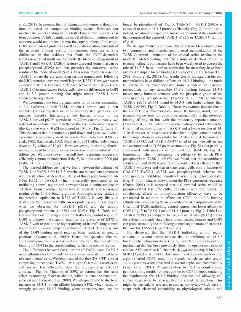
et al., 2011). In essence, the trafficking control region is thought tofunction based on competitive binding events. However, ourmechanistic understanding of this trafficking control region is farfrom complete. A full quantitative model of this competition and itsoutcome would require insight into the copy numbers of the cargo,COPI and of 14-3-3 proteins as well as the dissociation constants ofthe pertinent binding events. Furthermore, there are strikingdifferences in the residues that flank the COPI-dependentretention–retrieval motif and the mode III 14-3-3-binding motif ofTASK-1 and TASK-3; TASK-1 features a second serine that can bephosphorylated (S392) and that precedes the conserved serineresidue of the mode III motif (S393). This serine residue is absent inTASK-3, where the corresponding residue immediately followingthe KRR retention–retrieval motif is lysine (K372). Here, we presentevidence that this sequence difference between the TASK-1 andTASK-3 C-termini causes biologically relevant differences in COPIand 14-3-3 protein binding that might render TASK-1 moreamenable to regulation.We determined the binding parameters for all seven mammalian
14-3-3 isoforms to both TASK protein C-termini and to theirvariants (phosphorylated or non-phosphorylated peptides andmutants thereof ). Interestingly, the highest affinity of theTASK-1-derived pS393 peptide to 14-3-3 was approximately twoorders of magnitude lower than that of the TASK-3-derived peptide(the Kd value was ∼10 µM compared to 100 nM; Fig. 2, Table 1).This illustrates that the numerous pull-down and yeast two-hybridexperiments previously used to assess this interaction (O’Kellyet al., 2002; Rajan et al., 2002; Smith et al., 2011) effectively operatedown to Kd values of 10 µM. However, owing to their qualitativenature, the yeast two-hybrid experiments obscure substantial affinitydifferences. We also showed that pull-down experiments cease toefficiently capture an interaction if the Kd is in the order of 200 µM(Table S1, Fig. 5) or higher.The marked differences that we found between the affinities of
TASK-1 or TASK-3 for 14-3-3 proteins are in excellent agreementwith the structure (Anders et al., 2013) of this peptide bound to 14-3-3σ; K372 of TASK-3, which is centrally positioned in thetrafficking control region and corresponds to a serine residue inTASK-1, forms hydrogen bonds with an aspartate and asparagineresidue of the 14-3-3-binding groove. A phosphoserine residue inthe position equivalent to K372 of TASK-3 is very likely todestabilize the interaction with 14-3-3 proteins, and this is exactlywhat we observed for TASK-1 pS392 and the doublyphosphorylated protein (at S392 and S393) (Fig. 5, Table S1).Because the exact binding site for the trafficking control region onCOPI is unknown, we cannot interpret the relevance of K372 inTASK-3 with respect to the higher affinity of its trafficking controlregion to COPI when compared to that of TASK-1. The consensusof the COPI-binding motif requires basic residues in specificpositions (Zuzarte et al., 2009). Hence, we speculate that theadditional lysine residue in TASK-3 contributes to the high-affinitybinding of COPI to the corresponding trafficking control region.The differences between the C-termini of TASK-1 and TASK-3
in the affinities for COPI and 14-3-3 proteins were also found to berelevant in intact cells. We demonstrated that the CD8–CFP reportercomprising the exposed TASK-1 wild-type C-terminus reaches thecell surface less efficiently than the corresponding TASK-3construct (Fig. 6). Mutation of S392 to alanine has the sameeffect as mutating K389 to alanine, which mutates the retention–retrieval motif (Zuzarte et al., 2009).We interpret this to be due to anincrease in 14-3-3 protein affinity because S392, which results instrongly reduced 14-3-3 binding when phosphorylated, can no
longer be phosphorylated (Fig. 5, Table S1). TASK-1 S392A isexpected to recruit 14-3-3 proteins efficiently (Fig. 2, Table 1) and,indeed, we observed equal cell surface expression of the constructsthat comprised the exposed TASK-1 S392A or TASK-3 C-termini(Fig. 6).
We also quantitatively compared the effects on 14-3-3 binding fortwo commonly and interchangeably used manipulations of theTASK-C-termini – mutation of the crucial serine residue in themode III 14-3-3-binding motif to alanine or deletion of the C-terminal valine. Both variants have been widely used to dissect therole of 14-3-3 in cell surface expression because they have beenassumed to impair 14-3-3 binding (O’Kelly et al., 2002; Rajan et al.,2002; Smith et al., 2011). Our results clearly indicate that the twomanipulations have different effects on 14-3-3 binding – the modeIII serine in its phosphorylated form is indeed an essentialpre-requisite for any detectable 14-3-3 binding because 14-3-3makes many intricate contacts with the phosphate group of thecorresponding phosphoserine (Anders et al., 2013), whereasTASK-3 pS373 ΔV374 bound to 14-3-3 with higher affinity thanTASK-1 pS393 (Fig. 2, Table 1). These observations indicate that inthe context of a phosphorylated mode III binding motif, the C-terminal valine does not contribute substantially to the observedbinding affinity, in line with the previously reported structure(Anders et al., 2013), which shows one hydrogen bond between theC-terminal carboxy group of TASK-3 and a lysine residue of 14-3-3σ. However, we also observed that the biological outcome of thetwo manipulations is very similar for TASK-3 (Fig. 4) because bothTASK-3 S373A and TASK-3 ΔV374 failed to reach the cell surfaceand accumulated in COPI-positive structures (Fig. S1) that partiallycolocalized with markers of the cis-Golgi (GM130; Fig. 4).Importantly, when investigating the efficiency by which PKAphosphorylates TASK-3 ΔV374, we found that the recombinantcatalytic subunit of PKAmodifies this construct less efficiently thanTASK-3 wild type and that in transfected cells only a fraction ofCD8–CFP–TASK-3 ΔV374 was phosphorylated, whereas thecorresponding wild-type construct was fully phosphorylated(Fig. 4). From what is known about the PKA consensus sequence(Shabb, 2001), it is expected that a C-terminal serine would bephosphorylated less efficiently, consistent with our results. Inconclusion, effects on phosphorylation efficiency have to beconsidered in addition to effects on COPI or 14-3-3 bindingaffinity when comparing the in vivo outcome of manipulations in theC-terminal TASK trafficking control region. The lower affinity ofCOPI (Fig. 7) to TASK-1 and of 14-3-3 proteins (Fig. 2, Table 1) toTASK-1 pS393 (as compared to TASK-3 or TASK-3 pS373) allowsfor a dynamic steady state where phosphatases, kinases and COPIcan probe or modify the trafficking control region more often than isthe case for TASK-3 (Figs 6B and 7C).
Our discovery that the TASK-1 trafficking control regioncontains a second serine residue that is inhibitory to 14-3-3binding when phosphorylated (Fig. 5, Table S1) is reminiscent of amechanism that has been previously shown to operate on a class ofcardiac ATP-sensitive K+ channels (KATP) comprising Kir6.2 andSUR1 (Arakel et al., 2014). Both subunits of these channels exposearginine-based COPI recognition signals, which can also recruit14-3-3 proteins when presented in several copies and close vicinity(Yuan et al., 2003). Phosphorylation by PKA uncouples thesepeptide-sorting motifs from recognition by COPI, thereby renderingthe requirement for 14-3-3 binding obsolete and allowing cellsurface expression to be regulated by signal transduction. Thismight be particularly relevant in cardiac myocytes, which have toadapt their electrical excitability to physiological stimuli and
839
RESEARCH ARTICLE Journal of Cell Science (2016) 129, 831-842 doi:10.1242/jcs.180182
Journal
ofCe
llScience

therefore might rely on preassembled ion channels that are stored inthe Golgi because COPI retains them there (Arakel et al., 2014).Interestingly, cardiac myocytes also express TASK-1 (Putzke et al.,2007). Hence, the TASK-1 trafficking control region might lead tothe accumulation of a Golgi-localized TASK-1 population that canbe deployed upon β-adrenergic stimulation and concomitantphosphorylation by PKA, similar to the scenario that has beenreported for KATP. In this situation, double phosphorylation of theTASK-1 trafficking control region could trigger COPI- and 14-3-3-independent cell surface expression of a previously synthesized andassembled TASK-1 pool.In conclusion, our results illustrate how the binding parameters of
protein–protein interactions with a trafficking control region of amembrane protein can be poised to enable physiological regulationof cell surface expression. They also underscore the need to studynot only the kinases but also the phosphatases that have access tocargo membrane proteins in the secretory pathway of different celltypes.
MATERIALS AND METHODSReagentsTables detailing antibodies (Table S2), plasmids (Table S3) and primers(Table S4) used in this study are available as supplementary material.
Molecular biologyGST-conjugated C-termini of TASK-1 and TASK-3, and different mutantswere created according to Mant et al. (2011). A detailed list of plasmids andprimers can be found in Tables S1 and S2.
Reporter constructs containing the extracellular domain of CD8, afluorophore (eCFP) and the last 15 amino acids of the TASK-1 andTASK-3 C-termini or different mutated C-termini were created by initialsub-cloning of CD8 from constructs that have been previously describedby Zuzarte et al. (2009). The PCR product was digested with BamHI andEcoRI, and ligated into the multiple cloning site of pcDNA3.1. eCFP wasamplified by using PCR from pECFP-N1 (Clontech, Heidelberg,Germany). PCR products were digested with EcoRI and NotI, andsubsequently ligated into the pcDNA3.1 vector, fusing CD8 and CFP.Oligonucleotides encoding the last 15 amino acids of TASK-1, TASK-3and the different mutants were annealed and ligated into pcDNA3.1between NotI and XbaI restriction sites. Open reading frames encodinghuman 14-3-3 proteins were inserted into the pMAL2Cx vector at EcoRIand HindIII restriction sites.
Purification of proteinsGST proteins fused to the TASK-1 and TASK-3 C-termini were expressedand purified from the E. coli strain BL21(pREP4). Protein expression wasinduced with 1 mM IPTG for 3 h. Cells were subsequently lysed in GSTlysis buffer containing 20 mM HEPES pH 6.5, 150 mM KOAc, 5 mM Mg(OAc)2, 1 mM EDTA, 1 mM DTT and 1 mM PMSF. Crude cellular lysateswere centrifuged at 100,000 g for 30 min at 4°C. Cleared lysates wereincubated for 90 min with equilibrated glutathione Sepharose beads. Thebead slurry was transferred to a gravity column and washed with GST lysisbuffer. Bound proteins were eluted with GST elution buffer containing20 mM HEPES pH 9.5, 150 mM KOAc, 5 mM MgOAc, 1 mM EDTA,1 mM DTT and 15 mM glutathione. Cells expressing MBP-tagged 14-3-3proteins were lysed in buffer containing 150 mM NaCl, 5 mM MgCl2,50 mM Tris-HCl pH 7.5 (MBP lysis buffer). Further purification steps wereexecuted analogous to the procedure described for GST fusion proteins.Bound proteins were eluted using MBP lysis buffer supplemented with20 mMD-maltose. To remove theMBP tag, purified proteins were incubatedwith Factor Xa for 24 h at 4°C. Both GST fusion proteins and processedMBP-tagged proteins were subjected to size exclusion chromatographyusing an Äkta purifier (GE Healthcare, Chalfont St Giles, UK) equippedwith a Superdex75 size exclusion column. Recombinant human PKAcatalytic subunit was expressed and purified with an IP20 resin, as describedpreviously (Olsen and Uhler, 1989; Knape et al., 2015).
Fluorescence polarization measurementsInteractions between 14-3-3 proteins and TASK-1 and TASK-3 C-terminalpeptides and variants were investigated using a fluorescence polarizationassay (Moll et al., 2006; Muda et al., 2014). Fluorescein-labeled peptideswere purchased from Peps4Life Sciences (Heidelberg, Germany). Allmeasurements were performed in buffer containing 20 mM MOPS,150 mM NaCl, 0.005% (v/v) CHAPS pH 7.0, using a Fusion™ α-FPmicrotiter plate reader at room temperature in a 384-well microtiter plater(Optiplate 384, black; Packard, Meriden, CT). 14-3-3 proteins were seriallydiluted from 100 µM to 1 nM and incubated with 10 nM of fluorescentlylabeled peptide. The fluorescence polarization signal was detected for 2 s atan excitation wavelength of 485 nm with an emission fluorescencepolarization filter wavelength of 535 nm and a photomultiplier voltage of1100. Data were analyzed with GraphPad Prism 6.0 (GraphPad Software,San Diego, CA) by plotting the obtained fluorescence polarization signal inmillipolarization units (mPol) against the logarithm of the 14-3-3 proteinconcentration. The data were then fitted using a monophasic fit.
Surface plasmon resonance measurementsBinding of 14-3-3 proteins to the C-terminus of TASK-3 was analyzedusing surface plasmon resonance. An HC1000m SPR sensor chip wasactivated with 1-ethyl-3-(3-dimethylaminopropyl)-carbodiimide andN-hydroxysuccinimide (EDC–NHS) according to the manufacturer’sinstructions (Xantec Biotechnologies, Düsseldorf, Germany). Anantibody against GST (Carl Roth, Karlsruhe, Germany; catalog number3998.1) was coupled at the N-terminus to the activated chip surface at a flowrate of 15 µl/min and a concentration of 30 µg/ml to a surface density of4000–8000 µRIU (depending on the maximal capacity of each chip and oneach batch). The chip surface was subsequently deactivated with a 180-sinjection of 1 M ethanolamine pH 8.5, at a flow rate of 30 µl/min. GSTfusion proteins were captured at a flow rate of 30 µl/min to a surface densityof 200–400 µRIU onto the ligand and reference channel. Proteins capturedon the ligand channel were phosphorylated in vitro by injecting aphosphorylation buffer containing 20 mM HEPES, 150 mM NaCl,10 mM MgCl2, 100 µM ATP, 200 nM PKA catalytic subunit and 0.005%(v/v) Tween 20 pH 7.5 at a flow rate of 15 µl/min for 20 min. A serialdilution of the analyte [14-3-3 proteins or PKA-phosphorylated-substrateantibody (Cell Signaling Technology, Danvers, MA; clone 100G7E,catalog number 9624) was injected, and association was followed for4.5 min. Dissociation of the analyte from the ligand was monitored for7 min. The surface was regenerated after each experiment with glycine pH2.2. Data analysis was performed using Scrubber 2.0c. Equilibrium bindingisotherms were analyzed using GraphPad Prism 6.0. All bindingexperiments were performed on a Reichert SR 7500DC biosensorinstrument at 20°C and a flow rate of 40 µl/min on HC1000mSPR sensor chips (Xantec Bioanalytics, Düsseldorf, Germany) inbuffer containing 150 mM NaCl, 20 mM HEPES, 0.005% (v/v) Tween20, pH 7.5.
Cell culture and transfectionCOS-7 cells were obtained from European Collection of Authenticated CellCultures via Sigma-Aldrich (Taufkirchen, Germany; catalog number87021302) and tested for mycoplasma contamination at regular intervals.For western blotting analysis, COS-7 cells were transiently transfected usingthe calcium phosphate method. 5 µg of DNA was mixed with 95 µl of0.25 M CaCl2 and 100 µl of BES-buffered saline (BBS; 50 mM BES,273 mM NaCl, 2 mM Na2HPO4) and incubated for 30 min at roomtemperature before adding to cells. Cells were incubated for 20 h at 37°Cunder 3% CO2, washed twice with 1× PBS and then grown for 48 h at 37°Cunder 5% CO2. For indirect immunofluorescence staining, transienttransfection was performed on coverslips using FuGENE (Promega,Madison, WI). 150 µl of Opti-MEM (Life Technologies, Karlsruhe,Germany) was mixed with 10 µl of FuGENE and incubated at roomtemperature for 5 min for equilibration, and added to 5 µg of DNA. Afterincubation for 15 min at room temperature, 2 ml of Dulbecco’s modifiedEagle’s medium (DMEM; Life Technologies, Karlsruhe, Germany) wasadded to each tube, and the mixture was transferred to the cells. The cells
840
RESEARCH ARTICLE Journal of Cell Science (2016) 129, 831-842 doi:10.1242/jcs.180182
Journal
ofCe
llScience
were incubated with transfection solution for 10 h at 37°C under 5% CO2,then washed once with 1× PBS, followed by 38 h of incubation at 37°Cunder 5% CO2.
In vitro phosphorylation assaysGST fusion proteins of the TASK-1 and TASK-3 C-terminus were in vitrophosphorylated using recombinant PKA catalytic subunit in 150 mMNaCl,50 mM Tris-HCl pH 7.5, 10 mM MgCl2, 20 nM PKA, 100 µM ATP, asdescribed previously by Mant et al. (2011). Proteins were phosphorylatedfor either 10 min at room temperature (Fig. 4A,B) or overnight at 4°C. Thebuffer was supplemented with an ATP regeneration system (Yuan et al.,2003).
In vivo phosphorylation assays and western blotting1×105 COS-7 cells were transiently transfected using calcium phosphatewith 5 µg of DNA and washed twice with 1× PBS, resuspended in 500 µl ofmembrane preparation buffer [50 mMNaCl, 0.32 M sucrose, 2 mM EDTA,20 mM HEPES, pH 7.4, cOmplete EDTA-free protease inhibitor cocktail(Roche, Mannheim, Germany) and 50 µM PKA inhibitor H-89dihydrochloride hydrate (Sigma-Aldrich)], homogenized using aMICCRA-D-1 homogenizer disperser (ART Prozess- & Labortechnik,Müllheim, Germany), followed by 15 strokes with a Dounce homogenizer.After separation of the insoluble fraction with centrifugation for 30 min at100,000 g, the pellet was solubilized in 250 µl of solubilization buffer (1.5%Triton X-100, 0.75% Na-deoxycholate, 0.1% SDS, 50 mM Tris-HCl,100 mM NaCl, 5 mM EDTA, 2.5 mM EGTA, pH 7.5) for 30 min on ice,followed by a second centrifugation step. Subsequently, the samples wereprecipitated with trichloroacetic acid (TCA) and resuspended in λphosphatase buffer (1× PMP buffer, catalog number P0753, New EnglandBiolabs, Frankfurt, Germany). For each construct, one sample was treatedwith 1600 U λ phosphatase for 30 min at 30°C, supplemented with 5× SDSloading buffer and separated on 12% SDS gels containing 75 µM Phostagand 75 µM MnCl2 reagent. Transfer to a membrane was performed at 4°Cfor 18 h at 30 V. CD8–CFP–TASK fusion proteins were detected using aCD8-α antibody (Santa Cruz Biotechnology, Santa Cruz, CA; clone H-160,catalog number SC-7188, lot number E2213). Fluorescently labeledsecondary antibodies were detected using an Odyssey LiCOR imagingsystem (LiCOR, Bad Homburg, Germany).
Quantification of gelsTo quantitatively compare the intensity of Coomassie-stained protein bands(Fig. 4A) or signals obtained by detecting specific proteins with westernblotting (Fig. 6B), the LiCOR Studio-Lite software was used.
Flow cytometryFlow cytometry was performed with a FACS Calibur™ flow cytometer andFACS DIVA™ software (BD Biosciences, Heidelberg, Germany). Cellsurface expression of a reporter protein containing the extracellular domainof CD8, an eCFP fluorophore and the last 15 amino acids of the TASK-1 orTASK-3 C-termini, and different mutants was examined using a mousemonoclonal antibody against CD8 (Sigma-Aldrich; catalog number C7423;5 µl/106 cells in 500 µl PBS) and an Alexa-Fluor-647-coupled secondaryantibody (Invitrogen). The ratio between the anti-CD8 antibody staining andthe CFP signal was used to determine the relative cell surface expression ofconstructs bearing different TASK protein C-termini.
Indirect immunofluorescence stainingTransiently transfected COS-7 cells were fixed 48 h post transfection using atwo-step fixation protocol with 2% (w/v) formaldehyde, 0.125 M sucrose in1× PBS for 20 min at room temperature following fixation with 1% (w/v)formaldehyde in 1× PBS for 10 min. Cells were permeabilized for 10 min atroom temperature with 0.3% Triton X-100 and 0.05% SDS in 1× PBS,rinsed twice with PBS and blocked for 30 min at room temperature with10% (w/v) FBS in 1× PBS. Primary anti-CD8 antibody (Sigma-Aldrich;catalog number C7423, 1:50 dilution), anti-GM130 antibody (Abcam,Cambridge, UK; catalog number ab52649, 1:100 dilution), CD8-α antibody(Santa Cruz Biotechnology, Santa Cruz, CA; clone H-160, dilution 1:200)
and anti-βCOP antibody (Sigma-Aldrich; catalog number G6160, dilution1:2000) were used. Alexa-Fluor-647- or Alexa-Fluor-488-conjugated(Invitrogen) anti-mouse IgG or anti-rabbit IgG secondary antibodies wereused at a dilution of 1:500. Coverslips were mounted using Mowiolmounting medium. For Fig. 4D, to stain cell surface-exposed CD8, unfixedcells were washed twice with ice-cold PBS, stained on ice for 30 min withprimary antibodies diluted in 5% FBS in 1× PBS, then washed with PBS;the normal fixation protocol was then followed.
Light microscopyWide-field microscopy analysis of fixed and stained cells was performed ona Delta Vision microscope system (GE Healthcare) using a 60×UplanSApoobjective. Images were acquired using Coolsnap HQ2 CCDcamera (RoperScientific), SoftWorx software and were processed using ImageJ software.
COPI and 14-3-3 pull-down experimentsCOPI was purified as described previously (Yip and Walz, 2011). 10 µg ofpurified GST–Mst27–Task1-C15 or GST–Mst27–Task3-C15 (Sandmannet al., 2003; Michelsen et al., 2007) was phosphorylated by recombinantPKA (phosphorylation buffer: 20 mM HEPES pH 6.8, 2% glycerol,150 mM potassium acetate, 5 mMmagnesium acetate, 1 mM EDTA, 1 mMDTT supplemented with an ATP regeneration system comprising 10 mMphosphocreatine, 0.5 mM ATP, 0.5 mM GTP, 50 µg/ml creatinephosphokinase). Following phosphorylation, the baits were immobilizedon∼20 µl of glutathione–agarose (slurry volume). COPI or purified 14-3-3γwas added as indicated and incubated for a minimum period of 60 min. Thebaits were washed four times with phosphorylation buffer and eluted with1× SDS sample buffer (containing 100 mMDTT) and analyzed using SDS-PAGE or Phostag PAGE.
AcknowledgementsWe thank F. W. Herberg for generous support and extensive scientific advice,J. Daut for critical discussions and extremely helpful comments on the manuscript,and Anne Spang (Biozentrum Basel, Switzerland) for the gift of the anti-yeastCOPI antiserum.
Competing interestsThe authors declare no competing or financial interests.
Author contributionsM.K., E.C.A., D.B. and B.S. designed research; M.K., O.L. and E.C.A. performedresearch; M.K., O.L., E.C.A., D.B. and B.S. analyzed data; M.K., E.C.A. and B.S.wrote the paper.
FundingThis study was supported by the Deutsche Forschungsgemeinschaft [grantnumbers FOR 1086, TP9; SFB 1002, B01] to B. Schwappach; by the EuropeanUnion FP7 Health Programme 241481 AFFINOMICS; and the Federal Ministry ofEducation and Research [grant number 0316177F No Pain to D. Bertinetti].Deposited in PMC for immediate release.
Supplementary informationSupplementary information available online athttp://jcs.biologists.org/lookup/suppl/doi:10.1242/jcs.180182/-/DC1
ReferencesAnders, C., Higuchi, Y., Koschinsky, K., Bartel, M., Schumacher, B., Thiel, P.,
Nitta, H., Preisig-Muller, R., Schlichthorl, G., Renigunta, V. et al. (2013). Asemisynthetic fusicoccane stabilizes a protein-protein interaction and enhancesthe expression of K+ channels at the cell surface. Chem. Biol. 20, 583-593.
Arakel, E. C., Brandenburg, S., Uchida, K., Zhang, H., Lin, Y.-W., Kohl, T.,Schrul, B., Sulkin, M. S., Efimov, I. R., Nichols, C. G. et al. (2014). Tuning theelectrical properties of the heart by differential trafficking of KATP ion channelcomplexes. J. Cell Sci. 127, 2106-2119.
Coblitz, B., Shikano, S., Wu, M., Gabelli, S. B., Cockrell, L. M., Spieker, M., Hanyu,Y., Fu, H., Amzel, L. M. and Li, M. (2005). C-terminal recognition by 14-3-3 proteinsfor surface expression of membrane receptors. J. Biol. Chem. 280, 36263-36272.
Kagan, A., Melman, Y. F., Krumerman, A. and McDonald, T. V. (2002). 14–3–3amplifies and prolongs adrenergic stimulation of HERG K+ channel activity.EMBO J. 21, 1889-1898.
841
RESEARCH ARTICLE Journal of Cell Science (2016) 129, 831-842 doi:10.1242/jcs.180182
Journal
ofCe
llScience

Kilisch, M., Lytovchenko, O., Schwappach, B., Renigunta, V. and Daut, J.(2015). The role of protein-protein interactions in the intracellular traffic of thepotassium channels TASK-1 and TASK-3. Pflugers Arch. 467, 1105-1120.
Knape, M. J., Ahuja, L. G., Bertinetti, D., Burghardt, N. C. G., Zimmermann, B.,Taylor, S. S. and Herberg, F. W. (2015). Divalent metal ions Mg2+ and Ca2+ havedistinct effects on protein kinase A activity and regulation. ACS Chem. Biol. 10,2303-2315.
Mant, A., Elliott, D., Eyers, P. A. and O’Kelly, I. M. (2011). Protein kinase A iscentral for forward transport of two-pore domain potassium channels K2P3.1 andK2P9.1. J. Biol. Chem. 286, 14110-14119.
Michelsen, K., Schmid, V., Metz, J., Heusser, K., Liebel, U., Schwede, T., Spang,A. and Schwappach, B. (2007). Novel cargo-binding site in the beta and deltasubunits of coatomer. J. Cell Biol. 179, 209-217.
Moll, D., Prinz, A., Gesellchen, F., Drewianka, S., Zimmermann, B. and Herberg,F. W. (2006). Biomolecular interaction analysis in functional proteomics. J. NeuralTransmission 113, 1015-1032.
Muda, K., Bertinetti, D., Gesellchen, F., Hermann, J. S., von Zweydorf, F.,Geerlof, A., Jacob, A., Ueffing, M., Gloeckner, C. J. and Herberg, F. W. (2014).Parkinson-related LRRK2 mutation R1441C/G/H impairs PKA phosphorylation ofLRRK2 and disrupts its interaction with 14-3-3. Proc. Natl. Acad. Sci. USA 111,E34-E43.
O’Kelly, I. and Goldstein, S. A. (2008). Forward transport of K 2p 3.1: mediation by14-3-3 and COPI, modulation by p11. Traffic 9, 72-78.
O’Kelly, I., Butler, M. H., Zilberberg, N. and Goldstein, S. A. N. (2002). Forwardtransport. 14-3-3 binding overcomes retention in endoplasmic reticulum by dibasicsignals. Cell 111, 577-588.
Olsen, S. R. and Uhler, M. D. (1989). Affinity purification of the C alpha and C betaisoforms of the catalytic subunit of cAMP-dependent protein kinase. J. Biol. Chem.264, 18662-18666.
Putzke, C., Wemhoner, K., Sachse, F. B., Rinne, S., Schlichthorl, G., Li, X. T.,Jae, L., Eckhardt, I., Wischmeyer, E., Wulf, H. et al. (2007). The acid-sensitivepotassium channel TASK-1 in rat cardiac muscle. Cardiovasc. Res. 75, 59-68.
Rajan, S., Preisig-Muller, R., Wischmeyer, E., Nehring, R., Hanley, P. J.,Renigunta, V., Musset, B., Schlichthorl, G., Derst, C., Karschin, A. et al.(2002). Interaction with 14-3-3 proteins promotes functional expression of thepotassium channels TASK-1 and TASK-3. J. Physiol. 545, 13-26.
Renigunta, V., Schlichthorl, G. and Daut, J. (2015). Much more than a leak:structure and function of K(2)p-channels. Pflugers Arch. 467, 867-894.
Sandmann, T., Herrmann, J. M., Dengjel, J., Schwarz, H. and Spang, A. (2003).Suppression of coatomer mutants by a new protein family with COPI and COPIIbinding motifs in Saccharomyces cerevisiae. Mol. Biol. Cell 14, 3097-3113.
Shabb, J. B. (2001). Physiological substrates of cAMP-dependent protein kinase.Chem. Rev. 101, 2381-2412.
Smith, A. J., Daut, J. and Schwappach, B. (2011). Membrane proteins as 14-3-3clients in functional regulation and intracellular transport. Physiology 26, 181-191.
Yip, C. K. and Walz, T. (2011). Molecular structure and flexibility of the yeastcoatomer as revealed by electron microscopy. J. Mol. Biol. 408, 825-831.
Yuan, H., Michelsen, K. and Schwappach, B. (2003). 14-3-3 dimers probe theassembly status of multimeric membrane proteins. Curr. Biol. 13, 638-646.
Zuzarte, M., Heusser, K., Renigunta, V., Schlichthorl, G., Rinne, S.,Wischmeyer, E., Daut, J., Schwappach, B. and Preisig-Muller, R. (2009).Intracellular traffic of the K+ channels TASK-1 and TASK-3: role of N- and C-terminal sorting signals and interaction with 14-3-3 proteins. J. Physiol. 587,929-952.
842
RESEARCH ARTICLE Journal of Cell Science (2016) 129, 831-842 doi:10.1242/jcs.180182
Journal
ofCe
llScience