a dosimetric comparison of the use of equally spaced beam
TRANSCRIPT

R AD I A T I ON ONCO LOG Y PH Y S I C S
A dosimetric comparison of the use of equally spaced beam(ESB), beam angle optimization (BAO), and volumetricmodulated arc therapy (VMAT) in head and neck cancerstreated by intensity modulated radiotherapy
Wan Shun Leung1 | Vincent W. C. Wu1 | Clarie Y. W. Liu2 | Ashley C. K. Cheng2
1Department of Health Technology and
Informatics, The Hong Kong Polytechnic
University, Kowloon, Hong Kong
2Department of Oncology, Princess
Margaret Hospital, Kowloon, Hong Kong
Author to whom correspondence should be
addressed. Wan S. Leung
E‐mail: [email protected]; Telephone:
852‐34008655; Fax: 852‐23624365
Abstract
Introduction: Previous studies have shown that the beam arrangement had signifi-
cant influence on plan quality in intensity modulated radiotherapy (IMRT). This study
aimed to evaluate the dosimetric performance of beam arrangement methods by
employing equally spaced beams (ESB), beam angle optimization (BAO), and volu-
metric modulated arc therapy (VMAT) in the planning of five types of head and neck
(H&N) cancers treated by IMRT.
Methods: Five plans of different beam arrangement methods were optimized for
119 H&N cancer patients with the prescription of 66–70 Gy for high‐risk planning
target volume (PTV), 60 Gy for intermediate risk PTV, 54 Gy for low‐risk PTV using
a simultaneously integrated boost method. The five‐beam arrangement methods
were: ESB, coplanar BAO (BAOc), noncoplanar BAO (BAOnc), two‐arc VMAT
(VMAT2), and three‐arc VMAT (VMAT3). The H&N cancers included cancers of
nasopharynx, oral cavity, larynx, maxillary sinus, and parotid. Although the partial arc
VMAT could be used in cases where the PTVs were situated at one side of the head
such as the parotid, this arrangement was not included because it was intended to
include only the beam arrangements that were applicable to all the types of head
and neck cancers in the study. The plans were evaluated using a “figure‐of‐merit”
known as uncomplicated target conformity index (UTCI). In addition, PTV conforma-
tion number and homogeneity index, normal tissue integral dose, and organ at risk
(OAR) doses were also used. The mean values of these parameters were compared
among the five plans.
Results: All treatment plans met the preset dose requirements for the target vol-
umes and OARs. For nasopharyngeal cancer, VMAT3 and BAOnc demonstrated sig-
nificantly higher UTCI. For cancer of oral cavity, most beam arrangement showed
similar UTCI except ESB, which was relatively lower. For cancer of larynx, there was
no significant difference in UTCI among the five‐beam arrangement methods. For
cancers of maxillary sinus and parotid gland, the two BAO methods showed margin-
ally higher UTCI among all the five methods.
- - - - - - - - - - - - - - - - - - - - - - - - - - - - - - - - - - - - - - - - - - - - - - - - - - - - - - - - - - - - - - - - - - - - - - - - - - - - - - - - - - - - - - - - - - - - - - - - - - - - - - - - - - - - - - - - - - - - - - - - - - - - - - - - - - - - - - - - - - - - - - - - - - - - - - - - - - - - - - - - - - - - - -This is an open access article under the terms of the Creative Commons Attribution License, which permits use, distribution and reproduction in any medium,
provided the original work is properly cited.
© 2019 The Authors. Journal of Applied Clinical Medical Physics published by Wiley Periodicals, Inc. on behalf of American Association of Physicists in Medicine.
Received: 10 May 2019 | Revised: 2 September 2019 | Accepted: 13 September 2019
DOI: 10.1002/acm2.12748
J Appl Clin Med Phys 2019; 1–10 wileyonlinelibrary.com/journal/jacmp | 1

Conclusion: Individual methods showed dosimetric advantages on certain aspects,
and the UTCI of the BAO treatment plans are marginally greater in the case of max-
illary sinus and parotid gland. However, if treatment time was included into consid-
eration, VMAT plans would be recommended for cancers of the nasopharynx, oral
cavity, and larynx.
K E Y WORD S
beam angle optimization, head and neck radiation therapy, IMRT, VMAT
1 | INTRODUCTION
Head and neck cancer is one of the most complicated sites for radio-
therapy planning because the planning target volume (PTV) is usually
irregular in shape and surrounded by many important organs. Irradia-
tion of the organs at risk (OARs) may cause irreversible side effects
such as xerostomia, hearing loss, and trismus that degrade the
patient's quality of life.1–3 Furthermore, head and neck cancers at
different locations such as nasopharynx and larynx may lead to dif-
ferent considerations in treatment planning because of their varia-
tions in anatomy, body contour, and tissue density combinations.
Intensity modulated radiotherapy (IMRT) has been the main
treatment modality for many head and neck cancers due its rela-
tively high target dose conformity and steep dose gradient at target–normal tissue interfaces compared with the conventional three‐di-mensional conformal radiotherapy (3DCRT). While the dose distribu-
tion in IMRT is largely controlled by the beam modulation using
dynamic multileaf collimators (MLC), the beam arrangement including
beam number and beam angle employed in the treatment plan have
been reported to have significant dosimetric influence in the plan
quality in IMRT of many cancers including oesophagus,4 lung,5 phar-
ynx and larynx,6 and nasopharynx.7
Equally spaced beam (ESB) arrangement has been commonly
used in the early application of IMRT in head and neck cancers after
replacing 3DCRT in the early nineties.8–10 Volumetric modulated arc
therapy (VMAT) and beam angle optimization (BAO) in the Eclipse
treatment planning system (Varian Medical System, Palo Alto, USA)
are the two more recent options in assigning IMRT beams. VMAT is
the delivery of IMRT using rotating arc beams,11,12 while BAO is the
use of a specific optimization algorithm to select the optimum angles
of static beams, either coplanar or noncoplanar.
Previous dosimetric studies on the applications of VMAT and
ESB in head and neck cancers13,14 reported that dual arc VMAT
improved the target coverage and OAR sparing in cancers of
oropharynx, hypopharynx, and larynx6,15 and VMAT produced similar
plan quality as ESB arrangement with marked reduction of monitor
unit (MU) and shorter treatment delivery time.13,14 Studies on BAO
are limited. Some of them reported that coplanar BAO arrangement
when applied to glioblastoma, prostate, and pancreatic cancers
resulted in similar plan quality as ESB arrangement with reduced MU
and number of fields.16,17 Aside from coplanar beams in IMRT, BAO
can also generate noncoplanar beam arrangement. Although
noncoplanar IMRT has been reported to reduce the doses to OARs
and normal tissues in prostate cancer patients,18 its use in the clini-
cal department is uncommon mainly due to the treatment setup
inconvenience. However, with the recent emergence of 4Pi radio-
therapy with compatible linear accelerators,19 it is expected that the
use of noncoplanar IMRT will be increased and its potential advan-
tages can be better exercised.
To date, studies on IMRT beam arrangement for head and neck
cancers have been limited to specific sites or just any two of the
beam arrangement techniques. The optimum beam arrangement, in
terms of dosimetric quality, for individual sites of head and neck can-
cers remains uncertain. Therefore we aimed to conduct a more com-
prehensive study that evaluated the dosimetric performance of five
main IMRT beam arrangement methods on five types of common
head and neck cancers that covered the various sites of this body
region.
2 | MATERIALS AND METHODS
A total of 119 adult head and neck cancer patients treated by radical
IMRT were randomly selected. They included cancers of the
nasopharynx, oral cavity, larynx, parotid gland, and maxillary sinus.
Each cancer type consisted of a sample size of 25 except for maxil-
lary sinus, which had 19 due to the limited number of cases available
in the clinical department. The distributions of the T and N stages of
the patients in each cancer group are summarized in Table 1.
TAB L E 1 The distribution of T and N stages of the selectedpatients.
Nasopharynx(n = 25)
Oralcavity(n = 25)
Larynx(n = 25)
Maxillarysinus(n = 19)
Parotid(n = 25)
T‐stage
T1 2 2 0 0 7
T2 3 10 0 0 7
T3 15 11 16 12 8
T4 5 2 9 7 3
N‐stage
N0 8 3 5 3 10
N1 11 15 17 16 6
N2 6 7 3 0 9
2 | LEUNG ET AL.

All patients' planning CT data with contoured structures were
retrieved from the treatment plan database. Five hypothetical plans,
one for each beam arrangement method, were computed for each
patient using the Eclipse treatment planning system Version 13.6
(Varian Medical System, Palo Alto, US) by the same dosimetrist. The
five‐beam arrangement methods were: equally spaced beams (ESB),
coplanar beam angle optimization (BAOc), noncoplanar beam angle
optimization (BAOnc), two volumetric modulated arcs (VMAT2), and
three volumetric modulated arcs (VMAT3). The ESB arrangement
employed nine equally spaced beams at 40° apart as suggested by
previous literature.20 BAO was performed using Plan Geometry
Optimizer (Version 13.6.23) in the Eclipse treatment planning sys-
tem. BAOc used only coplanar beams while BAOnc included non-
coplanar beams. The total number of beams used in both methods
ranged between 5 and 9 depending on the beam selection process
by the optimization algorithm. VMAT2 consisted of two full arcs,
whereas VMAT3 consisted of three full arcs. All treatment plans
were planned with 6 MV photon and Millennium MLC. The PTVs
were delineated by the oncologist in‐charge. The prescription was
66–70 Gy in 30–35 fractions for high‐risk PTV (PTVH) involving pri-
mary tumor or tumor bed and positive nodal involvement. The other
two PTVs were intermediate‐risk PTV (PTVI) and low‐risk PTV
(PTVL) in the nodal region with prescriptions of 60 and 54 Gy
respectively. The PTVs were treated using simultaneously the inte-
grated boost method. The Anisotropic Analytical Algorithm (Version
13.6.23) was used for volume dose calculation and the Photon Opti-
mizer (Version 13.6.23) was used for optimization. For each patient,
all the plans were arranged using the same prescribed dose and
same set of dose objectives for the target volumes and OARs.
Plans were evaluated by the dose parameters generated from
the dose–volume histogram (DVH) of each structure. For the target
volumes, the dose parameters were the homogeneity index (HI) and
conformation number (CN). The HI was calculated according to the
Equation 1 as reported in ICRU 83,21 while the calculation of CN as
shown in the Equation 2 was adopted from the equation suggested
by van't Riet et al.22
HI ¼ D2% �D98%ð Þ �D50% (1)
CN ¼ VT; ref
VT� VT; ref
Vref; (2)
where VT, ref = volume of target receiving a dose equal to or greater
than the reference dose, VT = volume of target, Vref = volume
receiving a dose equal to or greater than the reference dose.
The OARs considered for dosimetric comparison were the spinal
cord, brain stem, and parotid glands (contralateral only for the parotid
cancer group) as they were the relatively more critical organs and
small changes in dose level would affect the risk of complications.
For the spinal cord and brain stem, D2% was used for dose recording,
whereas for the parotid gland, D50% and Dmean were used. For other
OARs such as the optic nerve, cochlea, and pituitary gland, it was
expected that they received relatively lower doses; slight differences
would not have clinical significance and therefore were not included
in this study. The normal tissue dose, which was expressed as the
integral dose (in Gy*cm3), was calculated by multiplying the Dmean of
the patient body included while planning CT scan excluding PTVs
with the volume of this body region (normal tissue).23 In addition, a
“Figure of merit”, also known as the uncomplicated target conformity
index (UTCI), was used to rank the overall plan quality.24 It was calcu-
lated by CN × Penalty of organs at risk (POAR) × Penalty of integral
dose (PID). The higher the score, the better was the overall plan qual-
ity. The CN component of the UTCI was adopted from the Van't Riet
study.22 The POAR and PID were calculated by e �0:05 Di�Dtolð Þ½ � with Di
representing the actual received dose and Dtol representing the toler-
ance dose. Since there was no established tolerance for integral dose,
the Dtol in the calculation of PID was taken as the lowest achieved
integral dose within the group of the same cancer. The role of POAR
and PID in the equation was to penalize the UTCI score when the
actual OARs of normal tissue dose exceeded their tolerance dose.
Statistical analysis was performed using the SPSS version 20
(IBM Corp, Armonk, NY). All the dose parameters and the UTCI
scores were first tested for normality using the Shapiro–Wilk test.
The mean values of the dose parameters and UTCI scores for each
beam arrangement group were calculated and compared. One‐way
repeated measures ANOVA test was used to analyze the differences
among the five‐beam arrangement methods. When there was signifi-
cant difference among them, post hoc Tukey test was applied to fur-
ther determine the ranking of each method.
3 | RESULTS
All treatment plans met the preset dose requirements for the target
volumes and OARs. Examples of dose distribution for the five‐beamarrangement methods for each of the five cancers are shown in Fig. 1.
3.A | Cancer of nasopharynx
For PTVH, ESB demonstrated the lowest HI and highest CN
(Table 2). ESB and VMAT3 performed relatively better in PTVI, in
which ESB showed the highest CN, and VMAT3 showed the lowest
HI. For PTVL, VMAT3 showed the lowest HI, whereas both VMAT2
and VMAT3 showed the highest CN. On comparing BAOc and
BAOnc, the latter produced higher CN. In addition, VMAT2 and
VMAT3 also showed similar target dose distributions. For the OARs
and normal tissues, BAO methods delivered relatively lower doses,
with BAOnc being the lowest. On the contrary, ESB delivered the
highest dose to all OARs and normal tissues. In terms of overall plan
quality, BAOnc and VMAT3 demonstrated significantly higher UTCI
than the other three beam arrangement methods.
3.B | Cancer of oral cavity
For the target volume doses, VMAT3 in general performed better as
it achieved lower HI in PTVH and PTVL, highest CN in PTVI and
PTVL, and lowest D2% in PTVH and PTVL (Table 3). The rest of the
LEUNG ET AL. | 3

F I G . 1 . Five types of head and neck (H&N) cancers. Diagram showing the dose distribution of the five types of H&N cancers using differentbeam arrangement methods. (A) Nasopharynx; (B) Oral cavity; (C) Larynx; (D) Maxilla; (E) Parotid.
4 | LEUNG ET AL.
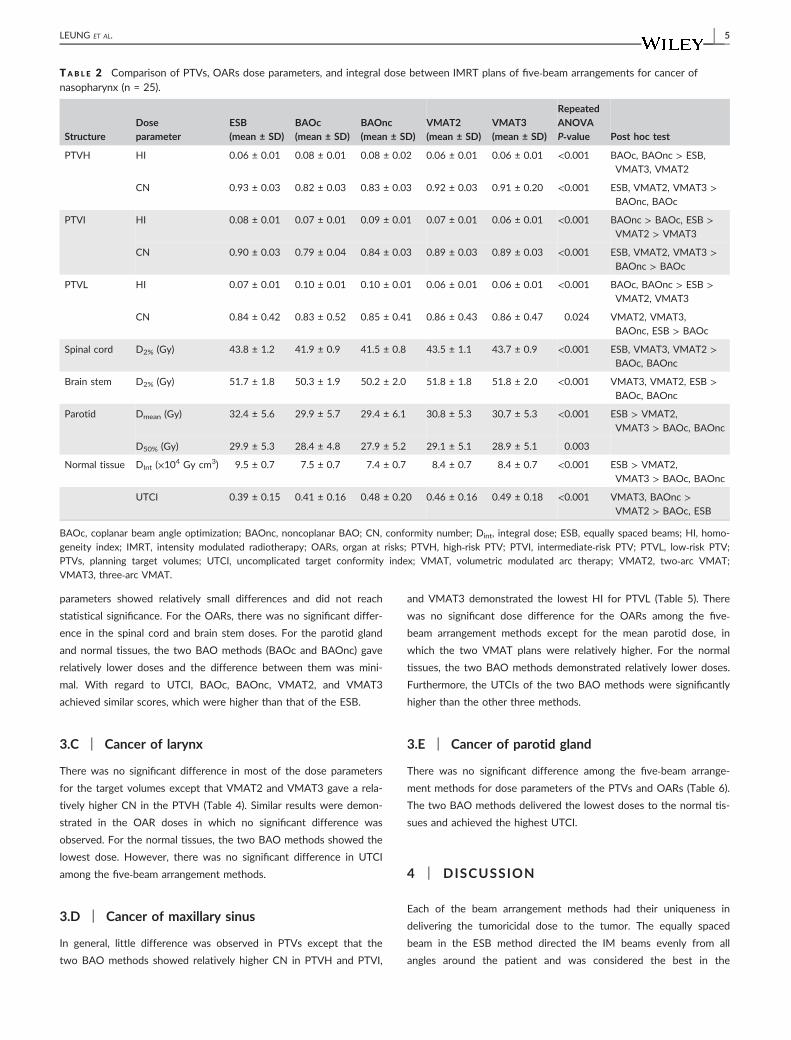
parameters showed relatively small differences and did not reach
statistical significance. For the OARs, there was no significant differ-
ence in the spinal cord and brain stem doses. For the parotid gland
and normal tissues, the two BAO methods (BAOc and BAOnc) gave
relatively lower doses and the difference between them was mini-
mal. With regard to UTCI, BAOc, BAOnc, VMAT2, and VMAT3
achieved similar scores, which were higher than that of the ESB.
3.C | Cancer of larynx
There was no significant difference in most of the dose parameters
for the target volumes except that VMAT2 and VMAT3 gave a rela-
tively higher CN in the PTVH (Table 4). Similar results were demon-
strated in the OAR doses in which no significant difference was
observed. For the normal tissues, the two BAO methods showed the
lowest dose. However, there was no significant difference in UTCI
among the five‐beam arrangement methods.
3.D | Cancer of maxillary sinus
In general, little difference was observed in PTVs except that the
two BAO methods showed relatively higher CN in PTVH and PTVI,
and VMAT3 demonstrated the lowest HI for PTVL (Table 5). There
was no significant dose difference for the OARs among the five‐beam arrangement methods except for the mean parotid dose, in
which the two VMAT plans were relatively higher. For the normal
tissues, the two BAO methods demonstrated relatively lower doses.
Furthermore, the UTCIs of the two BAO methods were significantly
higher than the other three methods.
3.E | Cancer of parotid gland
There was no significant difference among the five‐beam arrange-
ment methods for dose parameters of the PTVs and OARs (Table 6).
The two BAO methods delivered the lowest doses to the normal tis-
sues and achieved the highest UTCI.
4 | DISCUSSION
Each of the beam arrangement methods had their uniqueness in
delivering the tumoricidal dose to the tumor. The equally spaced
beam in the ESB method directed the IM beams evenly from all
angles around the patient and was considered the best in the
TAB L E 2 Comparison of PTVs, OARs dose parameters, and integral dose between IMRT plans of five‐beam arrangements for cancer ofnasopharynx (n = 25).
StructureDoseparameter
ESB(mean ± SD)
BAOc(mean ± SD)
BAOnc(mean ± SD)
VMAT2(mean ± SD)
VMAT3(mean ± SD)
RepeatedANOVAP‐value Post hoc test
PTVH HI 0.06 ± 0.01 0.08 ± 0.01 0.08 ± 0.02 0.06 ± 0.01 0.06 ± 0.01 <0.001 BAOc, BAOnc > ESB,
VMAT3, VMAT2
CN 0.93 ± 0.03 0.82 ± 0.03 0.83 ± 0.03 0.92 ± 0.03 0.91 ± 0.20 <0.001 ESB, VMAT2, VMAT3 >BAOnc, BAOc
PTVI HI 0.08 ± 0.01 0.07 ± 0.01 0.09 ± 0.01 0.07 ± 0.01 0.06 ± 0.01 <0.001 BAOnc > BAOc, ESB >VMAT2 > VMAT3
CN 0.90 ± 0.03 0.79 ± 0.04 0.84 ± 0.03 0.89 ± 0.03 0.89 ± 0.03 <0.001 ESB, VMAT2, VMAT3 >BAOnc > BAOc
PTVL HI 0.07 ± 0.01 0.10 ± 0.01 0.10 ± 0.01 0.06 ± 0.01 0.06 ± 0.01 <0.001 BAOc, BAOnc > ESB >VMAT2, VMAT3
CN 0.84 ± 0.42 0.83 ± 0.52 0.85 ± 0.41 0.86 ± 0.43 0.86 ± 0.47 0.024 VMAT2, VMAT3,
BAOnc, ESB > BAOc
Spinal cord D2% (Gy) 43.8 ± 1.2 41.9 ± 0.9 41.5 ± 0.8 43.5 ± 1.1 43.7 ± 0.9 <0.001 ESB, VMAT3, VMAT2 >BAOc, BAOnc
Brain stem D2% (Gy) 51.7 ± 1.8 50.3 ± 1.9 50.2 ± 2.0 51.8 ± 1.8 51.8 ± 2.0 <0.001 VMAT3, VMAT2, ESB >BAOc, BAOnc
Parotid Dmean (Gy) 32.4 ± 5.6 29.9 ± 5.7 29.4 ± 6.1 30.8 ± 5.3 30.7 ± 5.3 <0.001 ESB > VMAT2,
VMAT3 > BAOc, BAOnc
D50% (Gy) 29.9 ± 5.3 28.4 ± 4.8 27.9 ± 5.2 29.1 ± 5.1 28.9 ± 5.1 0.003
Normal tissue DInt (×104 Gy cm3) 9.5 ± 0.7 7.5 ± 0.7 7.4 ± 0.7 8.4 ± 0.7 8.4 ± 0.7 <0.001 ESB > VMAT2,
VMAT3 > BAOc, BAOnc
UTCI 0.39 ± 0.15 0.41 ± 0.16 0.48 ± 0.20 0.46 ± 0.16 0.49 ± 0.18 <0.001 VMAT3, BAOnc >VMAT2 > BAOc, ESB
BAOc, coplanar beam angle optimization; BAOnc, noncoplanar BAO; CN, conformity number; Dint, integral dose; ESB, equally spaced beams; HI, homo-
geneity index; IMRT, intensity modulated radiotherapy; OARs, organ at risks; PTVH, high‐risk PTV; PTVI, intermediate‐risk PTV; PTVL, low‐risk PTV;
PTVs, planning target volumes; UTCI, uncomplicated target conformity index; VMAT, volumetric modulated arc therapy; VMAT2, two‐arc VMAT;
VMAT3, three‐arc VMAT.
LEUNG ET AL. | 5

TAB L E 3 Comparison of PTVs, OARs dose parameters, and integral dose between IMRT plans of five‐beam arrangements for cancer of oralcavity (n = 25).
Structure Dose parameterESB(mean ± SD)
BAOc(mean ± SD)
BAOnc(mean ± SD)
VMAT2(mean ± SD)
VMAT3(mean ± SD)
RepeatedANOVAP‐value Post hoc test
PTVH HI 0.07 ± 0.01 0.08 ± 0.02 0.08 ± 0.02 0.06 ± 0.01 0.06 ± 0.01 <0.001 BAOc, BAOnc > ESB,
VMAT2, VMAT3,
CN 0.84 ± 0.04 0.80 ± 0.04 0.80 ± 0.04 0.88 ± 0.03 0.88 ± 0.30 <0.001 VMAT2, VMAT3 >ESB > BAOc, BAOnc
PTVI HI 0.10 ± 0.02 0.10 ± 0.02 0.10 ± 0.02 0.10 ± 0.02 0.10 ± 0.02 0.378
CN 0.84 ± 0.04 0.80 ± 0.04 0.80 ± 0.04 0.86 ± 0.04 0.86 ± 0.04 <0.001 VMAT2, VMAT3 >ESB > BAOc, BAOnc
PTVL HI 0.10 ± 0.01 0.11 ± 0.01 0.11 ± 0.01 0.10 ± 0.02 0.10 ± 0.02 <0.001 BAOnc, BAOc > ESB,
VMAT2, VMAT3
CN 0.84 ± 0.45 0.81 ± 0.56 0.80 ± 0.44 0.84 ± 0.53 0.84 ± 0.36 <0.001 VMAT3, ESB, VMAT2 >BAOc, BAOnc
Spinal cord D2% (Gy) 40.3 ± 2.5 39.8 ± 2.8 39.4 ± 2.6 39.1 ± 2.9 39.0 ± 3.1 0.088
Brain stem D2% (Gy) 46.2 ± 6.3 46.4 ± 5.2 46.8 ± 5.8 45.7 ± 6.0 45.9 ± 6.2 0.425
Parotid Dmean (Gy) 28.1 ± 4.7 26.0 ± 5.8 26.0 ± 5.1 28.2 ± 4.6 28.0 ± 4.8 <0.001 VMAT2, ESB, VMAT3 >BAOc, BAOnc
D50% (Gy) 28.7 ± 4.8 26.9 ± 5.9 27.2 ± 4.6 27.8 ± 4.8 28.0 ± 5.0 0.002 ESB > VMAT3, VMAT2,
BAOnc, BAOc
Normal tissue DInt (×104 Gy cm3) 9.7 ± 0.4 8.0 ± 0.6 8.0 ± 0.6 8.7 ± 0.5 8.8 ± 0.5 <0.001 ESB > VMAT2,
VMAT3 > BAOc,
BAOnc
UTCI 0.63 ± 0.45 0.77 ± 0.48 0.74 ± 0.48 0.70 ± 0.42 0.70 ± 0.44 0.003 BAOc, BAOnc, VMAT2,
VMAT3 > ESB
BAOc, coplanar beam angle optimization; BAOnc, noncoplanar BAO; CN, conformity number; Dint, integral dose; ESB, equally spaced beams; HI, homo-
geneity index; IMRT, intensity modulated radiotherapy; OARs, organ at risks; PTVH, high‐risk PTV; PTVI, intermediate‐risk PTV; PTVL, low‐risk PTV;
PTVs, planning target volumes; UTCI, uncomplicated target conformity index; VMAT, volumetric modulated arc therapy; VMAT2, two‐arc VMAT;
VMAT3, three‐arc VMAT.
TAB L E 4 Comparison of PTVs, OARs dose parameters, and integral dose between IMRT plans of five‐ beam arrangements for cancer oflarynx (n = 25).
Structure Dose parameterESB(mean ± SD)
BAOc(mean ± SD)
BAOnc(mean ± SD)
VMAT2(mean ± SD)
VMAT3(mean ± SD)
RepeatedANOVAP‐value Post hoc test
PTVH HI 0.06 ± 0.01 0.08 ± 0.02 0.07 ± 0.01 0.06 ± 0.03 0.06 ± 0.02 0.100
CN 0.88 ± 0.05 0.88 ± 0.04 0.88 ± 0.05 0.90 ± 0.04 0.90 ± 0.40 0.013 VMAT2, VMAT3 >BAOc, ESB, BAOnc
PTVI HI 0.12 ± 0.02 0.12 ± 0.02 0.13 ± 0.04 0.11 ± 0.03 0.11 ± 0.02 0.040
CN 0.82 ± 0.07 0.82 ± 0.08 0.84 ± 0.06 0.86 ± 0.05 0.86 ± 0.05 0.034
PTVL HI 0.08 ± 0.03 0.08 ± 0.04 0.08 ± 0.03 0.08 ± 0.03 0.07 ± 0.03 0.482
CN 0.84 ± 0.07 0.86 ± 0.05 0.86 ± 0.06 0.84 ± 0.81 0.86 ± 0.06 0.692
Spinal cord D2% (Gy) 41.0 ± 2.1 42.2 ± 1.5 42.2 ± 1.8 42.2 ± 1.8 42.3 ± 1.9 0.222
Brain stem D2% (Gy) 35.9 ± 19.4 35.1 ± 19.0 35.8 ± 19.5 35.1 ± 19.7 35.1 ± 19.9 0.554
Parotid Dmean (Gy) 28.5 ± 4.6 27.4 ± 5.1 26.4 ± 4.5 28.2 ± 4.8 27.4 ± 4.2 0.147
D50% (Gy) 22.8 ± 4.9 20.3 ± 5.3 20.1 ± 5.5 22.0 ± 6.1 22.2 ± 7.1 0.040
Normal tissue DInt (×104 Gy cm3) 8.0 ± 0.4 7.3 ± 0.6 7.2 ± 0.6 8.1 ± 0.6 8.3 ± 0.6 <0.001 VMAT3, ESB, VMAT2 >
BAOc, BAOnc
UTCI 1.80 ± 1.61 1.73 ± 1.48 1.99 ± 1.47 1.81 ± 1.87 1.73 ± 1.94 0.527
BAOc, coplanar beam angle optimization; BAOnc, noncoplanar BAO; CN, conformity number; Dint, integral dose; ESB, equally spaced beams; HI, homo-
geneity index; IMRT, intensity modulated radiotherapy; OARs, organ at risks; PTVH, high‐risk PTV; PTVI, intermediate‐risk PTV; PTVL, low‐risk PTV;
PTVs, planning target volumes; UTCI, uncomplicated target conformity index; VMAT, volumetric modulated arc therapy; VMAT2, two‐arc VMAT;
VMAT3, three‐arc VMAT.
6 | LEUNG ET AL.

treatment of central uniform‐shaped tumors. The VMAT method
shared similar characteristics but employed more beams from all
angles around the patients and reduced the treatment time.13
Because of this, they were expected to deliver higher integral dose
to normal tissues.25 The VMAT3 had the potential to produce more
conformal dose distribution than VMAT2 but required one additional
gantry rotation and therefore increased the treatment time. In this
study, the BAO methods used 5–9 beams directed from selected
angles. Beams that did not have contribution to the plan were elimi-
nated and the beam angles could be tailor‐made for individual
patients. As a result, the integral dose and total monitoring units
(MU) were lower.26 BAOnc had greater freedom to direct the beams
to the patient compared with BAOc, but in the expense of longer
treatment setup time. Overall, the clinical merit of the current study
is that it provides evidence‐based recommendations on the beam
arrangement for planners to use in the five types of head and neck
cancers.
4.A | Cancer of nasopharynx
With regard to the target conformity and homogeneity, ESB per-
formed better in PTVH and PTVI. This could be due to the fact that
these target volumes were relatively less irregular in shape than the
PTVL and, being more centrally situated at the skull, the evenly dis-
tributed intensity modulated IM beams were able to produce
relatively more conformal dose distribution. However, for PTVL
which extended to both sides of the neck, and was more irregular
with an inverted U‐shape (Fig. 1), the BAOnc and VMAT plans
demonstrated relatively better dose coverage. The main reason was
that with the use of noncoplanar beams in BAO and the greater
number of effective beam angles from VMAT, they were both more
effective in creating conformal high‐dose volumes covering the irreg-
ular target. By the same argument, the OARs were better spared by
these two beam arrangement methods.18 By combining the perfor-
mance in the target volumes and OARs, it was logical to see that
both BAOnc and VMAT3 achieved relatively better plans among the
five‐beam arrangement methods. Overall, VMAT3 would be recom-
mended because it would have a much shorter treatment delivery
time compared to BAOnc.
4.B | Cancer of oral cavity
In terms of dose coverage to the target volume, VMAT plans in gen-
eral performed better among the five‐beam arrangement methods as
it demonstrated the highest CN in all the PTVs. Since the oral cavity
was a relatively large structure, tumors could arise from different
locations in the oral cavity ranging from the periphery to the center.
The results showed evidence that the VMAT beam arrangement was
more flexible to deal with target volume location variation, and its
average performance on target coverage was better than the other
TAB L E 5 Comparison of PTVs, OARs dose parameters, and integral dose between IMRT plans of five‐beam arrangements for cancer ofmaxilla sinus (n = 19).
Structure Dose parameterESB(mean ± SD)
BAOc(mean ± SD)
BAOnc(mean ± SD)
VMAT2(mean ± SD)
VMAT3(mean ± SD)
RepeatedANOVAP‐value Post hoc test
PTVH HI 0.06 ± 0.01 0.07 ± 0.02 0.07 ± 0.02 0.07 ± 0.02 0.07 ± 0.02 0.206
CN 0.87 ± 0.03 0.90 ± 0.02 0.90 ± 0.03 0.87 ± 0.03 0.88 ± 0.03 0.003 BAOc, BAOnc,
VMAT3 > VMAT2, ESB
PTVI HI 0.08 ± 0.01 0.09 ± 0.02 0.09 ± 0.01 0.08 ± 0.03 0.08 ± 0.03 0.286
CN 0.79 ± 0.04 0.83 ± 0.06 0.83 ± 0.03 0.81 ± 0.03 0.79 ± 0.03 <0.001 BAOnc, BAOc,
VMAT2 > VMAT3, ESB
PTVL HI 0.09 ± 0.01 0.10 ± 0.01 0.10 ± 0.01 0.09 ± 0.02 0.08 ± 0.02 0.001 BAOnc > BAOc,
ESB, VMAT2 > VMAT3
CN 0.77 ± 0.04 0.79 ± 0.05 0.79 ± 0.03 0.79 ± 0.05 0.78 ± 0.04 0.046
Spinal cord D2% (Gy) 41.6 ± 2.2 40.7 ± 2.6 40.9 ± 2.7 41.7 ± 2.5 41.7 ± 2.7 0.044
Brain stem D2% (Gy) 51.1 ± 2.1 51.6 ± 1.4 51.6 ± 1.4 50.9 ± 1.7 50.2 ± 3.7 0.266
Parotid Dmean (Gy) 25.7 ± 5.9 23.9 ± 6.4 23.6 ± 6.7 26.2 ± 6.0 26.3 ± 6.0 <0.001 VMAT3, VMAT2 >ESB, BAOc, BAOnc
D50% (Gy) 25.7 ± 6.2 25.0 ± 5.7 24.0 ± 6.2 26.6 ± 5.3 26.2 ± 5.7 0.008
Normal tissue DInt (×104 Gy cm3) 7.5 ± 0.8 6.6 ± 0.8 6.6 ± 0.8 7.3 ± 0.8 7.2 ± 0.8 <0.001 ESB > VMAT2, VMAT3 >
BAOnc, BAOc
UTCI 0.74 ± 0.43 0.99 ± 0.62 1.10 ± 0.72 0.71 ± 0.40 0.71 ± 0.40 <0.001 BAOnc, BAOc > ESB,
VMAT2, VMAT3
BAOc, coplanar beam angle optimization; BAOnc, noncoplanar BAO; CN, conformity number; Dint, integral dose; ESB, equally spaced beams; HI, homo-
geneity index; IMRT, intensity modulated radiotherapy; OARs, organ at risks; PTVH, high‐risk PTV; PTVI, intermediate‐risk PTV; PTVL, low‐risk PTV;
PTVs, planning target volumes; UTCI, uncomplicated target conformity index; VMAT, volumetric modulated arc therapy; VMAT2, two‐arc VMAT;
VMAT3, three‐arc VMAT.
LEUNG ET AL. | 7

beam arrangements. Regarding the OARs, the spinal cord and brain
stem were located at some distance from the target volume, their
doses were relative low, and therefore their differences were small.
For the parotid gland, BAO plans provided relatively better sparing
and were able to keep the average mean dose below 26 Gy, which
was reported to be within the acceptable range of tolerance dose
(mean dose 25–30 Gy).27 Since the VMAT plans and BAO plans per-
formed better in target volume coverage and parotid gland sparing
respectively, it was logical to see their plans achieve similar rank in
the UTCI scores. Furthermore, since there was no significant differ-
ence between VMAT3 and VMAT2, the addition of extra arc in
VMAT3 did not bring any dosimetric advantage and therefore was
not necessary. Moreover, BAOc was adequate when compared with
BAOnc as including noncoplanar beams did not significantly improve
the plan quality. Taking the treatment time into consideration,
VMAT2 would be recommended as it shared similar plan quality as
the BAOc plans but offered shorter treatment time.
4.C | Cancer of larynx
Target volumes in laryngeal cancer were more regular in shape and
further away from OARs. All five‐beam arrangement methods per-
formed well on this relatively simple target volume geometry. This
was the reason why there was no significant difference in most of
the dose parameters of the target volumes, OARs, and UTCI scores.
This implied that any one of the beam arrangement methods was
effective in treating this cancer. It was worth noting that since the
BAO plans restricted the number of beams to below nine, it deliv-
ered relatively lower integral dose which might reduce the risk of
secondary cancer when compared to the VMAT plans. However, in
terms of treatment delivery time, the VMAT plans have the advan-
tage.
4.D | Cancer of maxillary sinus
Since tumor of the maxillary sinus was usually located at one side of
the head, the evenly distributed beams in ESB and VMAT would
irradiate the contralateral structures such as the parotid gland. This
phenomenon was reflected in the dosimetric results of the VMAT
plans that delivered higher mean parotid gland doses. Besides, the
BAO plans which allowed beams mainly directed from the ipsilateral
side performed better overall plan quality with the UTCI scores sig-
nificantly higher than the other three methods. It is logical to con-
sider whether the use of the partial arc VMAT which could also
avoid the direct beam entry from the contralateral side could be
comparable to the BAO plans. Unfortunately, it was one of the limi-
tations of the current study that the use of the partial arc VMAT
was not included, because it was intended to include only the beam
arrangements that were applicable to all the types of head and neck
cancer in the study. In the current study, VMAT has shown to have
comparable results with BAO in the HI and CN of PTV and in the
dose to brain stem and spinal cord. Although the advantage of the
use of the partial arc VMAT in the cancer of maxillary sinus was not
explicitly deduced, the use of partial arc VMAT could possibly
achieve the comparable results as in the full arc VMAT while reduc-
ing the disadvantages attributable to the beam entry from the con-
tralateral side. Therefore, the current results were not against the
use of partial arc VMAT in the cancer of maxillary sinus.
TAB L E 6 Comparison of PTVs, OARs dose parameters, and integral dose between IMRT plans of five‐beam arrangements for cancer ofparotid gland (n = 25).
Structure Dose parameterESB(mean ± SD)
BAOc(mean ± SD)
BAOnc(mean ± SD)
VMAT2(mean ± SD)
VMAT3(mean ± SD)
RepeatedANOVAP‐value Post hoc test
PTVH HI 0.06 ± 0.01 0.06 ± 0.01 0.06 ± 0.01 0.07 ± 0.02 0.07 ± 0.01 0.296
CN 0.86 ± 0.04 0.88 ± 0.04 0.87 ± 0.4 0.86 ± 0.03 0.86 ± 0.03 0.263
PTVI HI 0.07 ± 0.03 0.07 ± 0.02 0.07 ± 0.02 0.09 ± 0.01 0.08 ± 0.02 0.070
CN 0.83 ± 0.05 0.87 ± 0.04 0.85 ± 0.03 0.84 ± 0.02 0.82 ± 0.03 0.090
PTVL HI 0.09 ± 0.02 0.09 ± 0.03 0.09 ± 0.04 0.10 ± 0.02 0.11 ± 0.03 0.045
CN 0.83 ± 0.05 0.84 ± 0.03 0.85 ± 0.04 0.82 ± 0.05 0.84 ± 0.03 0.242
Spinal cord D2% (Gy) 34.5 ± 9.4 33.9 ± 8.3 34.2 ± 9.1 34.9 ± 8.9 34.5 ± 9.0 0.319
Brain stem D2% (Gy) 35.7 ± 13.4 35.5 ± 12.9 35.5 ± 12.9 36.1 ± 13.2 36.5 ± 11.9 0.390
Parotid Dmean (Gy) 7.0 ± 3.3 6.5 ± 2.3 6.1 ± 2.4 6.9 ± 3.0 6.8 ± 3.2 0.274
D50% (Gy) 8.0 ± 6.2 6.9 ± 4.0 6.8 ± 4.0 7.3 ± 4.4 7.5 ± 4.8 0.051
Normal tissue DInt (×104 Gy cm3) 8.7 ± 0.7 7.6 ± 1.1 7.7 ± 0.9 8.2 ± 1.1 8.3 ± 0.9 0.002 ESB > VMAT3,
VMAT2, BAOnc, BAOc
UTCI 4.55 ± 1.68 5.66 ± 1.72 5.68 ± 1.81 4.80 ± 1.53 4.88 ± 1.89 <0.001 BAOnc, BAOc,
VMAT3, VMAT2 > ESB
BAOc, coplanar beam angle optimization; BAOnc, non‐coplanar BAO; CN, conformity number; Dint, integral dose; ESB, equally spaced beams; HI, homo-
geneity index; IMRT, intensity modulated radiotherapy; OARs, organ at risks; PTVH, high‐risk PTV; PTVI, intermediate‐risk PTV; PTVL, low‐risk PTV;
PTVs, planning target volumes; UTCI, uncomplicated target conformity index; VMAT, volumetric modulated arc therapy; VMAT2, two‐arc VMAT;
VMAT3, three‐ arc VMAT.
8 | LEUNG ET AL.
4.E | Cancer of parotid gland
Target volumes of the parotid gland tumor were usually followed a
triangular shape and would not poses great difficulty to the various
beam arrangements. This was reflected in the dosimetric results of
the target volumes in which there was no significant difference in all
the dosimetric parameters among the five‐beam arrangement meth-
ods. Similar to the maxillary sinus cancer, parotid tumors are situated
at the lateral aspect of the head, and this would be a disadvantage
for the ESB and VMAT beam arrangement. Relatively higher doses
were found in the contralateral parotid gland in these plans although
the differences did not reach statistical significance. Similar to the
explanation for the maxillary sinus cancer, the integral dose in the
BAO plans were significantly lower, and this led to overall better
UTCI scores in these two plans. This echoed the study by Yir-
mibesoglu et al28 who reported that four‐field ipsilateral IMRT tech-
niques provided excellent coverage while maximally sparing the
contralateral parotid gland and submandibular gland. As a result,
BAOc plans would be recommended as it achieved the same plan
quality as the BAOnc but offered simpler treatment setup procedure.
Furthermore, by the same argument as stated for the cancer of max-
illary sinus, the results of the present study were not against the use
of partial arc VMAT in the cancer of parotid gland.
5 | CONCLUSION
The five‐beam arrangement methods produced acceptable plans for
all the five groups of head and neck cancer patients. Partial arc
VMAT was not included in the beam arrangement methods because
it was not commonly applied in centrally located cancers, therefore
it could not be used for comparison among the five groups of head
and neck cancers. The results showed that individual methods pro-
duced dosimetric advantages on certain aspects, and the UTCL
scores were marginally greater in the BAO method in the cancers of
the maxillary sinus and the parotid gland. However, if the treatment
time was included into consideration, VMAT plans would be recom-
mended for cancers of nasopharynx (VMAT3), oral cavity, and larynx
(VMAT2).
CONFLICT OF INTEREST
The authors declare no conflicts of interest with respect to the con-
tent of this manuscript.
REFERENCES
1. Eisbruch A, Kim HM, Terrell JE, Marsh LH, Dawson LA, Ship JA.
Xerostomia and its predictors following parotid‐sparing irradiation of
head‐and‐neck cancer. Int J Radiat Oncol Biol Phys. 2001;50(3):695–704.
2. Pan CC, Eisbruch A, Lee JS, Snorrason RM, Ten Haken RK, Kileny
PR. Prospective study of inner ear radiation dose and hearing loss in
head‐and‐neck cancer patients. Int J Radiat Oncol Biol Phys. 2005;61
(5):1393–1402.
3. Dijkstra PU, Kalk WWI, Roodenburg JLN. Trismus in head and neck
oncology: a systematic review. Oral Oncol. 2004;40(9):879–889.4. Fu Y, Deng M, Zhou X, et al. Dosimetric effect of beam arrangement
for intensity‐modulated radiation therapy in the treatment of upper
thoracic esophageal carcinoma. Med Dosim. 2017;42(1):47–52.5. Fitzgerald R, Owen R, Barry T, et al. The effect of beam arrange-
ments and the impact of non‐coplanar beams on the treatment plan-
ning of stereotactic ablative radiation therapy for early stage lung
cancer. J Med Radiat Sci. 2016;63(1):31–40.6. Vanetti E, Clivio A, Nicolini G, et al. Volumetric modulated arc radio-
therapy for carcinomas of the oro‐pharynx, hypo‐pharynx and larynx:
A treatment planning comparison with fixed field IMRT. Radiother
Oncol. 2009;92(1):111–117.7. Budrukkar AN, Hope G, Cramb J, Corry J, Peters LJ. Dosimetric
study of optimal beam number and arrangement for treatment of
nasopharyngeal carcinoma with intensity‐modulated radiation ther-
apy. Australas Radiol. 2004;48(1):45–50.8. Vlachaki MT, Teslow TN, Amosson C, Uy NW, Ahmad S. IMRT ver-
sus conventional 3DCRT on prostate and normal tissue dosimetry
using an endorectal balloon for prostate immobilization. Med Dosim.
2005;30(2):69–75.9. Ahmed M, Hansen VN, Harrington KJ, Nutting CM. Reducing the risk
of xerostomia and mandibular osteoradionecrosis: the potential ben-
efits of intensity modulated radiotherapy in advanced oral cavity car-
cinoma. Med Dosim. 2009;34(3):217–224.10. Gupta T, Agarwal J, Jain S, et al. Three‐dimensional conformal radio-
therapy (3D‐CRT) versus intensity modulated radiation therapy
(IMRT) in squamous cell carcinoma of the head and neck: A random-
ized controlled trial. Radiother Oncol. 2012;104(3):343–348.11. Earl MA, Shepard DM, Naqvi S, Li XA, Yu CX. Inverse planning for
intensity‐modulated arc therapy using direct aperture optimization.
Phys Med Biol. 2003;48(8):1075–1089.12. MacKenzie MA, Robinson DM. Intensity modulated arc deliveries
approximated by a large number of fixed gantry position sliding win-
dow dynamic multileaf collimator fields. Med Phys. 2002;29
(10):2359–2365.13. Verbakel WFAR, Cuijpers JP, Hoffmans D, Bieker M, Slotman BJ,
Senan S. Volumetric intensity‐modulated arc therapy vs. conven-
tional IMRT in head‐and‐neck cancer: a comparative planning and
dosimetric study. Int J Radiat Oncol Biol Phys. 2009;74(1):252–259.14. Tsai C‐L, Wu J‐K, Chao H‐L, Tsai Y‐C, Cheng JC‐H. Treatment and
dosimetric advantages between VMAT, IMRT, and helical tomother-
apy in prostate cancer. Med Dosim. 2011;36(3):264–271.15. Clivio A, Fogliata A, Franzetti‐Pellanda A, et al. Volumetric‐modu-
lated arc radiotherapy for carcinomas of the anal canal: A treatment
planning comparison with fixed field IMRT. Radiother Oncol. 2009;92
(1):118–124.16. Srivastava SP, Das IJ, Kumar A, Johnstone PAS. Dosimetric compar-
ison of manual and beam angle optimization of gantry angles in
IMRT. Med Dosim. 2011;36(3):313–316.17. Craft DL, Hong TS, Shih HA, Bortfeld TR. Improved planning time and
plan quality through multicriteria optimization for intensity‐modulated
radiotherapy. Int J Radiat Oncol Biol Phys. 2012;82(1):e83–e90.18. Tran A, Zhang J, Woods K, et al. Treatment planning comparison of
IMPT, VMAT and 4π radiotherapy for prostate cases. Radiat Oncol.
2017;12(1):10.
19. Rwigema J‐CM, Nguyen D, Heron DE, et al. 4π noncoplanar stereo-
tactic body radiation therapy for head‐and‐neck cancer: potential to
improve tumor control and late toxicity. Int J Radiat Oncol Biol Phys.
2015;91(2):401–409.20. Wu Q, Manning M, Schmidt‐Ullrich R, Mohan R. The potential for
sparing of parotids and escalation of biologically effective dose with
intensity‐modulated radiation treatments of head and neck cancers:
a treatment design study. Int J Radiat Oncol Biol Phys. 2000;46
(1):195–205.
LEUNG ET AL. | 9

21. DeLuca P, Jones D, Gahbauer R, et al. Prescribing, recording, and
reporting photon-beam intensity-modulated radiation therapy
(IMRT). J ICRU. 2010;10:1–93.22. van't Riet A, Mak AC, Moerland MA, Elders LH, van der Zee W. A
conformation number to quantify the degree of conformality in
brachytherapy and external beam irradiation: Application to the
prostate. Int J Radiat Oncol Biol Phys. 1997;37(3):731–736.23. Aoyama H, Westerly DC, Mackie TR, et al. Integral radiation dose to
normal structures with conformal external beam radiation. Int J
Radiat Oncol Biol Phys. 2006;64(3):962–967.24. Miften MM, Das SK, Su M, Marks LB. A dose‐volume‐based tool for
evaluating and ranking IMRT treatment plans. J Appl Clin Med Phys.
2004;5(4):1–14.
25. Higby C, Khafaga Y, Al‐Shabanah M, et al. Volumetric‐modulated arc
therapy (VMAT) versus 3D‐conformal radiation therapy in supra‐di-aphragmatic Hodgkin's Lymphoma with mediastinal involvement: A
dosimetric comparison. J Egypt Natl Canc Inst. 2016;28(3):163–168.26. Shukla A, Kumar S, Sandhu I, Oinam A, Singh R, Kapoor R. Dosimet-
ric study of beam angle optimization in intensity‐modulated radiation
therapy planning. J Cancer Res Ther. 2016;12(2):1045.
27. Ortholan C, Benezery K, Bensadoun R‐J. Normal tissue tolerance to
external beam radiation therapy: salivary glands. Cancer Radiother.
2010;14(4–5):290–294.28. Yirmibesoglu E, Fried DV, Kostich M, et al. Dosimetric evaluation of
an ipsilateral intensity modulated radiotherapy beam arrangement
for parotid malignancies. Radiol Oncol. 2013;47(4):411–418.
10 | LEUNG ET AL.