a diverse new assemblage of late eocene …pardomv/pe/2006_2/dakota/dakota.pdf · hallmark of...
TRANSCRIPT

Palaeontologia Electronica http://palaeo-electronica.org
PE Article Number: 9.2.5ACopyright: Society of Vertebrate Paleontology September 2006Submission: 7 August 2005. Acceptance: 9 April 2006.
Smith, Krister T. 2006. A Diverse New Assemblage of Late Eocene Squamates (Reptilia) from the Chadron Formation of North Dakota, U.S.A. Palaeontologia Electronica Vol. 9, Issue 2; 5A:44p, 6.2MB; http://palaeo-electronica.org/paleo/2006_2/dakota/index.html
A DIVERSE NEW ASSEMBLAGE OF LATE EOCENE SQUAMATES (REPTILIA) FROM THE CHADRON FORMATION OF
NORTH DAKOTA, U.S.A.
Krister T. Smith
ABSTRACT
The current fossil record of squamates across the Eocene/Oligocene boundary incentral North America is perplexing in that it shows a marked increase in species rich-ness in response to climatic cooling and drying, which is contrary to both the Europeanrecord and expectation. A diverse, new squamate assemblage from the late EoceneChadron Formation of the Medicine Pole Hills, North Dakota, provides new insight intothese changes. Nineteen squamate species are described, fourteen of which were pre-viously unknown. The acrodontan, varanid, and diploglossine anguid are previouslyunreported holdovers from the early Eocene. The Medicine Pole Hills local fauna isone of the most species-rich squamate assemblages yet reported from the fossilrecord.
Late Eocene iguanids are remarkable for both their species-level and higher taxo-nomic diversity: among their numbers are a phrynosomatine (fence lizards and rela-tives), a polychrotine (anoles and relatives), and a crotaphytine (collared and leopardlizards). The polychrotine, interpreted as the earliest-known representative of the Poly-chrus lineage, indicates that at least one subgroup of Iguanidae shows a historical bio-geographic pattern similar to that of North American scincomorph and anguimorphlizards. The increasing numerical dominance of iguanid species has been considered ahallmark of herpetofaunal modernization in central North America, but this measureappears inadequate in light of these data.
Including the new fauna, the record of central North American squamates, like theEuropean record, shows a decrease in richness across the Eocene/Oligocene bound-ary. However, many uncorrected sampling biases remain that may distort our picture ofthe squamate response to Eocene/Oligocene climate change. Sampling-standardizedstudies are indicated.
Krister T. Smith. Department of Geology and Geophysics, Yale University, P.O. Box 208109, New Haven, Connecticut, 06520-8109, USA. [email protected]
KEY WORDS: Squamata, informal taxonomy, Eocene/Oligocene boundary, diversity, climate change, bio-geography, divergence times

SMITH: SQUAMATES FROM THE LATE EOCENE
2
INTRODUCTION
The past decades have witnessed consider-able multidisciplinary attention to climate changearound the Eocene/Oligocene boundary and thecauses and consequences of associated bioticchange. This is partly due to a re-calibration of theNorth American terrestrial record (Prothero andSwisher 1992; Swisher and Prothero 1990), exten-sive study of deep-sea cores (e.g., Thomas 1992;Lear et al. 2000), and debate over bolide impacts(see discussion in Prothero 1994).
Wolfe (e.g., 1978, 1992) argued early on frompaleobotanical data that climatic deterioration wasnot limited to the Eocene/Oligocene boundaryinterval: the middle/late Eocene boundary was alsomarked by a decrease in mean annual temperature(MAT) and an increase in mean annual range oftemperature (MART) in many areas on the fringe ofthe North American continent. According to hisstudies, amelioration during the late Eocene wasfollowed by another marked deterioration in theearliest Oligocene.
How apparent climatic change along the NorthAmerican periphery was reflected in its interior is amatter of some contention. Hutchison (1982, 1992)could accept a moderate drop in MAT (<5°C) as anexplanation for the observed decrease in maximumcarapace length in testudinid turtles; but he alsoargued that in the early Oligocene, mean wintertemperature could not have been below 13°C,because the relatively large terrestrial turtles (>30cm carapace length) known from that time couldnot burrow to escape lower temperatures. Simi-larly, Evanoff et al. (1992) concluded that there wasa minor drop in MAT in the early Oligocene (from16.5°C in the late Eocene) based on the terrestrialgastropod fauna near Douglas, Wyoming. Contrari-wise, Wolfe (1992) found that, between the lateEocene and early Oligocene in Colorado, MATdecreased from ~12.5 to ~4.5°C; the magnitude ofthis drop (8°C) is comparable to what he found inthe Pacific Northwest and elsewhere in NorthAmerica (Wolfe 1978). The late Chadronian Rubyflora of Montana had a great many conifers andindicates a MAT of ~12°C and a probable MART of<16°C (Wolfe 1992). These pre-deterioration val-ues already exceed the limits suggested by Hutchi-son (1982) for the early Oligocene.
A change in mean annual precipitation (MAP)across the Eocene/Oligocene boundary seemsless controversial. Studies of palaeosols (e.g.,Retallack 1992; Sheldon and Retallack 2004) implythat MAP gradually dropped by ~50% between 36
and 27 Ma, by which time MAP values in Nebraskalikened modern-day values.
The transformation of North American squa-mate assemblages during the Tertiary has notbeen investigated in detail (Hutchison 1992), butbroad shifts in assemblage composition may relateto climate change. Gauthier (1982) noted a generalreplacement of Paleogene mesic-adapted lizards(especially anguimorphs) by Neogene xeric-adapted lizards (especially iguanids). Holman(2000) described an analogous shift from booid- tocolubroid-dominated assemblages around thesame time. Many of the common Paleogene squa-mate taxa moved south and are found today in thesoutheastern United States and Central America(Estes 1970; Savage 1960).
Despite increasing aridity and cooler tempera-tures, squamate diversity appears to increaseacross the Eocene/Oligocene boundary in centralNorth America (10 to 17 species: Sullivan and Hol-man 1996)1. A temperature decrease of only ~5°Cis related to a moderate decrease in lizard speciesrichness in modern North American deserts(Pianka 1967). The pattern seen in the NorthAmerican fossil record, however, may be vitiatedby differences in sampling, indicated by Lazarustaxa in the Chadronian (Sullivan and Holman1996).
In this paper, I report on a new and verydiverse fauna from the Chadronian of NorthDakota, the Medicine Pole Hills local fauna (l.f.).Many new lizard morphotypes are identified, andthree are formally named. Extensive comparisonsare made with the disarticulated skeletons ofextant taxa in order to estimate their phylogeneticposition. I then evaluate the effects of this newfauna on our understanding of squamate speciesrichness across the Eocene/Oligocene boundary.
GEOLOGIC SETTING AND PALEOENVIRONMENT
The White River Group of the Great Plainsincludes the Chadron and Brule Formations of lateEocene to Miocene age. It reaches its maximumthickness in northwestern Nebraska, decreases inthickness northward, and disappears in northernNorth Dakota (Larson and Evanoff 1998). PioneerTrails Regional Museum (PTRM) (Bowman, NorthDakota) locality V89002 is located in the Medicine
1. Actually, the increase is even greater than reported, for thehelodermatid Lowesaurus matthewi, known from two Orellanspecimens (Estes 1983; Pregill et al. 1986), was not included inSullivan and Holman's (1996) tally.

PALAEO-ELECTRONICA.ORG
3
Pole Hills of southwestern North Dakota (Figure 1;South Rhame quadrangle). The lower part (ChalkyButtes Member) of the Chadron Formation, rem-nant of a former sedimentary "blanket" (Trimble1980), caps those hills in many places (Murphy etal. 1993; Pearson 1993), and V89002 is located onone of the hilltops. Fossils were first reported fromthe area some 80 years ago (Leonard 1922), andsince 1989 the Pioneer Trails Regional Museumhas conducted excavations at V89002.
The sediment is a brownish, poorly sorted,poorly consolidated, generally medium- to fine-grained sandstone (Pearson 1993). A 2.5 m localsection (Figure 2) shows two trough cross-beddedunits (units 2 and 6); unit 5 is composed of sedi-ment indistinguishable from that of unit 2. Along thebedding planes are strewn gray mudballs, oftencomprising 15% of total rock volume. The troughsof units 2 and 6 have trends at high angle (~80°) toone another. Unit 3 is a fine-grained, parallel-bed-ded sandstone. Unit 4 displays planar cross-bed-ding.
Rapidly shifting channels that deposit planarand trough cross-bedded sandstone units (lithofa-cies Sp and St of Miall 1996) are characteristic ofthe distal reaches of some modern braided rivers(Einsele 1992), and I postulate that this sectionwas deposited as part of a braidbelt environment.Parallel-bedded units, like unit 3, are also depos-ited in such environments during waning floodstages (Einsele 1992: 45). The mudballs probablyderive from the underlying Paleocene strata (Kihmpersonal commun., 2004). Many fossils are quite
worn, presumably by stream abrasion. The similarcolor (creamy to tannish yellow) and preservationof nearly all specimens argue for a single prove-nance.
The Medicine Pole Hills l.f. is taphonomicallysimilar in many ways to the Calf Creek l.f. ofSaskatchewan (Holman 1972, 1976). The CypressHills Formation, which yields the Calf Creek l.f., isalso interpreted as a braidbelt deposit (Leckie andCheel 1989). As at V89002, the bone in theCypress Hills Formation is set in a poorly sortedmatrix that also contains mudballs torn from inter-fluves (Leckie and Cheel, op. cit.), although I inter-pret the Medicine Pole Hills fossil horizons asfluvial in origin rather than as debris flows. All fos-sils were collected from units 1 and 2 by dry-screening quarried sediment; fossils of the CalfCreek l.f. were also collected by screening (Hol-man 1972). Neither deposit appears to excludeparticular size classes, both preserving everythingfrom large brontotheriids to small marsupials(Storer 1996; Kihm et al. 2001; Pearson, personalcommun., 2001).
The Chadron Formation in the Medicine PoleHills and the Cypress Hills Formation in the CalfCreek area are both Chadronian in age, althoughmore carefully considered, they may differ by 1m.y. or so. The leptomerycid artiodactyls present inthe Medicine Pole Hills l.f., including a probableearly form of Leptomeryx yoderi, suggest an earlyChadronian age for these deposits (Heaton andEmry 1996; Hoganson et al. 1998). In contrast, theCalf Creek l.f. contains L. speciosus and L. mam-
Figure 1. Geographic map of the northern plains states, showing the location of the Medicine Pole Hills in NorthDakota (after Hoganson et al. 1998). New specimens described in this paper are deposited in the Pioneer TrailsRegional Museum in Bowman, North Dakota.

SMITH: SQUAMATES FROM THE LATE EOCENE
4
mifer (Storer 1996), which are indicative of amedial Chadronian age (Emry et al. 1987).
INSTITUTIONAL ABBREVIATIONS
American Museum of Natural History(AMNH), Carnegie Museum of Natural History(CM), Florida Museum of Natural History (UF), Pio-neer Trails Regional Museum (PTRM), Sencken-berg-Museum, Frankfurt-am-Main (SMF),Saskatchewan Museum of Natural History(SMNH), University of California Museum of Pale-
ontology (UCMP), United States National Museum(USNM), Yale Peabody Museum – Princeton Col-lection (YPM-PU), Yale Peabody Museum – Verte-brate Paleontology (YPM-VP), Yale PeabodyMuseum – Vertebrate Zoology (YPM-VZ).
SYSTEMATIC PALEONTOLOGY
When fossils of poor quality are given formaltaxonomic designations several difficulties mayarise. The type material of the new taxon may bediagnostic when described even though in absoluteterms it is not, leading to later abandonment of thename. Considered in this way, "diagnostic" is reallya time-dependent descriptor that could be definedas "distinguishable from all known (Recent and fos-sil) forms whose relevant parts have been studiedthus far." For example, the taxon Lestophis ancepsMarsh (1885) was named on the basis of vertebraefrom the Bridger basin of Wyoming, but later workhas shown that these rhineurid vertebrae possessno unique features, rendering L. anceps a nomendubium (Estes 1983). How diagnostic a particularspecimen is depends on how many distinctive fea-tures it displays and can be expected to correlatewith completeness.
More problems may arise when the descrip-tion of a new taxon on the basis of undiagnosticmaterial is later supplemented by information frommuch more complete specimens, which subse-quently become closely associated with the nameof the taxon. The discovery of a new taxon, fromwhich the type material of the first cannot be distin-guished, would thus render the first name invalid,but one hesitates to strike down the first namebecause most scientists in considering the taxonthink not of the type but rather of the more com-plete and well-described specimens.
Many of the taxa described herein, especiallyon the basis of jaws, are currently diagnostic—thatis, there are currently no other taxa whose mor-phology is known (by me) to match theirs. Butgiven their incomplete nature and the incompletestudy of the skeletal material of extant species, itseems likely that future workers could find otherspecies that, were they fragmentary, would lookidentical to those from Medicine Pole Hills. Whenthey cannot even reasonably be compared ("cf.")to an existing genus or species, I use an informalnomenclatural system. Each name designates theprobable phylogenetic affinity and includes anabbreviation, appended with a number, for thelocality name from which the specimens derive.For example, "Iguanid MPH-1" indicates that the
Figure 2. Stratigraphic section at PTRM locality V89002.Grain size (clay, silt, and fine, medium, and coarse sand)is indicated by width of the section. Fine intra-unit linesillustrate sedimentary structure (planar and trough cross-bedding and horizontal lamination). Numbers 1–6 on theright correspond to unit numbers used in text. The blackmarks in units 1, 2, 4, and 5 represent mudballs in theirrelative volumetric proportions.

PALAEO-ELECTRONICA.ORG
5
taxon is probably an iguanid and derives from theMedicine Pole Hills locality.
These appellations are viewed only as a tem-porary solution to the need for a name. The taxadescribed herein were almost certainly distinct spe-cies, regardless of whether they can be linked tospecies from another location (e.g., Smith 1994).The informal name refers only to material from the"type" (that is, original) locality, where distinctionsin informal designation likely represent real biologi-cal divisions and, conversely, where similarity prob-ably indicates common membership in a realbiological set. This treatment thus differs from thatof Hutchison (e.g., 1998), whose early Eocene tur-tle taxon Emydid P, for instance, is based on amuch better geographic assessment and samplesize.
In this section, I follow the metataxon conven-tion of Gauthier et al. (1988) and Schulte et al.(2003), whereby an asterisk following a taxonname indicates that the monophyly of that taxon aspresently constituted is open to reasonable dis-pute. Because the use of a rankless hierarchy in afaunal description is likely to confuse any readernot intimately familiar with the taxonomy of a groupof organisms, the following rankless hierarchy isprovided, with the lowest taxonomic level, species,in bold. If more than two taxa appear at the samelevel, this indicates that relationships among themare not resolved. The hierarchy is not repeated inthe systematic treatment: only the lowest taxo-nomic level is given.
I have attempted to restrict taxon names tothose that refer to clades. My use of these namesis equivalent to that of Estes et al. (1988). For thepurposes of the discussion, Amphisbaenia isviewed as nested within Scincomorpha, related toLacertoidea (e.g., Camp 1923; Schwenk 1988;Townsend et al. 2004). With only two exceptions,taxon names refer to the crown clade; for Rhineu-ridae, I follow Kearney (2003), and for Heloderma-tidae, Pregill et al. (1986). Within Iguanidae, Ifollow the conventions recommended by Macey etal. (1997), Zug et al. (2001), and most recentlySchulte et al. (2003). Polychrotinae* and Tropiduri-nae* sensu these authors are consideredmetataxa, following Frost et al.'s (2001) andSchulte et al.'s (2003) analyses. Note in particularthat I am using Iguanidae in the old sense for all liv-ing pleurodont iguanians, not in the sense of Frostand Etheridge (1989), who restricted that name tocertain genera (Iguaninae sensu de Queiroz 1987).Despite the author's discontent in following the Lin-naean system generally and the binomial in partic-
ular, and the attendant necessity to create"dummy" higher taxa—e.g., genera—for the recep-tion of new species that cannot be accommodatedin existing taxa, no official alternative yet exists;thus, with some reluctance, one new genus for anew species is proposed.Squamata Oppel (1811)
Iguania Cuvier (1817)Acrodonta Cope (1864)
Tinosaurus* Marsh (1872)Tinosaurus sp.
Iguanidae Oppel (1811)Polychrotinae* (Frost and Etheridge1989)
Polychrus Merrem (1820)Polychrus charisticus sp. nov.
Iguanid MPH-1Iguanid MPH-2Phrynosomatinae (Frost and Etheridge1989)
Tuberculacerta pearsoni gen. etsp. nov.
cf. Aciprion sp.Iguanid MPH-3Iguanid MPH-4Cypressaurus Holman (1972)
Cypressaurus sp.Autarchoglossa Wagler (1830)
Scincomorpha Camp (1923)Scincoidea Oppel (1811)
Scincoid MPH-1Xantusiidae Baird (1858)
Palaeoxantusia Hecht (1956)"Palaeoxantusia" borealisHolman (1972)
Amphisbaenia Gray (1844)Rhineuridae (Vanzolini 1951)
Spathorhynchus Berman(1973)
cf. Spathorhynchus sp.Rhineura Cope (1861)
cf. Rhineura sp.Anguimorpha Fürbringer (1900)
Anguimorph MPH-1Anguidae Gray (1825)
Diploglossinae Cope (1864)Diploglossine MPH-1
Glyptosaurinae (Marsh 1872)Peltosaurus Cope (1872)
cf. Peltosaurus sp.Helodermoides Douglass(1903)
Helodermoides sp.nov.??Xenosauridae Cope (1866)

SMITH: SQUAMATES FROM THE LATE EOCENE
6
Xenosaurid MPH-1Varanidae Gray (1827)
Saniwa* Leidy (1870)Saniwa edura sp. nov.
Tinosaurus sp.(Figure 3)
Referred specimens. PTRM 2038 (jaw fragment),Figure 3Description. A thorough description of this speci-men was provided by Pearson (1998), who wrote:
The referred specimen PTRM-2038 is a fragment of bone … withtwo tricuspid, acrodont teeth. Theanterior tooth measures 0.87 mmin width and the posterior tooth1.06 mm, as measured at theirwidest point. The teeth are later-ally compressed with three antero-posteriorly aligned cusps, themedian being the tallest at 0.95mm for the anterior and 1.12 mmfor the posterior tooth. The teethare separated with a spacing of0.27 mm at the narrowest pointand 0.35 mm at the level of the jawparapet. The total space occupiedby both teeth … equals 2.07 mm.The anterior tooth extends 0.50mm above the parapet of the jawand the posterior tooth 0.64 mm.Both teeth exhibit sub-equal lateralcusps…. The bases of the teethare broad and fused to the dorsalmargin of the [jaw] at a slightangle. … The presence of lateralcusps on both teeth indicates thespecimen originated from the mid-
dle, or more posterior portion, ofthe [jaw]. (p. 37)
The specimen is illustrated in Figure 3.Remarks. Tinosaurus* is a poorly diagnosed taxonerected for small- to medium-sized Paleogenesquamates that evince acrodont tooth implantationand tricuspid posterior teeth (Estes 1983). It repre-sents one or more incursions of acrodont iguaniansinto North America during a time of widespreadfaunal interchange (Gauthier 1982) that resultedfrom decreased latitudinal temperature gradients(Greenwood and Wing 1995). The type specimen(now at USNM) of the type species (T. stenodonMarsh 1872) is from the Bridgerian of North Amer-ica. Barring multiple invasions by Acrodonta, theNorth American taxa may constitute an exclusivelymonophyletic group (though this cannot be demon-strated with apomorphies at present). The relationsof these taxa to Asian and European speciesreferred to Tinosaurus* are also unclear, but theNorth American taxa would remain the name-bear-ers.Pearson (1998) compared PTRM 2038 to T. steno-don. There is presently not enough material to jus-tify specific assignment. This specimen is the latestknown record of Acrodonta in the New World(Pearson 1998).1
Polychrus charisticus sp. nov.(Figure 4.1–5)
Holotype. PTRM 1841 (partial right maxilla), Fig-ure 4.1–2
1. Though Hutchison (1992) carefully included the faunal list ofEmry (1973), which had been neglected by other authors (Estes1983; Sullivan and Holman 1996), he mistakenly stated (p. 456)that Emry listed "cf. Tinosaurus" from the Flagstaff Rim area ofWyoming; the taxon in question was actually cf. Thinosaurus,considered synonymous with Saniwa* (Estes 1983).
Figure 3. Tinosaurus sp., jaw fragment in lingual view (PTRM 2038). Scale bar equals 1 mm.

PALAEO-ELECTRONICA.ORG
7
Paratypes. PTRM 2637 (right dentary fragment);1990 (partial right dentary); 2596 (left dentary frag-ment); 1418 (right dentary fragment); 2673 (rightdentary fragment); 5351 (edentulous right dentary),Figure 4.4–5; 5400 (jaw fragment); 1997 (rightmaxilla fragment); 5372 (left maxilla fragment);5373 (right maxilla fragment), Figure 4.3; 5374(partial right maxilla)Locality and horizon. PTRM V89002, ChalkyButtes Member, Chadron Formation (late Eocene),North Dakota, USADistribution. Thus far known only from type local-ity.Diagnosis. A polychrotine iguanid with a dorsolat-erally projecting lip of bone on lateral margin ofpremaxillary process of maxilla; dorsal surface ofpremaxillary process concave, bounded mediallyby crista transversalis; anterior inferior alveolarforamen of maxilla small; base of nasal processexcavated anteriorly; anterior margin of nasal pro-cess vertical, overhangs premaxillary process;nasal process rugose, with small facet on anterior
margin for articulation with nasal; teeth conical,tapering, with striated crowns and squarish cross-section posteriorly; Meckelian groove closed andextensively fused anteriorly and posteriorly; and aweak, discontinuous sulcus dentalis along most ofthe tooth row that is bounded medially by a sharplip of bone.Differs from Polychrus acutirostris, P. gutturosusand P. marmoratus in having conical, unicuspidteeth and greater overlap of the premaxilla onto thedorsal surface of the premaxillary process of themaxilla. Also differs from P. gutturosus and P. mar-moratus in having a rugose nasal process of themaxilla. Also differs from P. acutirostris in having amore vertical nasal process and a less extendedpremaxillary process of the maxilla.Etymology. charistikos, Gr. "giving freely," in refer-ence to the permission given by the landowner, JeffOakland, for excavation of the specimens.Description of holotype. The teeth of the maxillaare moderately robust, high-crowned and, as far asthey are preserved, unicuspid, tapering apically.
Figure 4. Polychrus charisticus sp. nov., (1) Illustration and (2) photograph of holotype, partial right maxilla in lateralview (PTRM 1841). (3) Paratype, premaxillary process of a right maxilla in dorsolateral view (PTRM 5373). Anterior toright. Note lip overhanging the lateral margin. (4) Illustration and (5) photograph of paratype, edentulous right dentaryin medial view (PTRM 5351). The AIAF is represented by the notch at the posterior margin of the medial face of thedentary. The anterior-most one or two teeth are not discernible in this view. The specimen has subsequently beendamaged. (6) Right maxilla of extant Polychrus marmoratus in lateral view (YPM R13556). Scale bars equal 1 mm.

SMITH: SQUAMATES FROM THE LATE EOCENE
8
Weak labial and occasionally lingual striae are visi-ble on the crowns despite heavy wear to the tips.The anterior teeth are slightly recurved, the moreposterior teeth essentially straight (Figure 4.1–2).The palatal shelf is not wide. The palatine processis very weak, but there is a relatively large facet,developed on its dorsal surface and extending ontothe main of the palatal shelf, that marks the articu-lation with the palatine. The superior alveolar fora-men (SAF) is not well separated from the jugalgroove, and it is set in a short gutter (see alsobelow under Cypressaurus sp.). The nasal pro-cess, covered with irregular rugosities (inter-spersed with small foramina) above the labialforamina, has a nearly vertical anterior margin thateven projects out over the premaxillary process(Figure 4.1). On the anterodorsal margin of thenasal process is a small but distinct facet thatmarks the articulation with the nasal. The maxillaryanterior inferior alveolar foramen (AIAF) opensalong this margin above the level of the premaxil-lary process.Description of paratypes. Further information onthe maxilla comes from PTRM 1841, 5373, and5374. The crest for attachment of the vomer andthe lamina transveralis anterior (Oelrich 1956)—here called the crista transversalis—is well devel-oped, crossing the palatal shelf anteromedially,then extending nearly to the anterior end of thebone along its medial margin. The dorsal surface ofthe premaxillary process is concave, bounded byridges medially and laterally (Figure 4.3). A secondforamen—filled with sediment and continued by asmall groove anteriorly—lies just lateral to thecrista transversalis; it is probably for the subnarialartery (see Oelrich, 1956). A small lip of boneprojects dorsolaterally to overhang part of the max-illary wall in all specimens. The premaxillary pro-cess is especially long, extending well beyond theanterior limit of the concavity on its dorsal surface(Figure 4.3). This anterior extension bears a facetdorsally for articulation with the premaxilla. Itsanterior margin is somewhat concave in dorsalaspect. A ventral facet for articulation with the pre-maxilla is also present.PTRM 5351 (Figure 4.4–5) is one of the largestdentaries. Though the bone itself is well preserved,only the bases of non-resorbed teeth remain; theircrowns were likely broken during the screeningprocess. Eighteen tooth spaces occupy 14.0 mm;the more anterior teeth are mesiodistally com-pressed (their bases are elliptical). The posteriorteeth have broader bases and a squarish outline.There is a narrow, shallow, and discontinuous sub-
dental gutter (sulcus dentalis) bordered medially bya sharp lip of bone. The dentary does not growmuch taller posteriorly, and the dental parapet islow. Posteriorly, the dentary is broken at the level ofthe AIAF. Only the anterior portion of this foramenis preserved, and it is slit-like and narrow; a smallgroove extends anteriorly from it. The foramen islocated in the upper half of the medial face of thedentary. The Meckelian groove is closed andextensively fused anteriorly. The anterior-most endof the dentary curves only slightly medially, notstrongly and abruptly. There are five irregularlyspaced labial foramina, the last of which is posi-tioned between the twelfth and thirteenth teeth(from symphysis). There are a few fine, irregularlyoriented grooves on the external surface of thedentary, which are probably artifactual. There isalso a shallow, longitudinal groove on the ventralsurface of the dentary. The ventral border of thedentary is nearly flat. The anterior opening of theMeckelian canal is highly restricted.Other referred specimens supplement the descrip-tion above. Tooth crowns in PTRM 5200 (onlyslightly worn) taper considerably toward their tips,even as posteriorly as tooth 13 (the last tooth pre-served in this specimen). There is no indication atthis point of tricuspid crowns. PTRM 1990 has tworeasonably well-preserved teeth, at positions fourand five (from symphysis). Each has a faint mesialcrest descending the tooth crown, disappearingabove the dental parapet. The fifth tooth has fine,vertical striations about midway up the lingual sideof the crown and coarser striae on the labial side;the tip of the crown, though slightly worn, bears noindication of accessory cusps.Remarks. The extensive (>50%) closure andfusion of the Meckelian groove in Polychrus charis-ticus is a derived feature it shares with the poly-chrotine taxa Anolis and Polychrus as well as withmany iguanines and tropidurines (Etheridge and deQueiroz 1988). The extent of anterior fusion of thegroove is matched, among living taxa, only by Ano-lis, Polychrus, and some Leiocephalus. The den-tary of P. charisticus also appears (dorsoventrally)short posteriorly, but as the end of the bone is notpreserved, it is not known how much taller it wouldbecome; nor is a dentary tooth count obtainable.Dentary tooth counts vary from 21 to 26 teeth in liv-ing Polychrus I have seen (26 in P. acutirostris, 24in P. gutturosus, 21 in P. marmoratus).The maxilla of Polychrus charisticus is distinctiveand furnishes a number of useful characters. Espe-cially noteworthy is the evident overlapping of the

PALAEO-ELECTRONICA.ORG
9
premaxilla onto the premaxillary process of themaxilla. In the few living iguanians where a dorsaloverlap surface is evident on the premaxillary pro-cess—only Polychrus and the hoplocercineEnyalioides oshaughnessyi (where it is highlydeveloped)—, the premaxilla has a posterolateralrecess formed by the development above the prim-itive articulation surface of a flange that overlapsthe maxilla. The premaxillary overlap was evidentlymore highly developed in P. charisticus than in P.marmoratus and P. gutturosus.The moderately concave dorsal surface of the pre-maxillary process, and the dorsolaterally projectinglip of bone on its lateral margin, are seen in allexamined Polychrus (see Appendix). A dorsalexcavation is also found in many tropidurines(Plica, Uracentron, Tropidurus, Microlophus). In thelatter taxa, however, the premaxillary processcurves strongly medially toward its anterior endand lacks a dorsolaterally projecting ridge. A dorsalexcavation is also seen in Enyalioides oshaugh-nessyi and many iguanines; but in iguanines thelateral crest bounding this dorsal excavation curvesmedially across the dorsal surface of the process.The excavation of the anterior base of the nasalprocess proceeds to such an extent in Polychrusmarmoratus (Figure 4.6) and P. gutturosus (espe-cially) that the lateral surface of the nasal processslightly overhangs the premaxillary process, as inP. charisticus (Figure 4.1). This was not observedin any other iguanian and is not well developed inP. acutirostris.The superior alveolar foramen (SAF) is partially"roofed" in Polychrus charisticus, as in other disar-ticulated specimens of Polychrus, Anolis, manytropidurins (Microlophus, Plica, Tropidurus, Ura-centron; less so in Uranoscodon), and phrynoso-matines (see Appendix). In most other iguaniansexamined, including the agamid Uromastyx hard-wickii and Leiocephalus, there is a gutter devel-oped on the dorsal surface of the palatal shelf ofthe maxilla that extends far anteriorly toward theanterior end of the nasal process, and the superioralveolar nerve penetrates the maxilla in severalplaces along the floor and anterior end of this gut-ter (see, e.g., Figure 11.3). Limited out-group com-parison and the restricted distribution of thecovered or "roofed" condition of the SAF inIguanidae suggest that the former condition isderived and could serve to unite Anolis and Poly-chrus. The state of the SAF is unknown in leio-saurs and para-anoles (sensu Etheridge and deQueiroz 1988).
The irregular, nearly pustolose rugosities on thelateral surface of the nasal process in Polychruscharisticus also occur in some Anolis, some Poly-chrus (e.g., P. acutirostris), some Leiocephalus,some Phrynosoma, and some Laemanctus. Inmost iguanians, however, rugosities, whenpresent, are restricted to the frontal and perhapsparietal. Extensive development of rugosities isconsidered derived in Iguania (Etheridge and deQueiroz 1988), but their complicated distribution incertain taxa (Anolis, Polychrus) disallows, atpresent, the use of this feature as an apomorphyuniting them.The nasal facet on the anterior margin of the nasalprocess, seen in Polychrus charisticus, is alsofound in other Polychrus (Figure 4.6), corytopha-nines, Oplurus cuvieri, and Enyalioides oshaugh-nessyi. The derived nature of the maxilla inAnolis—which shows, for instance, a strong, low-angle canthal crest and a sharply medially bentnasal process—makes it difficult to ascertainwhether this feature is primitive for Anolis, Poly-chrus, and Corytophaninae but lost due to transfor-mation in Anolis. Stem representatives of Anoliswould help to clarify this problem. In other iguanids,the nasal generally does not leave a distinct faceton the anterior margin of the nasal process; if itleaves a mark at all, it is an arcuate depression onthe anteromedial surface of the nasal process.Out-group comparison with Agamidae and Sphen-odon punctatus suggests that the presence of ananterior facet is primitive. It appears to have beenlost, then, in Anolis, Pristidactylus torquatus(unknown in other leiosaurs), Chalarodon mada-gascariensis, Crotaphytinae, Phrynosomatinae,and Tropidurinae*.Striations of the tooth crown are uncommon inIguania, yet not as rare as commonly conceived. Ihave observed them labially and lingually in a num-ber of Anolis (e.g., A. biporcatus, A. extremus, A.garmani), in all Polychrus, and in some Crotaphy-tus. They are also found in the early Eocene Para-sauromalus olseni (see Gilmore 1928) and someSceloporus (e.g., S. cyanogenys) and Tropidurus(e.g., T. hispidus). Distinct striations are interpretedas primitive for Polychrus and may independentlyhave evolved in the other taxa listed above. Thenew species also likens Polychrus in the posses-sion of a distinct if discontinuous sulcus dentalisthat extends far posteriorly and whose medial bor-der is a sharp lip of bone.Several other features are primitive or of uncertainpolarity and can do no more than exclude Poly-

SMITH: SQUAMATES FROM THE LATE EOCENE
10
chrus charisticus from crown Anolis. For instance,the anterior margin of the nasal process of themaxilla is very steep in P. charisticus, as it is inmany iguanids; but in Anolis, as well as some Poly-chrus (e.g., P. acutirostris, but not P. marmoratus orP. gutturosus), the margin rises more graduallyposteriorly. As in Anolis and Polychrus, the cristatransversalis is tall, a feature also seen in taxa likePhrynosomatinae.In summary, Polychrus charisticus is united withAnolis and Polychrus by (1) extensive fusion of theMeckelian groove, both anteriorly and posteriorly,and (2) at least partial roofing of the SAF on thedorsal surface of the maxilla. Polychrus charisticusis united with living Polychrus by (1) extension ofthe premaxillary process of the maxilla, togetherwith (2) dorsal overlap of the premaxillary processby the premaxilla, (3) slight overhang of the pre-maxillary process by the anterior margin of thenasal process, (4) the dorsolaterally projecting lipof bone on the lateral margin of the premaxillaryprocess, and (5) lingual and labial tooth crown stri-ations. The evident tuberculation of the dermalskull bones is not grossly inconsistent with thisplacement, as this feature appears in many taxa inboth Anolis and Polychrus (though it cannot beconcluded to be primitive for them). Known autapo-morphies of P. charisticus are (1) the simplifiedtooth crowns, and (2) possibly the greater extent ofoverlap of the premaxillary process by the premax-illa. There are presently no features that wouldplace the new species inside or outside crownPolychrus, and I have conservatively placed the
new species in that taxon. If further informationwere to show the new species to lie outside crownPolychrus, charisticus could be removed.Oddly, Polychrus is a predominantly South Ameri-can taxon. Only one of six species, P. gutturosus, isfound north of the Isthmus of Panama (accordingto the EMBL Database; see Köhler 2000). The sis-ter-taxon of P. gutturosus, according to Frost et al.(2001), is P. femoralis, known exclusively fromSouth America (EMBL Database; Lehr 2002). Thissmall clade, in turn, was found to be the sister-taxon to remaining species of Polychrus. Polychrusgutturosus may be a fairly recent immigrant to Cen-tral America. If Polychrus charisticus is correctlyreferred, the story becomes more complex.Regardless of whether Polychrotinae* is paraphyl-etic (Frost et al. 2001), it seems that some mem-bers of the group had a longer history in NorthAmerica than previously thought. P. charisticuswould provide a good candidate for the estimationof divergence times in Iguania using molecularmethods, provided that the position of Polychrus inIguanidae is secured.Size alone would not contravene the referral ofVertebra Type 1 (see below) to this species.
Iguanid MPH-1(Figure 5)
Referred specimens. PTRM 1822 (left dentary),Figure 5; 2605 (right dentary fragment); 2743(edentulous left dentary); 5231 (partial right den-tary); 5334 (left dentary fragment); 5352 (partial
Figure 5. Iguanid MPH-1, (1) illustration and (2) photograph of left dentary in medial view (PTRM 1822). The notch onthe posterior margin of the medial face of the dentary does not represent the AIAF. The first tooth is scarcely visible inthis view. Scale bar equals 1 mm.

PALAEO-ELECTRONICA.ORG
11
right dentary); 5353 (right dentary fragment); 5354(partial right dentary); 5724 (maxilla fragment)Description. The most complete specimen, PTRM1822, is also one of the larger dentaries. There arespaces for 20 teeth in the ~10.8 mm of tooth row;at least one more tooth appears to have beenpresent behind the twentieth. The teeth are rela-tively tall, and both anterior and posterior teeth aremesial-distally compressed (Figure 5). The eighthtooth (from symphysis) has distinct mesial and dis-tal "shoulders," separated from the central cusp byvery weak, vertical grooves that anticipate moreposterior tooth morphology. Posterior teeth havestrong mesial and distal accessory cusps, bothseparated from the central cusp by grooves. Thecrowns are parallel-sided (i.e., not flared). There isa slight concavity on the lingual side of each tooth,matched by a slight convexity on the labial side;thus, the cutting edges are directed somewhat lin-gually. The bases of the posterior teeth are slightlybulbous or protrusive. There is a weak sulcus den-talis anteriorly, but no subdental shelf or gutter pos-teriorly. The Meckelian groove is closed and fused,though it is impossible to determine precisely howfar posteriorly, because the dentary is broken. Anarrow, longitudinal groove extends anteriorly fromthe break, just ventral to the tooth row; this groovepenetrates entirely through the medial wall of thedentary and may be artifactual. The external sur-face of the dentary is quite smooth; it is perforatedby eight irregularly spaced labial foramina, the pos-terior-most of which is located at the level of theboundary between teeth 18 and 19 (from symphy-sis). The dentary does not curve strongly mediallyat its anterior end (i.e., it is not "hooked"). Theanterior opening of the Meckelian canal is elon-gate.Other specimens provide more information. InPTRM 5231, approximately the same size asPTRM 1822, the fifth and sixth teeth (from symphy-sis) have distinct "shoulders" separated bygrooves. (The crown of the fourth tooth is broken;the third is resorbed.) PTRM 5353 is the medialsegment of a small dentary, with tooth crown mor-phology similar to that of PTRM 1822. Interestingly,the crowns of the teeth are nearly translucent; thispeculiarity is matched by PTRM 5354, a small par-tial dentary. PTRM 5354 also confirms the pres-ence of posterior labial foramina. PTRM 2743, ahighly weathered, edentulous partial dentary, pre-served no indication of a medial, longitudinalgroove on the dentary.
Remarks. This species is considered an iguanidbecause it possesses distinctly tricuspid teeth(grooves separating accessory cusps from the cen-tral cusp are present both labially and lingually),which are mesiodistally compressed and showpleurodont implantation. A number of other squa-mates have been described as having "tricuspid"teeth, such as scincoids, but in most of these taxalabial grooves bounding accessory cusps are notdeveloped (the xantusiid Lepidophyma is anexception). In other taxa with distinctly tricuspidteeth, such as some teiids, they are mesiodistallyexpanded and tend to show considerable basalcementum. Most acrodont iguanians also have dis-tinctly tricuspid teeth, but they show acrodont toothimplantation and are mesiodistally expanded.These remarks apply to other iguanids describedbelow.Iguanid MPH-1 is easily distinguished from Poly-chrus charisticus by several features. The teeth ofthe former are weakly tricuspid anteriorly (essen-tially unicuspid), and are closely spaced distally(widely spaced); the dental parapet is relativelyhigher (lower); the labial foramina extend far poste-riorly (not far posteriorly); and the anterior openingof the Meckelian canal is elongate (constricted).Fusion of the Meckelian groove is a derived char-acter in Iguania (Etheridge and de Queiroz 1988).Because this state was achieved independentlymultiple times, it is by itself of little systematic util-ity; as noted above, extensive fusion of the Mecke-lian groove appears also in Iguaninae, Anolis,Polychrus, and many tropidurines. There are other-wise few derived characters in the jaw elementsthat would constrain the affiliation of Iguanid MPH-1.Size cannot disbar the referral of Vertebra Type 1(see below) to this taxon.
Iguanid MPH-2(Figure 6)
Referred specimens. PTRM 2602 (right dentary),Figure 6Description. The tooth row of this dentary is 15.0mm long, and the depth of the dentary at the ulti-mate tooth is 4.6 mm. Twenty-three tooth positions,with one complete and one partial tooth in place,are preserved (Figure 6). The complete tooth islocated in the latter half of the tooth row. It is rela-tively low-crowned. As preserved, the crown is rel-atively blunt, in part due to wear and to breakage ofits mesial edge; there is no trace of a distal acces-sory cusp on the crown. Thus, cheek teeth in

SMITH: SQUAMATES FROM THE LATE EOCENE
12
Iguanid MPH-2 were probably unicuspid or weaklytricuspid (this cannot be better constrained, due tothe extent of wear). There is a narrow subdentalshelf that spans much of the tooth row. The Mecke-lian groove is open, except for a space of ~2.5 mmin the anterior half of the dentary, where the dorsaland ventral flanges close on one another. A smallgroove (Figure 6) is present on the ventral surfaceof the dorsal flange at the level of the eighteenthtooth (from symphysis), marking the position of theAIAF, which would also then have been in contactwith the splenial. The Meckelian groove is widelyopen posteriorly. The anterior end of the dentary ismarkedly inturned ("hooked").Immediately posterior to the tooth row is a notch forthe coronoid (Figure 6) which continues for a shortdistance onto the labial surface of the dentary.There was evidently no strong, anterior blade ofthe coronoid in this species, nor is there a largedorsal process of the dentary to brace the coronoidanteriorly. Several labial foramina penetrate thedentary’s external surface; these tiny holes areirregularly spaced and extend >50% along thetooth row. The lateral surface of the dentary is oth-erwise smooth.Remarks. The nearest relatives of Iguanid MPH-2are obscure. Primitively, species of the phrynoso-matine taxon Phrynosoma tend to have especiallydeep dentaries (Van Devender and Eshelman1979), which may also apply to the Phrynosomastem [e.g., Paraphrynosoma greeni (Holman 1987)from the early Oligocene of Colorado]. PTRM 2602appears relatively deep, but the depth/length ratio
is actually much closer to iguanids generally anddoes not suggest a relationship to Phrynosoma.The small, deep, labial coronoid notch is likewisenot currently diagnostic. A strong labial coronoidblade on the dentary is typical in members of Ano-lis (pers. obs.), Iguaninae (pers. obs.), Leioceph-alus (Etheridge 1966; Pregill 1992; pers. obs.), andhoplocercines (Estes et al. 1988, figure 16; pers.obs.); a weaker one is found in some phrynosoma-tines and tropidurines (pers. obs.), and a blade isgenerally absent in members of Polychrus, Pristi-dacylus, Corytophaninae, Crotaphytinae, andOplurinae that I have examined.Finally, partial closure without fusion of the Mecke-lian groove is a derived feature also appearing inseveral iguanid clades. It is evident in a number ofcrotaphytines, Basiliscus, phrynosomatines, andoplurines (Etheridge and de Queiroz 1988) and insome liolaemins (Etheridge, personal commun.,2002). While on biogeographic grounds it isunlikely that Iguanid MPH-2 is a tropidurine or oplu-rine, this apomorphy cannot itself diagnose thespecies described here. Better specimens are alsodesirable before the species is formally named.
Tuberculacerta gen. nov.Type species. Tuberculacerta pearsoni sp. nov.(by monotypy)Diagnosis. As for type and only known species.Etymology. tuberculum, L., diminutive of tuber, L."swelling, bulb," plus lacerta, L. "lizard," referring tothe tuberculated snout of this squamate.
Figure 6. Iguanid MPH-2, (1) illustration and (2) photograph of right dentary in medial view (PTRM 2602). Note thecoronoid notch that continues onto the labial surface of the dentary. The first tooth is not visible in this view. Scale barequals 1 mm.
PALAEO-ELECTRONICA.ORG
13
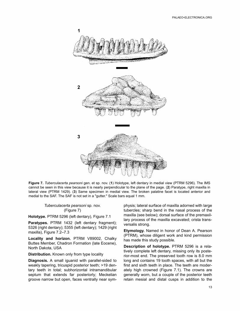
Tuberculacerta pearsoni sp. nov.(Figure 7)
Holotype. PTRM 5296 (left dentary), Figure 7.1Paratypes. PTRM 1432 (left dentary fragment);5326 (right dentary); 5355 (left dentary); 1429 (rightmaxilla), Figure 7.2–7.3Locality and horizon. PTRM V89002, ChalkyButtes Member, Chadron Formation (late Eocene),North Dakota, USADistribution. Known only from type localityDiagnosis. A small iguanid with parallel-sided toweakly tapering, tricuspid posterior teeth; >19 den-tary teeth in total; subhorizontal intramandibularseptum that extends far posteriorly; Meckeliangroove narrow but open, faces ventrally near sym-
physis; lateral surface of maxilla adorned with largetubercles; sharp bend in the nasal process of themaxilla (see below); dorsal surface of the premaxil-lary process of the maxilla excavated; crista trans-versalis strong.Etymology. Named in honor of Dean A. Pearson(PTRM), whose diligent work and kind permissionhas made this study possible.Description of holotype. PTRM 5296 is a rela-tively complete left dentary, missing only its poste-rior-most end. The preserved tooth row is 8.0 mmlong and contains 19 tooth spaces, with all but thefirst and sixth teeth in place. The teeth are moder-ately high crowned (Figure 7.1). The crowns aregenerally worn, but a couple of the posterior teethretain mesial and distal cusps in addition to the
Figure 7. Tuberculacerta pearsoni gen. et sp. nov. (1) Holotype, left dentary in medial view (PTRM 5296). The IMScannot be seen in this view because it is nearly perpendicular to the plane of the page. (2) Paratype, right maxilla inlateral view (PTRM 1429). (3) Same specimen in medial view. The broken palatine facet is located anterior andmedial to the SAF. The SAF is not set in a "gutter." Scale bars equal 1 mm.
SMITH: SQUAMATES FROM THE LATE EOCENE
14
main central cusp. The posterior crowns are paral-lel-sided to weakly tapering. Anteriorly there is aweak subdental shelf, but this shelf gradually dis-appears posteriorly. The Meckelian groove isentirely open; for most of its length it is directedventromedially, but anteriorly it is directed ventrally.There is a prominent medial "hook" to the dentaryat its anterior end. The intramandibular septum(IMS) extends far posteriorly and is present as farback as the dentary itself is preserved. The IMS isoriented nearly horizontally. Externally, there aresix labial foramina, the last at the level of the four-teenth tooth.Description of paratypes. PTRM 1432 and 5326are smaller than PTRM 5296 and poorly pre-served, but PTRM 5355, a still smaller specimen,provides more information on the pattern of toothcrown morphology in the dentary. The ninth andtenth teeth (from symphysis) are partially pre-served, and weak grooves mesially and distallyindicate that those teeth had at least weak "shoul-ders," if not distinct cusps. PTRM 5355 also has asomewhat better-developed subdental shelf poste-riorly. All three of these specimens show a long,subhorizontal IMS, as seen in PTRM 5296.PTRM 1429 is a well-preserved right maxilla(referred to the same taxon as the dentaries on thebasis of size and tooth morphology). Twelve toothpositions and nine teeth are preserved (Figure 7.2–3). The teeth are unicuspid anteriorly, but the tenthand eleventh teeth are tricuspid, with parallel-sidedto weakly tapering crowns; where precisely thetransition occurs is difficult to estimate, because ofwear on some more anterior teeth. The crowns aremoderate in height. The nasal process is broadand shows a strong canthal crest (Figure 7.2); it is"bent," in other words, divided into an anterodor-sally facing surface and a laterally facing one. Pos-teriorly, there is a facet on the medial surface of thenasal process for articulation with the prefrontal.Anteriorly, the nasal process is continuous with thewell-developed crista transversalis. The nasal pro-cess shows large, well-developed dermal rugosi-ties, which presumably indicate the margins ofepidermal scales, down to just above the main rowof labial foramina, of which there are six. The pre-maxillary process is broken medially at the level ofthe subnarial arterial foramen. The dorsal surfaceof the process is excavated. The AIAF lies just dor-sal and medial to this pit, at the base of the nasalprocess. The palatine process, as preserved, isinsignificant, but the sharp-bordered facet visibleon its dorsal surface (Figure 7.3) and the taperingof the palatal shelf medial to this facet suggest that
it has been broken. Its original extent is unknown.The SAF is "roofed" (see above under Polychruscharisticus) and opens posteriorly near the end ofthe nasal process (Figure 7.3). The posterior endof the maxilla is broken, but it retains part of themoderately deep groove for articulation with thejugal. This groove extends anteriorly to the level ofthe SAF.Remarks. The elongate IMS of Tuberculacertapearsoni is noteworthy. To evaluate this feature iniguanids, I measured two lengths in each of over50 isolated dentaries, including representatives ofall major clades except Liolaemini. The first lengthis that from the symphysis to the end of the IMS;the second, from the symphysis to the end of thetooth row. The ratio between these measurementswas ≥0.69 in all phrynosomatines (usually >0.80)and >0.80 in Anolis, Pristidactylus torquatus,Oplurinae, and Leiocephalus. A ratio of <0.69 wasfound in all other clades, including Polychrus,Corytophaninae, Crotaphytinae, Enyalioidesoshaughnessyi, Iguaninae, and Tropidurini (Ura-centron, Uranoscodon, Microlophus, Tropidurus,Plica). In cases where two specimens of the samespecies were examined (Phrynosoma asio, Gam-belia wislizenii), the ratio differed by no more than7%. In the four agamids examined, the ratio wasbetween 0.60 and 0.70. An extended IMS—defined here as one showing a ratio of greater than~0.7—is tentatively interpreted as independentlysynapomorphic of some part of Polychrotinae*,Oplurinae, Leiocephalus, and Phrynosomatinae,pending evaluation in Liolaemini. The posterior endof the most complete dentary in the sample, PTRM5296, is not preserved; one can nevertheless saythat the IMS extends to the level of the nineteenthdentary tooth, further than in all iguanids with a"short" IMS (ratio less than ~0.7).In the maxilla of Tuberculacerta pearsoni, as in allphrynosomatines examined, the SAF is "roofed."As discussed above, this morphology appears tobe synapomorphic of Anolis and Polychrus, ofparts of Tropidurinae*, and of Phrynosomatinae.The nasal process of the maxilla of Tuberculacertapearsoni is distinctive in being strongly bent medi-ally and continuous with the crista transversalis atits anterior base. A bend like this, which corre-sponds to a strong canthal crest, is well developedin all examined members of Phrynosomatinae withthe exception of some Phrynosoma (P. cornutum,P. platyrhinos). As discussed below, the nasal pro-cess in these species of Phrynosoma departsstrongly from the primitive condition. Among other

PALAEO-ELECTRONICA.ORG
15
iguanians examined, a sharp bend is present inTropidurini but is weak in Leiocephalus and oplu-rines; it is weak or absent in many Polychrus (P.gutturosus, P. marmoratus), Crotaphytinae, Iguani-nae, and Enyalioides oshaughnessyi. In the casewhere a sharp bend is absent, the nasal process ofthe maxilla is essentially a vertical flange that maycurve slightly medially at its dorsal terminus. Onthe other hand, a sharp bend is present in Anolis(even better developed here) and in Corytophani-nae; in Anolis, the anterodorsal surface of thenasal process is continuous with the crista trans-versalis, as in Phrynosomatinae, whereas in Cory-tophaninae the bend occurs high and in a nearlycoronal plane, such that the (small) dorsally facingsurface is not continuous with the crista. Outgroupcomparison suggests that the presence of a sharpbend is derived in Iguanidae where it occurs. Thus,the distribution within Iguania of a sharp bend inthe maxilla suggests that it is primitive for Phryno-somatinae (and also Tropidurini and Anolis) andhas been transformed beyond recognition in somemembers of the eponymous clade Phrynosoma.Absence in T. pearsoni of a closed and fusedMeckelian groove (primitive for Tropidurinae andPolychrotinae*) suggests in turn that it is related toPhrynosomatinae; this conclusion is consistentwith the extended IMS and "roofed" SAF discussedabove.Rugosities on the nasal process of the maxilla, asseen in Tuberculacerta pearsoni, also occur insome species of Phrynosoma. Rugosities on thedermal bone in Phrynosoma generally extend farbeyond the frontal, regardless of whether theyreach the nasal process, which is highly reduced inmany species. Among other iguanids examined,some members of Polychrotinae* and Crotaphytushave the best-developed dermal rugosities. Pregill(1992) has also discussed them in some species ofLeiocephalus but determined them not to be primi-tive for that clade; Etheridge and de Queiroz (1988)and Lang (1989) furthermore noted them in Lae-manctus serratus. Extreme development of rugosi-ties on the dermal skull bones may be asynapomorphy of Phrynosoma or part of its stem,and their presence in T. pearsoni might suggestthat this species belongs on that stem.This evidence tying Tuberculacerta pearsoni to thestem of Phrynosoma is tenuous. In any case, thenew taxon lacks at least two synapomorphies of(crown) Phrynosoma, namely, a medially facinganterior extent of the Meckelian groove (Van Dev-ender and Eshelman 1979) and a high dentarydepth/length ratio. Many species of Phrynosoma
furthermore possess an open Meckelian groove (P.modestum is an exception), a characteristic thatdistinguishes them from other phrynosomatines.An open Meckelian groove is primitive for Iguania(Etheridge and de Queiroz 1988), but it is importantto determine whether its occurrence in Phryno-soma is symplesiomorphic or secondary. If closureof the Meckelian groove was acquired twice inPhrynosomatinae—once in the Uta-Urosaurus-Sceloporus clade and once in "sand lizards"(sensu Frost and Etheridge 1989)—then an openMeckelian groove in T. pearsoni only excludes itfrom these clades; if closure is a synapomorphy ofPhrynosomatinae and reversed in Phrynosoma,then the open Meckelian groove could unite T.pearsoni with Phrynosoma. The (uncertain) phylo-genetic position of Petrosaurus is crucial toaddressing this question.A maxilla of Tuberculacerta pearsoni preservingthe palatine process is desirable. If it is a phrynoso-matine, as the evidence above suggests, it shouldhave a strong and triangular palatine process. Inmost phrynosomatines (excepting Petrosaurusmearnsi and some Sceloporus like S. clarkii and S.serrifer) the palatine process is moderate to largein size and triangular. This morphology alsoobtains in oplurines, crotaphytines, and many tropi-durines (although in Leiocephalus and Ura-noscodon the process tends to be blunt), butcontrasts with the morphology in Polychrotinae*,Corytophaninae, Crotaphytinae, Iguaninae, andEnyalioides oshaughnessyi, where the process isblunt, rounded, and poorly developed. One canconclude, minimally, that a strong, triangularpalatine process is primitive for Phrynosomatinae.In summary, Tuberculacerta pearsoni possesses alarger suite of characters uniting it with Phrynoso-matinae than with any other taxon, including (1) anelongate IMS, (2) a sharp bend in the nasal pro-cess of the maxilla, creating an anterodorsally fac-ing surface that is continuous with the cristatransversalis, and (3) a "roofed" SAF. WithinPhrynosomatinae, T. pearsoni is tenuously unitedwith Phrynosoma by the well-developed rugositiesof the facial bones. The open Meckelian groovedoes not contravene this placement.Tuberculacerta pearsoni has at least one autapo-morphy that distinguishes it from all extant phryno-somatines examined, namely, the dorsalexcavation of the premaxillary process of the max-illa. A slight depression is evident in the premaxil-lary process of Cophosaurus texana andUrosaurus graciosus, but it is not as well devel-

SMITH: SQUAMATES FROM THE LATE EOCENE
16
oped as in T. pearsoni; in most phrynosomatinesexamined, the premaxillary process is flat and mayor may not slope slightly ventrolaterally.Thus far, the earliest record of Phrynosomatinaeseems to be the early Oligocene Paraphrynosomagreeni (Holman 1987), which was also suggestedto lie on the stem of Phrynosoma.
cf. Aciprion sp.(Figure 8)
Referred specimens. PTRM 5198 (right dentary),Figure 8; 2054 (left dentary fragment); 2594 (partialleft dentary); 5202 (partial right dentary); 2095 (leftmaxilla fragment); 5356 (right maxilla fragment);5401 (left maxilla fragment); ?5396 (left maxillafragment)Description. PTRM 5198 is a right dentary missingonly its posterior-most end (Figure 8). It preserves23 tooth spaces (with nine teeth); the length of thetooth row as preserved is 10.9 mm. The teeth aremoderate in height. There are well-developedmesial and distal accessory cusps on the crowns ofthe posterior teeth, but the crowns are not flared.These cusps are much weaker on the more ante-rior teeth but nevertheless visible on tooth nine.The posterior crowns do not abut one another. Theposterior-most tooth is somewhat shorter than theanterior-most, reflecting a decrease in tooth sizetoward the back of the tooth row. Five middle teethoccupy a length of 5.4 mm. Anteriorly, a moder-ately developed subdental gutter is present, whichis reduced, but still present, posteriorly. The IMSextends to under the twentieth tooth space (9.2mm along the tooth row). The Meckelian groove isopen along the entire dentary; however, it narrowsconsiderably in the middle. The anterior end of thedentary turns abruptly medially (it is "hooked"). Onthe external surface, there is a linear indentation,running longitudinally just below the parapet of thejaw. There are five labial foramina, the last locatedat the level of the fourteenth tooth space. The
remaining specimens provide little more informa-tion on dentary morphology, although it is worthnoting that PTRM 2594, nearly the same size asPTRM 5198, lacks an external, longitudinal inden-tation.The maxilla of cf. Aciprion sp. is not represented byequally well-preserved material. (Maxillae werereferred to the same taxon as the dentaries on thebasis of size and tooth morphology.) PTRM 2095has essentially the same tooth morphology as thedentaries described above. It is a middle fragmentof maxilla; it is quite worn and does not preservethe nasal process. The roof of bone covering theposterior course of the superior alveolar foramen isbroken, giving the foramen the superficial appear-ance of being set in a "gutter"; it was not. PTRM5356 is a slightly more posterior fragment that pre-serves the anterior part of the jugal groove, whichis only moderately deep.Remarks. Cf. Aciprion sp. compares closely inmany details with the equi-sized Aciprion formo-sum from the early Oligocene of Colorado (YPM4663, YPM-PU 10015). Cf. Aciprion sp. had >23teeth in the dentary, versus 25 in YPM-PU 10015.Tooth morphology, especially the position andnature of the transition from unicuspid to tricuspidcrowns, and tooth spacing are similar in the twotaxa. The only significant dental differencebetween the two appears to be tooth crown height,which is lower in cf. Aciprion sp. The two taxa addi-tionally share a similar symphyseal structure. A.formosum does differ from cf. Aciprion sp. in that ithas only a weak subdental shelf; additionally, in A.formosum the Meckelian groove is closed (but notfused) for several millimeters in the anterior half ofthe dentary, while in cf. Aciprion sp., the groovemerely narrows.As in Aciprion formosum (see Estes 1983), thereare few features of cf. Aciprion sp. that permitready phylogenetic placement. The apparentlyhigh IMS/tooth row length ratio (~0.8?) is sugges-
Figure 8. cf. Aciprion sp., right dentary in medial view (PTRM 5198). The first tooth position is not discernible in thisview. Scale bar equals 1 mm.

PALAEO-ELECTRONICA.ORG
17
tive of affinities with Phrynosomatinae/Oplurinae/Tropidurini or with Anolis (see above under Tuber-culacerta pearsoni).
Iguanid MPH-3(Figure 9)
Referred specimens. PTRM 5357 (partial left den-tary), Figure 9; 5178 (partial left dentary); 5267 (leftdentary fragment)Description. PTRM 5357 is the posterior segmentof a left dentary (Figure 9). It is generally well worn,with bone edges and tooth crowns rounded. Tentooth positions and seven teeth are preserved. Theteeth are moderately high-crowned. The anterior-most tooth was either unicuspid or weakly tricus-pid. The teeth become more strongly tricuspid pos-teriorly; the crowns begin to flare and actuallycontact one another to form a continuous cuttingsurface. Due to the incomplete preservation, it isunclear where along the dentary the transition intooth crown morphology occurs. Five middle teethoccupy 4.6 mm. There is a very weak subdentalshelf anteriorly. The Meckelian groove is openthroughout the preserved length of dentary. PTRM5178 and 5267 provide no further details.Remarks. Iguanid MPH-3 is very similar to cf.Aciprion sp. By comparison with the latter, how-
ever, Iguanid MPH-3 is distinguished by havingmore closely spaced teeth and slightly flared toothcrowns that, in the posterior part of the jaw, contactone another. The IMS appears to be posteriorlyextensive in this taxon, but better-preserved andmore complete specimens must be found beforeany systematic determination can be made.
Iguanid MPH-4(Figure 10)
Referred specimen. PTRM 2041 (partial left den-tary), Figure 10Description. PTRM 2041, the middle segment of aleft dentary, is heavily worn. Fourteen tooth posi-tions and nine teeth are preserved. The teeth areworn to rounded stumps, so no distinct morphologyis apparent (Figure 10); they are, however, notclosely spaced. There is the slightest of subdentalgutters along most of the length of the tooth row.The Meckelian groove gently closes anteriorly. TheIMS extends posteriorly to under the tenth pre-served tooth.Remarks. Iguanid MPH-4 is poorly preserved, andlittle can be said about the characteristics of thismorphotype. As the tooth crowns are worn away,tricuspid teeth cannot be used to refer it toIguanidae; instead, complete closure (without
Figure 9. Iguanid MPH-3, (1) illustration and (2) photograph of partial left dentary in medial view (PTRM 5357).Crowns of teeth, like the bone itself, are heavily worn but clearly abut on one another. Scale bar equals 1 mm.
SMITH: SQUAMATES FROM THE LATE EOCENE
18
fusion) of the Meckelian groove must be used (I amunaware of any non-iguanid that shows this fea-ture). It is distinguished from most other taxadescribed here by gentle closure, without fusion, ofthe Meckelian groove and from Cypressaurus sp.(see below) by the more robust teeth.
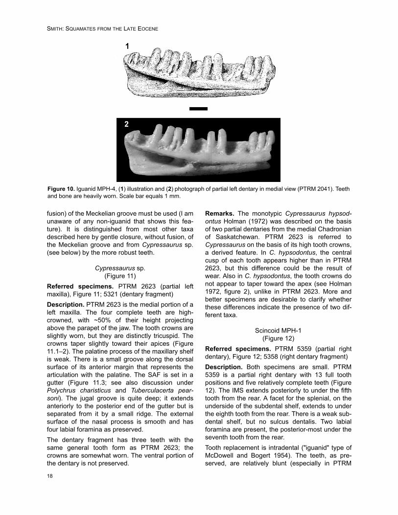
Cypressaurus sp.(Figure 11)
Referred specimens. PTRM 2623 (partial leftmaxilla), Figure 11; 5321 (dentary fragment)Description. PTRM 2623 is the medial portion of aleft maxilla. The four complete teeth are high-crowned, with ~50% of their height projectingabove the parapet of the jaw. The tooth crowns areslightly worn, but they are distinctly tricuspid. Thecrowns taper slightly toward their apices (Figure11.1–2). The palatine process of the maxillary shelfis weak. There is a small groove along the dorsalsurface of its anterior margin that represents thearticulation with the palatine. The SAF is set in agutter (Figure 11.3; see also discussion underPolychrus charisticus and Tuberculacerta pear-soni). The jugal groove is quite deep; it extendsanteriorly to the posterior end of the gutter but isseparated from it by a small ridge. The externalsurface of the nasal process is smooth and hasfour labial foramina as preserved.The dentary fragment has three teeth with thesame general tooth form as PTRM 2623; thecrowns are somewhat worn. The ventral portion ofthe dentary is not preserved.
Remarks. The monotypic Cypressaurus hypsod-ontus Holman (1972) was described on the basisof two partial dentaries from the medial Chadronianof Saskatchewan. PTRM 2623 is referred toCypressaurus on the basis of its high tooth crowns,a derived feature. In C. hypsodontus, the centralcusp of each tooth appears higher than in PTRM2623, but this difference could be the result ofwear. Also in C. hypsodontus, the tooth crowns donot appear to taper toward the apex (see Holman1972, figure 2), unlike in PTRM 2623. More andbetter specimens are desirable to clarify whetherthese differences indicate the presence of two dif-ferent taxa.
Scincoid MPH-1(Figure 12)
Referred specimens. PTRM 5359 (partial rightdentary), Figure 12; 5358 (right dentary fragment)Description. Both specimens are small. PTRM5359 is a partial right dentary with 13 full toothpositions and five relatively complete teeth (Figure12). The IMS extends posteriorly to under the fifthtooth from the rear. A facet for the splenial, on theunderside of the subdental shelf, extends to underthe eighth tooth from the rear. There is a weak sub-dental shelf, but no sulcus dentalis. Two labialforamina are present, the posterior-most under theseventh tooth from the rear.Tooth replacement is intradental ("iguanid" type ofMcDowell and Bogert 1954). The teeth, as pre-served, are relatively blunt (especially in PTRM
Figure 10. Iguanid MPH-4, (1) illustration and (2) photograph of partial left dentary in medial view (PTRM 2041). Teethand bone are heavily worn. Scale bar equals 1 mm.

PALAEO-ELECTRONICA.ORG
19
5358, which is worn) and low-crowned. Many teethare especially broad at the base, tapering towardtheir crowns. The crowns themselves show a mod-erate central cusp bordered by strong mesial andweaker distal crests that curve labially around themain cusp, forming "shoulders." The lingual, butnot labial, surfaces of the crowns are striated.Remarks. Scincoid MPH-1 is distinguished fromheretofore described Paleogene scincoid lizards bythe tapering of the teeth toward their apices, and
the absence of a sulcus dentalis. The primary rea-son for allying it with Scincoidea is general toothform, which, as described, consists of a main cen-tral cusp, striated lingually and bordered by "shoul-ders" (not distinct accessory cusps). Clearly, betterspecimens are desirable.
“Palaeoxantusia” borealis Holman (1972)(Figure 13)
Figure 11. Cypressaurus sp., (1) illustration and (2) photograph of partial left maxilla in medial view (PTRM 2623).Teeth taper slightly toward their tips. (3) Same specimen in dorsomedial view. The jugal groove is deep, and thesuperior alveolar foramen is set in a "gutter." Scale bar equals 1 mm.

SMITH: SQUAMATES FROM THE LATE EOCENE
20
Referred specimens. PTRM 2720 (partial left den-tary), Figure 13.1; 5360 (edentulous left dentary);5361 (partial right dentary), Figure 13.2; 2074(medial portion of right maxilla); 5284 (left dentaryfragment); 5288 (partial right dentary); 2074 (right
maxilla fragment); 2713 (partial right maxilla); 5298(right maxilla fragment); 5387 (right dentary frag-ment)Amended diagnosis. As in Holman (1972), exceptthat the AIAF is located at or behind the ultimate
Figure 13. “Palaeoxantusia” borealis, (1) partial left dentary in medial view (PTRM 2720). No indication of a splenial ispreserved in this specimen. The thin line extending anteriorly from the AIAF is a fracture. (2) Partial right dentary inmedial view (PTRM 5361). Scale bars equal 1 mm.
Figure 12. Scincoid MPH-1, (1) illustration and (2) photograph of partial right dentary in medial view (PTRM 5359).Scale bar equals 1 mm.

PALAEO-ELECTRONICA.ORG
21
dentary tooth in most members (see below) andtooth crowns are striated lingually.Description. None of the Medicine Pole Hills spec-imens approaches the quality of the holotype sple-niodentary of "Palaeoxantusia" borealis (SMNH1435). The most complete specimen, PTRM 5360,is edentulous and broken just posterior to the ulti-mate tooth and lacks a splenial; it displays manyfeatures seen in the other specimens from theMedicine Pole Hills l.f. It is relatively small, with atooth-row length of 4.0 mm; the height of the den-tary at the ultimate tooth is 1.1 mm. A sulcus denta-lis is well developed. Twelve tooth positions arepresent. The AIAF is situated just behind the ulti-mate tooth (more clearly seen in PTRM 2720: Fig-ure 13.1). There are five labial foramina. PTRM5361 shows the long, horizontal posterior exten-sion of the dentary beyond the tooth row (Figure13.2).The teeth of most specimens are rather worn; theyare blunt and unicuspid and do not preserve detailsof crown morphology. The teeth of a couple ofspecimens retain traces of the weak "shoulders"that lie mesial and distal to the central cusp. Theultimate maxillary tooth of PTRM 5298 and 5387present clear examples of weak striations near thebase of the crown.Remarks. This Medicine Pole Hills xantusiid com-pares well to "Palaeoxantusia" borealis, and it isreferred to that taxon. The specimens describedhere are smaller than those from the Calf Creek l.f.(Medicine Pole Hills: mean dentary depth at ulti-mate tooth ~1.1 mm, n = 4; Calf Creek: meandepth ~1.3 mm, n = 6). However, there is muchoverlap at the lower end of the Calf Creek sizerange, and a 20% difference in linear dimensionswould translate into an approximately 75% differ-ence in volume (and hence, mass), which accountsfor the overall slender appearance of the MedicinePole Hills specimens. It is possible that, if the spec-imens are correctly referred, "P." borealis exhibitsan inverse correlation of size (mass) to latitude—and, hence, temperature—conforming to Berg-mann’s rule (Bergmann 1847), which is disputed insquamates (e.g., Angilletta et al. 2004). Alterna-tively, none of the specimens in the sample may befully grown. Only one of the largest specimens fromCalf Creek (depth ~1.5 mm) evinces completefusion of the splenial to the dentary and can beinterpreted as more-or-less fully adult. Unfortu-nately, none of the specimens preserves the sple-nial at all (this fact may indicate that the splenial
was not yet fused, i.e., that all the specimens arefrom juveniles).Fossil xantusiid species are typically based on thedentary or spleniodentary, and there are preciousfew features in this bone upon which phylogenetichypotheses can be based (Vicario et al. 2003).Thus, it is important to point out a feature not yetdescribed that may provide some constraint on therelationships of fossil xantusiids. In the earliest-known of these, “Palaeoxantusia” fera, from themiddle Paleocene of Wyoming (Estes 1976; Sulli-van 1982), the AIAF is located under the ultimateor penultimate dentary tooth in all specimens forwhich this character is clearly scorable (n = 4 and9, respectively). The same holds true for the largeBridgerian form "P. fera" (YPM-PU 17506, n = 3)and the Bridgerian type of P. fera (AMNH 3815;Hecht 1956). The only specimen that appears toviolate this generalization is AMNH 3821, from thesame locality as the type of P. fera; it appears,then, that middle Eocene "Palaeoxantusia" exhibitssome polymorphism. "Palaeoxantusia" kyrentosand “P.” allisoni from the Uintan of California alsodisplay an anterior foramen (see Schatzinger 1980,text-figures 1,4). Among extant xantusiids, theanterior position of the AIAF is seen in Lepido-phyma flavimaculatum (pers. obs.); some Xantusiariversiana (three of eight) also approach the condi-tion. In all "Palaeoxantusia" borealis from both theMedicine Pole Hills (Figure 13.1) and Calf Creek, incontrast, the AIAF is located just behind the ulti-mate dentary tooth. This posterior position—it maybe defined as the case in which the foramen isentirely located behind the midline of the ultimatetooth—is also seen in the living basal (Hedges andBezy 1993; Vicario et al. 2003) member Cricosauratypica (see Savage 1963, figure 8), Xantusiadownsi (see Norell 1989, figure 14), and X. vigilis,X. arizonae, X. henshawi, and a significant propor-tion of X. riversiana (pers. obs.). Given Vicario etal.’s (2003) recent phylogeny of Xantusiidae, itseems possible that the posterior position is primi-tive for Xantusia and partially reversed in X. riversi-ana. The only other small xantusiid, Cricosauratypica, must independently have acquired this fea-ture, implying that it might simply be related toabsolute size. On the other hand, the posteriorposition in one paratype of P. fera and many X. riv-ersiana indicates that this need not be the case.Size correlation also suggests that an anterior shift-ing of the foramen ought to be visible during devel-opment.Estes (1983: 125) concluded that "Palaeoxantusia"borealis was inadequately diagnosed and might be

SMITH: SQUAMATES FROM THE LATE EOCENE
22
synonymous with P. fera. The validity of "P." borea-lis is confirmed, and its diagnosis is amendedabove.
cf. Spathorhynchus sp.(Figure 14)
Referred specimens. PTRM 1793 (right dentary),Figure 14; 1825, 5364–5370, 5638, 5639, 5662,5667, 5671, 5672, 5691 (trunk vertebrae)Description. The worn dentary is relatively large,the tooth row measuring 6.2 mm in length. Thereare eight tooth positions, but only the sixth andseventh teeth are even partially preserved (Figure14). They are subpleurodont and slightly separatedfrom the interior edge of the dentary by a weaksubdental shelf. Their bases are inflated and con-fluent, rather square in coronal section. The firstand fourth tooth bases are the largest. The splenialnotch extends to the boundary between the fourthand fifth teeth. The interior of the dentary has asmall ridge that extends ventrally from the top ofthe Meckelian canal; an intramandibular septum istherefore not well developed posteriorly. The Meck-elian groove is closed and fused. There is a small,medially facing notch, just posterior to the lasttooth, which is confluent with the well-defined coro-noid fossa on the labial surface of the bone, thusexcluding the possibility of a long labial process ofthe dentary. The coronoid fossa actually comprisestwo conjoined fossae, the boundary between whichis marked by a faint ridge. The medial, shallowerone is confluent with the aforementioned notchposterior to the last tooth; the lateral, deeper oneextends anteriorly to the level of the boundarybetween the sixth and seventh teeth. The coronoidfossa as a whole is triangular in shape, with theapex pointed forward. Two, widely spaced labialforamina are present, one each below the secondand fifth teeth. The mandibular symphysis is weak.
The best-preserved vertebra (PTRM 5364) is large,measuring 7.5 mm in length (along neural arch)and somewhat wider anteriorly than posteriorly(10.2 mm at prezygapophyses vs. 9.1 mm atpostzygapophyses). The prezygapophyses are ori-ented in a nearly coronal plane. A midline keel runsalong the entirety of the neural arch; the keel endsin a projection (partially broken) from the posteriorend of the arch. The posterior end is flared andmarkedly taller than the anterior end. On the dorsalsurface of the posterior half of the neural arch runlinear ridges (or "flutings": Gans 1978) whichdiverge posteriorly from the midline. The ridgesmatch corresponding structures on the ventral sur-face of the arch (a convexity above matches a con-cavity below), and thus, viewed from behind, theedge of the arch appears crinkled. (This ventralmirroring of flutings is present in many, but not all,vertebrae referred to cf. Rhineura sp.) The centrumis depressed. The condyle has a constricted neck.There is no hypapophyseal ridge; only the rightsubcentral arterial foramen is present, and it islocated close to the synapophysis, well away fromthe midline. The synapophyses are hemispherical.The vertebrae (including PTRM 5364) typicallyhave a small indentation just above the synapo-physis on their lateral side. Most referred vertebraeare only two-thirds the size of PTRM 5364.Remarks. The low tooth count and subpleurodonttooth implantation are sufficient to refer this spe-cies to Amphisbaenia. Unfortunately, the polarity ofmany character state changes seen within Amphis-baenia is equivocal because of uncertain in-groupand out-group relationships (Estes et al. 1988;Kearney 2003); this complicates discussion of rela-tionships. The determination of whether many fea-tures are primitive or derived in Rhineuridaedepends crucially on the position that that taxonoccupies. Despite these phylogenetic difficulties, aplethora of rhineurid amphisbaenians has been
Figure 14. Cf. Spathorhynchus sp., right dentary in medial view (PTRM 1793). The bone is heavily worn, and nodetails of tooth morphology are preserved. Scale bar equals 1 mm.

PALAEO-ELECTRONICA.ORG
23
named from the late Eocene through Oligocene ofNorth America, generally based on more-or-lesscomplete skulls. PTRM 1793 shares with many ofthese fossil rhineurids—including the middleEocene Spathorhynchus fossorium Berman(1973), early Oligocene Rhineura hatcheri (Kear-ney et al. 2005), Miocene Rhineura sepultura Hol-man (1979), and extant R. floridana (Cope 1892,figure 5; pers. obs.)—enlargement of the first andfourth dentary teeth. In "Rhineuridae sp. indet."from the late Paleocene of Wyoming (Estes 1975),only the fourth tooth appears to be enlarged (pers.obs.). Hyporhina tertia has enlarged first and fourth"maxillary teeth" (Berman 1972: 8); whether thisapplies to the dentary is unclear. The first andfourth dentary teeth appear to be enlarged in Jeps-ibaena minor (YPM-PU 13460; pers. obs.). It isprobable that specified tooth enlargement is diag-nostic of a clade including part of the stem of R.floridana, though precisely how much of the stem isat this point difficult to assess. The late Paleocene"Rhineuridae sp. indet." is the earliest taxon to dis-play apomorphy one (fourth tooth enlarged); apo-morphy two (first tooth enlarged) is first known inthe early Eocene J. minor. Kearney (2003) con-cluded that all these North American forms weremore closely related to R. floridana than to anyother living amphisbaenian.Cf. Spathorhynchus sp. can be excluded fromdescribed species of Rhineura by the primitiveretention of eight dentary teeth. Spathorhynchusfossorium and the late Eocene S. natronicus alsoretain the presumably primitive state of eight den-tary teeth (Berman 1973, 1977) and are both rela-tively large, with tooth row lengths somewhat lessthan 10 mm. Thus, of described coeval rhineurids,PTRM 1793 compares best to Spathorhynchus
natronicus; due to poor preservation, however, Irefer the specimens to cf. Spathorhynchus sp.The largest vertebra, PTRM 5364, is considerablylarger than one would expect in relying on the den-tary alone. This fact suggests that the animal rep-resented by PTRM 1793 was not yet fully grown.Cf. Spathorhynchus sp. may be the same as thelarge rhineurid taxon described as Lestophis sp. nr.L. anceps by Holman (1972) based on vertebrae.[Estes (1983), with good cause, declared Lestophisa nomen dubium.] Berman (1972) stated thatridges are present on the neural arches of verte-brae in rhineurids, to which list Estes (1976) addedAmphisbaena (in A. alba, this is particularly true ofthe more anterior vertebrae). Kearney (2003)recently found dorsal ridges to be present also inAulura, Dalophia, Leposternon, and Monopeltis,which she regarded as basal in Rhineuroidea. Flut-ings are not always reflected on the underside ofthe neural arch, but this is conceivably related tosize.Interestingly, amphisbaenian vertebrae—from bothcf. Spathorhynchus sp. and cf. Rhineura sp. (seebelow)—appear to be unusually abundant in theMedicine Pole Hills l.f. Iguanids, though they aremuch more diverse and numerically abundant(based on jaw elements), are represented by (rela-tively) far fewer vertebrae. The same might be trueof the amphisbaenians in the Calf Creek l.f., whichare represented by 58 and 43 vertebrae (small andlarge, respectively; Holman 1972). Even consider-ing that a given amphisbaenian typically has morethan three times as many vertebrae as a non-fos-sorial squamate (<30 precloacal vertebrae in non-fossorial taxa versus often >100 in amphisbae-nians; Hoffstetter and Gasc 1969), the differenceappears anomalous.
Figure 15. Cf. Rhineura sp., left dentary in medial view (PTRM 5248). The thin line extending anteriorly from the AIAFis a fracture. Scale bar equals 1 mm.

SMITH: SQUAMATES FROM THE LATE EOCENE
24
cf. Rhineura sp.(Figure 15)
Referred specimens. PTRM 5248 (left dentary),Figure 15; 5322 (right dentary fragment); 5725 (leftdentary); 1422, 1434, 1438, 1443–1445, 1553,1796, 1812, 1813, 1828, 1831, 1851, 2023, 2042,2067, 5171, 5180, 5216, 5235, 5238, 5348 (12specimens), 5362, 5363, 5384–5386, 5668, 5673,5674, 5686–5688 (trunk vertebrae)Description. The most complete specimen, PTRM5248, has a tooth row length of 3.0 mm. There areseven tooth positions (Figure 15), although thefourth tooth is missing. Of these seven, the firstand fourth are enlarged relative to the others. Theteeth are subpleurodont and conical, tapering uni-formly toward their crowns; these crowns, pre-served only on the third and fifth teeth, are slightlyrecurved. There is a narrow, shallow sulcus denta-lis. The symphysis is relatively large, expanded rel-ative to the part just distal to it and has a smallposteroventral projection. The Meckelian groove isclosed and fused, and the splenial notch extendsanteriorly to the level of the boundary between thefifth and sixth teeth. A fossa for the labial coronoidblade extends anteriorly to the level of the seventh(last) tooth.PTRM 5322 is approximately the same size asPTRM 5248, though it is broken (just?) in front ofthe anterior margin of the splenial notch and pre-serves only the first through fifth teeth. The firstand fourth teeth are enlarged. The symphysis isrelatively large, and it has a posteroventral projec-tion as in PTRM 5248.The vertebrae of cf. Rhineura sp. are quite abun-dant and typical of rhineurid vertebrae. Vertebralflutings (see above under cf. Spathorhynchus sp.)are present but are only rarely reflected on theunderside of the neural arch (e.g., in PTRM 5171),and then only weakly. The subcentral arterialforamina are typically located along the anteriorthird to fourth of the centrum. As in cf. Spathorhyn-chus sp., there is typically an indentation dorsal tothe synapophysis on the lateral side of the verte-brae.Remarks. The referral of these specimens to cf.Rhineura sp. is based on small size, tooth count,and age. The reduction to seven dentary teeth is aderived feature within Rhineuridae (see above). Cf.Rhineura sp. is therefore closer to Rhineura thanare various Paleogene forms like Jepsibaenaminor (8 dentary teeth) and Spathorhynchus spp.(8). No known North American Eocene rhineuridhas seven dentary teeth, but Gilmore (1928)
describes early Oligocene Rhineura hatcheri (CM423A and B) as having seven. Hyporhina antiqua(YPM-PU 11390) also has seven dentary teeth, butthat species lacks the expanded symphysis seen inJ. minor and cf. Spathorhynchus sp. (pers. obs.)The enlargement of the first and fourth teeth placescf. Rhineura sp. firmly on the rhineurid line, and itspossession of only seven dentary teeth indicatesthat it is derived relative to Spathorhynchus.Sullivan and Holman (1996) synonymized manyearly Oligocene rhineurid taxa, arguing that thecharacters used to distinguish fossil amphisbae-nians, including tooth count, may be related solelyto ontogenetic stage and therefore "taxonomicallyinsignificant" (p. 362). This point is well taken, for itis widely recognized, for instance, that tooth countincreases with absolute size during development inmany squamates. (I am not aware of any suchstudy specifically on amphisbaenians.) Estes(1983) was correct in stating that taxonomic andsystematic revision of Paleogene amphisbaeniansis needed.Neverthelesss, the dismissal of tooth count data inrhineurid systematics seems not yet to be war-ranted. Granted, extant Rhineura floridana hasonly six dentary teeth, and it is also smaller thanmany of its presumed early Paleogene relatives,which have eight dentary teeth. But Rhineura flori-dana’s small size is itself an evolutionary change.Its small size and low tooth count may be informa-tive for both alpha taxonomy and systematics,even if the two characters are developmentally cor-related and might be considered a single charactercomplex. In other words, ontogenetic correlation isby itself an insufficient basis for discarding charac-ter information in the referral of fossil forms. Addi-tionally, in the particular case of Rhineuridae,absolute size is clearly a poor predictor of toothcount: the large Eocene Spathorhynchus as wellas small Jepsibaena minor have eight dentaryteeth, whereas the large Miocene Macrorhineuraand small extant Rhineura floridana have but six.Cf. Rhineura sp. might be the same as the smallrhineurid species noted by Holman (1972) in theCalf Creek l.f., which was represented only by ver-tebrae.
Anguimorph MPH-1(Figure 16)
Referred specimens. PTRM 5375 (partial left den-tary), Figure 16; PTRM 1824 (partial right dentary);PTRM 5376 (anterior left dentary fragment)

PALAEO-ELECTRONICA.ORG
25
Description. The most complete specimen of thistaxon is PTRM 5375. It is large, the tooth row being13.7 mm long as preserved. A weak, narrow sulcusdentalis (Figure 16.1–2) is present at the anteriorend of the bone. Fourteen or fifteen tooth positionsare preserved. The teeth are pleurodont and low-crowned, less than one-third of their height project-ing above the parapet of the jaw. The tooth crownsare striated lingually, and small mesial and distola-bial crests descend the crowns (the mesial crest ismuch stronger), creating a V-shaped cutting edge.Except for the most posterior teeth, the crowns arediscolored and somewhat transparent. They arebluntly pointed (in part due to wear), slightlyrecurved, weakly labiolingually compressed, and alittle concave lingually near the juncture betweencrown and shaft. The teeth become progressivelysmaller and more blunt posteriorly and lose thecrown-shaft concavity. The excavated base of thethird preserved tooth with the incoming replace-ment tooth not directly below it indicates an inter-dental mode of replacement. The Meckeliangroove is open. Though much of the shelf that sup-ports the teeth has been broken posteriorly, a thin
upper remnant of the IMS is evident; its originalextent is unknown.Externally, labial foramina are not readily identifi-able in PTRM 5375, but a groove runs along muchof the length of the dentary in the expected positionof these foramina (Figure 16.3). The posterior, ven-trolateral surface of the dentary is flattened, orplanated; the plane of flattening is parallel to thelongitudinal axis and oblique to a sagittal plane.Articulation surfaces with the postdentary bonesare not apparent.The two other specimens are far less complete, butboth of them confirm the tooth morphology of thistaxon. Additionally, PTRM 1824 displays the plan-ated, external dentary surface and labial groove inthe position of the labial foramina. There appear tobe a couple of foramina present along the groove;whatever their number, they are small. Its tworemaining teeth also exhibit discoloration of thecrowns. PTRM 5376 exhibits a weak anterior sul-cus dentalis but does not preserve the portion ofthe dentary that is planated in the other two speci-mens. No discoloration of the tooth crowns is evi-dent in this specimen.
Figure 16. Anguimorph MPH-1, (1) illustration and (2) photograph of partial left dentary in medial view (PTRM 5375).The crowns of the teeth are striated lingually. (3) Same specimen in lateral view. Note the planation of the ventrolat-eral wall of the dentary. Scale bar equals 1 mm.

SMITH: SQUAMATES FROM THE LATE EOCENE
26
Remarks. The interdental mode of tooth replace-ment unites this new species with Anguimorpha(cf. Estes et al. 1988). Its tooth morphology isunique, but the V-shaped cutting edge is found inAnguidae and some anterior teeth in Xenosaurus(Gauthier, 1982; pers. obs.). In possessing lin-gually striated tooth crowns it also likens anguids,Xenosaurus, and many Varanus (pers. obs.). Manyother features, such as osteodermal characteristicsand the state of the IMS posteriorly, cannot yet beevaluated. Oddly, nearly all anguimorphs possessat least a moderate subdental shelf along most ofthe dentary. That this shelf is essentially lacking inAnguimorph MPH-1 would seem to be autapomor-phic.Relationships among anguimorphs generally andanguid subclades in particular are not well resolved(e.g., Camp 1923; Rieppel 1980; Gauthier 1982;Macey et al. 1999; Wiens and Slingluff 2001;Townsend et al. 2004), which also limits our under-standing of character evolution within the group.Preserved features in Anguimorph MPH-1 appearonly to disbar it from membership in particularanguid subclades: from Diploglossinae and Ger-rhonotinae, by its lack of bicuspid teeth; from Glyp-tosaurinae by its tooth morphology; and fromAnniella, by its higher tooth count and tooth mor-phology. In all these respects, Anguimorph MPH-1is primitive relative to known representatives of
these groups. Although its tooth form and lack of asignificant subdental shelf render AnguimorphMPH-1 distinctive, better specimens are requiredbefore a formal new name can be applied.
Diploglossine MPH-1(Figure 17)
Referred specimens. PTRM 1807 (left dentaryfragment), Figure 17.1; 1823 (jaw fragment); 5378(osteoderm), Figure 17.2Description. PTRM 1823 is a fragment of either aright dentary or a left maxilla. The bit of bone towhich the two preserved teeth are attached isworn. The crown of the first tooth is also worn, butthe second does not project as high—it is probablyan incompletely emplaced replacement tooth—andappears largely undamaged. This second (poste-rior) tooth bears a distinct mesial crest, offset fromthe rest of the tooth crown by a shallow groove.The main tooth cusp is distally displaced andslightly labiolingually compressed. The lingual sur-face of the crown is smooth, but near the base ofthe crown, on its mesial side, there are coarse stri-ations; the striae do not extend onto the lingualside, possibly a result of wear. The bases of bothteeth are missing, so it is difficult to estimate crownheight.There are 13 tooth positions, with eight teeth, pre-served in PTRM 1807, a left dentary fragment (Fig-
Figure 17. Diploglossine MPH-1, (1) Left dentary fragment in medial view (PTRM 1807). (2) Osteoderm in externalview (PTRM 5377). Scale bars equal 1 mm.

PALAEO-ELECTRONICA.ORG
27
ure 17.1). The teeth are considerably smaller thanin PTRM 1823. They are of moderate height, andthe lingual surfaces of the crowns are apparentlyunstriated. All but the anterior-most teeth havestrong mesial crests; on posterior teeth thesecrests are so expanded that the crowns are mesio-distally longer than the shafts. A few teeth appearfully implanted, their labial margins flush with theparapet of the jaw; lingual to these teeth is a mod-erate subdental shelf. Many of the preserved teethare incompletely emplaced replacement teeth andhave bulbous, rounded bases that are not stronglyfused to the dentary; they also sit away from theparapet of the jaw over the subdental shelf. Therelative position of replacement teeth is difficult todetermine posteriorly, where the tooth pattern isdistorted. The anterior-most tooth, however, isexcavated distolingually, indicating an interdental(anguimorph) pattern of tooth replacement.The osteoderm, PTRM 5378 (Figure 17.2), is rect-angular and unkeeled, ~2.2 mm in length. Its sculp-turing consists of variably linked, apically flattenedtesserae that are separated by grooves; wherethey merge, they create a vermiculate sculpture.The anterodorsal edge of the gliding surface wasoblique to the anterior margin, but there is no indi-cation of a posterior projection of the gliding sur-face. This surface is approximately one-fourth thelength of the whole osteoderm.Remarks. The jaw fragments constitute the pri-mary evidence for the presence of a diploglossinein this assemblage. Strong development of themesial tooth crests, often with the development ofa secondary cusp, diagnoses Diploglossinae andpossibly Gerrhonotinae (within Anguidae) as wellas Xenosaurus and its stem (Gauthier 1982); onlydiploglossine anguids have apomorphically shorter,stocky teeth, often with expanded crowns that aremesiodistally longer than their shafts. An undam-aged osteoderm with a posterior projection of thegliding surface would be desirable to buttress thisconclusion (cf. Hoffstetter 1962; Gauthier 1982).The sculpturing of the osteoderm, however, is simi-lar to that of the (stem) diploglossine Eodiploglos-sus borealis Gauthier (1982) from the early Eoceneof Wyoming; and in living diploglossines and E.borealis, not all the osteoderms have a posteriorprojection. There is presently little that would per-mit a more specific determination of the taxon, noris it known whether it lies on the stem of Diploglos-sinae or in the crown. It represents the latest-known occurrence of the diploglossine lineage incentral North America.
cf. Peltosaurus sp.Referred specimens. PTRM 2606 (partial left den-tary); PTRM 2617 (partial right maxilla); 5272 (jawfragment); 5381 (right maxilla); 5685 (right frontalfragment)Description. PTRM 2606 is the anterior two-thirdsof a left dentary. Fourteen tooth positions and twoteeth (numbers 4 and 10, from symphysis) are pre-served. Both teeth, like the dentary itself, arestreamworn, and details of crown morphology arelacking. The apex of tooth 10, however, is some-what labiolingually compressed. The crowns aremoderate in height, projecting less than 50%above the parapet of the jaw. There is a moder-ately developed subdental shelf, without attendantsulcus dentalis; this shelf is largely broken anteri-orly. The Meckelian groove is ventrally located onthe anterior portion of the dentary. There are six,irregularly spaced labial foramina on the externalsurface. There is also a small indentation at theposterolateral margin of the preserved portion, butthe generally streamworn character of the speci-men suggests that this feature may represent arounded fracture rather than the anterior edge ofan articular surface or fossa.PTRM 5381 is a small, nearly complete but some-what worn right maxilla. The premaxillary processis broken near the base of the nasal process.Eleven tooth positions and seven complete or par-tial teeth are preserved. The teeth are widelyspaced, but it is impossible to estimate crownheight. Both the crowns and the shafts are some-what recurved. The crowns are unicuspid, blunt,and without labial or lingual striations, which mayor may not be due to abrasion. They decrease inheight posteriorly such that the posterior-mostteeth are exceedingly small. The dorsal surface ofthe maxilla is divided into anterior and posteriorsegments by an oblique ridge on the interior sur-face of the nasal process and dorsal surface of themain body of the maxilla. The SAF is located at thelevel of the posterior edge of the nasal process.The foramen is set in a short groove and is coveredanteriorly by the dorsal roof of the maxilla. Thepalatine process is only weakly developed and islocated at about the same level as the superioralveolar foramen; on its dorsal surface is a shallowfacet for articulation with the palatine. Laterally,there are four labial foramina and several otherforamina, irregularly spaced, on the slightly irregu-lar surface of the nasal process. There are noosteoderms fused to the surface of the nasal pro-cess, probably a result of the small size of the indi-

SMITH: SQUAMATES FROM THE LATE EOCENE
28
vidual. Another poorly preserved maxilla (PTRM2617) appears to have weak striations on the lin-gual surface of one anterior tooth.The frontal fragment (PTRM 5685) is heavily worn,but it certifies the existence of a non-glyptosauringlyptosaurine in the Medicine Pole Hills l.f. A thinstrip along the orbital margin is devoid of the tuber-culated osteodermal crust that otherwise coversthe bone. A small foramen pierces the lateral mar-gin of the orbit.Remarks. These specimens are compared to Pel-tosaurus on the basis of glyptosaurine tooth form,size, and geologic age. PTRM 2606 shares withspecimens of Peltosaurus granulosus robust teeth,similar tooth spacing, and similar size, but morecomplete teeth, as on the maxilla, seem rathermore recurved than in P. granulosus. The lack ofstriations on most teeth may be attributable tostream abrasion, a phenomenon that may alsoaccount for the lack of striations on Peltosaurus sp.from the Cypress Hills Formation of Saskatchewan(see Lambe 1908; cf. Estes 1983). Peltosaurus israre in late Eocene sediments, though one wouldexpect a "melanosaurin" glyptosaurine to bepresent somewhere on stratigraphic grounds.
Helodermoides sp. nov.?(Figure 18)
Referred specimens. PTRM 1825 (left parietalfragment): Figure 18; 5647, 5709 (cephalic osteo-derms)Description. PTRM 1825 is a left, anterior frag-ment of parietal. Its edges and surfaces are inplaces smoothed by abrasion, including the tops ofthe osteoderms (Figure 18.1). There are four rela-tively complete, tall, bulbous, tuberculated osteo-derms coössified with the dorsal surface. Theosteoderms are vaguely polygonal, with sub-rounded corners, and separated from one anotherby deep grooves. They are undercut in places, andsome isolated tubercles occur in the grooves. Sev-
eral osteoderms are missing from the anterolateralpart of the bone. The left edge of the parietal fora-men (Figure 18.1–2, pf) is visible, perforating thefirst, large, median osteoderm. In medial view, theparietal fragment has a hemispherical cavity, about2.5 mm in diameter, that excavates both osteo-derm and underlying bone. It opens dorsallythrough the osteoderm and ventrally through ashort canal into the cranial cavity. On the ventralsurface of the parietal, the anterior ridges (Figure18.2) are well developed. The distance betweenthe median plane and the anterior ridge is less thanthat between the anterior ridge and the lateral edgeof the parietal (i.e., the triangular inter-ridge area issmall). There are two notches, rounded at theirmedial apices, on the anterolateral part of the ven-tral surface; they are confluent with the anterolat-eral edge of the bone and constitute articulationfacets for the postfrontal and possibly also the pos-torbital.PTRM 5647 and 5709, hexagonal cephalic osteo-derms, are also referred to this taxon.Remarks. The more-or-less hexagonal osteo-derms of PTRM 1825 are sufficient to refer it toGlyptosaurini. As Sullivan (1979) noted, tall, bul-bous osteoderms, among other features, serve todistinguish the presently monotypic Helodermoidestuberculatus from other glyptosaurins. Sullivan alsoargued (op. cit.) that H. tuberculatus is diagnosedby "closure of the supratemporal fenestra," a phe-nomenon that in Glyptosaurini deserves furtherinvestigation. The primitive condition is illustratedby Paraglyptosaurus princeps ("Glyptosaurushillsi": Gilmore 1928, figure 63; and "G. rugosus":Meszoely 1970, figure 16B; see Sullivan 1979 fornomenclature), in which the postfrontal articulationon the parietal is a single notch restricted to the lat-ter bone’s anterior-most end. Sullivan (1979, figure10h) figures complete closure of the supratemporalfenestra in one specimen of Helodermoides(USNM 214736). The condition in PTRM 1825 is
Figure 18. Helodermoides sp. nov.?, Partial left parietal in dorsal (1) and ventral (2) views (PTRM 1825). pf, parietalforamen. Scale bar equals 5 mm.

PALAEO-ELECTRONICA.ORG
29
intermediate, for although the two notches on theventral surface suggest some closure, the bonethins out laterally, and there are no other obviousarticulation surfaces. Sullivan is surely correct thatextensive closure of the supratemporal fenestra byincreased articulation of the temporal bones is aderived feature in Anguidae. The seemingly signifi-cant difference in supratemporal closure betweenspecimens (PTRM 1825 and AMNH 8706 vs.USNM 214736) suggests specific difference. How-ever, because the type specimen of H. tubercula-tus (CM 707) consists only of frontals, adetermination of which form is new is not yet possi-ble. Helodermoides is a previously known and in
this sense unproblematic taxon from the Chadro-nian.
Xenosaurid MPH-1(Figure 19)
Referred specimens. PTRM 1986 (premaxilla),Figure 19; 5371 (dermal skull bone fragment)Description. PTRM 1986 is a slightly worn but rel-atively complete premaxilla. The dorsal surface iscovered by coössified osteoderms, worn on theirouter surfaces (Figure 19.1). The osteoderms arebroad and rounded and separated by narrow val-leys. They tend to be flatter toward the midline andmore conical laterally and do not extend to theextreme lateral margins of the bone. Tiny pocks
Figure 19. Xenosaurid MPH-1, Premaxilla in dorsal (1) and ventral (2) views (PTRM 1986). Scale bar equals 1 mm.

SMITH: SQUAMATES FROM THE LATE EOCENE
30
and perhaps foramina pierce them. The anteroven-tral margin of the bone, below the fused osteo-derms, is perforated by several small foramina.The posterolateral borders of the premaxillaappear unbroken. The dorsal nasal process is bro-ken near its tip. On the ventral margin, there arespaces for seven teeth, including one midline tooth(Figure 19.2). Only one tooth, the first, right, pairedtooth, is preserved, and it is short, the crown wornand blunt. The incisive process is narrow andbilobed, unfused even basally, where the right andleft portions are strongly apposed; it lies immedi-ately behind the median tooth alveolus. The ventralpremaxillary foramina are located lateral to the inci-sive process and posterior to the first pair of pre-maxillary tooth alveoli. The posterior premaxillaryforamina are larger than the ventral ones, withwhich they connect internally (Bahl 1937). Broaddorsal wings extend from the midline to the lateraledge of the bone, covering the typical location ofthe external nares. The wings are thinner betweenthe thick midline and the thick lateral margins.Small grooves present on both sides of the ventralsurface mark articulations with the premaxillaryprocesses of the maxillae.PTRM 5371 is a thin, rectangular fragment of askull bone, 5.5 × 2.5 mm. It is referred to the sametaxon as the premaxilla on the basis of size andsimilar osteoderms. All of the osteoderms are flat-ter than on the premaxilla, however: none is bul-bous. One long edge is unbroken and is groovedfor an articulation along the whole of its preservedlength. In internal (ventral?) aspect, this sameedge is distinguished by a moderately high ridge.Its appearance suggests that the element maybound the supratemporal fenestra (a parietal frag-ment, or postfrontal), which might have been par-tially closed.Remarks. These specimens are referred to anundetermined xenosaurid with some hestitation. Inpossessing multiple anterior foramina the premax-illa likens Xenosaurus but differs from most otheranguimorphs. But unlike Xenosaurus platycepsand X. grandis, which have nine premaxillary teeth(like most anguimorphs), Xenosaurid MPH-1 hasbut seven. Uncertain phylogenetic relationshipswithin Anguimorpha, however, and the presence ofonly seven premaxillary teeth in Shinisaurus croco-dilurus, render it uncertain whether nine teeth inXenosaurus is primitive or derived. Gilmore (1941)reported only eight in Exostinus serratus.An alternative hypothesis is that this species is ahelodermatid, which would be consistent with the
more bulbous character of the osteoderms on thepremaxilla. The absence of the "coralline" sculptur-ing characteristic of Heloderma might then be inter-preted as artifactual (i.e., it has been removed byabrasion). The premaxilla is too small (especiallyconsidering the fusion of the osteoderms) to beattributed to Helodermoides and moreover lacksany indication of tubercles. There is presently noevidence that would tie the specimens to any otherspecies in the fauna.Regardless of its affinities, the broad posterolateralwings of the premaxilla of Xenosaurid MPH-1 areunlike anything known in squamates. The externalnares must have been displaced a considerabledistance posteriorly as a result of their presence;furthermore, the maxilla necessarily articulates onthe ventral surface of the premaxilla rather than thedorsal surface, as in most lizards.
Saniwa edura sp. nov.(Figure 20)
Holotype. PTRM 5379 (partial left dentary), Figure20.1Paratype. PTRM 5380 (partial parietal), Figure20.2–3Locality and horizon. PTRM V89002, ChalkyButtes Member, Chadron Formation (late Eocene),North Dakota, USADistribution. Thus far only known from type local-ity.Etymology. L. eduro, meaning "to last, endure,persist," in reference to its status as the last knownvaranid in North AmericaDiagnosis. A varanid lizard with teeth that are lat-erally compressed somewhat obliquely to the jawaxis and have strongly recurved tooth crowns, ven-tromedially expanded tooth bases, and coarse ifdiscontinuously expressed infoldings along thetooth base labially and lingually; dentary with awide subdental shelf posteriorly; frontoparietalsuture weakly anteriorly convex; and dorsal sur-face of parietal highly rugose.Description of holotype. PTRM 5379 is themedial portion of a large left dentary, preserving sixtooth positions with three partial to near-completeteeth (Figure 20.1). The bases of the teeth areexpanded posteromedially. They show coarse butdiscontinuously expressed infoldings; they are visi-ble externally at the very base and then further upthe shaft, but are subdued in between. Each toothhas one small, basal nutrient foramen, located dis-tal to the anteroposterior mid-point of the tooth.

PALAEO-ELECTRONICA.ORG
31
The crowns lack serrations and are stronglyrecurved and labiolingually compressed, giving riseto mesial and distal carinae. The best-preservedtooth is the fourth, which shows a peculiar crownmorphology. In the upper crown of this tooth, theplane of compression is not quite parallel to thedentary; rather, the anterior edge is rotated labially~20°. A rigorous tooth count is unobtainable owingto incomplete preservation; by comparison withVaranus, the decreasing size of the posterior-mosttooth bases and the rise of the subdental shelf indi-cate that the last preserved tooth is close to or atthe end of the tooth row, and the narrowing of theMeckelian groove anteriorly suggests that probably5–7 more teeth were present there. Tooth countmay not have differed much from extant Varanus.Posteriorly, the subdental shelf is wide, about equalto the width of the tooth bases there. The Mecke-lian groove is in a ventral position anteriorly, andthere is no indication how far anteriorly the splenialextended. The IMS extends posteriorly to the endof the preserved part of the dentary. The posterior,dorsolateral surface is strongly beveled, presum-ably just anterior to where the coronoid fit. Severallabial foramina are present, the posterior-mostbeing the largest.Description of paratype. PTRM 5380 is the well-preserved, right side of a large, azygous parietal,referred to the same species on the basis of sizeand varanid morphology. The anterior margin ofthe bone—the frontoparietal suture—is weaklyconvex. Its dorsal surface is distinctly rugose, per-haps reflecting a strong attachment of epidermal
scales (Figure 20.2). On the anterior portion of thebone, rugosities are bounded by widely spacedgrooves, perhaps indicating the presence of rela-tively large scales. Posterolateral to the parietalforamen, tuberculae are smaller and more clearlydeveloped (Figure 20.2) but probably smaller thanindividual epidermal scales. The parietal foramenis surrounded by a raised ridge nearly hexagonal inshape. The foramen was probably covered by asingle, hexagonal interparietal scale and is rela-tively large (1.9 × 1.1 mm). A weak, longitudinalridge extends anteriorly from the ridge surroundingthe foramen to the frontoparietal suture (where itmay have continued onto the frontal). Posteriorly,the jaw adductor musculature originates in thesupratemporal fossa, whereas anteriorly the pari-etal table overhangs the fossa. On the anterolat-eral surface of the bone is a triangular notch for theattachment of the postfrontal (Figure 20.3); its limitis marked by a bump in the margin of the parietaltable in dorsal view (Figure 20.2). Along the edgeof ventral crista runs a small groove.Remarks. Saniwa edura was probably close insize to, or slightly larger than, Saniwa ensidensLeidy (1870), the most well-known North Americanfossil varanid. The dentary of S. edura is slightlylarger than that of the type specimen of S.ensidens: Gilmore (1928) notes that in the holotypeof S. ensidens, three dentary teeth take up a totalof 11.0 mm, whereas in PTRM 5379, three middledentary teeth take up ~12 mm. There are at leastthree partial parietals of Saniwa* known [YPM1074 (illustrated in Gilmore 1928, plate 9, figure
Figure 20. Saniwa edura sp. nov., (1) Holotype, partial left dentary in medial view (PTRM 5379). Partial right parietal indorsal (2) and lateral (3) views (PTRM 5380). Scale bar equals 5 mm.

SMITH: SQUAMATES FROM THE LATE EOCENE
32
11), YPM 1059 (mentioned by Gilmore), and YPM613], which Estes (1983) suggested were referableto S. ensidens. PTRM 5380 is smaller than themand clearly belonged to an animal smaller than theone that produced the dentary.The dentary of Saniwa edura is similar to that of S.ensidens in having a wide subdental shelf posteri-orly. In contrast, in living Varanus (except V. komo-doensis) the shelf is much narrrower toward theposterior end of the dentary. The parietal of Saniwaedura distinguishes it from Saniwa ensidens. InYPM 1074 and 613, the lateral edges of the pari-etal table are sharp and upturned (Gilmore 1928),whereas in PTRM 5380 they are flat. This featureis virtually unknown in extant Varanus (it was onlyobserved in V. niloticus and large V. exanthemati-cus, which have blunt, upturned edges). Addition-ally, the parietal table is rugose in S. edura,whereas it is smooth in S. ensidens and most Vara-nus (less well-developed rugosities of similar formare seen in V. prasinus). Comparison of tooth mor-phology is difficult owing to incomplete preserva-tion of the type material of S. ensidens (Caldwell2003).Some aspects of the anatomy of Saniwa edura arerendered uncertain due to its poor preservation.For example, some modern species (e.g., Varanusmelinus) and Palaeosaniwa canadensis have ser-rations on the mesial and distal tooth carinae; it ispossible that such were present on PTRM 5379 buthave been lost to stream abrasion. Similarly, manyliving Varanus show distinct lingual striations onthe tooth crown; these were not observed in theholotype of S. edura. Several other features areprimitive. For instance, the narrow grooves,extending anteriorly along ventrolateral edges ofthe parietal, were found in all varanoids examinedand in extant taxa appear to correspond to the tae-niae marginales of the synotic tectum. Likewise,the dorsal origin of the adductor musculature isshared with Lanthanotus and Varanus (Estes et al.1988) together with fossil "necrosaurids" (Estes1983).Several medium-sized varanid species have beendescribed from the middle Eocene, all nowassigned to Saniwa* (see Gilmore 1928). The newspecies is also assigned to that taxon on the basisof overall similarity. Although Saniwa* presentlyhas no known synapomorphies (Estes 1983), nei-ther can it be shown to be paraphyletic. Assign-ment of edura to Saniwa* is a conservativesolution: should new evidence later show Saniwa*to be paraphyletic, edura can always be removed.
These specimens constitute the latest certainrecord of Varanidae in North America (cf. Estes1983; Sullivan and Holman 1996). Brattstrom(1955) and Schatzinger (1975) have describedSaniwa brooksi from the Uintan of California on thebasis of vertebrae and a jaw fragment. Emry(1973) reported Saniwa sp. and ?Thinosaurus (=Saniwa) sp. from the White River Formation(Chadronian) of Wyoming but did not describe orcite specimens.
ADDITIONAL MATERIAL
A great many "lizard" vertebrae are knownfrom V89002. One morphotype deserves specialattention. Its description separate from the taxadescribed above is not intended to imply that it rep-resents a different taxon, only that their attributionto a particular taxon is not yet fully warranted.
Vertebra Type 1(Figure 21)
Referred specimens. PTRM 1829, 2073, 5629,5633, 5657 (posterior presacral vertebra, Figure21), 5663Description. PTRM 5657 is a well-preserved pos-terior presacral vertebra. The neural arch sur-rounds a large, rounded neural canal. The neuralcrest is low anteriorly but rises posteriorly into amoderately developed, posteriorly inclined spine(Figure 21.1, 3). The anterior border of the neuralarch is strongly concave. Posteriorly, the neuralspine is mirrored on the underside of the neuralarch by a small ridge that does not extend into theneural canal. Accessory articulations(zygosphenes and zygantra) are well developed.The zygosphenes are oriented ventrolaterally, atan angle of ~40° to the prezygapophyses. Thezygophenes are continuous with the prezygapo-physes; that is, there is no notch separatingzygosphene from prezygapophysis (Figure 21.3).The articular surfaces of the pre- and postzygapo-physes are rounded (Figure 21.1–2). There is apair of tiny foramina below the prezygapophyseson the anteromedial edge of the neural arch.
On the ventral surface are well-developed,bilateral subcentral impressions, giving rise to amid-sagittal ridge (Figure 21.2). The ridge is notrounded but rather squared off in cross-section,and a narrow, shallow, longitudinal groove runsalong its length. In other words, the ridge appearsweakly paired (Figure 21.2). No subcentral foram-ina are present. Ridges of bone run posteroven-trally from the small, rounded synapophyses

PALAEO-ELECTRONICA.ORG
33
toward the condyle, bounding the subcentralimpressions laterally. The centrum is moderatelydepressed. Half of the specimens referred herehave a paired ventral ridge (PTRM 5629, 5657,5663).Remarks. It is tempting to try to refer these lizardvertebrae to a species described above (since theywere surely produced by one of them). The largestiguanids in the locality are Polychrus charisticus,Iguanids MPH-1 and -2, and cf. Aciprion sp. P.charisticus, Iguanid MPH-1, and cf. Aciprion sp.,represented by 12, 9, and 8 specimens (respec-tively), are also the most abundant iguanids in thelocality, rendering them good candidates for refer-ral. (Certain other lizard taxa in the fauna are alsolarge enough to have produced the vertebrae—even too large—but the clades to which these spe-cies generally lack accessory articulations and/orsubcentral impressions. For a discussion of verte-brae in Squamata, see Hoffstetter and Gasc 1969).For the moment, I decline (semi-)formally to asso-ciate the vertebrae.
The most notable feature of Vertebra Type 1 isthe development of zygosphene–zygantral acces-sory articulations. Zygosphenes and zygantra haveevolved several times within Squamata, most nota-bly in Iguania, Teiidae, and Serpentes (Hoffstetterand Gasc 1969; Estes et al. 1988). WithinIguanidae, they appear in all iguanines, some"Enyalioides," crotaphytines, and corytophanines(de Queiroz 1987) as well as some members ofTropidurinae* (e.g., Uranoscodon superciliosus)and Polychrotinae* (e.g., Anolis equestris, Poly-
chrus). In lacking complete separation ofzygosphene from prezygapophysis, Vertebral Type1 has a condition intermediate between having noaccessory articulations and having advanced ones(see de Queiroz 1987, figure 36), as in the extantiguanine Dipsosaurus dorsalis.
The squaring and pairing of the mid-sagittalventral ridge is more restricted in distribution. Ihave observed it in some corytophanines, crota-phytines, some Sauromalus obesus, and Dipsos-aurus dorsalis, especially on the more posteriorpresacral vertebrae; it appears to be absent in thelarge iguanines (including juveniles). The signifi-cance of this feature is uncertain.
DISCUSSION
Referral of Fragmentary Specimens
The difficulty in systematizing many of thetaxa described above reflects not only the fragmen-tary nature of the specimens but also the fact thatpublished anatomical data on isolated bones ofsquamates is meager. Fossil iguanids are knownfrom a few good Tertiary specimens (e.g., Parasau-romalus olseni, Aciprion formosum), and morpho-logical cladistic analyses of Iguania have focusedon gross skeletal characteristics of extant species.Thus, there are no characters in the maxilla per sethat have been argued to diagnose iguanid clades.I have attempted to redress the problem by com-parison with disarticulated Recent and fossil speci-mens. Much work, however, remains to beaccomplished.
Figure 21. Vertebra Type 1, Posterior presacral vertebra in (1) dorsal, (2) ventral and (3) anterodorsolateral views(PTRM 5657). Scale bar equals 3 mm.

SMITH: SQUAMATES FROM THE LATE EOCENE
34
Known Richness in Squamate Assemblages
The fossil record has yielded squamateassemblages of greatly differing species richness,in part because sample sizes differ. In the MedicinePole Hills l.f., 17 lizard and 2 amphisbaenian spe-cies have been described. Additionally, four snaketaxa have been identified on the basis of numerousvertebrae. Thus, when all squamate taxa areincluded—that is, when squamates are not splitinto paraphyletic, partially ecomorphic groups like"Lacertilia"—the Medicine Pole Hills l.f. has about23 species and is one of the most diverse fossilsquamate assemblages heretofore described. Thesecond most diverse assemblage is the Ukhaa Tol-god assemblage from the Late Cretaceous of Mon-golia, from which 21 taxa have been described.Notably, the Ukhaa Tolgod assemblage completelylacks snakes (Gao and Norell 2000).
Squamate Species Richness across the Eocene/Oligocene Boundary
Sampling and richness. Particularly relevant tothis inquiry is the fact that there are no microverte-brate quarries from which squamate assemblageshave been described in the lower Oligocene. Theone well-studied quarry from the upper Eocene,which produced the Calf Creek l.f. (Holman 1972),was not very diverse. This fact has two effects onthe study of richness. First, it greatly increases thenumber of known specimens for a few late Eocenetaxa without greatly increasing the total number ofspecies. Second, it might lead one to conclude thatthe late Eocene is in fact better studied than theearly Oligocene, because microvertebrate quar-ries often produce much higher diversities of smalltaxa than surface collections (e.g., Silcox and Rose2001). However, this conclusion is not necessarilywarranted. Calf Creek is the most northerly localityof those compiled by Sullivan and Holman (1996).Negative latitudinal gradients in species richnessare a pervasive feature of nearly all higher taxa inthe modern world (e.g., Pianka 1966; Rosenzweig1995), including reptiles (Kiester 1971; Pianka1977), and they have also been described from thefossil record (e.g., Crane and Lidgard 1989). Onecan reasonably assume that squamate richnesswas likewise deployed in the late Eocene andtherefore conclude that the Calf Creek l.f. shouldbe the least rich of all sampled faunas. In otherwords, it may contribute relatively little to total rich-ness while superficially bettering sampling by inflat-ing the number of specimens.
Sampling statistics for the late Eocene andearly Oligocene clarify these points. Counting skull
elements alone, there are 109 published squamatespecimens from the Chadronian and 93 from theOrellan (compiled from Estes 1983; Holman 1972;Sullivan and Holman 1996). In terms of specimencounts, the Chadronian appears to be better sam-pled. However, when large specimens—especiallyglyptosaurine anguids—are removed from thesamples (they are much easier to pick up in sur-face collecting), then sampling counts decline to 66specimens from the Chadronian and 78 from theOrellan. Even so, frequency distributions of thenumber of specimens per species (glyptosaurinesremoved) show that in the Chadronian, two (25%)of the species are represented by ≥10 specimens,both of them from the Calf Creek l.f. In the Orellen,two (only 11%) of the species are represented by≥10 specimens.
The situation appears to be quite different inEurope, where the major terrestrial faunal break isknown as the Grande Coupure (Stehlin 1909).Rage (1984; 1986) and Rage and Augé (1993)have argued that squamate richness in Europeplummets across the Eocene/Oligocene boundary.Richness drops from 16–17 taxa in level MP19(see Schmidt-Kittler 1987), to 3–5 in MP20 and 4 inMP21 (Figure 22) (Rage and Augé 1993). (These
Figure 22. Sampling and the European record of squa-mate diversity. The Grande Coupure and Eocene/Oli-gocene boundary are basically the boundary betweenstandard levels MP 20 and MP 21 (MP = MammifèresPaléogène). Solid yellow line is number of localities;solid (dashed) red line is maximum (minimum) numberof taxa per level. The number of fossiliferous localitiesfor each level was compiled from Schmidt-Kittler (1987).Data on squamate diversity come from Rage and Augé(1993); "maximum" number of taxa includes question-able occurrences excluded from the "minimum," follow-ing these authors.

PALAEO-ELECTRONICA.ORG
35
estimates do not include boid taxa A–M that Rageincluded in his 1984 tabulation and would increasethe magnitude of the change.) One way to examinesampling issues is by comparing the number ofwell-correlated localities at each level to speciesrichness. There are 17 such localities for MP19, 9for MP20, and 15 for MP21 (Figure 22; compiledfrom Schmidt-Kittler 1987). It is not necessarily thecase that all of these localities have produceddescribed squamate fossils, but the numbers dosuggest a decrease in sampling from MP19 toMP20, which may account for the drop in richnessat that time. However, richness remains low inMP21 despite many more localities for that level. Adrop in richness near the Eocene/Oligoceneboundary in Europe is probably robust to increasedsampling.North American Record of Species RichnessRevisited. With these new data, the current centralNorth American record of squamates is at leastqualitatively consonant with the European recordand is furthermore consistent with expectation.There are now 25 known Chadronian squamates incomparison with 18 in the Orellan (Table 1). Of theChadronian taxa, 11 were previously described,and 14 are new. Other Lazarus taxa remain unac-counted for (Table 1). Snake vertebrae from theMedicine Pole Hills l.f. have not yet beendescribed, which will further increase known squa-mate richness in the Chadronian. However, sam-pling in the Chadronian is now also better than inthe Orellan, so it is difficult to know whether thesedifferences are real.
The apparent central North American drop inrichness (~30%) is not as dramatic as that seen inEurope (>50%; Rage and Augé 1993). Climaticconditions across the Eocene/Oligocene boundaryhave not been as well studied in Europe, at least inpart because of the discontinuous nature of thefossil record there. Collinson (1992) noted a gener-ally large decrease in the richness of tropical andsubtropical plant species and a sudden increase inthe proportion of temperate palynomorphs at theGrande Coupure in the Paris Basin. These floralchanges probably reflect cooling, but it is unclearhow cooling in Europe compares with that in conti-nental North America. Grimes et al. (2005) andMosbrugger et al. (2005) recently demonstratedthat MAT decreased by a few degrees celcius fromthe late Eocene to early Oligocene, primarily as aresult of increased MART. Other factors besidestemperature are doubtlessly involved in producingdifferent results, such as the relatively large num-ber of Asian immigrants in the earliest Oligocene of
Europe (Rage and Augé 1993) and the geographyof late Eocene Europe.
Poor sampling in North America remains agreat impediment to our understanding (see alsoEstes 1970; Hutchison 1992; Tihen 1964). There islittle locality or stratigraphic information for oldercollections, screenwashing has been sporadic atbest, and samples are generally geographicallydisjunct (geography, independently of time, cannow influence the pattern). In this situation tech-niques like rarefaction that merely address thenumber of specimens rather than the pattern oftheir occurrence in space are inapplicable. Thus,an understanding of the response of squamaterichness to Eocene/Oligocene climate change willrequire sampling-standardized collections fromstratigraphically controlled localities in geographi-cally restricted areas.Composition of Late Eocene Squamate Assem-blages. The Medicine Pole Hills l.f. is over threetimes as diverse as the Calf Creek l.f. It is possiblethat the difference partially reflects the age differ-ence (~1 m.y.) between the localities. However, thedifference in local richness is not as great as itappears. Rarefaction is a statistical technique thatnormalizes richness for sample size (e.g., Hurlbert1971; Heck et al. 1975). It is appropriate herebecause we are comparing individual localities,and the results are directly interpretable becausethe deposits are roughly isotaphonomic. The twoamphisbaenian taxa from the Calf Creek l.f. areonly known from (a disproportionate number of)vertebrae and constitute outliers in the specimenfrequency distribution. Because differing specimenseparate rarefaction curves were calculated andwere derived for cranial elements alone and for allspecimens using the program Analytic Rarefaction1.3 (Holland 2001) (Figure 23). If the specimensample size from the Medicine Pole Hills l.f. wasthe same as that from Calf Creek, the richness ofthe former would be reduced to 10.1 ± 2.8 species,for cranial specimens, versus 18.6 species whenall specimens are considered; only three speciesare known from cranial remains at Calf Creek. Thecranial comparison, because it does not rely on afrequency distribution with extreme outliers, isprobably the more meaningful of the two analyses.Rarefaction reduces the apparent difference inspecies richness for cranial remains alone—16species (19–3)—by more than half, to 7.1 species(10.1–3). The results are still consistent with a lati-tudinal gradient in local species richness.
Depending on the specific status of the taxapresent, the Calf Creek and Medicine Pole Hills

SMITH: SQUAMATES FROM THE LATE EOCENE
36
Table 1. List of squamate taxa currently known from Chadronian and Orellan strata in North America (compiled fromEstes 1983; Sullivan and Holman 1996; this study). An asterisk indicates that the taxon is known exclusively from theMedicine Pole Hills l.f.; a dagger indicates an uncertain record. Listing of taxa in the same row is not meant to implydirect descendance, only that they cannot be excluded from the same lineage. Species-level taxon names are givenwhere available; in cases where specimens from the Medicine Pole Hills l.f. cannot adequately be diagnosed to spe-cies level, but also cannot be excluded from a species already known from the Chadronian, only the previously knownspecies is listed, with the conservative assumption that the specimen(s) in question do represent that species.
Chadronian Orellan
Tinosaurus sp.*
Polychrus charisticus sp. nov.*
Iguanid MPH-1*
Iguanid MPH-2*
Tuberculacerta pearsoni gen. et sp. nov.* Paraphrynosoma greeni
cf. Aciprion sp.* Aciprion formosum
Iguanid MPH-3*
Iguanid MPH-4*
Cypressaurus hypsodontus
"Crotaphytus" oligocoenicus
Scincoid MPH-1* Scincid, new taxon
"Palaeoxantusia" borealis
cf. Rhineura sp. Rhineura hatcheri
Spathorhynchus natronicus
Hyporhina tertia Hyporhina antiqua
Exostinus serratus
Anguimorph MPH-1* Parophisaurus pawneensis
Diploglossine MPH-1*
cf. Peltosaurus sp. Peltosaurus granulosus
Helodermoides sp. nov.?* Helodermoides sp.
Helodermoides tuberculatus
Xenosaurid MPH-1*
Lowesaurus matthewi
Saniwa edura sp. nov.*
Coprophis dakotaensis
Boavus sp. Boavus cf. occidentalis
Calamagras weigeli Calamagras angulatus
Calamagras murivorus
Helagras orellanensis
Ogmophis compactus Ogmophis compactus†
Geringophis vetus
Texasophis galbreathi

PALAEO-ELECTRONICA.ORG
37
local faunas have up to five taxa in common, viz., asmall and a large rhineurid, Peltosaurus, "Palaeox-antusia" borealis, and Cypressaurus. Calf Creek isconspicuous in lacking a xenosaurid, an acrodon-tan, a varanid, and a scincoid. (The latter four arerepresented by few specimens in the MedicinePole Hills l.f., so their absence at Calf Creek is notaltogether surprising.) Additionally, the largestanguid (Helodermoides) is seemingly absent in theCalf Creek l.f. Gilmore (1928: 120) noted the pres-ence of "Glyptosaurus sp." in the "Cypress Hills";Holman (1976) also listed this taxon without provid-ing information on new specimens. Since theCypress Hills Formation spans both the Duch-esnean and Chadronian land-mammal "ages," andGilmore provided no locality data for the specimen(a single cephalic osteoderm), the definitive pres-ence of "Glyptosaurus" or Helodermoides cannotbe supported with present data. Finally, the CalfCreek l.f. lacks the great diversity of iguanids seenin North Dakota.
The Medicine Pole Hills squamate assem-blage is in some respects intermediate betweenearly Eocene and Miocene faunas. There are twoholdovers of early Eocene immigrants, the pre-sumed relatives of the varanid Saniwa and the acr-odontan Tinosaurus. These taxa are thought tohave entered North America during the wet, warm
climate of the early Eocene (Estes 1983); they evi-dently persisted until near the end of the Eocene.Additionally, there are a number of more ancientNorth American lineages that remain, including axantusiid and (probably) a xenosaurid. The formerapparently disappears in central North America bythe beginning of the Oligocene. Despite some simi-larities to early Eocene squamate assemblages,the Medicine Pole Hills l.f. is remarkable in itsdiversity of iguanids, in this respect more like theMiocene (Tihen 1964). Over 40% of the lizards areiguanids, as compared with an estimated 20% inthe early Eocene Wasatch Formation of Wyoming(Gauthier 1982).
The transformation of central North Americansquamate assemblages over time has beendescribed as showing a growing prominence ofxeric-adapted lizards (especially iguanids). Tihen(1964) argued that modernization began in the lateOligocene and was centered in the early Miocene(Arikareean). Regardless of the poverty of sam-pling in the medial and late Oligocene, the datapresented here superficially contradict Tihen's the-sis in the high proportion of iguanids present in theMedicine Pole Hills l.f., but only if we assume thatby modernization is meant "phrynosomatization."In other words, there is contradiction only if we arewed to the idea that late Eocene iguanids are ofthe type now inhabiting the western interior (i.e.,phrynosomatine and crotaphytine). Of those taxathat can possibly be related to extant clades, one isa phrynosomatine (this study) and one a crotaphyt-ine (Holman 1972; Estes 1983; McGuire 1996), butone is a polychrotine. Thus, at least one iguanidclade may show the same pattern as many scinco-morph and anguimorph lizards, in which Eocenetaxa retreated or became restricted to the south tobecome members of the modern Sierra-Madreanand Austroriparian herpetofaunas. An increasedproportion of iguanids does not by itself imply thatthe Medicine Pole Hills squamate fauna is anymeaningful sense "modern." That Iguanidae mayshow such a historical biogeographical patternshould not be surprising in view of the great vari-ability in autecology within the clade.
CONCLUSIONS
A full understanding of change in squamateassemblage composition during the Tertiary is frus-trated by sampling problems. It is very difficult evento assess the biases that currently affect the squa-mate record, much less to correct for them. Never-theless, the squamates of the Medicine Pole Hillsl.f. provide a great deal of new information on the
Figure 23. Rarefaction curves for the Calf Creek (C.C.)and Medicine Pole Hills (M.P.H.) local faunas. The blueline is for all specimens at M.P.H.; the red, for M.P.H. cra-nial elements only (see text), with associated the 95%confidence intervals in yellow. The purple line is for allelements at C.C.; the black, for C.C. cranial elements atonly. Data from the Calf Creek l.f. derive from Holman(1972); data from the Medicine Pole Hills l.f. are from thisstudy.

SMITH: SQUAMATES FROM THE LATE EOCENE
38
character of late Eocene assemblages. Fourteenpreviously unreported species are identified in thisChadronian fauna, including an acrodont iguanian,seven iguanids, a scincoid, a new anguimorph, a?xenosaurid, a varanid, a diploglossine, and Helo-dermoides; another five are likely already knownfrom that time (see Table 1). Of particular interestare the relatives of early Eocene immigrants fromthe Old World (Tinosaurus, Saniwa*), a phrynoso-matine (fence lizards and relatives), a polychrotine(anoles and relatives), and the last record of thestem-clade of Diploglossinae (galliwasps) in cen-tral North America. Three of the new species arenamed formally. The polychrotine, apparently amember of the Polychrus stem-clade, indicatesthat the transformation of squamate assemblageswas not simply a gradual increase in the richnessof iguanid taxa that are currently present in centralNorth America (Phrynosomatinae, Crotaphytinae).Rather, some iguanids, like their counterparts inother parts of the squamate tree, may have hadlong histories in the area. This conclusion is com-pletely consistent with the explicit recognition thatIguanidae has a great deal of intraclade variabilityin autecological preference. The fossil record nowsuggests that squamates in central North America,like those in Europe, suffered a drop in richness inresponse to Eocene/Oligocene climatic deteriora-tion, but because of remaining sampling problemsthis observation must be treated with some circum-spection. Sampling-standardized studies on newand existing collections are indicated.
ACKNOWLEDGMENTS
I gratefully acknowledge D.A. Pearson of thePioneer Trails Regional Museum (Bowman County,North Dakota), under whose direction the MedicinePole Hills locality has been excavated, for exten-sive discussion and for his kind permission to workon these fossils. I am also indebted to J. Oakland,who allowed excavations on his land. D. Pearsonand members of the 2001 PTRM field school pro-vided invaluable assistance in the field. L.J. Hickeywas helpful in interpreting the local stratigraphy aswell as suggesting the name Tuberculacerta; I alsothank A. Kihm for helpful discussions about thetaphonomy and stratigraphy of the Medicine PoleHills deposits. L. Gauthier skillfully executed theillustrations shown in Figures 3.1–3, 4–17, 19, and21; T. Colantonio, Figure 3.6. I am grateful to J.Gauthier, R. Etheridge, K. de Queiroz, and B.-A.Bhullar for discussion. This paper, or parts thereof,also benefited from the comments of J. Botha, K.de Queiroz, J. Downs, W. Joyce, A. Kihm, D. Polly,
T. Tsuihiji, M. Wills, and from two thoughtful andanonymous reviewers. Financial support camefrom a Yale University Fellowship, an NSF Gradu-ate Research Fellowship, a GSA StudentResearch Grant (#7022-01), and a grant from theECOSAVE Foundation of the Yale Institute for Bio-spheric Studies.
REFERENCES
Angilletta, M.J., Jr., Niewiarowski, P.H., Dunham, A.E.,Leaché, A.D., and Porter, W.P. 2004. Bergmann’sclines in ectotherms: illustrating a life-history per-spective with sceloporine lizards. American Natural-ist, 164:E168-E183.
Bahl, K.N. 1937. Skull of Varanus monitor (Linn.).Records of the Indian Museum, 39:133-174.
Baird, S.F. 1858. Description of new genera and speciesof North American lizards in the Museum of theSmithsonian Institution. Proceedings of the Academyof Natural Sciences of Philadelphia, 1858:253-256.
Bergmann, C. 1847. Ueber die Verhältnisse derWärmeökonomie der Thiere zu ihrer Größe. Göt-tinger Studien, 1:595-798.
Berman, D.S. 1972. Hyporhina tertia, new species (Rep-tilia: Amphisbaenia), from the early Oligocene (Chad-ronian) White River Formation of Wyoming. Annals ofCarnegie Museum, 44:1-10.
Berman, D.S. 1973. Spathorhynchus fossorium, a mid-dle Eocene amphisbaenian (Reptilia) from Wyoming.Copeia, 1973:704-721.
Berman, D.S. 1977. Spathorhynchus natronicus, a newspecies of rhineurid amphisbaenian (Reptilia) fromthe early Oligocene of Wyoming. Journal of Paleon-tology, 51:986-991.
Brattstrom, B.H. 1955. New snakes and lizards from theEocene of California. Journal of Paleontology,29:145-149.
Caldwell, M.W. 2003. Holotype snout elements ofSaniwa ensidens reassigned to cf. Restes sp. indet.(Xenosauridae). Journal of Paleontology, 77:303-306.
Camp, C.L. 1923. Classification of the lizards. Bulletin ofthe American Museum of Natural History, 48:289-481.
Collinson, M.E. 1992. Vegetational and floristic changesaround the Eocene/Oligocene boundary in westernand central Europe, p. 437-450. In Prothero, D.R.and Berggren, W.A. (eds.), Eocene-Oligocene Cli-matic and Biotic Evolution. Princeton UniversityPress, Princeton, New Jersey.
Cope, E.D. 1861. Specimens of Lepidosternum Flori-danum Baird were exhibited. Proceedings of theAcademy of Natural Sciences of Philadelphia,1861:75.
Cope, E.D. 1864. On the characters of the higher groupsof Reptilia Squamata–and especially of the Diploglo-ssa. Proceedings of the Academy of Natural Sci-ences of Philadelphia, 1864:224-231.

PALAEO-ELECTRONICA.ORG
39
Cope, E.D. 1866. Fifth contribution to the herpetology ofTropical America. Proceedings of the Academy ofNatural Sciences of Philadelphia, 1866:317-323.
Cope, E.D. 1872. Descriptions of some new Vertebratafrom the Bridger group of the Eocene. Palaeontologi-cal Bulletin, 1:1-6.
Cope, E.D. 1892. The osteology of the Lacertilia. Pro-ceedings of the American Philosophical Society,30:185-221.
Crane, P.R. and Lidgard, S. 1989. Angiosperm diversifi-cation and paleolatitudinal gradients in Cretaceousfloristic diversity. Science, 246:675-678.
Cuvier, G. 1817. Le règne animal distribué d'après sonorganisation, pour servir de base a l'histoire naturelledes animaux et d'introduction a l'anatomie com-pareé. Paris.
de Queiroz, K. 1987. Phylogenetic systematics of igua-nine lizards: a comparative osteological study. Uni-versity of California Publications in Zoology, 118:1-203.
Douglass, E. 1903. New vertebrates from the MontanaTertiary. Annals of the Carnegie Museum, 2:145-199.
Einsele, G. 1992. Sedimentary Basins: Evolution, Facies,and Sediment Budget. Springer-Verlag, Berlin.
Emry, R.J. 1973. Stratigraphy and preliminary biostratig-raphy of the Flagstaff Rim area, Natrona County,Wyoming. Smithsonian Contributions to Paleobiol-ogy, 18:1-43.
Emry, R.J., Russell, L.S., and Bjork, P.R. 1987. TheChadronian, Orellan, and Whitneyan North Americanland mammal ages, p. 118-152. In Woodburne, M.O.(ed.), Cenozoic Mammals of North America: Geo-chronology and Biostratigraphy. University of Califor-nia Press, Berkeley, California.
Estes, R. 1970. Origin of the Recent North Americanlower vertebrate fauna: An inquiry into the fossilrecord. Forma et Functio, 3:139-163.
Estes, R. 1975. Lower vertebrates from the Fort UnionFormation, late Paleocene, Big Horn Basin, Wyo-ming. Herpetologica, 31:365-385.
Estes, R. 1976. Middle Paleocene lower vertebratesfrom the Tongue River Formation, southeastern Mon-tana. Journal of Paleontology, 50:500-520.
Estes, R. 1983. Sauria Terrestria, Amphisbaenia (Hand-buch der Paläoherpetologie, v. 10A). Gustav FischerVerlag, Stuttgart.
Estes, R., de Queiroz, K., and Gauthier, J.A. 1988. Phy-logenetic relationships within Squamata, p. 119-281.In Estes, R. and Pregill, G.K. (eds.), PhylogeneticRelationships of the Lizard Families. Stanford Univer-sity Press, Stanford, California.
Etheridge, R. 1966. The systematic relationships of WestIndian and South American lizards referred to theiguanid genus Leiocephalus. Copeia, 1966:79-91.
Etheridge, R. and de Queiroz, K. 1988. A phylogeny ofIguanidae, p. 283-367. In Estes, R. and Pregill, G.K.(eds.), Phylogenetic Relationships of the Lizard Fam-ilies. Stanford University Press, Stanford.
Evanoff, E., Prothero, D.R., and Lander, R.H. 1992.Eocene-Oligocene climatic change in North America:the White River Formation near Douglas, east-cen-tral Wyoming, p. 116-130. In Prothero, D.R. andBerggren, W.A. (eds.), Eocene-Oligocene Biotic andClimatic Evolution. Princeton University Press, Princ-eton, New Jersey.
Frost, D.R. and Etheridge, R. 1989. A phylogenetic anal-ysis and taxonomy of iguanian lizards (Reptilia:Squamata). Miscellaneous Publications, University ofKansas Museum of Natural History, 81:1-65.
Frost, D.R., Etheridge, R., Janies, D., and Titus, T.A.2001. Total evidence, sequence alignment, evolutionof polychrotid lizards, and a reclassification of theIguania (Squamata: Iguania). American MuseumNovitates, 3343:1-38.
Fürbringer, M. 1900. Zur vergleichenden Anatomie desBrustschulterapparates und der Schultermuskeln.Jenaische Zeitschrift für Naturwissenschaft, 34:215-718.
Gans, C. 1978. The characteristics and affinities of theAmphisbaenia. Transactions of the Zoological Soci-ety of London, 34:347-416.
Gao, K. and Norell, M.A. 2000. Taxonomic compositionand systematics of Late Cretaceous lizard assem-blages from Ukhaa Tolgod and adjacent localities,Mongolian Gobi Desert. Bulletin of the AmericanMuseum of Natural History, 249:1-118.
Gauthier, J.A. 1982. Fossil xenosaurid and anguid liz-ards from the early Eocene Wasatch Formation,southeast Wyoming, and a revision of the Anguio-idea. Contributions to Geology, University of Wyo-ming, 21:7-54.
Gauthier, J.A., Estes, R., and de Queiroz, K. 1988. Aphylogenetic analysis of Lepidosauromorpha, p. 15-98. In Estes, R. and Pregill, G.K. (eds.), PhylogeneticRelationships of the Lizard Families. Stanford Univer-sity Press, Stanford, California.
Gilmore, C.W. 1928. Fossil lizards of North America.Memoirs of the National Academy of Sciences, 22:1-201.
Gilmore, C.W. 1941. Some little-known fossil lizards fromthe Oligocene of Wyoming. Proceedings of theUnited States National Museum, 91:71-76.
Gray, J.E. 1825. A synopsis of the genera of Reptilesand Amphibia, with a description of some new spe-cies. Annals of Philosophy, 10:193-217.
Gray, J.E. 1827. A synopsis of the genera of saurian rep-tiles, in which some new genera are indicated andothers reviewed by actual examination. Annals ofPhilosophy, 2:54-58.
Gray, J.E. 1844. Catalogue of the Tortoises, Crocodiles,and Amphisbaenians, in the Collection of the BritishMuseum. Edward Newman, London.
Greenwood, D.R. and Wing, S.L. 1995. Eocene conti-nental climates and latitudinal temperature gradients.Geology, 23:1044-1048.

SMITH: SQUAMATES FROM THE LATE EOCENE
40
Grimes, S.T., Hooker, J.J., Collinson, M.E., and Mattey,D.P. 2005. Summer temperatures of late Eocene toearly Oligocene freshwaters. Geology, 33:189-192.
Heaton, T.H. and Emry, R.J. 1996. Leptomerycidae, p.581-608. In Prothero, D.R. and Emry, R.J. (eds.),The Terrestrial Eocene-Oligocene Transition in NorthAmerica. Cambridge University Press, Cambridge,UK.
Hecht, M.K. 1956. A new xantusiid lizard from theEocene of Wyoming. American Museum Novitates,1774:1-8.
Heck, K.L., van Belle, G., and Simberloff, D. 1975.Explicit calculation of the rarefaction diversity mea-surement and the determination of sufficient samplesize. Ecology, 56:1459-1461.
Hedges, S.B. and Bezy, R.L. 1993. Phylogeny of xan-tusiid lizards: concern for data and analysis. Molecu-lar Phylogenetics and Evolution, 2:76-87.
Hoffstetter, R. 1962. Observations sur les ostéodermeset la classification des anguidés actuels et fossiles(Reptiles, Sauriens). Bulletin du Museum Nationald'Histoire Naturelle, 34:149-157.
Hoffstetter, R. and Gasc, J.-P. 1969. Vertebrae and ribsof modern reptiles, p. 201-310. In Gans, C. (ed.),Biology of the Reptilia, v. 1: Morphology A. AcademicPress, New York.
Hoganson, J.W., Murphy, E.C., and Forsman, N.F. 1998.Lithostratigraphy, paleontology, and biochronology ofthe Chadron, Brule, and Arikaree Formations inNorth Dakota, p. 185-196. In Terry, D.O., LaGarry,H.E., and Hunt, R.M. (eds.), Depositional Environ-ments, Lithostratigraphy, and Biostratigraphy of theWhite River and Arikaree Groups (Late Eocene toEarly Miocene, North America) (Geological Societyof America Special Paper 325). Geological Society ofAmerica, Boulder, Colorado.
Holland, S.M. 2001. Analytic Rarefaction 1.3. http://www.uga.edu/~strata/software (accessed July 2005).
Holman, J.A. 1972. Herpetofauna of the Calf Creek localfauna (lower Oligocene: Cypress Hills Formation) ofSaskatchewan. Canadian Journal of Earth Sciences,9:1612-1631.
Holman, J.A. 1976. Cenozoic herpetofaunas ofSaskatchewan, p. 80-92. In Churcher, C.S. (ed.),Athlon: Essays on Palaeontology in Honour of LorisShano Russell. University of Toronto Press, Toronto,Canada.
Holman, J.A. 1979. A new amphisbaenian of the genusRhineura from the middle Miocene of South Dakota.Herpetologica, 35:383-386.
Holman, J.A. 1987. Some amphibians and reptiles fromthe Oligocene of northeastern Colorado. Dakoterra,3:16-21.
Holman, J.A. 2000. Fossil Snakes of North America: Ori-gin, Evolution, Distribution, Paleoecology. IndianaUniversity Press, Indiana.
Hurlbert, S.H. 1971. The nonconcept of species diver-sity: a critique and alternative parameters. Ecology,54:577-586.
Hutchison, J.H. 1982. Turtle, crocodilian, and champsos-aur diversity changes in the Cenozoic of the north-central region of western United States. Palaeogeog-raphy Palaeoclimatology Palaeoecology, 37:149-164.
Hutchison, J.H. 1992. Western North American reptileand amphibian record across the Eocene/Oligoceneboundary and its climatic implications, p. 451-463. InProthero, D.R. and Berggren, W.A. (eds.), Eocene-Oligocene Biotic and Climatic Evolution. PrincetonUniversity Press, Princeton, New Jersey.
Hutchison, J.H. 1998. Turtles across the Paleocene/Eocene Epoch boundary in west-central North Amer-ica, p. 401-408. In Aubry, M.-P., Lucas, S.G., andBerggren, W.A. (eds.), Late Paleocene–EarlyEocene Climatic and Biotic Events in the Marine andTerrestrial Records. Columbia University Press, NewYork.
Kearney, M. 2003. Systematics of the Amphisbaenia(Lepidosauria: Squamata) based on morphologicalevidence from Recent and fossil forms. Herpetologi-cal Monographs, 17:1-74.
Kearney, M., Maisano, J.A., and Rowe, T. 2005. Cranialanatomy of the extinct amphisbaenian Rhineurahatcherii (Squamata, Amphisbaenia) based on high-resolution X-ray computed tomography. Journal ofMorphology, 264:1-33.
Kiester, A.R. 1971. Species density of North Americanamphibians and reptiles. Systematic Zoology,20:127-137.
Kihm, A.J., Schumaker, K.K., Warner-Evans, C., andPearson, D.A. 2001. Marsupials from the MedicinePole Hills local fauna (latest Eocene) of NorthDakota. Journal of Vertebrate Paleontology, 21:67A-68A.
Köhler, G. 2000. Reptilien und Amphibien Mittelameri-kas. Band 1: Krokodile, Schildkröten, Echsen. Herpe-ton Verlag, Offenbach.
Lambe, L.M. 1908. The vertebrata of the Oligocene ofthe Cypress Hills, Saskatchewan. Contributions toCanadian Palaeontology, 3:1-65.
Lang, M. 1989. Phylogenetic and biogeographic patternsof basiliscine iguanians (Reptilia: Squamata:"Iguanidae"). Bonner Zoologische Monographien,28:1-172.
Larson, E.E. and Evanoff, E. 1998. Tephrostratigraphyand source of the tuffs of the White River sequence,p. 1-14. In Terry, D.O., LaGarry, H.E., and Hunt, R.M.(eds.), Depositional Environments, Lithostratigraphy,and Biostratigraphy of the White River and ArikareeGroups (Late Eocene to Early Miocene, North Amer-ica) (Geological Society of America Special Paper325). Geological Society of America, Boulder, Colo-rado.
Lear, C.H., Elderfield, H., and Wilson, I.A. 2000. Ceno-zoic deep-sea temperatures and global ice volumesfrom Mg/Ca in benthic foraminiferal calcite. Science,287:269-272.

PALAEO-ELECTRONICA.ORG
41
Leckie, D.A. and Cheel, R.J. 1989. The Cypress HillsFormation (upper Eocene to Miocene): a semi-aridbraidplain deposit resulting from intrusive uplift.Canadian Journal of Earth Sciences, 26:1918-1931.
Lehr, E. 2002. Amphibien und Reptilien in Peru. Natur-und Tier-Verlag, Münster.
Leidy, J. 1870. [Descriptions of Emys jeanesi, E. hay-deni, Baëna arenosa, and Saniwa ensidens] Pro-ceedings of the Academy of Natural Sciences ofPhiladelphia, 1870:123-124.
Leonard, A.G. 1922. The White River Formation in NorthDakota. North Dakota Quarterly Journal, 12:27-114.
Macey, J.R., Larson, A., Ananjeva, N.B., and Papenfuss,T.J. 1997. Evolutionary shifts in three major structuralfeatures of the mitochondrial genome among igua-nian lizards. Journal of Molecular Evolution, 44:660-674.
Macey, J.R., Schulte, J.A., Larson, A., Tuniyev, B.S.,Orlov, N., and Papenfuss, T.J. 1999. Molecular phylo-genetics, tRNA evolution, and historical biogeogra-phy in anguid lizards and related taxonomic families.Molecular Phylogenetics and Evolution, 12:250-272.
Marsh, O.C. 1872. Preliminary description of new Ter-tiary reptiles. American Journal of Science, Series 3,4:298-309.
Marsh, O.C. 1885. Names of extinct reptiles. AmericanJournal of Science, 29:169.
McDowell, S.B. and Bogert, C.M. 1954. The systematicposition of Lanthanotus and the affinities of theanguinomorphan lizards. Bulletin of the AmericanMuseum of Natural History, 105:1-142.
McGuire, J.A. 1996. Phylogenetic systematics of crota-phytid lizards (Reptilia: Iguania: Crotaphytidae). Bul-letin of the Carnegie Museum of Natural History,32:1-143.
Merrem, B. 1820. Versuch eines Systems der Amphib-ien. Johann Christian Krieger, Marburg.
Meszoely, C.A.M. 1970. North American fossil anguid liz-ards. Bulletin of the Museum of Comparative Zool-ogy, 139:87-150.
Miall, A.D. 1996. The Geology of Fluvial Deposits: Sedi-mentary Facies, Basin Analysis, and PetroleumGeology. Springer-Verlag, New York.
Mosbrugger, V., Utescher, T., and Dilcher, D.L. 2005.Cenozoic continental climatic evolution of CentralEurope. Proceedings of the National Academy of Sci-ences, USA, 102:14964-14969.
Murphy, E.C., Hoganson, J.W., and Forsman, N.F. 1993.The Chadron, Brule, and Arikaree Formations inNorth Dakota: the buttes of southwestern NorthDakota. North Dakota Geological Survey, Report ofInvestigations, 96:1-144.
Norell, M.A. 1989. Late Cenozoic lizards of the AnzaBorrego Desert, California. Contributions in Science,Natural History Museum of Los Angeles County,414:1-31.
Oelrich, T.M. 1956. The anatomy of the head of Ctenos-aura pectinata (Iguanidae). Miscellaneous Publica-tions, Museum of Zoology, University of Michigan,94:1-122.
Oppel, M. 1811. Die Ordnungen, Familien, und Gattun-gen der Reptilien. Munich.
Pearson, D.A. 1993. The Medicine Pole Hills local fauna- the first diverse Chadronian (latest Eocene)-agefauna from North Dakota, p. 24-25, 49-50. In UpperCretaceous and Lower Tertiary Geology and Paleon-tology of Extreme Southwestern North Dakota (Mar-shall Lambert Symposium). North Dakota Academyof Science.
Pearson, D.A. 1998. The agamid lizard Tinosaurus fromthe Medicine Pole Hills locality (Eocene–Chadro-nian), southwestern North Dakota. Proceedings ofthe North Dakota Academy of Science, 52:37.
Pianka, E.R. 1966. Latitudinal gradients in species diver-sity: A review of concepts. American Naturalist,100:33-46.
Pianka, E.R. 1967. On lizard species diversity: NorthAmerican flatland deserts. Ecology, 48:333-351.
Pianka, E.R. 1977. Reptilian species diversity, p. 1-34. InGans, C. and Tinkle, D.W. (eds.), Biology of the Rep-tilia, v. 7: Ecology and Behavior. Academic Press,New York.
Pregill, G.K. 1992. Systematics of the West Indian lizardgenus Leiocephalus (Squamata: Iguania: Tropidu-ridae). Miscellaneous Publications, University of Kan-sas Museum of Natural History, 84:1-69.
Pregill, G.K., Gauthier, J.A., and Greene, H.W. 1986. Theevolution of helodermatid squamates, with descrip-tion of a new taxon and an overview of Varanoidea.Transactions of the San Diego Society of Natural His-tory, 21:167-202.
Prothero, D.R. 1994. The Eocene-Oligocene Transition:Paradise Lost. Columbia University Press, New York.
Prothero, D.R. and Swisher, C.C. 1992. Magnetostratig-raphy and geochronology of the terrestrial Eocene-Oligocene transition in North America, p. 46-73. InProthero, D.R. and Berggren, W.A. (eds.), Eocene-Oligocene Biotic and Climatic Evolution. PrincetonUniversity Press, Princeton, New Jersey.
Rage, J.-C. 1984. La "Grande Coupure" éocene/oli-gocene et les herpétofaunes (Amphibiens et Rep-tiles): Problemes du synchronisme des événementspaléobiogéographiques. Bulletin de la SociétéGéologique de France, 26:1251-1257.
Rage, J.-C. 1986. The amphibians and reptiels at theEocene–Oligocene transition in western Europe: Anoutline of the faunal alterations, p. 309-310. InPomerol, C. and Premoli-Silva, I. (eds.), TerminalEocene Events (Developments in Palaeontology andStratigraphy 9). Elsevier, Amsterdam.
Rage, J.-C. and Augé, M. 1993. Squamates from theCainozoic of the western part of Europe: a review.Revue de Paléobiologie, Volume Spécial 7:199-216.
Retallack, G.J. 1992. Paleosols and changes in climateand vegetation across the Eocene/Oligocene bound-ary, p. 382-398. In Prothero, D.R. and Berggren,W.A. (eds.), Eocene-Oligocene Biotic and ClimaticEvolution. Princeton University Press, Princeton,New Jersey.

SMITH: SQUAMATES FROM THE LATE EOCENE
42
Rieppel, O. 1980. The phylogeny of anguinomorph liz-ards. Denkschriften der Schweizerischen Naturfor-schenden Gesellschaft, 94:1-86.
Rosenzweig, M.L. 1995. Species Diversity in Space andTime. Cambridge University Press, Cambridge, UK.
Savage, J.M. 1960. Evolution of a peninsular herpeto-fauna. Systematic Zoology, 9:184-212.
Savage, J.M. 1963. Studies on the lizard family Xantusi-idae. IV. The genera. Contributions in Science, Natu-ral History Museum of Los Angeles County, 71:1-38.
Schatzinger, R.A. 1975. Later Eocene (Uintan) lizardsfrom the greater San Diego area, California. M.Sc.Thesis, San Diego State University.
Schatzinger, R.A. 1980. New species of Palaeoxantusia(Reptilia: Sauria) from the Uintan (Eocene) of SanDiego Co., California. Journal of Paleontology,54:460-471.
Schmidt-Kittler, N. (ed.). 1987. European reference lev-els and correlation tables. Münchner Geowissen-schaftliche Abhandlungen, A (Geologie undPaläontologie), 10:13-32.
Schulte, J.A., Valladares, J.P., and Larson, A. 2003. Phy-logenetic relationships within Iguanidae inferredusing molecular and morphological data and a phylo-genetic taxonomy of iguanian lizards. Herpetologica,59:399-419.
Schwenk, K. 1988. Comparative morphology of the lepi-dosaur tongue and its relevance to squamate phylog-eny, p. 569-598. In Estes, R. and Pregill, G.K. (eds.),Phylogenetic Relationships of the Lizard Families.Stanford University Press, Stanford.
Sheldon, N.D. and Retallack, G.J. 2004. Regional paleo-precipitation records from the late Eocene and Oli-gocene of North America. Journal of Geology,112:487-494.
Silcox, M.T. and Rose, K.D. 2001. Unusual vertebratemicrofaunas from the Willwood Formation, earlyEocene of the Bighorn Basin, Wyoming, p. 131-164.In Gunnell, G.F. (ed.), Eocene Biodiversity: UnusualOccurrences and Rarely Sampled Habitats. KluwerAcademic/Plenum Publishers, New York.
Smith, A.B. 1994. Systematics and the Fossil Record:Documenting Evolutionary Patterns. Blackwell, Lon-don.
Stehlin, H.G. 1909. Remarques sur les faunules de mam-mifères des couces éocenes et oligocenes du Bassinde Paris. Bulletin de la Société Géologique deFrance, 9:488-520.
Storer, J.E. 1996. Eocene-Oligocene faunas of theCypress Hills Formation, Saskatchewan, p. 240-261.In Prothero, D.R. and Emry, R.J. (eds.), The Terres-trial Eocene-Oligocene Transition in North America.Cambridge University Press, Cambridge, UK.
Sullivan, R.M. 1979. Revision of the Paleogene genusGlyptosaurus (Reptilia, Anguidae). Bulletin of theAmerican Museum of Natural History, 163:1-72.
Sullivan, R.M. 1982. Fossil lizards from Swain Quarry"Fort Union Formation," middle Paleocene (Torrejo-nian), Carbon County, Wyoming. Journal of Paleon-tology, 56:996-1010.
Sullivan, R.M. and Holman, J.A. 1996. Squamata, p.354-372. In Prothero, D.R. and Emry, R.J. (eds.),The Terrestrial Eocene-Oligocene Transition in NorthAmerica. Cambridge University Press, New York.
Swisher, C.C. and Prothero, D.R. 1990. Single-crystal40Ar/39Ar dating of the Eocene-Oligocene transitionin North America. Science, 249:760-762.
Thomas, E. 1992. Middle Eocene - late Oligocenebathyal benthic foraminifera (Weddell Sea): faunalchanges and implications for ocean circulation, p.245-272. In Prothero, D.R. and Berggren, W.A.(eds.), Eocene-Oligocene Climatic and Biotic Evolu-tion. Princeton University Press, Princeton, New Jer-sey.
Tihen, J.A. 1964. Tertiary changes in the herpetofaunasof temperate North America. Senckenbergiana Bio-logica, 45:265-279.
Townsend, T.M., Larson, A., Louis, E., and Macey, J.R.2004. Molecular phylogenetics of Squamata: theposition of snakes, amphisbaenians, and dibamids,and the root of the squamate tree. Systematic Biol-ogy, 53:735-757.
Trimble, D.E. 1980. The geologic story of the GreatPlains. U.S. Geological Survey Bulletin, 1493:1-55.
Van Devender, T.R. and Eshelman, R.E. 1979. Referralof the fossil lizard Sceloporus holmani (late Plioceneof north-central Kansas) to the genus Phrynosoma.Herpetologica, 35:380-382.
Vanzolini, P.E. 1951. A systematic arrangement of thefamily Amphisbaenidae (Sauria). Herpetologica,7:113-123.
Vicario, S., Caccone, A., and Gauthier, J. 2003. Xan-tusiid "night" lizards: A puzzling phylogenetic prob-lem revisited using likelihood-based Bayesianmethods on mtDNA sequences. Molecular Phyloge-netics and Evolution, 26:243-261.
Wagler, J. 1830. Naturliches System der Amphibien, mitvorangehender Klassification der Säugethiere undVögel: Ein Beitrag zur vergleichenden Zoologie.Munich.
Wiens, J.J. and Slingluff, J.L. 2001. How lizards turn intosnakes: A phylogenetic analysis of body-form evolu-tion in anguid lizards. Evolution, 55:2303-2318.
Wolfe, J.A. 1978. A paleobotanical interpretation of Ter-tiary climates in the northern hemisphere. AmericanScientist, 66:694-703.
Wolfe, J.A. 1992. Climatic, floristic, and vegetationalchanges near the Eocene/Oligocene boundary inNorth America, p. 421-436. In Prothero, D.R. andBerggren, W.A. (eds.), Eocene-Oligocene Biotic andClimatic Evolution. Princeton University Press, Princ-eton, New Jersey.
Zug, G.R., Vitt, L.J., and Caldwell, J.P. 2001. Herpetol-ogy: An Introductory Biology of Amphibians and Rep-tiles (2nd ed.). Academic Press, New York.

PALAEO-ELECTRONICA.ORG
43
APPENDIX
Modern osteological specimens studied and referred to in this study are as fol-lows.
Iguania
Iguanidae:Corytophaninae: Basiliscus basiliscus (UF 99625,
99655), B. galeritus (UF 61491), B. plumifrons(UF 71704, 71725), B. vittatus (UF 44905,61307, 140835), Corytophanes cristatus (UF57011, 69072), C. hernandesii (UF 72492),Laemanctus longipes (UF 43115, 62082,62561)
Crotaphytinae: Crotaphytus baileyi (YPM R11535),C. collaris (UF 11483, 11696, 100012, YPMR11208), Gambelia wislizenii (UF 51166,54056, 56412, YPM R10367)
Hoplocercinae: Enyalioides oshaughnessyi (SMF67590)
Iguaninae: Amblyrhynchus cristatus (UF 54782),Brachylophus fasciatus (UF 37578), Conolo-phus subcristatus (UF 11583), Ctenosauraclarki (UF 61948), C. hemilopha (UF 11477),C. oedirhina (UF 28530), C. pectinata (UF62493), C. similis (UF 61966, 67982), Cycluracornuta (UF 99656), C. ricordi (UF 86782),Dipsosaurus dorsalis (UF 55334, YPMR12811), Sauromalus hispidus (UF 38333), S.obesus (YPM R11623–11625), S. varius (UF62313)
Oplurinae: Chalarodon madagascariensis (YPMR11951), Oplurus cuvieri (SMF 79191)
Phrynosomatinae: Callisaurus draconoides (UF43122, 45140, YPM R11037), Cophosaurustexana (YPM R11523), Holbrookia maculata(UF 43839, 45139, YPM R11521), Petrosaurusmearnsi (YPM R12809), Phrynosoma asio (UF60909, 62087), P. cornutum (UF 99225, YPMR11528), P. modestum (UF 55355, 56328,60917), P. platyrhinos (YPM R11017), Sce-loporus clarkii (UF 54914), S. cyanogenys (UF35011), S. formosus (UF 62083), S. gram-micus (UF 60671), S. jarrovii (UF 69154), S.megalepidurus (UF 99542), S. poinsettii (UF50264), S. serrifer (UF 54560), Uma notata(YPM R11536), Urosaurus graciosus (YPMR11077), Uta stansburiana (YPM R12146)
Polycrotinae*: Anolis acutus (UF 62414), A.armouri (UF 99503–99504), A. biporcatus (UF52563, YPM R12131), A. brevirostris (UF99349, 99616), A. capito (UF 60346), A. caro-linensis (UF 11132-1, 99671, 99750), A. chlo-
rocyanus (UF 42494, 99949), A. coelestinus(UF 99430–99431), A. conspersus (UF63863), A. cristatellus (UF 99457, YPMR12048), A. cybotes (UF 99513, 99927), A.distichus (UF 99525, 99621), A. equestris(YPM R11195), A. extremus (UF 15017,15019), A. garmani (UF 42404, 48915), A. gra-hami (UF 48997, 99166), A. marmoratus (UF11712), A. maynardi (UF 21738, 63862), A.lemurinus (UF 99315), A. lineatopus (UF11708-3, 48996), A. lividus (UF 11489, 48327),A. olssoni (UF 99681), A. opalinus (UF 48917–48918), A. ortonii (UF 68183, 68185), A. pulch-ellus (UF 99345), A. richardii (UF 144517), A.ricordi (UF 64820, 99672), A. sagrei (UF99521, 99995), A. scriptus (UF 99541, 99565),A. smaragdinus (UF 99357, 99381), A. wattsi(UF 11488, 24019), A. whitemani (UF 99686),Polychrus acutirostris (SMF 24870), P. gutturo-sus (UF 49377), P. marmoratus (YPMR13556), Pristidactylus torquatus (YPMR11031)
Tropidurinae*: Leiocephalus inaguae (UF 99112,99247), L. lunatus (UF 66071), L. melanochlo-rus (UF 60649, 71618), L. personatus (UF99341, 99478), L. punctatus (YPM R11291), L.schreibersii (YPM R12128), Microlophusoccipitalis (UF 99683), Plica plica (55718,63244), Plica umbra (43638), Tropidurus hispi-dus (UF 61630, 69147), T. torquatus (UF99338), Uracentron azureum (UF 66078,68302), Uranoscodon superciliosus (YPMR10489, 11872).
Agamidae:Leiolepidinae: Leiolepis belliana (YPM R10622,
12129), Uromastyx acanthinurus (YPMR13525), U. geeri (YPM R13701), U. hard-wickii (YPM R11539), U. maliensis (YPMR13524)
Agaminae: Acanthosaura cracigera (YPMR12142), Agama agama (YPM R11094), A.atricollis (YPM R13561), Calotes versicolor(YPM R12114), Hypsilurus auritus (YPMR12124), Japalura splendida (YPM R12100),Lophognathus temporalis (YPM R13891),Physignathus cocincinus (YPM R11218,14495), Xenagama taylori (YPM R11327)
Chamaeleonidae: Bradypodion fisheri (YPMR13266), Chamaeleo chamaeleon (YPM

SMITH: SQUAMATES FROM THE LATE EOCENE
44
R10536), C. oustaletii (YPM R11214), C. sene-galensis (YPM R12116), Rhampholeon brev-icaudata (YPM R13594, 13815)
Gekkota:
Gekkonidae: Eublepharis macularius (YPMR11248), Hemidactylus fasciatus (YPMR10436), Paroedura masobe (YPM R11402),Tarentola mauritanica (YPM R11829)
Scincomorpha:
Scincidae: Egernia cunninghami (UF 64825),Eumeces obsoletus (UF 62556), E. skilto-nianus (YPM R12151), E. taeneolatus (UF61467), Mabuya dissimilis (UF 67813), M.maculilabris (UF 55342), Scincus mitranus(YPM R13500, 13565), Tiliqua gigas (UF76243)
Cordylidae: Cordylus cataphractus (UF 68997), C.giganteus (YPM R12820), C. warreni (UF57845), Gerrhosaurus major (YPM R13888)
Lacertidae: Podarcis sicula (YPM R13553, 13555,13570)
Teiidae: Callopistes maculata (YPM R12825), Cne-midophorus tigris (YPM R11020, 13374), Dicr-odon guttulatum (UF 48447), Kentropyxborckianus (UF 99285)
Gymnophthalmidae: Neusticurus bicarinatus (UF54554)
Anguimorpha
Anguidae:Anguinae: Anguis fragilis (YPM R11030, 13572),
Ophisaurus attenuatus (UF 57121), O. com-pressus (UF 68916, YPM R13504), O. pseudo-
pus (YPM R12870), O. ventralis (UF 52539,YPM R10638)
Anniellinae: Anniella pulchra (UF 51810)Diploglossinae: Celestus carraui (UF 83555), C.
costatus (UF 55485, 99990), C. crusulus (UF21743), C. curtissi (UF 99395), C. warreni (UF99396)
Gerrhonotinae: Abronia aurita (YPM R11222), A.smithi (YPM R11646), Elgaria coerulea (UF61574), E. multicarinata (YPM R14098), Ger-rhonotus liocephalus (YPM R11299), Mesaspisgadovii (UF 62782), M. moreletii (UF 541214,YPM R13661)
Xenosauridae: Shinisaurus crocodilurus (UF68983, 71700, YPM R12817), Xenosaurusgrandis (YPM R12818), X. platyceps (UF60965)
Varanidae: Lanthanotus borneensis (YPMR12673), Varanus albigularis (YPM R13717,13981), V. beccari (YPM R13267), V. benga-lensis (YPM R11028, 11089), V. doriensis(YPM R13989), V. dumerilii (YPM R11203), V.exanthematicus (YPM R10812), V. griseus(YPM R10370), V. indicus (YPM R10381), V.jobiensis (YPM R11074), V. melinus (YPMR11202), V. mertensi (YPM R11658), V. niloti-cus (YPM R10775), V. prasinus (YPMR10328), V. panoptes (YPM R10394), V. rudi-collis (YPM R12235), V. salvator (YPMR10131, 11064), V. storri (YPM R11042), V.tristis (YPM R11175)
Helodermatidae: Heloderma horridum (YPMR10618), H. suspectum (UF 52565, YPMR11201, 14352)