a comparative study of laparoscopic versus open...
TRANSCRIPT
A COMPARATIVE STUDY OF LAPAROSCOPIC
VERSUS OPEN CHOLECYSTECTOMY
IN CMCH COIMBATORE
Dissertation submitted in Partial fulfillment
of regulations required for the award of
M.S Degree in General Surgery Branch – I
THE TAMILNADU
DR. M.G.R. MEDICAL UNIVERSITY
CHENNAI – 600 004
September 2006

BONAFIDE CERTIFICATE
This is to certify that the comparative study of
OPEN VS LAPARASCOPIC CHOLECYSTECTOMY
is a bonafide work done by
S.JOTHIKUMAR
for the award of the Degree Branch-1 M.S
(General Surgery) in Dr.MGR Medical University,Chennai.
Unit chief signature
HOD signature
Dean Signature
DECLARATION
This is consolidated report on a comparative study of OPEN VS
LAPAROSCOPIC CHOLECYSTECTOMY based on the cases treated at
CMCH Coimbatore during the period of 2004 – 2006. This is submitted
to THE TAMILADU DR.M.G.R MEDICAL UNIVERSITY CHENNAI in partial
fulfillment of rules, regulations of M.S Degree Examination in general
surgery to be held on September 2006.
S.JOTHIKUMAR
ACKNOWLEDGEMENT
I wish to express my sincere thanks to my Unit Chief
Dr. G. S. RAMACHANDRAN MS, MNAMS, of his encouragement and
valuable guidance during this study. I am also very much grateful to
my professor and Head of the Department Dr. K. P. ARUN KUMAR
and all Unit Chiefs for their encouragement and teaching for preparing
my study.
I wish to express my thanks to our Dean Dr. T. P. KALA NITHI
for permitting me to use the clinical materials of this hospital for the
study.
I am thankful to my all Assistant Surgeons in Surgery
Department for their cooperation for this study.
CONTENTS
S.No Page No
1. INTRODUCTION 1
2. AIM OF THE STUDY 2
3. REVIEW OF SURGICAL ANATOMY 3
4. SPECTRUM OF GALL STONE DISEASE 7
5. INDICATIONS FOR CHOLECYSTECTOMY 10
6. INVESTIGATIONS 11
7. OPEN CHOLECYSTECTOMY 14
8. LAPAROSCOPIC SURGERY – BASICS 18
9. LAPAROSCOPIC CHOLECYSTECTOMY – INDICATIONS AND
CONTRAINDICATIONS 22
10. LAPAROSCOPIC CHOLECYSTECTOMY 24
11. RECENT ADVANCES IN LAPARASCOPY 31
12. MATERIALS AND METHODS 33
13. DISCUSSION OF OUR STUDY 45
14. REVIEW OF LITERATURE 49
15. CONCLUSIONS 54
16. BIBLIOGRAPHY 55
17. PROFORMA 58
18. MASTER CHART 60
INTRODUCTION
The modern era of laparoscopic surgery has evoked remarkable
changes in approaches to surgical diseases. The trend toward minimal
access surgery (MAS) has prompted general surgeons to scrutinize
nearly all operations for possible conversion to laparoscopic
techniques.
HISTORICAL ASPECTS
The first open cholecystectomy was performed by langenbuch on
July 15-1882 in Berlin. The first laparoscopic cholecystectomy was
performed by Muhe in 1985. How ever the first laparoscopic
cholecystectomy recorded in medical literature was performed in
March 1987 by Mouret in Lyon, France. The technique was perfected a
year later in March 1988 by Dubois in Paris. With in a year leaders in
Europe and United States perfected the technique and are responsible
for unprecedented and rapid world wide expansion of the procedure.
The explosive success of laparoscopic cholecystectomy initiated a
revolution with in general surgery. At present nearly every abdominal
operations has been performed laparoscopically.
The sudden surge of Minimal Access Surgery (MAS) to all fields
has prompted to me to take this study.

AIM OF THE STUDY
Our aim of the study is to compare laparoscopic cholecystectomy
with that of open cholecystectomy by the following factors.
1. The technique of surgery.
2. Duration of surgery.
3. Post operative morbidity.
4. Analgesic requirement.
5. Antibiotic requirement.
6. Post operative hospital stay.
7. Complications.
8. Resumption of normal diet.
9. Return to normal activity.
10. Cosmesis.

1, 2REVIEW OF SURGICAL ANATOMY
GALL BLADDER
The gall bladder is pear shaped, 7.5-12cm long and a capacity of
about 50ml and is situated on the inferior surface of segment V of
right lobe of Liver. The anatomical divisions are a fundus, a body and a
neck that terminates in a narrow infundibulum. The muscle fiber in the
wall of the gall bladder are arranged in criss cross manner, being
particularly well developed in its neck. The mucous membrane
contains indentation of the mucosa that sinks into the muscle coat,
these are crypts of Luschka.
Arterial supply of the gall bladder is critical. The cystic artery, a
branch of right hepatic artery, is usually given off behind the common
hepatic duct. Venous drainage directly drain into quadrat lobe of Liver
or hepatic vein. The lymphatics of gall bladder drain into the cystic
lymph node of lund.
CYSTIC DUCT
The cystic duct is about 3cm in length but variable. Its lumen is
usually 1-3mm in diameter. The mucosa of the cystic duct is arranged
in spiral folds known as the valves of Heister. Its wall is surrounded by
a sphincteric structure of Lutkens. While the cystic duct joins the

Cystohepatic triangle of calot
common hepatic duct in its supraduodenal segment in 80 percentage
of cases,it may extent down into the retroduodenal or even
retropancreatic part of the bile duct before joining. Occasionally, the
cystic duct may join the right hepatic duct or even right hepatic
sectorial duct.
COMMON BILE DUCT
The Common hepatic duct is usually less than 2.5cms long and
is formed by the union of right and left hepatic duct. The common bile
duct is about 7.5cms long and 6-8mm in diameter. It is formed by the
junction of cystic and common hepatic ducts.
CYSTO HEPATIC TRIANGLE OF CALOT
It is formed by the cystic duct and neck of the gall bladder
inferiorly, the liver edge superiorly and the common hepatic duct
medially. It contains the cystic artery and cystic lymph node of lund
and the right hepatic artery as it emerges from behind the common
hepatic duct. The vast majority of anomalous bile ducts arise from the
right ductal system and 80% are located in the cysto hepatic triangle
of calot.
Every surgeon should know the variation in the anatomy of gall
bladder, cystic duct and cystic artery.
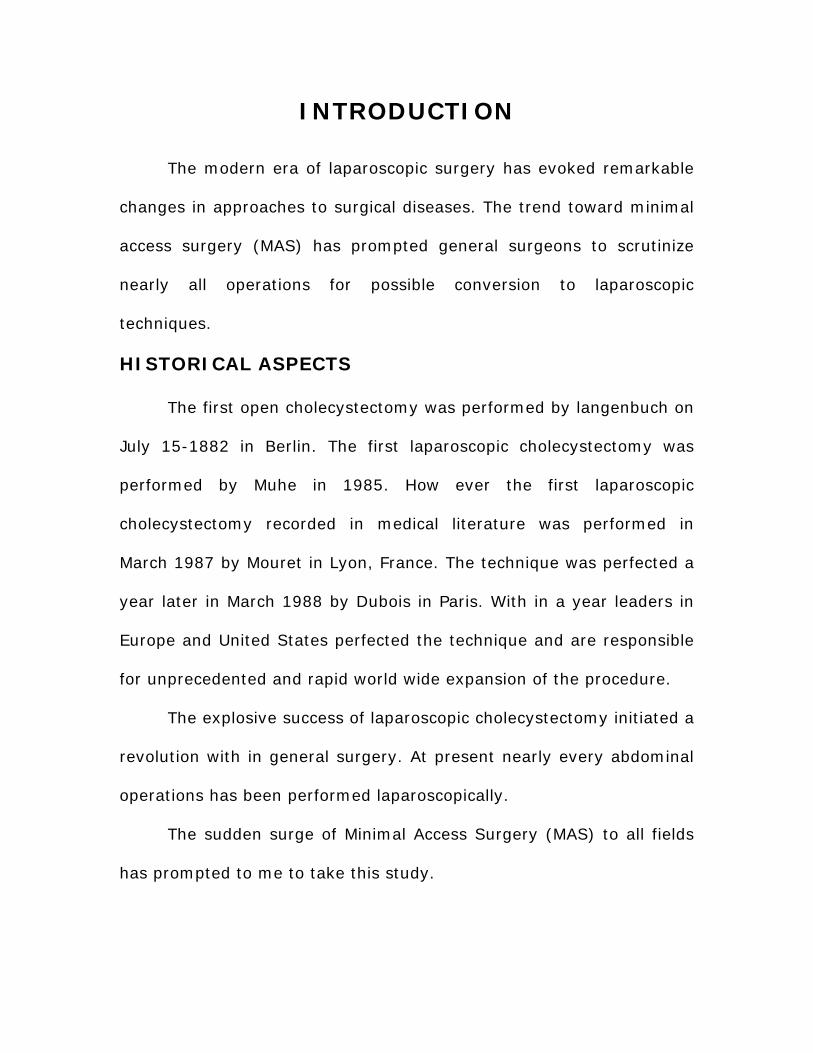
Anomlies of Gall bladder
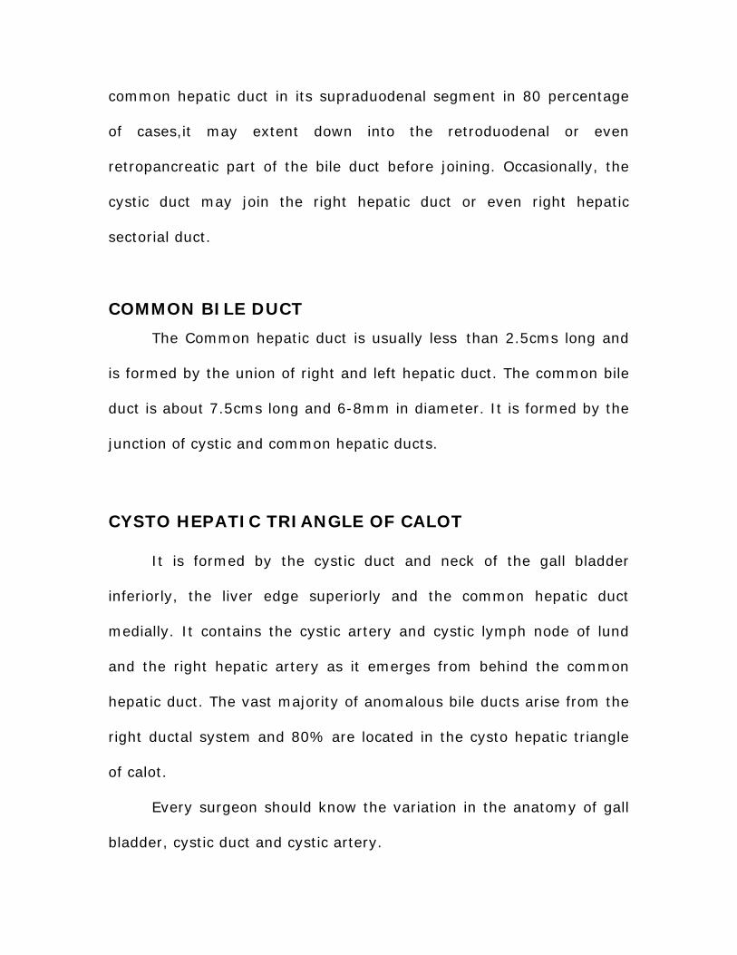
Anomlies of Cystic Duct

Anomalies of gall bladder
1. Absence of gall bladder
2. The Phrygian cap
3. Floating gall bladder
4. Double gall bladder
5. Septum of gall bladder
6. Diverticulam of gall bladder
Anomalies of cystic duct
1. Absence of cystic duct
2. Low insertion of cystic duct
3. An accessory of cholecystohepatic duct
4. Segment IV drainage into cystic duct
5. Drainage of right posterior sectorial duct (RP) into the neck
of gall bladder.
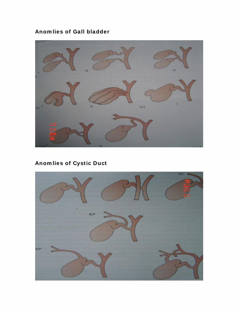
Anomalies of cystic artery
1. Cystic artery crossing in front of the common hepatic duct.
2. Low origin of cystic artery from common hepatic or
gastroduodenal arteries.
3. Accessory cystic artery arising from hepatic artery or
gastroduodenal arteries.
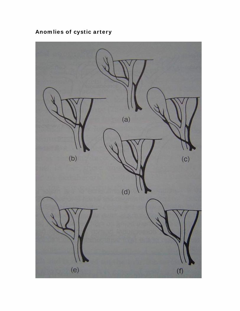
Anomlies of cystic artery
4. Tortuous right hepatic artery with a short cystic artery. This
most dangerous anomalie is called caterpillar turn or
Moyniham`s hump.
5. Right hepatic artery runs close to the cystic duct and neck of gall
bladder.
SPECTRUM OF GALLSTONE DISEASE
Gallstones are the most common billiary pathology. In UK, USA
and Australia, the prevalence rate varies from 15 to 25%. Male to
female ratio 1:2. In India the prevalence rate reported as 2% to 29%.
Seven times more common in the North India (stone belt) than in
South India. Male to female ratio 1:6.4, Mixed stones are more
common in India.3
Gallstones can be classified in various ways.4, 5, 6, 7
1. Present accepted classification:
Cholesterol stones, Black pigment & Brown pigment
stones.
2. Based on chemical composition:
Cholestrol stones, pigment stones, mixed stones.
3. Aschoff classification:
Inflammatory, metabolic, static& mixed stones.
CLINICAL SYNDROMES OF GALLSTONES DISEASE
a. In the gall bladder
1. Silent stones
2. Chornic cholecystitis
3. Acute billiary colic / acute cholecystitis
4. Gangrene
5. Perforation
6. Empyema
7. Mucocele
8. Carcinoma
b. In the bile ducts
1. Obstructive jaundice
2. Cholangitis / septicaemia
3. Acute gallstone pancreatitis
4. Billary fistulous disease
c. In the intestine
1. Gallstone illeus
The current consensus of surgical opinion is that here is no
indication for cholecystectomy in the management of patients
with asymptomatic gallstone disease except in the following
cases.5
i. Diabetic patients.
ii. Calcified gallbladder.
iii. Patients undergoing surgery for other
conditions & if patients general condition is
good.
iv. Acromegalic patients on long term treatment
with somatostatin analogues.
In our study the following group of patients are taken & compared
1. Chronic calculous cholecystitis
2. Cholelithiasis
3. Billiary colic
4. Acute cholecystitis
6,7 INDICATION FOR CHOLECYSTECTOMY
1. Acute Cholecystitis
2. Chornic Cholecystitis
3. Calculous Cholecystitis
4. Mucocele of gallbladder
5. Emphyema of gallbladder
6. Biliary colic
7. Polyp of gall bladder
8. Carcinoma of gallbladder
9. Perforation of gallbladder
10. Emphysematous Cholecystitis
11. Cholcysto enteric fistula
4,5,6,7 INVESTIGATION
1. Full blood count , hemoglobin & urine analysis
2. Blood sugar, blood urea
3. Serum creatinine
4. Liver function test:
Bilurubin Direct
Bilurubin Indirect
Alkaline phosphate
Aspartate Transaminase
Alanine Transaminase
Gamma –Glutamyl Transpeptidase
Prothrombin Time
Albumin
Urine Bile Salts and Bile Pigment, Urobilinogen
5. Plain Radiography
Radio opaque gall stones in 10% of patients.
Porcelain gall bladder –calcification of gall bladder.
6. Ultrasonography
Non- invasive
Now the standard initial imaging technique for the
investigation of the patient suspected of having a gall
stone and is also the prime investigation for the patient
presenting with jaundice.
7. Radio isotope scanning
99mTcHIDA, PIPIDA
They are excreted in the bile and are used to
visualize the billiary tree. In acute cholecystitis the gall
bladder is not seen. The technique is used when billiary
enteric anastamoses are functioning inadequately as it will
show the extent of obstruction at the anastamoses and
indicate the delay in excretion.
8. Computerized tomography
Useful in malignancy
9. Magnetic Resonance Cholangio Pancreatography (MRCP)
MRCP is the standard technique for the investigation of the
billiary tree.
10. Endoscopic Retrograde Cholangio-Pancreatography (ERCP)
Diagnostic & Therapeutic.
11. Percutaneus Transhepatic Cholangiography(PTC)
Meglumine iothalamate 60%
Chiba or okuda needle 15cm long 0.7mm diameter
In addition to diagnostic purpose this technique
enables placement of a catheter into the bile ducts to
provide external biliary drainage or the insertion of
indewelling stents. Fine flexible choledochoscope can also be
passed through the tract to diagnose strictures, take biopsy
and remove stones.
Routinely above first six investigations are performed in all
patients.
OPEN CHOLECYSTECTOMY
INDICATION:
1. Severe Acute Cholecystitis
2. Emphyema of Gallbladder
3. Gallbladder perforation
4. Chole Cysto Enteritic Fistulae
5. Carcinoma Gallbladder
6. Conversion from Laparoscopic Procedure
7. Emphysematous Cholecystitis
8. Severe COPD with Cholecystitis
9. Cirrhosis with Cholecystitis
10. Portal Hypertension with Cholecystitis
11. Previous Upper Abdominal Surgery with Cholecystitis
12. Pregnancy with Cholecystitis

PROCEDURE: 8, 9, 10
Anasthesia:
1. General Anaesthesia
Incision:
1. Kocher’s sub costal
2. Right paramedian
3. Mayo – Robson(hockey stick)
4. Right upper quadrant transverse
5. Upper midline
6. Mini laparotomy
Steps:
The first step consists in careful packing off. The first pack is
placed in the lower part of the wound –displacing duodenum,
transverse colon & small intestine downwards. A second pack is placed
medially to cover and retract the stomach. A third pack may be
inserted laterally to fill the right kidney pouch.
The Retrograde Method (Duct First Method):
A forceps is applied to the infundibulum of the gallbladder and is
used to draw the viscus gently forward and to the right. The junction
of the cystic and common ducts is now displayed by snipping the
overlying peritoneum and by gauze stripping. An absorbable ligature is
now placed loosely around the cystic duct close to the junction with
the common duct. Gentle traction on the cystic duct and careful sharp
and gauze dissection keeping close to the upper part of the cystic
artery. It should be doubly ligated with silk and divided.
The gallbladder is separated from the liver bed by dividing the
peritoneal reflection.
Fundus First Method:
This method is advised only when difficulties (particularly severe
inflammatory change) prevent the ducts being displayed in the first
steps of operation. So separation of gallbladder is commenced at the
fundus then the cystic duct and artery are ligated. Wound closed with
or without drains.
COMPLICATION
1. Wound infection
2. Intra abdominal abcess
3. Illeus
4. Haemorrage
5. Accumulation of bile
6. Injury to common bile duct and late stricture
7. Injury to right hepatic artery

8. Post cholecystectomy syndrome
9. Venus thrombosis and embolism
10. Portal pyaemia
11. Biliary fistula
12. Adhesive intestinal obstruction
13. Pulmonary complications

LAPAROSCOPIC SURGERY BASICS
SPECTRUM OF LAPROSCOPIC OPERATIONS 11
GROUP I:
Operations where the laparoscopic approach provides an
undoubted benefit and has replaced open intervention
cholecystectomy, cardiomyopathy, nerve sections, antireflux surgery,
splenectomy, adrenelectomy for nonmalignant tumours and operation
for varicocele.
GROUP II:
An operation where the laparoscopic approach appears to be
beneficial and safe by more information is needed.
Hernia repair, appendicectomy, adhesiolysis, surgical treatment
of duct calculi, segmental colonic resection for diverticular disease or
sessilepolpys, rectopexy, enucleation of insulinomas, nephrectomy for
benign disease, distal pancreatic resections, oesophagectomy for
cancer.
GROUP III:
Operations are currently under evaluation and should not be
attempted outside clinical trials.Resection for potentially curable
invasive cancer.
GROUP IV:
Unsuitable operations no benefit, increased risk. Pancreatico
duodenectomy, D2 resection for carcinoma stomach.
EQUIPMENT: 11, 12, 13
Image system:
Light sources and light cables
Xenon
metal handle (halogen)
glass fiber bundle
fluid light cables
Telescopes:
Rigid viewing type based on Hopkins rod-lens system.
10 mm with 0, 30, 45 degree viewing telescopes with
insufflating port and an instrumental channel.
optoelectronic telescopes with CCD.
CCD (charged couple device cameras).

Single chip
Three chip
Image display system
One or two monitors
Devices:
Automatic insufflators
CO2 and nitrous oxide
Pneumo peritoneum pressure 10-12 mm hg
4 to 12 l/min flow rate
Instrumentation:
Veress needle
To create pneumo –peritonium by closed method
Hassan canula
To create pneumoperitonium by open method
Access ports
Reusable or disposable
Grasping forceps
Angled dissecting forceps
Scissors

Clip appliers
Extraction forceps
Staplers
Suction irrigator
Needle holders
Energy sources:
Endocoagulators
Diathermy units
Unipolar
Bipolar
Lasers
Harmonic scalpel
Ultrasonic dissecting instrument

LAPROSCOPIC CHOLECYSTECTOMY
INDICATIONS AND CONTRAINDICATIONS
INDICATIONS FOR LAPROSCOPIC
CHOLECYSTECTOMY: 12, 13
1. Symptomatic Cholelithiasis
Biliary colic
Acute cholecystitis
2. Asymptomatic Cholelithiasis
Sickle cell disease
Total parentral nutrition
Chornic immuno suppression
Acromegalic patient on stomatostatin treatment
Calcified gall bladder
Diabetic patient with gall stones
Candidates for renal transplant
Incidental with other procedures.
3. Acalculous Cholecystitis
4. Gall Stone Pancreatitis
5. Gall bladder polpys
CONTRAINDICATION FOR LAPROSCOPIC
CHOLECYSTECTOMY: 12, 13
Absolute Contraindications
1. Patients unfit for general anaesthesia
2. Significant portal hypertension
3. Gallbladder carcinoma
Relative Contraindications
1. Multiple prior operations
2. Acute severe cholecystitis and peritonitis
3. Cirrhotic liver
4. Extensive scarring in calots triangle
5. Acute pancreatitis
6. Abnormal anatomy
7. Pregnancy
8. Morbid obesity
9. Evidence of generalized peritonitis
10. Septic shock from cholangitis.
Pregnancy Should no longer be considered as a contra indication to
Lap Cholecystectomy. All Pregnant patients requiring cholecystectomy
in 2nd & 3rd trimesters should be offered the advantages of the Lap
Cholecystectomy.14

LAPAROSCOPIC CHOLECYSTECTOMY
OPERATION ROOM SETUP
OPERATIVE TECHNIQUE 12, 13, 15
Preparation and Anaesthesia
ETGA
Ryles Tube Aspiration
Bladder Catheterization
Thrombo Embolic Prophylaxis
Positioning
North American approach, team surgeon and camera assistant
stand on the left side of the patient.
First assistant with staff nurse on the right side of the patient.
Monitor and instrument trolley on the right side of the patient.
Ports
First Port - Umblical 10 mm, camera port.
Second Port - Epigastric 10 mm, working port.
Third Port - Right subcostal port at midclavicular line 5 mm.
Fourth Port - Right lateral port at anterior axillary line 5 mm for
retraction of gall bladder.
In closed method by using verees needle, a small incision made
at the umblicus since it is thinnest portion of abdominal wall and
allows easy access. Confirmation of the intraperitoneal location of the
needle tip is made by the saline drop test.
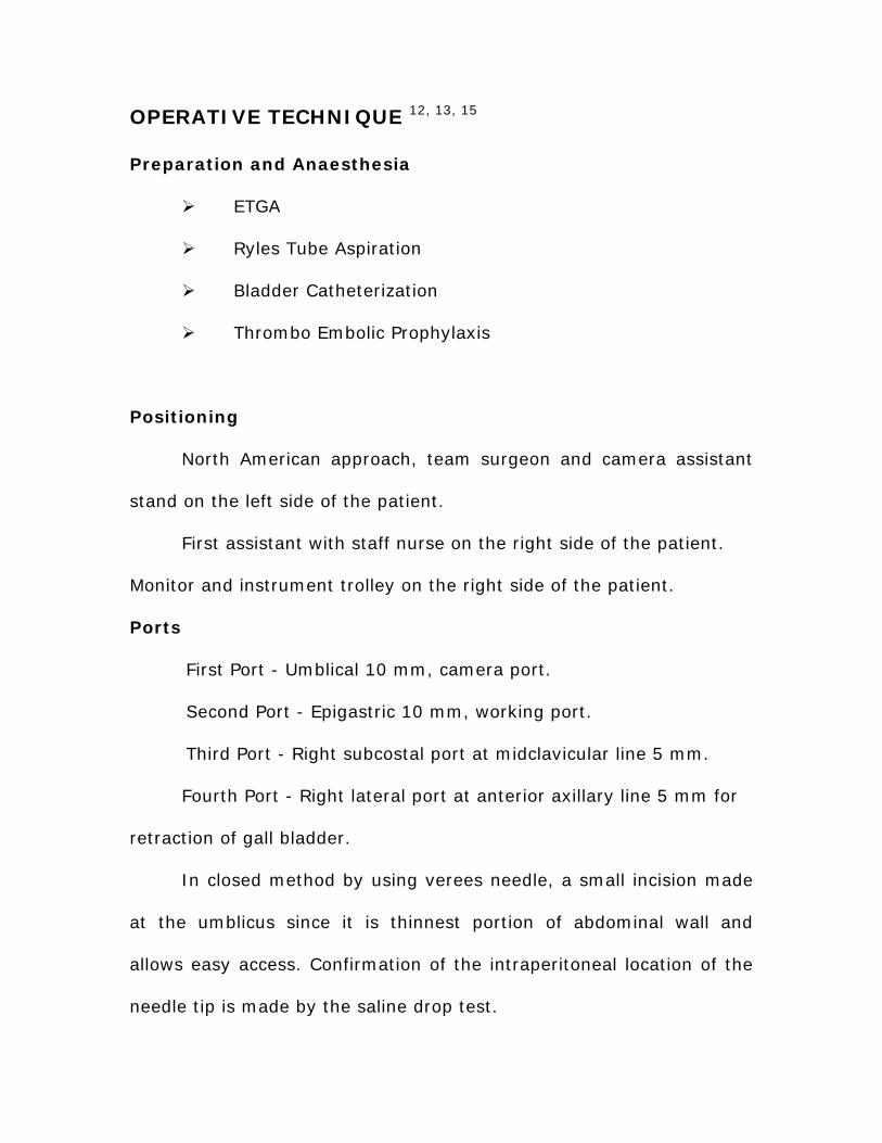
A -> Cranial retraction of Gallbladder. B -> Omental Adhesions.
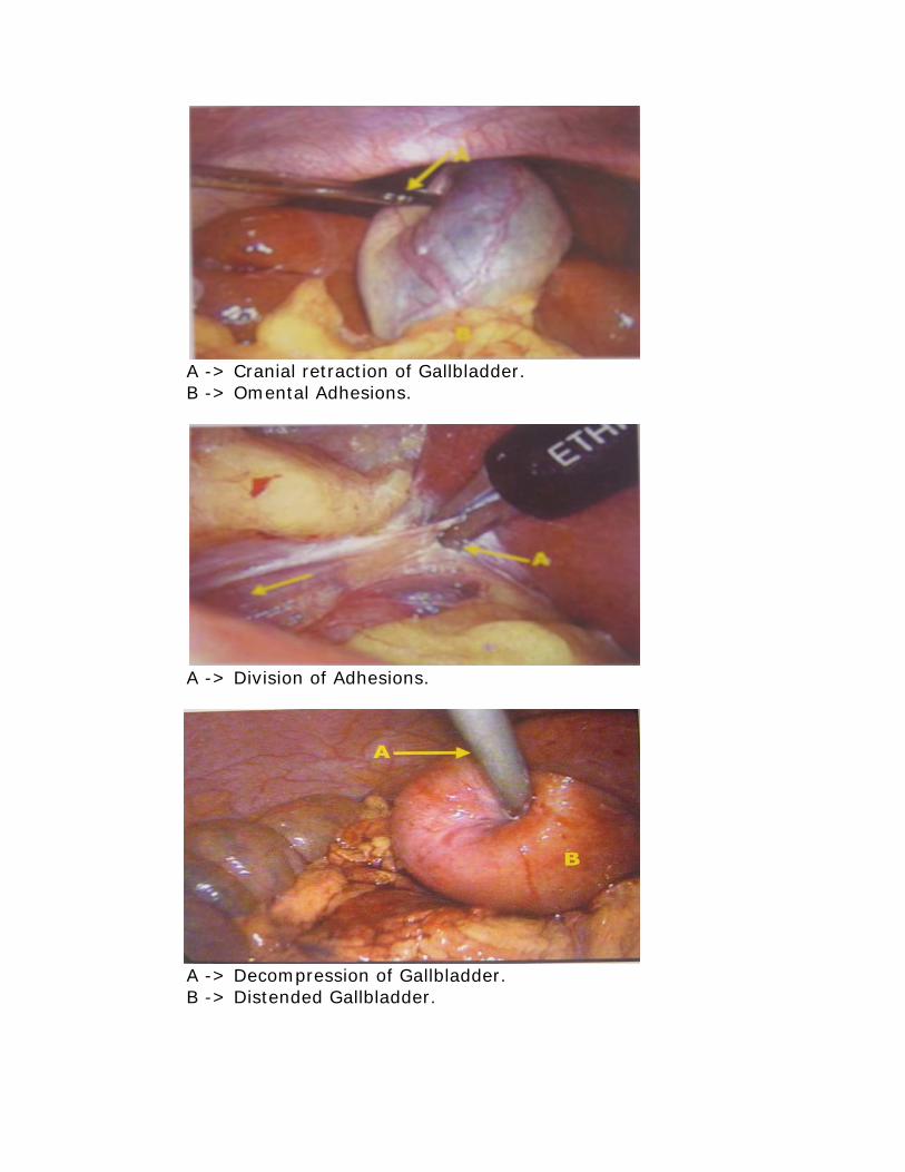
A -> Division of Adhesions.
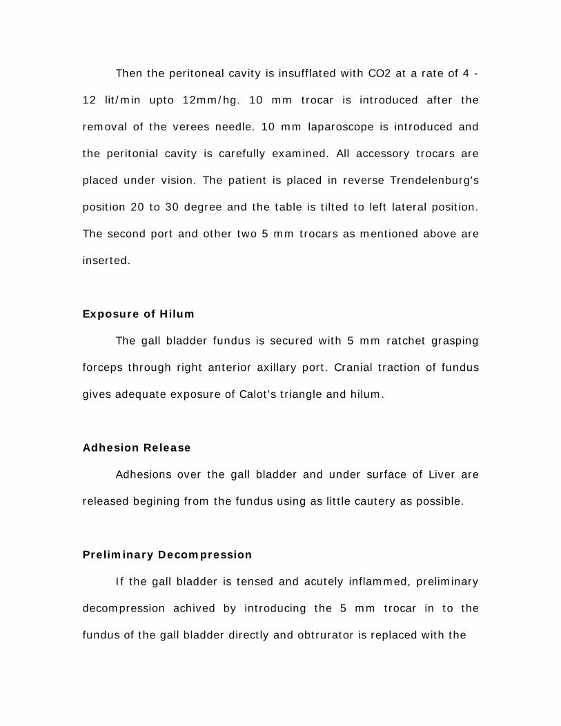
A -> Decompression of Gallbladder. B -> Distended Gallbladder.
Then the peritoneal cavity is insufflated with CO2 at a rate of 4 -
12 lit/min upto 12mm/hg. 10 mm trocar is introduced after the
removal of the verees needle. 10 mm laparoscope is introduced and
the peritonial cavity is carefully examined. All accessory trocars are
placed under vision. The patient is placed in reverse Trendelenburg's
position 20 to 30 degree and the table is tilted to left lateral position.
The second port and other two 5 mm trocars as mentioned above are
inserted.
Exposure of Hilum
The gall bladder fundus is secured with 5 mm ratchet grasping
forceps through right anterior axillary port. Cranial traction of fundus
gives adequate exposure of Calot's triangle and hilum.
Adhesion Release
Adhesions over the gall bladder and under surface of Liver are
released begining from the fundus using as little cautery as possible.
Preliminary Decompression
If the gall bladder is tensed and acutely inflammed, preliminary
decompression achived by introducing the 5 mm trocar in to the
fundus of the gall bladder directly and obtrurator is replaced with the

A -> Exposure of posterior side of Calot’s Triangle. B -> Posterior dissection of Calot’s Triangle.
A -> Exposure of anterior side of Calot’s Triangle. B -> Anterior dissection of Calot’s Triangle.
A -> Lateral traction of Infundibulum. B -> Cystic Duct. C -> Cystic Artery.
suction cannula through the trocar and complete suction of the
contents done. Then the trocar is removed from the toothed grasper is
placed on the wound of the gall bladder to hold it closed during the
cranial traction.
Calot’s Triangle Dissection
An atraumatic grasper is placed through right mid-calavicular
port on the Hartmann's pouch. The infundibular grasper is retracted
laterally to expose the anterior aspect of the calot's triangle and the
peritonium is peeled of then infundibular grasper is retracted
anteromedially to expose the posterior aspect of calot's triangle. The
junction of the cystic duct with the gall bladder (safety zone) is
identified by moving the infundibular grasper backward, forward and
side to side.
Skeletonisation of Cystic Pedicle
This is done using a curved dissector with the following
precautions
Monopolar electrocautery is commonly used.
Energy source is not used in close proximity to CBD.
Minumum electrocautery is used to keep adequate
haemostasis.
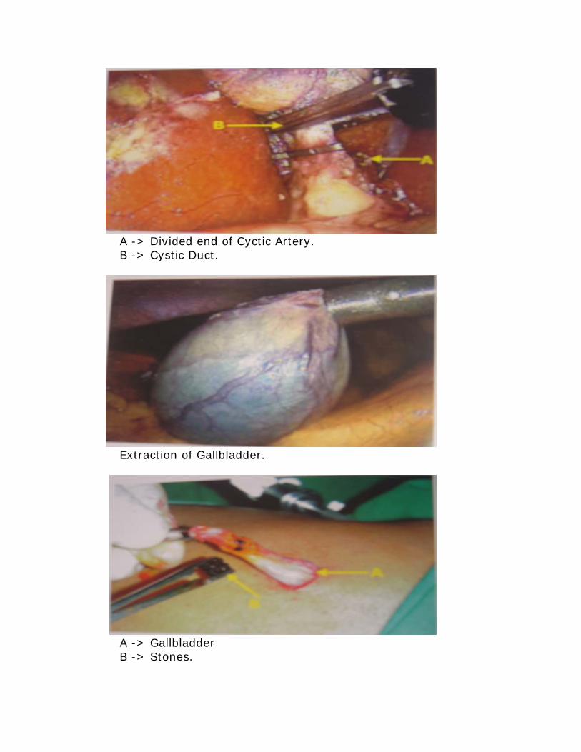
A -> Divided end of Cyctic Artery. B -> Cystic Duct.
Extraction of Gallbladder.
A -> Gallbladder B -> Stones.

It is not always necessary to identify the cystic and
common bile duct junction.
No arterial structure is assumed to be cystic until it is
clearly and unequivocally, shown to pass directly to gall
bladder.
Clipping and Division of Cystic Pedicle
Both the cystic duct and artery are clipped, two on the cystic
stump side one each close to gall bladder. Blind application of clips in
the calot's triangle should be avoided. In some selected situations
ducts needs to be divided first to expose the cystic artery. Care is
taken in such circumstances not to give excessive traction till the
cystic artery is divided.
Dissection of Gallbladder from its bed
Traction and counter traction with right lateral and left medial
twist facilitates the dissection. During separation of gall bladder, fundal
traction is gradually released down. Prior to complete detachment of
gall bladder liver bed is reinspected for adequate haemostasis or bile
leakage. Once achieved gall bladder is completely detached and
extracted.
Extraction of Gallbladder
Extraction of gallbladder is done through epigastric port.
Irrigation of the epigastric port is done in patients with infected
gallbladder to prevent port tract infection. A ready vac tube drain or
ryles tube drain is kept through lateral axillary port and placed in the
sub hepatic space.
The trocars are removed under direct visual control. The ports
are closed with vicryl stitches. All the trocar sites are injected with
bubivacaine for post operative pain relief.
COMPLICATION OF LAPRASCOPIC CHOLECYSTECTOMY
Intraoperative Complication
Haemorrhage – bleeding can occur in various ways.
At the site of trocar insertion.
During adhesion release from omental vessels.
During dissection in the calot’s triangle from cystic
artery and hepatic artery.
From the gall bladder fossa.
Perforation of gall bladder and contamination of peritoneal
cavity with potentially infected bile and gall stones.
Carcinoma of gall bladder must be recognized
preoperatively with a high index of suspicion and if
suspected consider for conversion to open procedure

Bile duct injury.
Postoperative Complications
Biliary leakage/ biliary fistulae.
Biliary peritonitis.
Biliary strictures.
Diathermy induced thermal injuries to the surrounding
structures can occur.
Omentum or bowel can herniated through the umbilical
port site. This can be avoided by suturing the linea alba
securely in all cases.
Port site metastasis can occur if carcinoma gallbladder is
not suspected preoperatively.

RECENT ADVANCES IN LAPARPSCOPIC
SURGERY
Three dimensional Video Systems.
Image Display System that project image onto a sterile
screen overlying the chest of the patient.
Abdominal Wall Lifting Systems.
Rubber Tube Slings Abdominal Wall Lift.
Planar Intraperitonial Abdominal Wall Retraction Lift.
Devices.
Harmonic Scalpel Curved Blade.
Extra Peritonial Abdominal Wall Lift Devices.
Robots and Master Slave Manipulators.
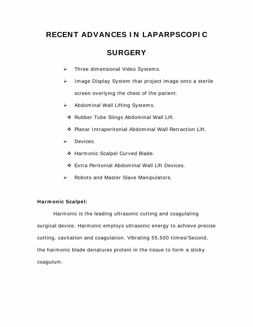
Harmonic Scalpel:
Harmonic is the leading ultrasonic cutting and coagulating
surgical device. Harmonic employs ultrasonic energy to achieve precise
cutting, cavitation and coagulation. Vibrating 55,500 tiimes/Second,
the harmonic blade denatures protein in the tissue to form a sticky
coagulum.
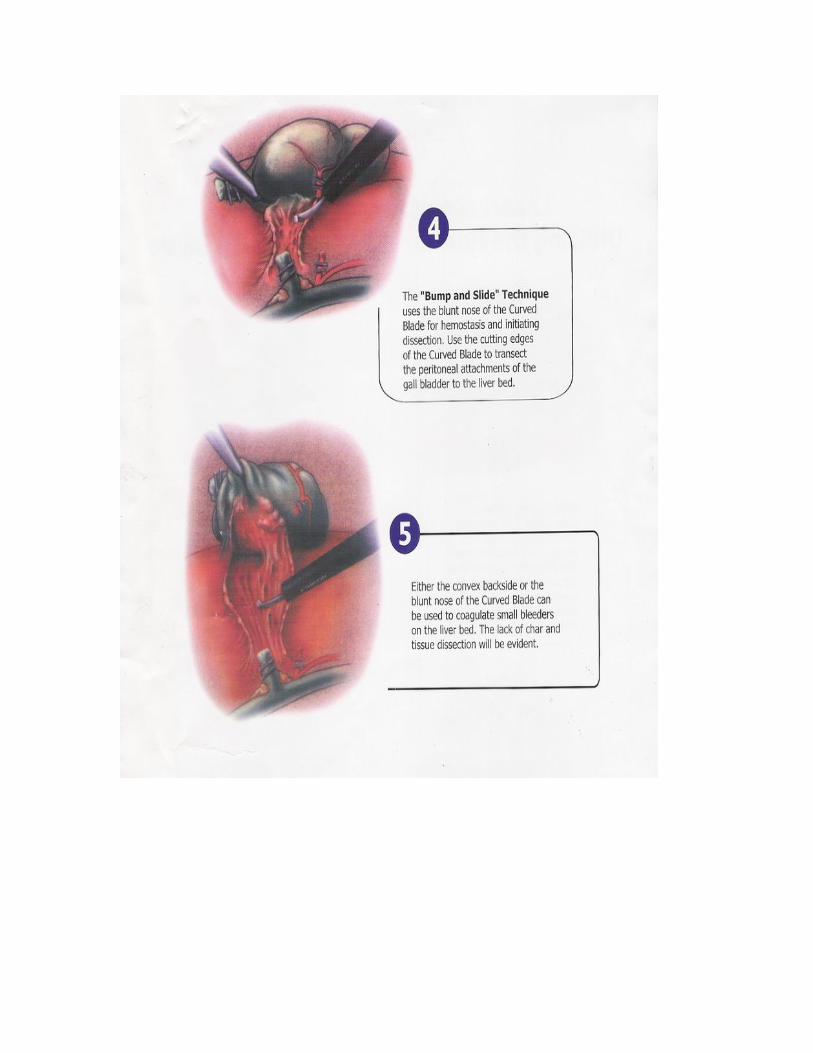

Some important benefits as follows.
No Electrical current to or through the patient.
Minimal lateral thermal damage.
Greater precision near vital structures.
Seals blood vessels up to 5 mm diameter.
Cavitational effect aids in tissue plane dissection.
MATERIALS AND METHODS
Case Selection
In our CMCH we are doing both open and laparoscopic
cholecystectomy. This study is done between January 2004 to
February 2006. In this period I have selected 25 cases of laparoscopic
cholecystectomy to compare with 25 cases of open cholecystectomy.
Common indications for surgery were chronic calculous cholecystitis,
acalculous cholecystitis, cholelithiasis, biliary colic and acute
cholecystitis.
The following factors are compared in laparoscopic and open
cholecystectomy
Technique of surgery
Duration of surgery
Post operative pain
Analgesic requirements
Duration of antibiotics given
Intra operative and post-op Complications
Resumption of normal diet
Post operative hospital stay
Return to normal activity
Cosmesis
CONVERSION TO OPEN METHOD
Procedure was converted to open method in two cases out of 25
patients due to the following reasons.
1. In one case there were plenty of thick adhesions between
gallbladder and surrounding structures particularly duodenum.
2. In another case there was excessive fat in the calot’s
triangle and cystic pedicle could not be identified.
Indication for Conversion16
1. Inability to identify anatomy.
2. Adhesions.
3. Severe inflammation.
4. Bleeding.
5. Spillage of stones.
6. Impacted cystic duct Stone.
7. Mass near Gall bladder.
8. Injury to Stomach.

CONVERSION RATE
Conversion Rate - Lap to Open [n=25]
Lap92%
Conversion to Open
8%
Lap
Conversion toOpen
Conversion Rate (%) 8%

DURATION OF SURGERY
0
20
40
60
80
100
120
140
TYPE OF SURGERY
TIM
E I
N M
INU
TE
S
OPENLAP
Average operating time for Open – 90 min
Average operating Time for Lap – 120 min
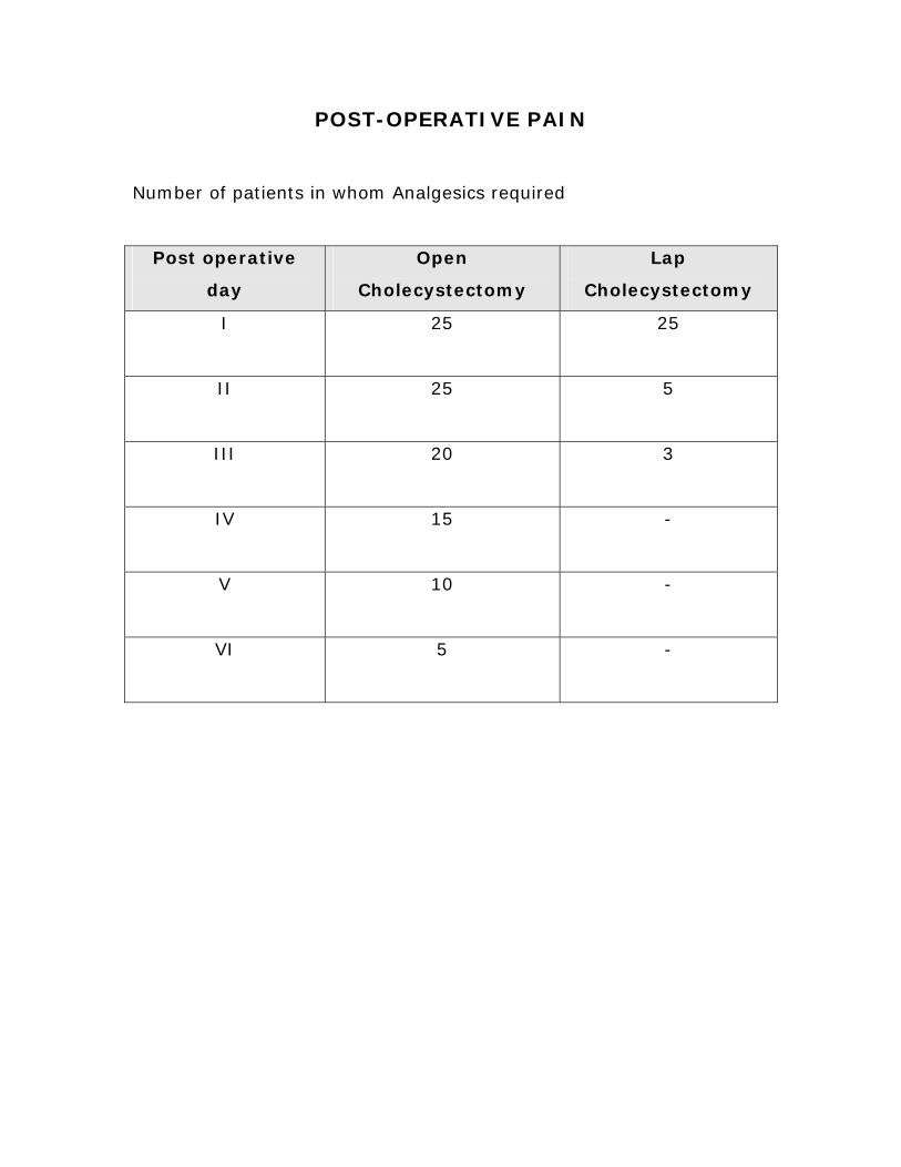
POST-OPERATIVE PAIN
Number of patients in whom Analgesics required
Post operative
day
Open
Cholecystectomy
Lap
Cholecystectomy
I 25 25
II 25 5
III 20 3
IV 15 -
V 10 -
VI 5 -

DURATION OF ANTIBIOTICS GIVEN
0123456789
10
POST-OP DAYS
TYPES OF SURGERY
OpenLap
Average Post op antibiotics given for Open Method – 7 Days
Average Post op antibiotics given for Lap Method – 4 Days.

INTRA OPERATIVE COMPLICATIONS
Complications Open Laparoscopic
Bleeding 2 1
Bile duct injury Nil Nil
Bowel injury Nil Nil
Others Nil Nil
POST OPERATIVE COMPLICATION
Complications Open Laparoscopic
Bleeding Nil Nil
Bile leak through drainage 2 1
Wound Infection 3 Nil
Jaundice Nil 1
Post cholecystectomy
syndrome
1 Nil
Pulmonary complications Nil Nil

RESUMPTION OF NORMAL DIET
POST OPERATIVE DAY
5
3
OpenLap
Average Post op resumption of normal diet for Open – 5 Days
Average Post op resumption of normal diet for Lap – 3 Days
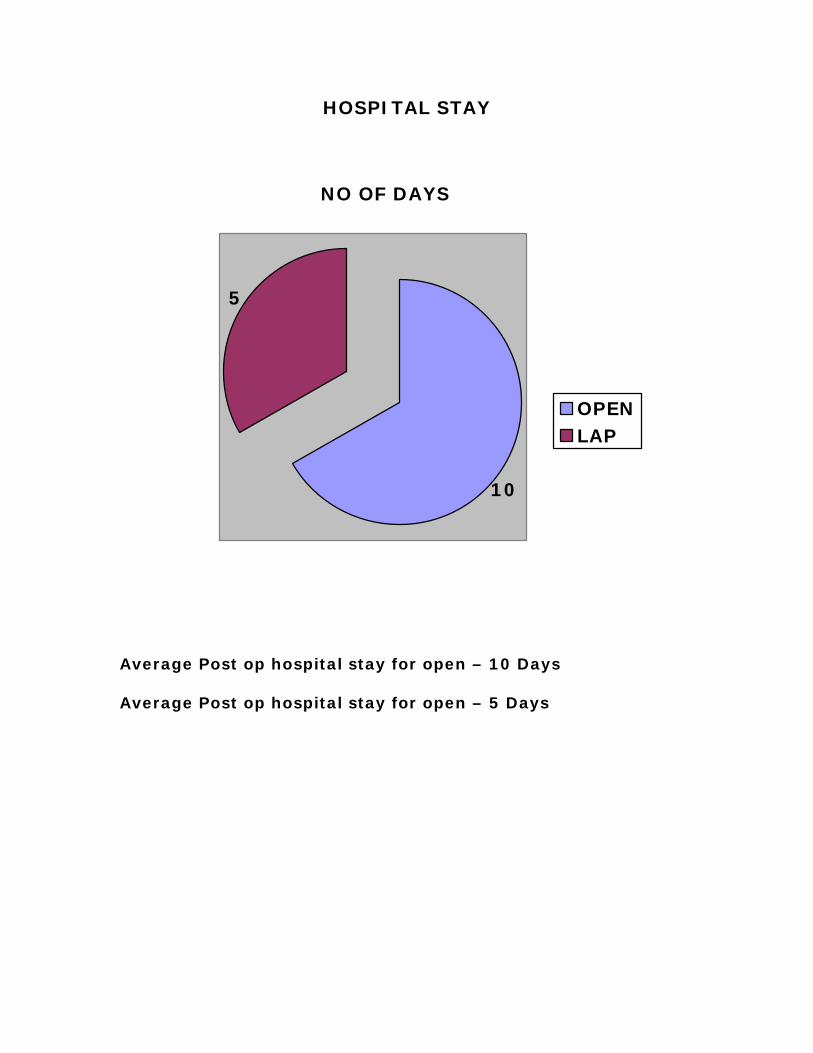
HOSPITAL STAY
NO OF DAYS
5
10
OPENLAP
Average Post op hospital stay for open – 10 Days
Average Post op hospital stay for open – 5 Days
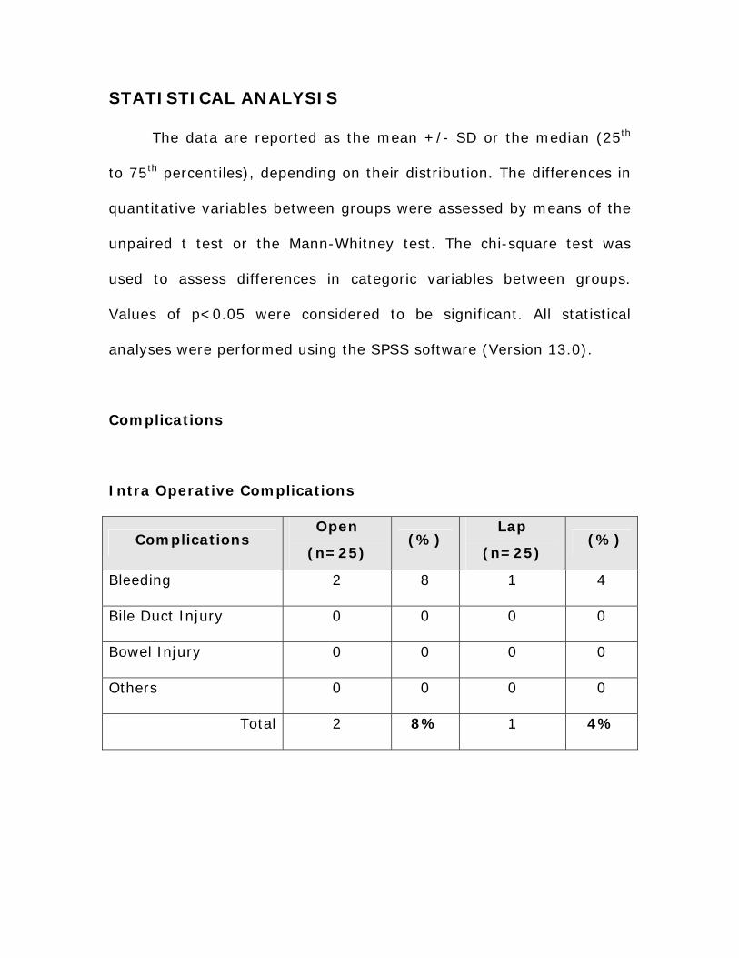
STATISTICAL ANALYSIS
The data are reported as the mean +/- SD or the median (25th
to 75th percentiles), depending on their distribution. The differences in
quantitative variables between groups were assessed by means of the
unpaired t test or the Mann-Whitney test. The chi-square test was
used to assess differences in categoric variables between groups.
Values of p<0.05 were considered to be significant. All statistical
analyses were performed using the SPSS software (Version 13.0).
Complications
Intra Operative Complications
Complications Open
(n=25) (%)
Lap
(n=25) (%)
Bleeding 2 8 1 4
Bile Duct Injury 0 0 0 0
Bowel Injury 0 0 0 0
Others 0 0 0 0
Total 2 8% 1 4%

Post Operative Complications
Complications Open (n=25) (%) Lap (n=25) (%)
Bleeding 0 0 0 0
Bile leak through drain 2 8 1 4
Wound Infection 3 12 0 0
Jaundice 0 0 1 4
Post cholecystectomy
syndrome 1 4 0 0
Pulmonary
complications 0 0 0 0
Total 6 24% 2 8%
Chi-Square Test
Complications
[n=50]
Open
cholecystectomy
Laparoscopic
cholecystectomy Total
2 1 3 Intra Operative
Post Operative 6 2 8
Total 8 [16%] 3 [6%] 11
P=0.023 significance between the
variables
chi-dist – 0.7822
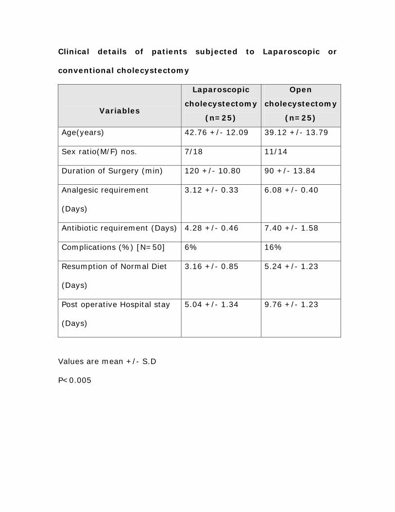
Clinical details of patients subjected to Laparoscopic or
conventional cholecystectomy
Variables
Laparoscopic
cholecystectomy
(n=25)
Open
cholecystectomy
(n=25)
Age(years) 42.76 +/- 12.09 39.12 +/- 13.79
Sex ratio(M/F) nos. 7/18 11/14
Duration of Surgery (min) 120 +/- 10.80 90 +/- 13.84
Analgesic requirement
(Days)
3.12 +/- 0.33 6.08 +/- 0.40
Antibiotic requirement (Days) 4.28 +/- 0.46 7.40 +/- 1.58
Complications (%) [N=50] 6% 16%
Resumption of Normal Diet
(Days)
3.16 +/- 0.85 5.24 +/- 1.23
Post operative Hospital stay
(Days)
5.04 +/- 1.34 9.76 +/- 1.23
Values are mean +/- S.D
P<0.005
DISCUSSION OF OUR STUDY
In our study I have selected cases for surgery based on
preoperative history, clinical examination, ultrasonography and liver
function test. We exclude the common bile duct stones by clinical
signs, LFT and ultrasonography.
A study of 25 open cholecystectomy patients of which 18 female
and 7 male patients were compared with that of 25 cases of
laparoscopic cholecystectomy of which 14 female and 11 male
patients.
The relative advantages and disadvantages of laparoscopic and
open surgery are measured primarily in terms of quality of life for the
patients involved. The study revealed the following findings.
By technique wise laparoscopic surgery provides
better visualization with magnification of surgical anatomy
in contrast to the open surgery.
Among the 25 laparoscopic cholecystectomies, two
cases were converted to open cholecystectomy due to
adhesions and inability to identify anatomy. Conversion
rate was 8%.
The mean operative time for laparoscopic
cholecystectomy is 120 minutes which is 30 minutes
longer than conventional open method (90 min).

Regarding post operative morbidity in terms of pain,
recovery from surgery and ambulance from bed the
laparoscopic patients faired better from open surgery.
Traditional major open abdominal operations have
potent effects on the immune system. Surgical trauma
induces an inflammatory state characterized by the release
of proinflammatory cytokines IL-1B, IL-6, IL-8, TNFalpha
and acute phase proteins such as C-reactive protein are
typically transiently increased. Surgical manipulation also
depresses cell mediated immunity by alteration in
recruitment, activation and function of circulating
lymphocytes, monocytes and other immune cells. After
open cholecystectomy, higher post operative plasma levels
of CRP, TNFalpha, IL-1B, IL-6 and higher leukocyte counts
relative to laparoscopic cholecystectomy.17 This was the
probable reasons for early recovery, less pain and early
ambulance in laparoscopic cholecystectomy patients.
Regarding analgesic requirement the open surgery
patients required analgesics even on the sixth post
operative day. While the laparoscopic patients didn’t
experienced pain in the immediate post operative period
because of less acute phase reactions and port site

infiltration of bupivacine and no patients required
analgesics on the fourth post operative day.
The mean duration of antibiotics given for open
cholecystectomy patients were around 7 days while for
laparoscopic patients it was only 4 days.
Regarding intra operative complications bleeding has
occurred in two open cholecystectomy and one open
laparoscopic cholecystectomy patients. Bile duct injury was
nil in both open and lap cholecystectomy. Regarding post
operative complication bile leak through drain has occurred
in two open and one lap patients. All the three patients
were treated conservatively and subsided, probably reason
due to bile leak from the gall bladder bed in the liver. Out
of 25 cases of open cholecystectomy 3 cases had got
wound infection, but it was nil in lap cholecystectomy.
Transient post op jaundice was developed in one lap case.
Persistent pain and dyspepsia after cholecystectomy (post
cholecystectomy syndrome) occurred in one open
cholecystectomy patient. Long term pain less common
after laparoscopic than open cholecystectomy.18 In our
study both groups patients there were no pulmonary
complications. But other studies revealed impairment in
pulmonary function after lap cholecystectomy was less
marked than after open cholecystectomy.19 The overall
complication rate for open method was 16% and for lap
only 6%.
The patients operated by conventional open method
resumed to normal diet only on 5th post operative day,
while those done by lap method resumed to normal diet
even on the 3rd post operative day.
Regarding post operative study in the hospital, for
open method patients it was totally 10 days after surgery,
while for lap patients it was only 5 days. The early
ambulance and even return to normal activity was quick
after lap method, so cost effective.20
Cosmesis is the greatest advantage after lap
cholecystectomy compared to open method.
REVIEW OF LITERATURE
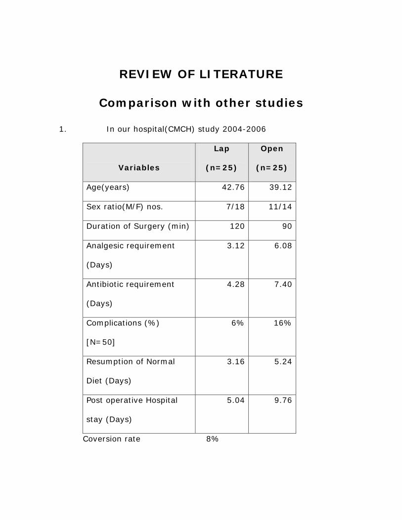
Comparison with other studies
1. In our hospital(CMCH) study 2004-2006
Variables
Lap
(n=25)
Open
(n=25)
Age(years) 42.76 39.12
Sex ratio(M/F) nos. 7/18 11/14
Duration of Surgery (min) 120 90
Analgesic requirement
(Days)
3.12 6.08
Antibiotic requirement
(Days)
4.28 7.40
Complications (%)
[N=50]
6% 16%
Resumption of Normal
Diet (Days)
3.16 5.24
Post operative Hospital
stay (Days)
5.04 9.76
Coversion rate 8%

2. U.Berggren et al. 1994 21
Variables Lap Open
Age(years) 41.4 42.8
Sex ratio(M:F) 5:10 4:8
Operating Time (min) 87 69.2
Hospital stay (days) 1.8 2.8
Sick Leave (days) 11.7 24
3. M.Johansson et al. 2005 22
Variables Lap Open
Age(years) 53 56
Sex ratio(M:F) 19:16 16:19
Operating Time (min) 90 80
Conversion rate (%) 23 -
Hospital stay (days) 2 2
Sick Leave (days) 11 14

4. P.Helligso et al. 1994 23
Variables Lap
Operating Time(min) 110
Conversion rate(%) 2.8%
Intra operative
complications (%)
0.9%
Post op complications (%) 7.1%
Hospital stay (days) 3.5
Time of recovery (days) 12.5
5. Prospective randomized trials of open versus laparoscopic
cholecystectomy 24, 25
Author No of
Pts
Operating
Time
(min)
Complicati
ons
(%)
Length
of Stays
(Days)
Return
to work
(Days)
Berggrenetal
1994
OC
LC
12
15
69
87
-
-
3
2
24
12
McMahonetal
1994
OC(minilap)
148
57
20
4
-
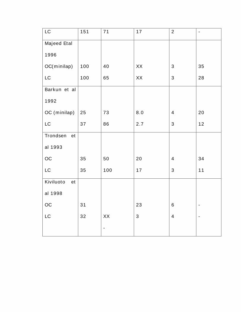
LC 151 71 17 2 -
Majeed Etal
1996
OC(minilap)
LC
100
100
40
65
XX
XX
3
3
35
28
Barkun et al
1992
OC (minilap)
LC
25
37
73
86
8.0
2.7
4
3
20
12
Trondsen et
al 1993
OC
LC
35
35
50
100
20
17
4
3
34
11
Kiviluoto et
al 1998
OC
LC
31
32
XX
-
23
3
6
4
-
-

6. Results of large series Laparoscopic Cholecystectomy
Study
No
of
Pts
Conversions
(%)
Mortality
(%)
Complic
ations
(%)
Bile duct
injuries
(%)
Fullarton et al
(1994)
1683 17.0 0.50 5.9 0.7
Newman et al
(1995)
1525 2.2 0.26 4.1 0.0
Southern
Surgeons Club
(Meyers 1991)
1518 4.7 0.07 1.5 0.5
Cuschieri et al
(1991)
1236 3.6 0.00 1.6 0.3
Brune et al
(1994)
800 1.2 0.00 2.8 0.2
Perissat et al
(1992)
777 5.5 0.10 3.3 0.4
Jatzko et al
(1995)
740 5.4 0.14 1.9 -
Cappucino et al
(1994)
563 4.8 0.00 6.9 0.3
Soper et al
(1998)
1200 2.1 0.10 2.7 0.2

CONCLUSION
In our study the laparoscopic cholecystectomy surpasses the
open cholecystectomy by the followings:
Better visualization and magnification of surgical anatomy.
Decreased post operative morbidity.
Shorter duration of analgesic requirements.
Shorter duration of antibiotic requirements.
Decreased wound infection.
Quicker ambulance, better compliance and rapid return to
normal activity.
Rapid resumption of normal diet.
Shorter post operative hospital stay.
Best cosmesis.
The only disadvantage is the prolonged operative time, which
can be minimized in due course of time as the learning curve
progresses.
We have also found that the conversion to open cholecystectomy
should be done in proper time with out any hesitation in case of
complications that could not be managed by laparoscopic surgery and
conversion in such case reflects sound judgment and should not be
considered as a complication.
BIBLIOGRAPHY
1. Lee McGregor’s Synopsis of Surgical Anatomy, 12th Edition, page
78 to 102.
2. Keith L.Moore, Clinically Oriented Anatomy, 4th Edition, Page 272
to 277.
3. H.Mohan, R.P.S Punia, S.B Dhawan, S.Ahal, M.S.Sekhon,
Morphological Spectrum of gall stone disease in 1100
cholecystectomies in North India, Indian Jou. Surgery, June
2005, Volume 68, Page 140 to 142.
4. Bailey & Love’s Short Practice of Surgery, 24th Edition, Page
1094 to 1113.
5. Sir Alfred Cuschieri’s Essential Surgical Practice Higher Surgical
Training in General Surgery, 4th Edition, Page 375 to 452.
6. Sabiston’s Text book of Surgery,16th Edition, Page 1076 to 1111.
7. Schwartz’s Principles of Surgery, 8th Edition, Page 1187 to 1219.
8. Robert J.Baker & Josef E.Fischer, Mastery of Surgery, 4th Edition,
Page 1142 to 1163.
9. Farquharson’s Text book of Operative Surgery, 8th Edition, Page
421 to 440.
10. L.H.Blumgert & Y.Fong, Surgery of the Liver & Biliary Track, 3rd
Edition, Page 697 to 707.
11. Sir Alfred Cuschieri’s Essential Surgical Practice Basic Surgical
Training, 4th Edition, Page 493 to 520.
12. Palanivelu’s Text book of Surgical Laparoscopy 1st Edition, Page
121 to 188.
13. Alfred Cuschieri & George Berci’s Laparoscopic Biliary Surgery,
2nd Edition, Page 69 to 142.
14. K. Singh, A. Olivi, S. Juneja, Laparoscopic Cholecystectomy
during Pregnancy. Indian Jou. Surgery June 2005, Volume 68,
page 131 to 134
15. The Surgical Clinics of North America, Minimal Access Surgery,
Part I, August 2000.
16. Lap Converted to open Cholecystectomy minimally prolongs
hospitalization. The American Journal of Surgery, Dec 2005, Vol
190, Page 879 to 881.
17. Patricia Sylla, Irena Kirman, Richard L. Whelan, Immunological
advantages of advanced Laparoscopy. The Surgical Clinics of
North America, Feb 2005, Vol 85, Page 1 to 18.
18. G.Stiff, M.Rhodes, A.Kelly, K.Telford, C. P. Armstrong & B. I.
Rees, Long Term Pain, Less common after laparoscopic than
open cholecystectomy, British Jou. Surgery 1994, vol 81, Page
1368 to 1370.
19. Post Op Pulmonary functions in Lap Vs Open Cholecystectomy, a
prospective, Comparative study, Indian Journal of
Gastroenterology, Jan – Feb 2005, Vol 24, Page 6 to 8.
20. Fullarton GM, Darling K, Williams J, Mac Million J, Bell G,
Evaluation of the cost Of Lap & Open Cholecystectomy, British
Jou. Surgery, 1994, Vol 81, Page 124 to 126.
21. U.Berggren, J. Rastad, T. Gordh, D.Grama, U. Haglund, D.
Arvidsso, Laparoscopic Vs Open Cholecystectomy:
Hospitalization, Sick Leave, Analgesic & Trauma responses,
British Jou. Surgery 1994, Vol 81, Page 1362 to 1365.
22. M.Johanson, A. Thune, L.Nelvin, M. Stiernstam, B. Westman &
Lundall, Randomized Clinical Trial of Open Vs Laparoscopic
Cholecystectomy for Acute Cholecystitis, British Jou. Surgery,
2005, Vol 92, Page 44 to 49.
23. P.Helligso, C. Freund & J. Nielsen, Department of Surgery Dan
Mark, Laparoscopic Cholecystectomy – A Prospective Evaluation
of early Results. British Jou. Surgery, Sep 1994, Vol 81, Page 11.
24. L.H.Blumgart, Y. Fong, Surgery of the Liver & Biliary Track, 3rd
Edition, Page 709 to 733.
25. Review of Literature from Medline.
Http://www.medlib.med.wayne.edu/ovidwe
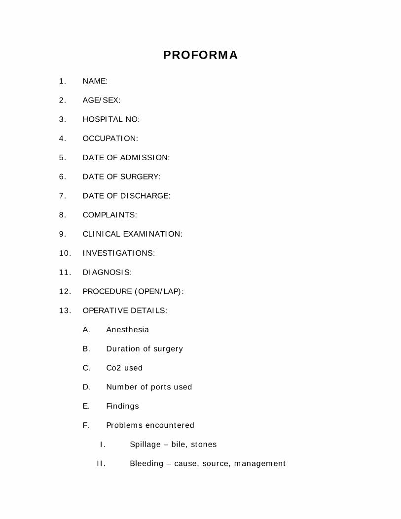
PROFORMA
1. NAME:
2. AGE/SEX:
3. HOSPITAL NO:
4. OCCUPATION:
5. DATE OF ADMISSION:
6. DATE OF SURGERY:
7. DATE OF DISCHARGE:
8. COMPLAINTS:
9. CLINICAL EXAMINATION:
10. INVESTIGATIONS:
11. DIAGNOSIS:
12. PROCEDURE (OPEN/LAP):
13. OPERATIVE DETAILS:
A. Anesthesia
B. Duration of surgery
C. Co2 used
D. Number of ports used
E. Findings
F. Problems encountered
I. Spillage – bile, stones
II. Bleeding – cause, source, management
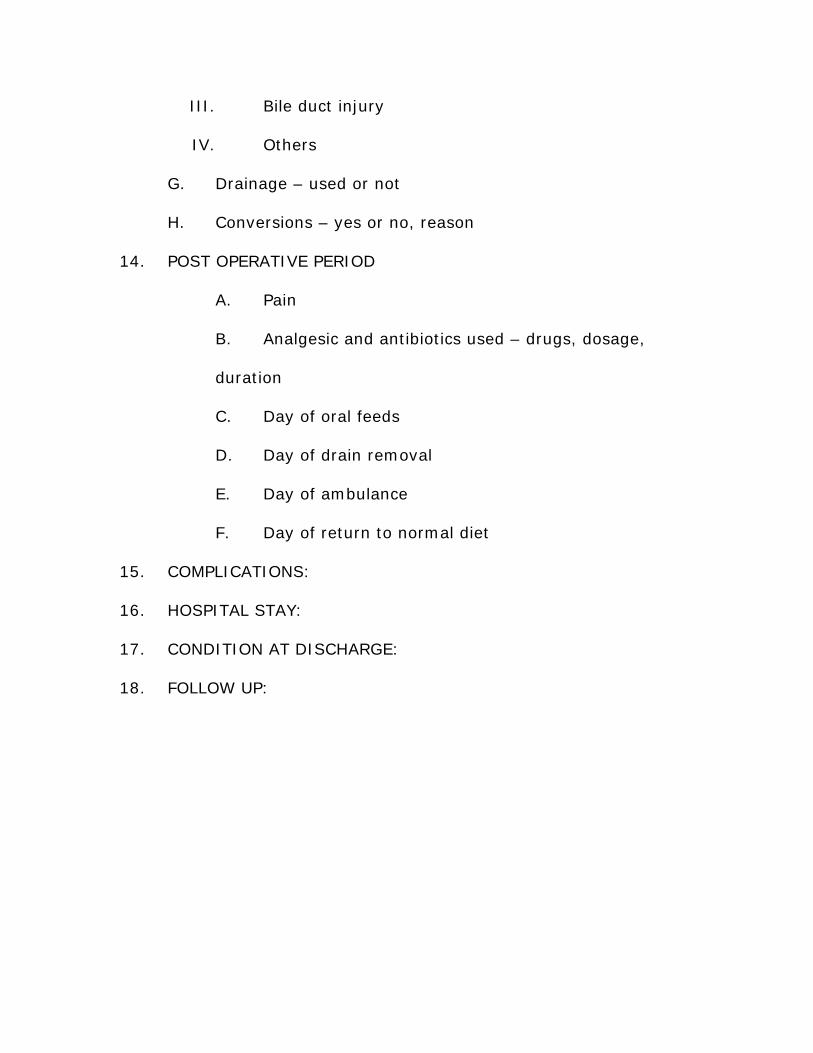
III. Bile duct injury
IV. Others
G. Drainage – used or not
H. Conversions – yes or no, reason
14. POST OPERATIVE PERIOD
A. Pain
B. Analgesic and antibiotics used – drugs, dosage,
duration
C. Day of oral feeds
D. Day of drain removal
E. Day of ambulance
F. Day of return to normal diet
15. COMPLICATIONS:
16. HOSPITAL STAY:
17. CONDITION AT DISCHARGE:
18. FOLLOW UP:
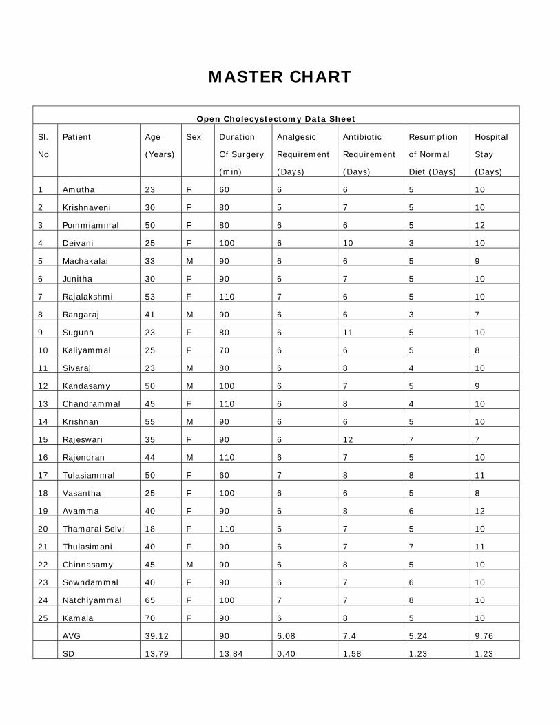
MASTER CHART
Open Cholecystectomy Data Sheet
Sl.
No
Patient Age
(Years)
Sex Duration
Of Surgery
(min)
Analgesic
Requirement
(Days)
Antibiotic
Requirement
(Days)
Resumption
of Normal
Diet (Days)
Hospital
Stay
(Days)
1 Amutha 23 F 60 6 6 5 10
2 Krishnaveni 30 F 80 5 7 5 10
3 Pommiammal 50 F 80 6 6 5 12
4 Deivani 25 F 100 6 10 3 10
5 Machakalai 33 M 90 6 6 5 9
6 Junitha 30 F 90 6 7 5 10
7 Rajalakshmi 53 F 110 7 6 5 10
8 Rangaraj 41 M 90 6 6 3 7
9 Suguna 23 F 80 6 11 5 10
10 Kaliyammal 25 F 70 6 6 5 8
11 Sivaraj 23 M 80 6 8 4 10
12 Kandasamy 50 M 100 6 7 5 9
13 Chandrammal 45 F 110 6 8 4 10
14 Krishnan 55 M 90 6 6 5 10
15 Rajeswari 35 F 90 6 12 7 7
16 Rajendran 44 M 110 6 7 5 10
17 Tulasiammal 50 F 60 7 8 8 11
18 Vasantha 25 F 100 6 6 5 8
19 Avamma 40 F 90 6 8 6 12
20 Thamarai Selvi 18 F 110 6 7 5 10
21 Thulasimani 40 F 90 6 7 7 11
22 Chinnasamy 45 M 90 6 8 5 10
23 Sowndammal 40 F 90 6 7 6 10
24 Natchiyammal 65 F 100 7 7 8 10
25 Kamala 70 F 90 6 8 5 10
AVG 39.12 90 6.08 7.4 5.24 9.76
SD 13.79 13.84 0.40 1.58 1.23 1.23
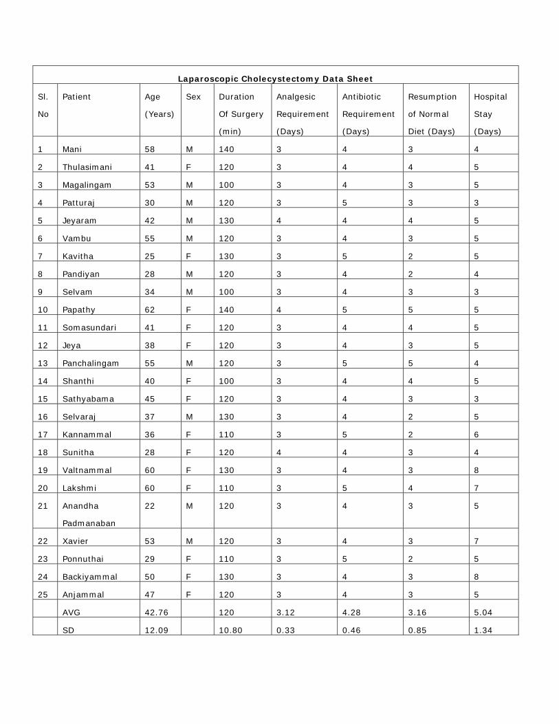
Laparoscopic Cholecystectomy Data Sheet
Sl.
No
Patient Age
(Years)
Sex Duration
Of Surgery
(min)
Analgesic
Requirement
(Days)
Antibiotic
Requirement
(Days)
Resumption
of Normal
Diet (Days)
Hospital
Stay
(Days)
1 Mani 58 M 140 3 4 3 4
2 Thulasimani 41 F 120 3 4 4 5
3 Magalingam 53 M 100 3 4 3 5
4 Patturaj 30 M 120 3 5 3 3
5 Jeyaram 42 M 130 4 4 4 5
6 Vambu 55 M 120 3 4 3 5
7 Kavitha 25 F 130 3 5 2 5
8 Pandiyan 28 M 120 3 4 2 4
9 Selvam 34 M 100 3 4 3 3
10 Papathy 62 F 140 4 5 5 5
11 Somasundari 41 F 120 3 4 4 5
12 Jeya 38 F 120 3 4 3 5
13 Panchalingam 55 M 120 3 5 5 4
14 Shanthi 40 F 100 3 4 4 5
15 Sathyabama 45 F 120 3 4 3 3
16 Selvaraj 37 M 130 3 4 2 5
17 Kannammal 36 F 110 3 5 2 6
18 Sunitha 28 F 120 4 4 3 4
19 Valtnammal 60 F 130 3 4 3 8
20 Lakshmi 60 F 110 3 5 4 7
21 Anandha
Padmanaban
22 M 120 3 4 3 5
22 Xavier 53 M 120 3 4 3 7
23 Ponnuthai 29 F 110 3 5 2 5
24 Backiyammal 50 F 130 3 4 3 8
25 Anjammal 47 F 120 3 4 3 5
AVG 42.76 120 3.12 4.28 3.16 5.04
SD 12.09 10.80 0.33 0.46 0.85 1.34