a case study of distal tibial fractures managed with
TRANSCRIPT

A CASE STUDY OF DISTAL TIBIAL FRACTURES
MANAGED WITH LOCKING COMPRESSION PLATE
USING MIPO TECHNIQUE
DISSERTATION SUBMITTED FOR
M.S. DEGREE
(BRANCH II - ORTHOPAEDIC SURGERY)
APRIL 2015
THE TAMILNADU
DR. M.G.R. MEDICAL UNIVERSITY,
CHENNAI, TAMILNADU.

CERTIFICATE
This is to certify that this dissertation titled “A CASE STUDY OF
DISTAL TIBIAL FRACTURES MANAGED WITH LOCKING
COMPRESSION PLATE USING MIPO TECHNIQUE” is a
bonafide work done by Dr.P.VANAJ KUMAR., Post graduate
student of the Department of Orthopaedics, Tirunelveli Medical
College Hospital, Tirunelveli, during the academic year 2012 – 2015.
This work did not form the basis for the award of any degree
previously.
Dean,
Tirunelveli Medical College,
Tirunelveli.
CERTIFICATE
This is to certify that the work entitled “A CASE STUDY OF
DISTAL TIBIAL FRACTURES MANAGED WITH LOCKING
COMPRESSION PLATE USING MIPO TECHNIQUE‖ is a
bonafide work done by Dr. P. VANAJ KUMAR in the Department
of Orthopaedics, Tirunelveli Medical College, Tirunelveli.
He has completed the necessary period of stay in the Department and
has fulfilled the conditions required for submission of this thesis
according to the university regulations. The study was undertaken by
the candidate himself and observations recorded have been
periodically checked by us.
Recommended and forwarded.
Prof. Dr. ELANGOVAN CHELLAPPA, M.S.Ortho., D.Ortho.,
Professor and Head of the Department
Department of Orthopaedics
Tirunelveli Medical College
Tirunelveli.

DECLARATION
I, Dr. P. VANAJ KUMAR, solemnly declare that the
dissertation entitled ―A CASE STUDY OF DISTAL TIBIAL
FRACTURES MANAGED WITH LOCKING COMPRESSION
PLATE USING MIPO TECHNIQUE‖ has been prepared by me
under the able guidance and supervision of my guide Prof.Dr.
Elangovan Chellappa, M.S.ORTHO., D.ORTHO., Prof. & HOD,
Department of Orthopaedics and Traumatology, Tirunelveli Medical
College, Tirunelveli, in partial fulfillment of the regulation for the
award of M.S. (ORTHOPAEDIC SURGERY) degree by the
Tamilnadu Dr. M.G.R. Medical University, Chennai in the
examination to be held in April 2015.
This work did not form the basis for the award of any
other degree or Diploma to me previously from any other university.
Place: Tirunelveli
Date: DR. P. VANAJ KUMAR

ACKNOWLEDGEMENT
I am deeply indebted to my beloved chief and my teacher, Prof.
Dr. Elangovan Chellappa, M.S.Ortho, D.Ortho., Professor and
H.O.D., Department of Orthopaedics, Tirunelveli Medical College,
Tirunelveli for the able guidance, inspiration and encouragement he
has rendered at every stage of this study.
I am grateful to my beloved teachers Prof. Dr.N.Manikandan,
Prof.Dr.K.Thanigaimani, Prof.Dr.N.Thanappan for their invaluable
help and guidance rendered to me in preparing this dissertation.
I express my heartfelt gratitude to Dr.M.Senthil Kumar,
Dr.M.S.Abul Kasim, Dr.R.Arokia Amalan, Dr.A.Suresh Kumar,
Dr.S.Mageswaran,Dr.R.SundaraPandian, Dr.V.ChandraSekaran,
Dr.AbrahamJames, Dr.Babu Aloy Assistant Professors of
Orthopaedics, Tirunelveli Medical College, Tirunelveli for their
valuable advice and help in carrying out this study.

My sincere thanks to Prof.Dr.L.D.Thulasiraman
M.S.Ortho., Dean, Tirunelveli Medical College, Tirunelveli for
permitting me to utilize the clinical materials of the hospital.
I would like to thank my patients, friends, colleagues and
Family, who have stood by me throughout this work and above all the
God for His kindness throughout this study.

Index:
Table of Contents
INTRODUCTION ............................................................................ 11
AIM .................................................................................................... 14
REVIEW OF LITERATURE .......................................................... 15
BIO MECHANICS ........................................................................... 29
ANATOMY OF DISTAL TIBIA .................................................... 35
CLASSIFICATION OF DISTAL TIBIAL FRACTURES .......... 41
MECHANISM OF INJURY ............................................................ 46
INVESTIGATIONS.......................................................................... 49
PRINCIPLES OF MANAGEMENT .............................................. 53
METHODS OF TREATMENT....................................................... 56
PRE OPERATIVE PLANNING ..................................................... 78
SURGICAL TECHNIQUE .............................................................. 79
COMPLICATIONS .......................................................................... 92
EVALUATION OF OUTCOME..................................................... 97
MATERIALS AND METHODS ..................................................... 98

PROCEDURE ................................................................................. 110
ANALYSIS OF FUNCTIONAL OUTCOME ............................. 112
DISCUSSION .................................................................................. 115
CONCLUSION ............................................................................... 122
BIBLIOGRAPHY ............................................................................ 123
ILLUSTRATIONS.......................................................................... 135
PROFORMA ................................................................................... 142
MASTER CHART .......................................................................... 144

11
1. INTRODUCTION
Increased incidence of Road Traffic Accidents claims most of human
mortality and morbidity in the current age. Hence, it forms the major
epidemic of Modern world. Of these, fractures of distal tibia have been
difficult to treat. In this era of increasing life expectancy, there is a rise
of elderly population which, increases the incidence of these fractures
in osteoporotic bones, adding to the morbidity. Due to the proximity of
these fractures to the ankle, regaining full ankle movement may be
difficult. Soft-tissue damage, comminution and fracture extension into
the ankle joint lead to unsatisfactory results in many cases regardless
of the treatment modality.
Better understanding of the injury patterns, availability of better
implants, the concept of early surgical fixation and early postoperative
mobilization of joint all have convincingly improved the functional
outcome of the patient to a large extent.
Main challenges encountered in the treatment of distal tibia fractures
are
- these are high energy fractures
12
- associated with extremely damaged soft tissue envelope
- increased incidence of compound injuries
-increased skin complications following surgery
-comminution of the metaphysis and articular surface makes
anatomical reduction difficult.
The resulting incongruency of articular surface leads to early
secondary OsteoArthritis.
In metaphysis the fixation is less satisfactory resulting in early
loosening of the implant. Achieving rigid fixation in comminuted
fractures is difficult due to poor purchase and hence the fixation is less
optimal to allow weight bearing or even early mobilization. Initially
conservative treatment with Plaster Of Paris was advocated as a
treatment option. But it leads to high incidence of malunion and
stiffness of ankle joint. Also prolonged recumbency resulted in high
incidence of thromboembolic diseases and pneumonia.

13
Open reduction and internal fixation with plate osteosynthesis lead to
skin necrosis and infection in > 40% of patients eventually leading to
malunion and implant failure. Intramedullary devices give inadequate
stability due to wide medullary cavity leading to implant failure and
screw breakage. For compound fractures, initial treatment with
external fixator for wound care followed by a definitive mode of
internal fixation was advocated. This involves multiple procedures
which increased economical and mental stress for the patients.
But minimally invasive plating offers the advantage of fracture
fixation without disturbing the soft tissue cover; less chances of
infection, early mobilization of patient. Using a locking compression
plate reduces the tendency for varus collapse and at the same time
affords better stability. The successful management of these injuries,
demands a thorough knowledge of fracture personality and technical
aspects of fracture fixation; and a tailored post-operative management.

14
2. AIM:
• To discuss the management of fractures of distal tibia
• To evaluate the biomechanical and biological advantages of
Locking compression plates
• To evaluate clinical, functional and radiological outcomes after
minimally invasive plate osteosynthesis using distal tibial locking
compression plates.

15
3. REVIEW OF LITERATURE:
Hansmann from Hamburg did the first plate osteosynthesis in the year
1886. During the 1950’s the AO / Association for the Study of Internal
Fixation standardized the use of plating systems. Then the main goal
of fracture treatment was to restore the function of the injured limb by
providing the bone with primary strength through stable internal
fixation. This resulted in a decrease of limb deformities and joint
stiffness.
The original AO/ASIF technique was based on the compression
principle using plates and screws. The dynamic compression plates
provided axial compression of the fracture (Fig. 1). Thus perfect
fracture reduction and compression using lag screws resulted in
primary bone healing without visible callus. So, even the smallest
fragments were reduced to restore the exact anatomy, often damaging
the vitality of bone and soft tissues. This highly traumatic technique
ultimately resulted in delayed bone healing, nonunion, and increased
chances of infection.

16
Fig. 1 : Dynamic Compression Plate.
This led to a change over from the concept of absolute stability to a
newer concept of bridge plate which provides relative stability. The
smaller fracture fragments are left untouched and bridged by
anchoring the plate only to the proximal and distal main fragments.
Lag screws are not used to achieve inter fragmentary compression.
Hence the fracture unites by secondary bone healing with the
production of thick external callus.
Preserving the circulation at the fracture zone was the main concern
than achieving anatomical reduction and stable fixation. Ganz3
named
this newer technique as biological plate osteosynthesis.

17
This indirect reduction technique with bridging stabilization did not
expose the fracture site and hence was less traumatic and more
biological resulting in good bone and soft tissue healing.
The evolution of Bone Plates:
Conventional plating techniques aimed to press the plate against the
bone fragments with high compressive force, to create a stable bone–
implant construct. It required adequate number of bicortical screws to
achieve adequate anchoring force. Lüthi et al.7 showed that the
periosteal blood supply was severely compromised by this highly
stable plate-bone construct. Gautier et al,2 Jörger,
6 and Vattalo,
16
showed that these circulatory disturbances can lead to bone necrosis.
This can be managed only by reaming the dead bone. This process
may temporarily perforate the bone.
Later it was proposed that reducing the contact surface of the plate and
bone minimized the damage to cortical blood supply. Based on this
concept a special plate which reduces the contact surface by more than

18
50% compared to conventional DCP10
was designed and was named as
Limited Contact DCP (Fig. 2). Even in LC-DCP, the principle of
compressive forces against the bone was still present.
Fig. 2: Limited Contact Dynamic Compression Plate.
Only the use of an angular-stable implant can obviate the need for the
frictional forces between plate and bone. This kind of device is an
internal fixator placed under the skin surface. The first internal fixator
was developed in the 1970s in Poland.11
Then the Association for the
Study of Internal Fixation (ASIF) developed ―The point contact
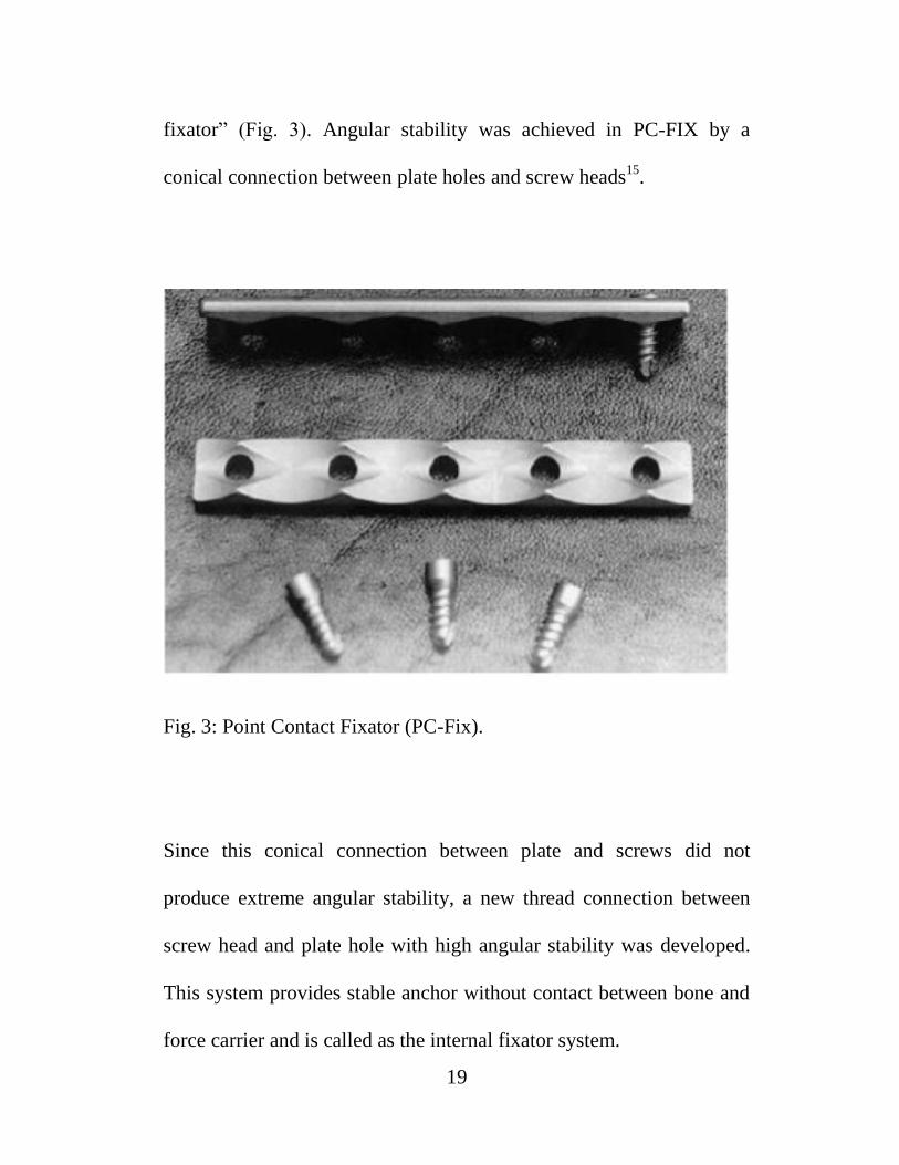
19
fixator‖ (Fig. 3). Angular stability was achieved in PC-FIX by a
conical connection between plate holes and screw heads15
.
Fig. 3: Point Contact Fixator (PC-Fix).
Since this conical connection between plate and screws did not
produce extreme angular stability, a new thread connection between
screw head and plate hole with high angular stability was developed.
This system provides stable anchor without contact between bone and
force carrier and is called as the internal fixator system.

20
The internal fixator system includes4,13
- The less invasive stabilization system (LISS) (Fig. 4)
-The locking compression plate (LCP) (Fig. 5).
Fig. 4: Less Invasive Stabilization System.

21
Fig. 5: Locking Compression Plate.
The conventional cortical screws were used as anchoring screws but
these locking head screws act as Schanz screws between the bone and
the force carrier.13
In conventional plating, with axial compression there exists a
transverse strain on the bone. But in case of internal fixators load
transfer occurs through the locking head screws.

22
Locking compression plates:
They bear a new screw-hole geometry called the combi- hole, which
can be filled with a conventional cortical screw or an angular-stable
locking head screw. At present almost all plate designs are available
with this combi-hole for use as internal fixators in metaphyseal
fractures 9.
While the cortical screws were used only as reduction screws, the
locking head screws are used as implant set screws. For a stable
construct at least three locking head screws must be used in each main
fragment. The screws must be bicortical in osteoporotic bone.
The drill bits used for LHS are
2.8-mm for 3.5 systems
4.3-mm for 4.5 systems
The drill guide can also be locked in the angular-stable hole of the
plate.
23
Indications for these internal fixator systems include, metaphyseal
fractures of
the proximal and distal humerus,
the distal radius and the distal tibia.
The biological advantages of this new system are
- Preservation of the periosteal blood supply
- Improved fixation in poor metaphyseal or osteoporotic bone
- Facilitation of minimally invasive and percutaneous techniques.
Minimally invasive plate osteosynthesis:
Distal tibia fractures with or without intra articular extension, are
difficult fractures to manage. Distal tibia is circular in cross section
and has a thin cortex while the diaphysis is triangular with thicker
cortex.
Since intramedullary nail is designed only for snug fit at the shaft, it
cannot provide stability at the distal tibia.3,16
Complications of
interlocking nailing in distal tibia fractures are

24
- malunion upto 29%
- implant failure upto 39%.1,13
Since tibia is a subcutaneous bone 2/3 rd of its blood supply comes
from periosteum. ORIF strips off this vital layer and results in
- Non union upto 35%
- Infection upto 25%.4-7,17
External fixators are recommended only as a temporary method of
stabilization in open fractures with severe soft tissue injury.1,18
MIPO technique using LCP
- preserves extraosseous blood supply
- respects the osteogenic fracture haematoma
- provides a biologically friendly and stable fixation
- allows indirect reduction methods
- permits sub-cutaneous tunneling of the plate avoiding large
incisions
- prevents iatrogenic injury to vascular supply of the bone19

25
- provides both angular and axial stability since it is a friction
independent self-stable construct
- minimizes risk of secondary loss of reduction because of the
locking mechanism between screw heads and plate holes.12
Comparative studies between interlocking nails and conventional
plating revealed a higher incidence of axial mal-alignment and higher
mean pain scores with interlocking nails (Vallier et al). But operative
time and radiation exposure were significantly higher in the MIPO
group.6,8
The MIPO group showed better functional outcome in terms of joint
mobility, less post-operative pain and early return to work.27
MIPO with LCP also requires anatomical reduction of the fracture
using indirect reduction maneuvers and image intensifier guidance,
before plate fixation.

26
The complications like
- delayed union,
- non-union,
- prominent hardware,
- malleolar skin irritation and
- post-operative pain.23,24,26
result from mal-reduction and suboptimal pre contouring of the plate.
The common indications for implant removal were
- prominent hardware
- malleolar skin irritation
- pain
The use of Low profile metaphyseal Locking Compression Plates
reduces hardware prominence.23
Polyaxial locking plates are useful options when the supramalleolar
anatomy mismatches with the pre contoured plate. It provides choice
of screw trajectories according to fracture pattern.24

27
The indirect reduction methods followed are
- calcaneal pin traction,
- external fixators,
- mechanical distractors22
- Kirschner wire used as a joystick at fracture site
- fibula fixation before tibia fixation when both are fractured at
the same level.1,23,24,28
Routine fixation of fibula is unnecessary unless the fracture has
involved the syndesmosis.
Rarely, following MIPO technique secondary procedures like bone
grafting or bone marrow injection may be needed for delayed union.29
Prolonged injury-hospital arrival interval results in
- gross swelling,
- skin injury and
- fracture blisters.
This causes a delay in the definitive fixation of the fracture, waiting
for the soft tissues to heal. But union time is not affected by this delay
in surgery.30

28
Late infections occurring after one month of complete wound healing
did not affect the union time (Lau et al). The patients with superficial
wound infection improved with antibiotics but patients with wound
breakdown and exposed implant required repeated wound debridemet
and long hospital stay.31
Injury to saphenous nerve and great saphenous vein can be avoided by
careful attention towards the placement of skin incision, dissection of
vein and atraumatic placement of the drill sleeve.26,31
LCP removal is usually a tough task as the conical extraction devices
may not work when stripping of hexagonal recess or thread happens.
In such cases the removal can be completed by cutting and bending of
the plate around the stripped screw.23,32
Thus MIPO with LCP is a highly fruitful method of treatment
in distal tibial fractures, both in terms of union rate and
complications rate.

29
4. BIOMECHANICS
Distal Tibia Locking Compression Plate:
Fig. 6: Distal Tibia Locking Compression Plate.
The locking plate system is basically similar to the traditional plate
fixation methods with few improvements. Locking head screws
provide fixed angle construct and improved fixation in osteoporotic
bones (Fig. 7).

30
Fig. 7: Locking Head Screws.
1. The screws are not dependent on plate bone compression
2. Multiple screw insertion in distal fragment allows
improved fixation
3. These plates are anatomically contoured to match the
surface of the bone and hence intra-operative contouring
is not needed.
4. Combi - holes have additional dynamic compression mode that
provides options for axial compression in addition
to the locking mechanism (Fig. 8).

31
Fig. 8: Combi holes in LCP.
CONVENTIONAL BONE PLATING VERSUS LOCKED
COMPRESSION PLATING
Conventional bone plates rely on direct plate -bone and screw-bone
friction to maintain the fixation. Therefore the plates must be perfectly
contoured prior to application to the bone. Fracture reduction can be
lost from axial loads causing excessive shear forces on the construct
that are greater than the frictional loads between the bone-plate-screw
construct.

32
The cortical screws may toggle leading to screw loosening and loss of
fixation. Each screw works independently; the construct depends on a
single screw's stiffness or pullout strength.
The biomechanical goal of the LCPs is to amplify the stiffness of the
construct in a biological environment. The Locking plate is a fixed
angle construct that does not depend on screw purchase in bone.
After locking the screw into the plate, the shear stress at the screw-
bone interface is converted into compressive force by the fixed-angle
construct. The load is now perpendicular to the screw axis. In order for
the construct to fail under an axial load, the bone must collapse in
compression. Therefore, the strength in the LCP is the sum of all the
screw and plate interfaces.
Locking screws:
-have a smaller thread design
-are not used to generate plate-bone compression

33
-have a larger core diameter for greater bending and shear strength
-dissipate the load over a larger area of bone
-have the new Star Drive head that permits 65% greater insertion
torque than hexagonal drivers.
-have a conical, double-lead thread design that enables alignment with
the threads in plate hole.
The indications for LCPs :
1. Patients with poor bone quality (osteoporosis, osteomyelitis)
2. Complex juxta-articular fracture (where contouring will be difficult
in the metaphyseal region)
3. fractures with difficulty in achieving adequate number of
conventional screw purchase
4. Polytrauma cases (especially when the fractures cannot be
anatomically reconstructed).
5. Failure of conventional plate systems (cortex or cancellous screw
stripping or screw back-out)
6. Peri-prosthetic fractures

34
Studies in bone models show that locked screw constructs fail at
higher loads compared to cortex screws. Hence they are very
advantageous in osteoporotic bones.
The ideal number of locked screws on both sides of the fracture, the
number of unicortical versus bicortical screws needed for stable
fixation, indications for plate contouring, the effects of adding
additional implants are not clearly determined at present.
It is possible to place locking plates and screws minimally invasively
with fluoroscopic guidance. In humans there is little mechanical
advantage in applying more than 2 locked screws on each side of the
fracture.

35
5. ANATOMY OF DISTAL TIBIA
The distal tibia has five surfaces:
- inferior,
- anterior,
- posterior,
- lateral,
- medial.
The inferior surface is the articular surface (Fig. 9).
It is concave antero-posteriorly, and slightly convex transversely.
Fig. 9: Inferior Surface of distal tibia and fibula.
36
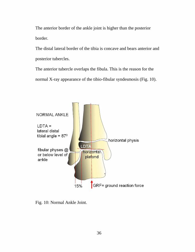
The anterior border of the ankle joint is higher than the posterior
border.
The distal lateral border of the tibia is concave and bears anterior and
posterior tubercles.
The anterior tubercle overlaps the fibula. This is the reason for the
normal X-ray appearance of the tibio-fibular syndesmosis (Fig. 10).
Fig. 10: Normal Ankle Joint.

37
The anterior tubercle bears the origin of the anterior tibio fibular
ligament. In the posterior tubercle lies the attachment of the deep
component of the posterior tibio fibular ligament.
The anatomy of the ankle, provides stability in dorsiflexion and
mobility in plantar flexion.
Dorsiflexion stability is provided by articular contact and plantar
flexion stability by the ligamentous structures.
The talar dome is narrow posteriorly and wider anteriorly.
During dorsiflexion, the fibula rotates externally to accommodate the
widened anterior part of talus.

38
a. Anatomy of Pilon Fracture
Fractures of distal tibia within 5cm of the ankle joint (Fig. 11).
Fig. 11: Pilon Fracture.

39
1. Pilon fractures involve the articular weight bearing portion of the
distal tibia.
2. Usually results from high energy axial load as in RTA, fall from
height and rarely from low-energy rotation/torsion.
b. Clinical Evaluation
1. Assess vascularity – look for dorsalis pedis and posterior tibial
pulses and distal capillary refill.
2. Evaluate associated soft tissue injury (swelling, blisters).
3. Look for compartment syndrome and closed degloving.
Blisters filled with blood denote more extensive skin and soft tissue
damage compared to blisters filled with clear fluid.

40
c. Pilon Fracture Radiology
i. Antero Posterior, lateral and mortise views of the ankle (Fig. 12)
ii. Antero Posterior and lateral views of entire Tibia
iii. CT scan of ankle joint with 3-dimensional reconstruction.
X Ray and CT imaging must be done with traction applied to the
limb.
Fig. 12: X Ray Ankle Mortise view.

41
6. CLASSIFICATION OF DISTAL TIBIA FRACTURES
Ruedi and Allgower’s classification
It was the first classification that came to use.
It was widely used in literatures on distal tibia fractures until the
introduction of AO classification.
It is based on the degree of articular comminution (Fig.13).
Type I - non displaced cleavage fractures of joint
Type II – displaced fractures with minimal comminution
Type III – displaced fractures with severe comminution.

42
Fig. 13: Ruedi Allgower’s classification.
This classification has a prognostic significance;
Prognosis being poor as the type increases from type I to type III.

43
AO/ OTA classification is now universally used for distal tibial
fractures (Fig. 14).
Type A – non-articular fractures
Type B – partial articular fractures
Type C – total articular fractures (tibial plafond fractures).
Fig. 14: AO / OTA classification.

44
Soft tissue injuries are classified based on
Tscherne and Gotzen classification.
Grade Description
0 Little or no soft tissue injury
1 Superficial abrasion with local contusion in skin / muscle
2 Deep contaminated abrasion with local contusion in skin /
muscle
3 Extensive crushing or contusion of skin / muscle destruction
Table 1: Tscherne classification.

45
Open fractures are classified as per Gustilo and Anderson.
Type I
o wound < 1 cm with minimal soft tissue injury
Type II
o wound >1cm with moderate soft tissue injury
Type III A
o high energy trauma with crushed tissue and contamination
o with adequate soft tissue coverage
Type IIIB
o extensive periosteal stripping and requiring soft tissue
transfer
Type IIIC
o with associated vascular injury

46
7. MECHANISM OF INJURY
- Low energy rotational forces (pronation dorsiflexion injury)
- High energy axial compression forces (road traffic accident, fall
from height)
Low energy injuries carry a good prognosis.
But high energy injuries with extensive soft tissue damage are
more common (Table. 2).
Rotation forces Axial load forces
Low energy high energy
Slow load rate Rapid load rate
Talus – translation Talus- proximal displacement
Minimal articular comminution Severe articular and metaphyseal
comminution
Minimal soft tissue damage Extensive soft tissue damage
Table. 2: Rotation forces vs Axial load forces.

47
Foot postion at the time of injury determines fracture pattern:
plantar flexion- posterior tibial fragment (Fig. 15),
neutral- entire articular surface (Fig. 16),
dorsiflexion- anterior fragment (Fig. 17).
Fig. 15: Plantar flexion injury

48
Fig. 16: Neutral flexion injury.
Fig. 17: Dorsiflexion injury.

49
8. INVESTIGATIONS
- Plain X Ray including entire Tibia AP and Lateral views.
- Plain X Ray Ankle AP, Lateral and Mortise views (Fig. 18,19).
- X Ray of contralateral ankle.
- X Ray foot AP and Oblique views.
- CT Ankle with 3 Dimensional reconstruction (Fig. 20,21,22).
Fig. 18: X Ray Ankle AP view.

50
Fig. 19: X Ray Ankle Lateral view.
Fig. 20: CT Ankle axial cut.

51
Fig. 21: CT Ankle Sagittal cut.
Fig. 22: 3-D reconstruction CT Ankle.

52
X Ray of the contralateral ankle may be useful as a template for
articular reconstruction.
CT scan images are useful in
- Identifying the direction of fracture lines
- Determining the extent of articular comminution and impaction
- Assessing the size and displacement of articular fragments
- Planning the skin incision
- Placing olive wires and lag screws.
X Rays and CT scans must be taken with traction applied to the
limb for better understanding of fracture morphology.

53
9. PRINCIPLES OF MANAGEMENT
The factors which play a dynamic role in management are
i. Degree of fracture displacement
ii. Extent of soft tissue injury
iii. Degree of comminution
iv. Neurovascular injuries
v. Severity of joint involvement
vi. Osteoporosis
vii. Associated ipsilateral fractures (ex: patella, tibial plateau, talus)
The main goal of fracture treatment is obtaining a stable, aligned,
mobile and painless joint to reduce post traumatic osteo arthritis.
The principles of treating distal tibia fractures are
Restoring length of fibula,
Anatomic reduction of articular surface,
Medial buttress plating to prevent varus malalignment
Bone grafting for metaphyseal defects,

54
The objectives of treatment are
i. To restore anatomical articular surface
ii. To obtain and maintain acceptable reduction and stable fixation
iii. To achieve normal mechanical alignment
iv. To treat the associated injuries
v. To provide a stable joint
vi. To achieve fracture union
vii. To regain a functional range of ankle motion
viii. To enable pain free weight bearing.
The prognosis of fracture depends on
i. degree of depression of articular surface
ii. extent of fracture separation
iii. degree of diaphyseal and metaphyseal comminution
iv. integrity of soft tissue coverage

55
The various treatment options are
1. Primary internal fixation of tibia alone or both tibia and
fibula in closed fractures without significant soft tissue swelling.
2. Primary internal fixation of the fibula with temporary
spanning external fixation for tibia and delayed conversion to
internal fixation when soft tissues permit.
3. Closed fractures with significant soft tissue swelling may be
placed in calcaneal pin traction.
4. Open fractures should be taken for emergency wound
debridement and ankle spanning external fixation which can
later be converted to definitive fixation.

56
10. METHODS OF TREATMENT
i. Open reduction and internal fixation to restore articular
congruency is must for an articular fracture with joint
instability.
ii. Anatomical reduction and stable fixation of articular fragments
is mandatory for articular cartilage regeneration.
iii. If open reduction and internal fixation is not possible due to
Patient conditions skeletal traction should be applied.
In the 1960s, conservative methods like traction and cast bracing,
produced better results than operative treatment, due to lack of
adequate internal fixation devices. It had a high occurrence of
malunion and Stiffness of adjacent joints. Also prolonged recumbency
caused thromboembolic disease and pneumonia.

57
With the development of advanced internal fixation devices, treatment
protocol changed in 1980s. Open reduction and internal fixation with
buttress plates was done. Due to the poor soft tissue cover it caused
skin necrosis in distal tibia which led to high chances of infection.
Also low profile plate led to implant failure and eventually malunion.
Intramedullary rods were used which had inadequate stability due to
wide medullary cavity in distal tibia which lead to Implant failure and
screw breakage. Also varus / valgus malalignment was common in
closed interlocking nailing..
External fixation can be used as either temporary or definitive fixation
in open distal tibia fractures especially those associated with vascular
injury. Since it is a form of Rigid fixation, delayed union was
common. As it Spans the joint, chances of joint stiffness are more.

58
A recent advance in technology for the treatment of distal tibial
fractures includes minimally invasive plate osteo synthesis using
locking compression plates.
This technique offers the advantages of
- bridging fixation of various fracture patterns without
disturbing the soft tissue cover
- less chances of infection
- early mobilization of patient
- reduced tendency for varus collapse
- greater stability.
Management of distal tibial fractures can be divided into two broad
categories.
i. conservative treatment
ii. operative treatment

59
In operative treatment, various modalities include
i. Open Reduction Internal Fixation with buttress plate
ii. Open Reduction Internal Fixation with locking compression
plate
iii. Minimally invasive plating with LCP
iv. Closed reduction & internal fixation with expert tibial locking
nails.
v. Ilizarov ring fixation
vi. Uni-planar external fixation.
vii. Hybrid external fixator application
CONSERVATIVE MANAGEMENT
Considerable controversy existed as to whether conservative (or)
surgical treatment leads to better results in the management of
periarticular fractures of distal tibia. Early attempts at internal fixation
of these complex injuries were associated with high incidence of
malunion, nonunion and infection.

60
Because of the increased risk of complications, numerous authors
concluded that closed methods were preferable to operative treatment.
With the improvement in surgical techniques, availability of better
implants, prevalence of better antibiotics, the operative management
has now got many recommendations in treatment of periarticular
fractures of distal tibia.
The indications for conservative therapy include
i. Undisplaced (or) Incomplete fractures with intact collateral
ligaments.
ii. Fractures displaced less than 5mm
iii. Elderly sedentary patients with depression less than 2 mm
iv. Impacted stable fracture in elderly osteoporotic patients.
v. Lack of modern internal fixation devices.
vi. Unfamiliarity or inexperience with surgical techniques.
vii. Significant underlying medical disease

61
The goal of conservative treatment is not anatomical reduction of
fracture fragments, but restoration of overall length and axial
alignment.
The criteria for acceptable fracture reduction include
i. < 70 malalignment in frontal plane
ii. < 100 malalignment in sagittal plane
iii. Limb shortening < 1.5 cm.
iv. Articular incongruity < 2 mm
If a patient is being treated with conservative plaster cast, he/she must
be closely observed for displacement of the fracture.
Weight bearing must not be allowed atleast for 8 weeks.

62
SURGICAL MANAGEMENT
a. INTRODUCTION:
In the past 25 years, various forms of treatment for fixation of
fractures of distal tibia have evolved. The combination of a better
understanding of fracture pattern, meticulous soft tissue handling,
judicious use of antibiotics and improved imaging techniques has
made different modes of minimally invasive fixation practically
possible.
Since 1980, various studies have compared the results of various
modes of fixation for periarticular fractures of distal tibia giving
variable results.
The goals of operative treatment of periarticular fractures of
distal tibia are
i. Anatomical Alignment
ii. Stable fixation
iii. Early Mobilization
iv. Early functional rehabilitation of the ankle.
63
Indications for operative management include
i. Displaced intraarticular fracture
ii. Patients with Multiple injuries
iii. Open fractures
iv. Associated vascular injuries requiring repair.
v. Severe ipsilateral limb injuries (patellar fracture, tibial plateau
fracture)
vi. Major associated ligamentous injuries.
vii. Irreducible fracture.
viii. Pathological fracture
Contraindications to internal fixation include
i. Active infection
ii. Inadequate facilities
iii. Inexperienced surgeons

64
b. TIMING OF SURGERY
i. For open injury, compartment syndrome and vascular injury
immediate treatment is needed
ii. For displaced unstable fractures surgery is done as early as the
condition of patient permits
iii. After stabilization of neurosurgical, abdominal and thoracic
injuries
iv. For critically ill patients, percutaneous fixation with temporary
joint spanning external fixator
v. Definitive internal fixation is done after soft tissue swelling
subsides and local skin condition permits.

65
c. PROCEDURE
Sequences in the surgical management of distal tibia fracture include
i. Restoration of articular surface
ii. Metaphyseal alignment.
iii. Defect filled with bone graft
iv. Early mobilization of joint.
I. Open Reduction Internal Fixation:
The fibula is fixed first through a postero lateral approach. Then the
articular surface of tibia is reduced through an antero medial approach.
Provisional K wires can be used to maintain articular reduction.
Atleast a 7 cm skin bridge must be maintained between the two
incisions. Narrow DCP, T Plate and Clover Leaf Plate with its tip cut
can be used to fix distal tibial fractures. T and Clover Leaf plates act
better in fractures with a large posterior fragment (Fig. 23,24).

66
Fig. 23: T Buttress plate.
Fig. 24: Clover Leaf Plate.
67
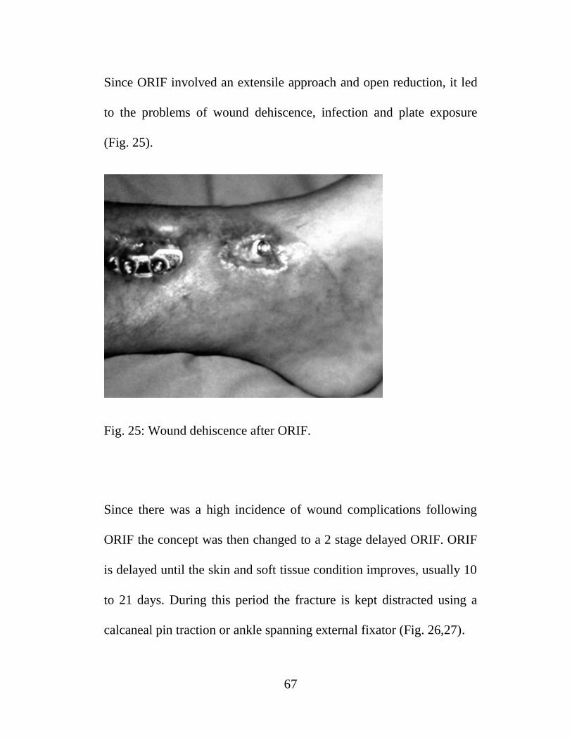
Since ORIF involved an extensile approach and open reduction, it led
to the problems of wound dehiscence, infection and plate exposure
(Fig. 25).
Fig. 25: Wound dehiscence after ORIF.
Since there was a high incidence of wound complications following
ORIF the concept was then changed to a 2 stage delayed ORIF. ORIF
is delayed until the skin and soft tissue condition improves, usually 10
to 21 days. During this period the fracture is kept distracted using a
calcaneal pin traction or ankle spanning external fixator (Fig. 26,27).

68
Fig. 26: Calcaneal pin traction on Bohler Braun frame.
Fig. 27: Ankle Spanning External Fixator.

69
Nowadays medial buttress plate is used after minimal open reduction
in which reduction is achieved using femoral distractor and articular
surface is elevated through a small cortical window made in the
anterior tibia (Fig. 28). The medial buttress plate is slid extra
periosteally and fixed (Fig. 29).
Fig. 28: Reduction done with the aid of femoral distractor.

70
Fig. 29: Medial and Antero Lateral Buttress Plating.
II. HYBRID EXTERNAL FIXATOR:
It is a Combination of wire (ilizarov) and pin fixation. It doesn’t span
the joints, so early mobilization can be done thus reducing the chances
of joint stiffness.

71
Also early mobilization helps in good healing of the articular cartilage.
It is also a more stable and less rigid fixation allowing axial micro
motion thus favoring good bony union.
But the fixator is cumbersome and uncomfortable for the patient
(Fig. 30,31).
Fig. 30: Clinical Picture-Hybrid External Fixator.

72
Fig. 31: X Ray-Hybrid External Fixator.
III. INTRAMEDULLARY NAIL:
Intramedullary nailing has recently received more attention in the
management of distal tibia fractures. These nails achieve more
biological fixation than plates because they are load sharing implants.
They enable greater soft tissue preservation.

73
But interlocking nail cannot be used in periarticular fractures of distal
tibia due to the difficulty in achieving anatomical alignment by closed
reduction and maintaining the reduction due to wide medullary canal
(Fig.32).
To overcome this, newer designs of nails such as expert tibial nail has
been introduced to deal with periarticular fractures of tibia.
Fig. 32: IL Nailing for Distal Tibia #.

74
IV. LOCKING COMPRESSION PLATE:
The screw holes in this plate have been specially designed to
accept either a standard cortical screw with a hemi spherical head or a
locking screw with a threaded head (Fig. 33).
It is used as a less invasive skeletal stabilization system by
sliding the plate without disturbing the soft tissues. A locked screw
plate construct can be compared to an implanted external fixation
device.
Fig. 33: Locking Compression Plate and Screw.

75
MIPO WITH LCP
a. Displaced type A fractures with minimal soft tissue injury
(Tscherne classification grade 0, grade 1)
These injuries may be reduced and fixed primarily, as a single stage
procedure, if the soft tissues are in truly excellent condition.
A distractor or external fixator may help reduction (Fig. 34). Fibular
reduction and fixation is the usual next step, but this reduction must be
accurate, so that it does not prevent tibial reduction. Finally, the tibial
plate is introduced with MIPO technique and final reduction of length,
alignment and rotation is achieved.
Fig. 34: Preliminary External Fixation.

76
b. Grossly displaced fractures with severe, closed soft-tissue
injury
(Tscherne classification, closed fracture grade 2 or 3)
It is generally advisable to proceed in two or more stages:
i. Closed reduction and joint bridging external fixation (Fig. 35)
ii. Definitive MIPO reconstruction after 5-10 days (wait for the
appearance of skin wrinkles)
Fig. 35: Pins for joint bridging external fixator.

77
c. compound distal tibial fractures
Rarely, with open fractures of the distal tibia, if the fracture pattern is a
simple type A1 fracture, direct reduction through the compound injury
and absolute stability with compression plating is possible. Secondary
soft-tissue reconstruction is still required. Often, definitive fracture
stabilization should be delayed, until the time of definitive soft-tissue
coverage.
The management includes several stages:
i. Emergency management: Wound debridement and lavage.
Joint-bridging external fixation and stabilization of the fibula (if
needed and soft tissues allow). Where possible, closure or
coverage of any opening into the joint should be achieved.
ii. After 48 hours: Plan soft-tissue coverage (local or free flap).
iii. Definitive stabilization at the time of soft-tissue coverage.

78
11. PRE OPERATIVE PLANNING
INVESTIGATIONS – X Ray Ankle AP, Lateral views, CT Ankle with
3 Dimensional reconstruction.
i. Examine for signs of vascular injury, closed degloving, fracture
blisters and compartment syndrome.
ii. Evaluate the soft tissue condition and plan the timing of surgery
accordingly.
iii. Assess fracture pattern and degree of comminution.
iv. Assess articular surface involvement.
v. Evaluate the need for distraction to aid reduction.
vi. Patient factors like diabetes, peripheral vascular disease,
alcoholism and smoking must be considered.
79
12. SURGICAL TECHNIQUE
Fibula or tibia first? Sequence of bone stabilization
In type 43-A1 fractures, the fibula may be fractured as well and needs
to be stabilized.
For simple fibular fractures, this is usually done first with ORIF and
stable plate fixation. Alternatively, for transverse fractures, consider a
small diameter, flexible intramedullary nail. Fibular reduction helps
realign the tibia fracture. The operation is completed by stable plate
fixation of the tibia. Finally, bone grafting is performed if required.
Some fibular fractures are complex and reduction may be difficult.
Their fixation will impede reconstruction of the tibia. In this situation,
fibular ORIF is better after the tibia has been fixed. The syndesmotic
ligaments are usually intact, so gross realignment of the fibula occurs
with reduction and fixation of the tibia.

80
An option, which is attractive for comminuted fibular fractures, is to
use a MIPO technique with a long bridging plate, or intramedullary
fixation of the fibula with a small diameter, flexible nail. Fibular
nailing is particularly applicable if the soft-tissue injury or complexity
of the fracture makes extensive exposure for internal fixation
hazardous.
Type A (extraarticular) fractures can often be reduced by
ligamentotaxis alone with indirect manipulation. Direct exposure is
therefore not often necessary. A properly contoured plate applied
according to a good preoperative plan improves your chances of a
good reduction as its shape itself acts as a reduction tool.
Implant choice and plate contouring
With MIPO plate constructs it is preferable to choose as long an
implant as possible for the widest distribution of load at the fracture
site (Fig. 36).

81
Fig. 36: Plate Contouring.
A variety of precontoured distal tibial plates are available. If such an
implant is not available, it is important to contour the plate prior to
insertion. A 3.5 or occasionally 4.5 mm standard or locking plate (LC-
DCP or LCP) can often be used, but distal purchase may be
compromised without a specially designed plate. For distal fractures
and osteoporosis, locking head screws (LHS) may be more stable
distally.
82
A non-contoured plate can be shaped prior to sterilisation, using a saw
bone model as a template. First, determine the length of the plate from
preoperative x-rays. Remember that the plate must be twisted to fit the
distal tibia. As illustrated, the medial tibia distally lies closer to the
sagittal plane while the shaft rotates externally above the metaphysis.
Preliminary reduction
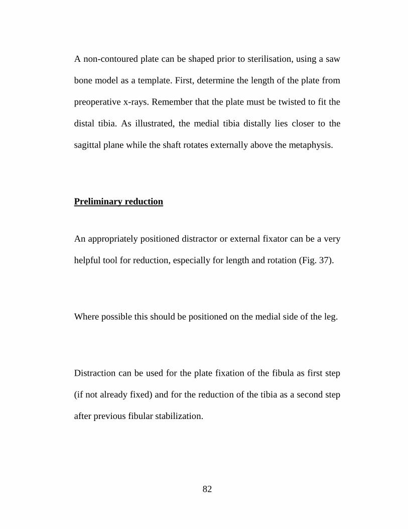
An appropriately positioned distractor or external fixator can be a very
helpful tool for reduction, especially for length and rotation (Fig. 37).
Where possible this should be positioned on the medial side of the leg.
Distraction can be used for the plate fixation of the fibula as first step
(if not already fixed) and for the reduction of the tibia as a second step
after previous fibular stabilization.
83
Fig. 37: Indirect reduction with a distractor
Schanz screws are positioned in safe zones of the tibial shaft and talar
neck (or the calcaneal tuberosity). In case of previously applied
joint-bridging fixator, the already existing Schanz screws can be used.
a. Plate insertion
Tibial length and rotation are restored indirectly with distractor or
external fixation. Angulation may be approximated in the same way,
but is definitively corrected by plate application.
84
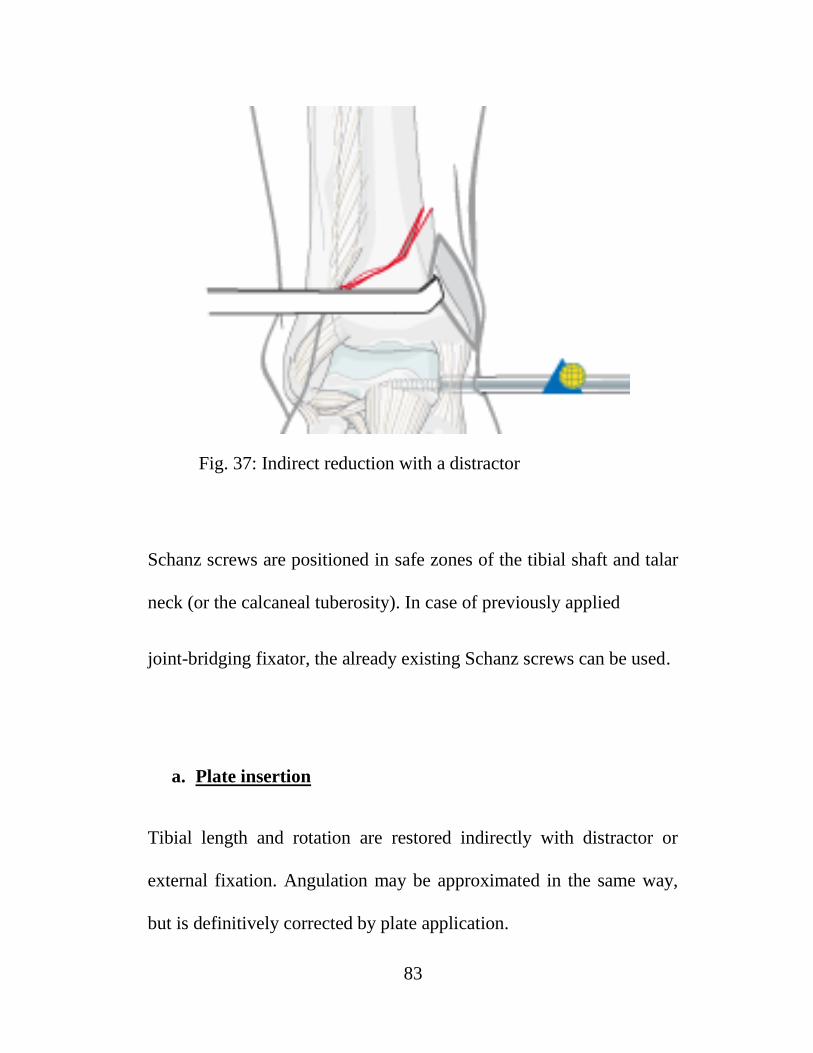
The plate is inserted through a distal medial oblique incision, after
proximal tunneling with a blunt instrument. Depending on the fracture
situation, the plate is usually positioned on the anteromedial aspect of
the tibia (Fig. 38).
Fig. 38: Inserting the plate.
Proximally, above the fracture zone, a small incision (2-3 cm) will aid
plate positioning. It is important that the plate and proximal screw be
centered on the tibia, particularly if locking head screws (LHS) are
planned.

85
Temporary fixation can be performed with K-wires through the screw
holes to approximate final plate position before insertion of screws
(Fig. 39).
Fig. 39: Preliminary plate stabilization.
After achieving accurate position of the plate, insert a conventional
cortical screw in one of the most distal plate holes to approximate the
plate to the bone.

86
Alternatively, the plate can be manually pressed to the bone, to allow
the insertion of a locking head screw instead of the conventional
screw.
It is crucial that the plate is positioned very close to the bone,
especially at the supramalleolar level, to prevent soft-tissue irritation
by the plate.
b. Applying compression
For spiral and short oblique fracture patterns (A1.1 and A1.2) that are
anatomically reduced, it is possible to place a lag screw through the
plate to enhance the overall construct stability (Fig. 40).

87
Fig. 40: Applying interfragmentary compression with a lag screw
It is possible to apply this screw in a percutaneous fashion under image
intensifier control. Alternatively, depending on the fracture plane, the
lag screw can be placed independent of the plate.

88
Compression with plate tension
For transverse type A1.3 fractures, fracture compression is achieved
by applying tension with the plate, using eccentric placement of
screws in non-locking holes, or an external tension device.
To ensure that the opposite side of the fracture remains compressed, it
is necessary to add a subtle convex prebend to the implant at the
fracture level.
c. Finish plate fixation
Further proximal and distal screw insertion is completed. The number
and position of the screws required depends on the fracture pattern
and bone quality. Ideally the concept of ―balanced‖ fixation should be
achieved (Fig. 41).

89
Fig. 41: Final fixation.
Usually, the metaphysis requires more screws (3-5) than the diaphysis
(2-3). In osteoporotic bone, the number of screws must be increased on
both sides of the fracture.
Locking head screws (LHS) may improve fixation in osteoporotic
bone and short periarticular segments.

90
Wound closure
Atraumatic skin sutures are used for closure of screw insertion
wounds. Occasionally, additional deeper sutures are needed for distal
and proximal incisions (Fig. 42,43).
Fig. 42: Wound before closure.

91
Fig. 43: Wound after closure.
d. Final assessment
Anatomical reduction and fixation can be confirmed by taking X-rays
at the end of the surgery.
It is important to check with imaging in both planes that a previously
unrecognized split into the articular surface has not been displaced
during this procedure.

92
13. COMPLICATIONS
The surgical treatment for periarticular fractures of distal tibia now has
a better outcome than in the past because of improved implants.
However the new methods are not without problems.
a. Complications of fractures:
i. Infection
ii. Vascular injuries
iii. Nerve injuries
iv. Nonunion
v. Malunion
vi. Missed ligamentous injuries
vii. Ankle stiffness
b. Complications of operative treatment:
i. Skin necrosis
ii. Incomplete reduction
iii. Incongruous reduction
iv. Loss of ankle motion
v. Infection

93
INFECTION:
The major drawback of operative fixation of peri articular fractures of
distal tibia is the risk of infection.
However it should not exceed 5%. If wound drainage develops
postoperatively, aggressive irrigation and debridement are indicated.
Appropriate antibiotics should be given intravenously for 3 to 6 weeks.
In case of infection, the implants are preferably retained because
stable infected fractures are easy to manage than unstable infected
fractures. If the implant is loose, it should be removed and the fracture
protected by external Fixation.
NONUNION:
It is much more common in conservatively treated cases than in
surgically treated cases, owing in part to the poor blood supply to the
distal tibia. Nonunion generally is due to presence of infection,
unstable fixation, mechanical failure of the implant or any combination
of these factors.

94
Treatment may be difficult owing to preexisting osteopenia, proximity
to ankle joint and prior surgical procedures. Aseptic nonunion should
be treated by repeat osteosynthesis. Septic nonunion should be treated
with external stabilization.
POST TRAUMATIC ARTHRITIS:
The incidence of post traumatic arthritis is unknown. However
incongruity of the joint surface is the leading cause of the early
arthritis. This complication can be reduced by anatomical reduction
and early mobilization. In patients with severe arthritis, ankle
arthrodesis may be indicated. Factors such as age, range of motion,
presence or absence of contractures and infection play a vital role in
surgical decision making.
ANKLE STIFFNESS:
Perhaps the most common complication that occurs after tibial pilon
fracture is loss of ankle motion. This complication results from
damage to joint surface due to initial trauma or surgical exposure for
fixation or both.
95
Arthrofibrosis of the ankle joint is thought to restrict ankle movement.
Both immobilization after fracture and internal fixation can magnify
these effects. Ankle immobilization for more than 3 weeks usually
results in some degree of stiffness.
Early fixation of the fracture with minimal soft tissue handling and
early mobilization increase the chance for an optimal outcome after
periarticular fractures of distal tibia. Patients should attain full range of
ankle movements 4 weeks postoperatively.
VASCULAR INJURIES:
The exact incidence of vascular injury accompanying distal tibial
fracture is unknown. Vascular injuries can be caused by direct
laceration (or) contusion of the artery or vein by fracture fragments or
indirectly by stretching leading to initial damage, clinical examination
for signs of ischemia with evaluation of pulses and motor and sensory
function is essential.

96
MALUNION:
Malunion of tibial pilon fractures, distorts the articular surface of the
ankle and produces much more severe disability. It should be corrected
and internally fixed maintaining the articular surface.
ASSOCIATED LIGAMENTOUS INJURIES:
Concomitant ligamentous injuries to the ankle are common but are
rarely diagnosed preoperatively. Initially non operative treatment is
advocated as repair (or) reconstruction may produce further
comminution, prolonged operation time and increases the risk of loss
of ankle motion and infection.
Protected motion in conjunction with vigorous rehabilitation may
obviate the need for late reconstructive surgery. If necessary late
reconstruction should be done after the fracture has healed.

97
14. EVALUATION OF OUTCOME
We follow Tornetta et al scoring system for evaluation of the ankle
joint.
GRADE PAIN ROM ANGULATION
EXCELLENT None Dorsiflexion > 50
Plantarflexion > 300
<30
valgus
GOOD Intermittent Dorsiflexion 0 - 50
Plantarflexion 20 - 300
3-50
valgus
< 30 varus
FAIR Limiting daily
Activities
Dorsiflexion -5 to 00
Plantarflexion 15-200
5-80 valgus
3-50 varus
POOR Intractable Dorsiflexion < -50
Plantarflexion < 150
>80 valgus
>50 varus
Table 3: Tornetta et al scoring system.

98
15. MATERIALS AND METHODS
The period of surgery and follow up extends from September 2012 to
September 2014.
Inclusion Criteria :
1. Age >/= 20 years
2. Closed, unstable fractures of distal tibia
3. Grade I & II compound distal tibia fractures
4. Fractures in which acceptable closed reduction can be achieved.
Exclusion Criteria :
1. Grade III Open fractures
2. Irreducible fracture deformity
3. Compartment Syndrome / poor local skin conditions
4. AO type C3 fractures (articular comminution).

99
The cases were analyzed as per the following criteria.
i. Age distribution
ii. Sex distribution
iii. Side of injury
iv. Mode of injury
v. Anatomy of fracture
vi. Associated injuries
vii. Open fractures
viii. Duration for surgery
ix. Time of union
100
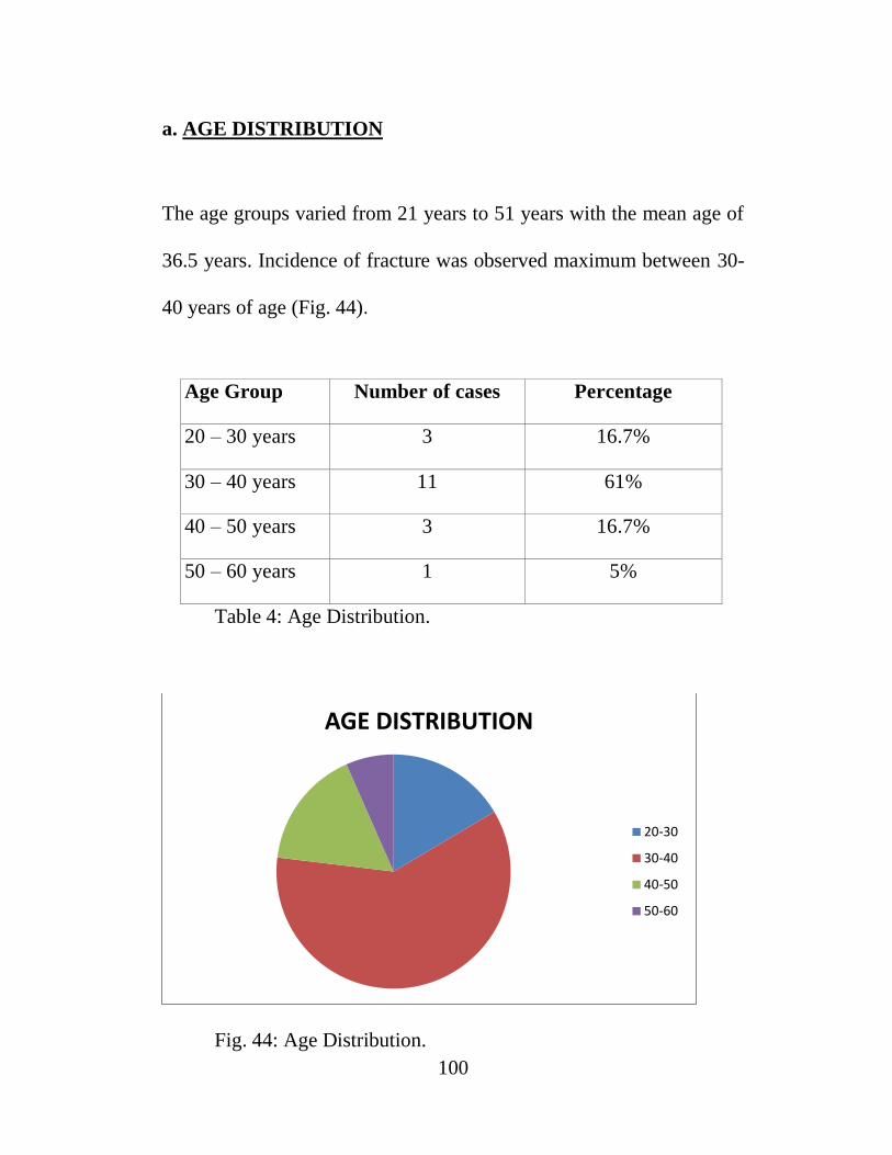
a. AGE DISTRIBUTION
The age groups varied from 21 years to 51 years with the mean age of
36.5 years. Incidence of fracture was observed maximum between 30-
40 years of age (Fig. 44).
Age Group Number of cases Percentage
20 – 30 years 3 16.7%
30 – 40 years 11 61%
40 – 50 years 3 16.7%
50 – 60 years 1 5%
Table 4: Age Distribution.
Fig. 44: Age Distribution.
AGE DISTRIBUTION
20-30
30-40
40-50
50-60
101
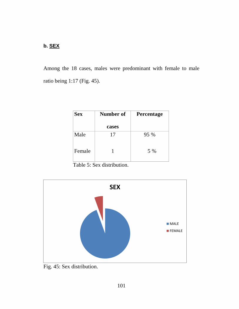
b. SEX
Among the 18 cases, males were predominant with female to male
ratio being 1:17 (Fig. 45).
Sex Number of
cases
Cases
Percentage
Male
Female
17
1
95 %
5 %
5 %
Table 5: Sex distribution.
Fig. 45: Sex distribution.
SEX
MALE
FEMALE

102
c. SIDE OF INJURY:
Right side was common in our series in the ratio of 5:4 (Fig. 46).
Sex Right Left
Male
Female
10
0
7
1 Total 10 8
Percentage 56% 44%
Table 6: Side of injury.
Fig. 46: Side of injury.
0
1
2
3
4
5
6
7
8
9
10
MALE FEMALE
RIGHT
LEFT
103
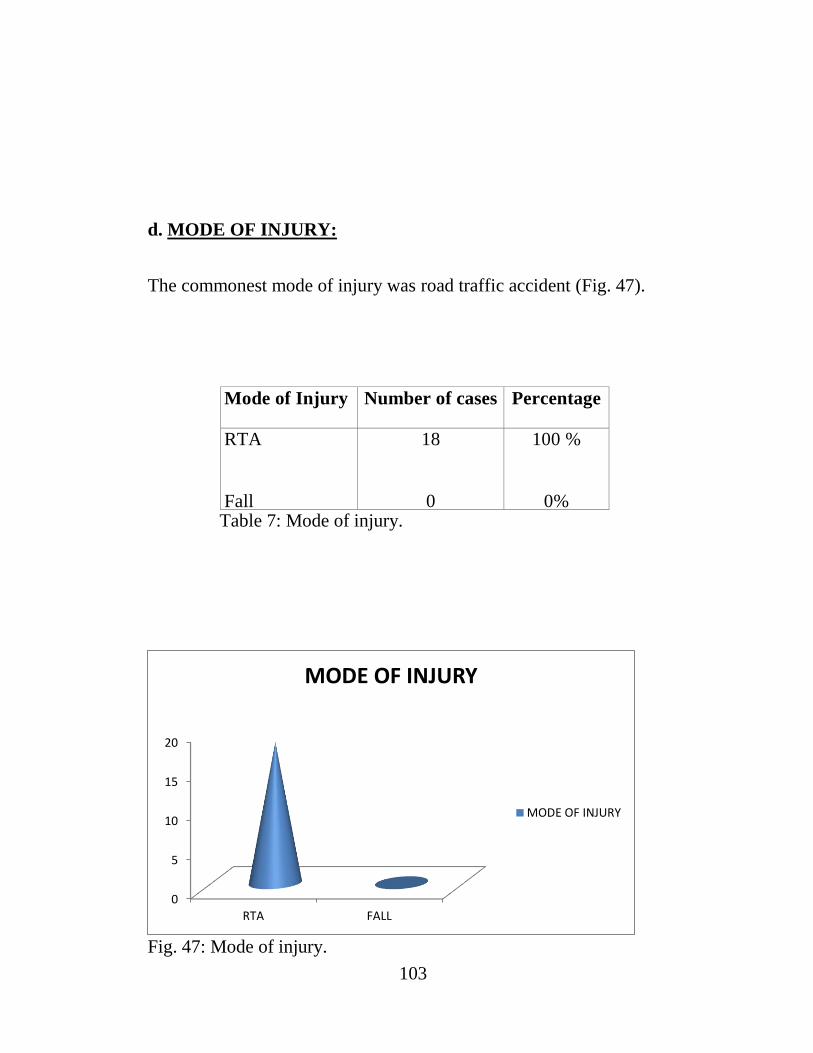
d. MODE OF INJURY:
The commonest mode of injury was road traffic accident (Fig. 47).
Mode of Injury Number of cases Percentage
RTA
Fall
18
0
100 %
0% Table 7: Mode of injury.
Fig. 47: Mode of injury.
0
5
10
15
20
RTA FALL
MODE OF INJURY
MODE OF INJURY

104
e. ANATOMY
The study contains equal number of intra articular and extra articular
fractures (Fig. 48).
SITE Number of cases Percentage
Intra Articular
Extra Articular
9
9
50%
50%
Table 8: Fracture Anatomy.
Fig. 48: Fracture Anatomy.
FRACTURE ANATOMY
INTRA ARTICULAR
EXTA ARTICULAR

105
f. ASSOCIATED INJURIES
Eight among the eighteen cases had associated injuries (Fig. 49).
Head injury – 2
Distal radius fractures - 1
Patella fracture - 4
Supracondylar fracture femur - 1
Table 9: Associated injuries.
Fig. 49: Associated injuries.
0
0.5
1
1.5
2
2.5
3
3.5
4
4.5
HEADINJURY
DISTALRADIUS
FRACTURES
PATELLAFRACTURES
SUPRACONDYLAR
FEMUR
ASSOCIATED INJURIES
ASSOCIATED INJURIES

106
g. OPEN FRACTURES
Eight out of the eighteen cases were open fractures (Fig. 50).
Type Number of cases Percentage
Simple 10 56 %
Compound Gr I 5 28 %
Compound Gr II 3 16 %
Table 10: Fracture type.
Fig. 50: Fracture type.
FRACTURE TYPE
SIMPLE
GRADE I COMPOUND
GRADE II COMPOUND

107
h. DURATION FOR SURGERY
There was a mean delay of 1 week for surgery (Fig. 51).
TIME INTERVAL Number of cases Percentage
1 day
< 1 week
<2 weeks
6
8
4
34
45
21
Table 11: Duration for surgery.
Fig, 51: Timing of surgery.
TIMING OF SURGERY
1 DAY
< 1 WEEK
< 2 WEEKS

108
i. TIME FOR UNION
The mean time for bone union was 18 weeks (Fig. 52).
Time of union Number of cases Percentage
< 16 weeks
16-24 weeks
>6 months
11
4
2
66
23
11 Table 12: Time for union.
Fig. 52: Time for union.
TIME FOR UNION
<16 WEEKS
16-24 WEEKS

109
OBSERVATIONS
i. 80% of the patients were between 30- 50 yrs.
ii. Both male and female were included , majority being males.
iii. Right side was common and no bilateral cases were studied.
iv. 44% of the fractures were compound injuries.
v. 44% of patients had associated injuries.
vi. Mean duration between injury and surgery was 1 week.
vii. Average time for bone union was 18 weeks.
viii. Average ankle dorsi flexion was 20 degrees.
ix. The results were excellent in 54%, good in 29% and fair in 17%
of patients.

110
16. PROCEDURE
a. General Measures
All the patients were received in the casualty department and were
resuscitated. After the general condition improved X rays AP and
lateral views were taken. A detailed preoperative work up was done.
All the cases were taken for surgical procedure as soon as possible.
Those cases which were compound were initially treated with external
fixator.
b. Post Operative Protocol:
Limb elevation is recommended for the first 2-5 postoperative days.
Physiotherapy with active assisted exercises is started immediately
after operation.
Immobilization is not necessary.
Clinical and radiological follow-up is advised after 2, 6 and 12 weeks.

111
Based on the fracture consolidation, weight bearing can be
progressively increased from 6-8 weeks with full weight bearing
usually after 3 months.
Supervised rehabilitation with intermittent clinical and radiographic
follow-up is advisable every 6-12 weeks until recovery reaches a
plateau, typically 6-12 months after injury
Weight-bearing radiographs are preferable to assess articular cartilage
thickness. Angular stable fixation may obscure signs of non-union for
many months.
c. Implant removal
Implant removal may be necessary in cases of soft-tissue irritation
by the implant (plate and screws). The best time for implant
removal is after complete remodeling, usually at least 12 months
after surgery.
In our study all the patients were followed up carefully looking for any
complication every fortnightly till fracture healing; and there after
every month up to 6 months; and every 6 months up to two years.

112
17. ANALYSIS OF FUNCTIONAL OUTCOME
Of the 18 patients included in the study one patient died in the late post
operative period due to co-morbid medical conditions . Other patients
are evaluated and studied for functional outcome (Fig. 53).
Normal bone union – 15
Delayed union – 2
Shortening – 2
Joint stiffness – 2
Valgus angulation – 2
Marginal Skin necrosis – 4
Deep infection -- 2

113
Fig. 53: Complications.
OVERALL RESULTS
The overall results of our study are much in favour of Minimally
Invasive Plate Osteosynthesis for distal tibia fractures (Fig. 54).
The post operative pain was minimal and the post operative ankle
function was very good.
Though we had marginal skin necrosis in 4 cases, they healed with
regular dressings and none of the cases went for skin and soft tissue
procedures.
0
2
4COMPLICATIONS

114
GRADING NO OF CASES PERCENTAGE
EXCELLENT 9 54
GOOD 5 29
FAIR 3 17
Table 13: Overall results.
Fig. 54: Overall results.
RESULTS
EXCELLENT
GOOD
FAIR
POOR
115
18. DISCUSSION
Ruedi and Allgower52
were the pioneers in open reduction and internal
fixation (ORIF) of pilon fractures. They changed the outlook of
management of distal tibia fractures in the early twentieth century.
They achieved 74% good functional results following ORIF for distal
tibia fractures. But it was later recognized that all their cases were
results of low velocity injuries.53,54
They could not reproduce similar
results following the principles of open reduction internal fixation in
high velocity injuries55
.
This led to the development of procedures that respect the soft tissue
envelope. These biological methods of fixation are currently the
procedures of choice in the challenging distal tibia fractures.
Two methods are currently popular in pilon fracture management.
- Hybrid external fixators are used in severely comminuted
pilon fractures with significant soft tissue damage.

116
- Minimally invasive plate osteosynthesis (MIPO), is used
in fractures without articular comminution and with
minimal soft tissue damage.55,56
ORIF for pilon fractures has a high complication rate of
- nonunion 18%,
- mal-unions 42%.
63-67
- superficial infections 20%,
- osteomyelitis 17%,
- post-traumatic osteoarthrosis 54%
- arthrodesis 27%,
- below knee amputation 6%,
Helfet et al.68
introduced a 2 stage MIPO for distal tibia fractures.
Stage 1 – fibular internal fixation and spanning external fixation of
tibia
Stage 2 – limited ORIF for distal tibia.

117
40% of their cases were intra articular fractures
60% were extra articular fractures.
They had a 10% incidence of >50 valgus deformity and
a 10% incidence of > 100 recurvatum deformity.
The average ankle dorsiflexion achieved was 14º and plantar flexion
was 42º.
Ours is a prospective study of 18 cases of distal tibial fractures
treated with MIPO using specially designed distal tibial LCP.
We did medial plating in all cases.
The age group of our patients varied from 21 years to 51 years with the
mean age of 36.5 years.
95% of our patients were males.
50% of our cases were extra articular and 50% intra articular fractures.
44% of the fractures were compound in nature.
44% of our cases had associated injuries.

118
We did not perform preliminary external fixation as in the Helfet et
al.'s68
series. We selected patients with apparently good soft tissue
condition. Thus a single stage MIPO protocol was followed thereby
providing a shorter duration of treatment. This single stage procedure
reduced the surgical insult thus preventing complications like wound
dehiscence, sepsis, delayed or non-union. The MIPO technique
enables a bridging fixation without disturbing the comminuted
segments and the surrounding soft tissue.
We used an anatomically prebent plate unlike Helfet et al.68
thus
achieving stronger fixation in the metaphyseal region as it permitted
insertion of 2 or 3 cancellous 6.5 mm screws in the small distal
segment.
The mean duration between injury and surgery in our study was 1
week.
The average time for bone union was 18 weeks.
We achieved 54% excellent, 29% good and 17% fair results.
The average ankle dorsiflexion was 200 .

119
The incidence of complications
- Delayed union – 11%
- Shortening - 11%
- Ankle stiffness – 11%
- Valgus angulation- 11%
- Marginal skin necrosis - 22%
- Deep infection - 11%
All our cases were followed for a mean period of 14.2 months
averaging from 28 months to 4 months.
Out of the 18 cases bony union was obtained in 17 cases ( one patient
died during follow up ) .
2 cases had delayed union. The prime reason for delayed union in both
the cases was intact fibula which made the fracture site to distract.
There was no case of implant failure.
The average time of bony union was 18 weeks compared to 18.5
weeks by Shrestha et al and 21.2 weeks by Hasenboehler et al.
120
There were 2 cases that were complicated by ankle stiffness. Both the
patients had poor compliance in the post operative period which was
the result of ankle stiffness.
Shortening of <2 cm was seen in two patients both of which had
highly communited distal tibial fractures with diaphyseal extension.
They were managed with heel raise.
Though we had marginal skin necrosis in 4 cases, they healed with
regular dressings and none of the cases went for skin and soft tissue
procedures.
Thus, with regards to functional outcome, our results are comparable
to those of Shrestha et al1 (Table 14). These results were possible
because of proper case selection, perfect articular reconstruction and
meticulous soft tissue handling.

121
S.NO
.
CRITERIA CURRENT
STUDY
SHRESTHA
ET AL1
HASENBOEHLER
ET AL9
1 STUDY
TYPE
PROSPEC
TIVE
PROSPEC
TIVE
RETROSPECTIVE
2 NO. OF
CASES
18 20 32
3 OPEN
FRACTURES
8 (44%) 8 (40%) 13 (40.6%)
4 INTRA
ARTICULAR
#
9 (50%) 2 (10%) 12 (37.5%)
5 SURGERY
DELAY
7 DAYS 4.45 DAYS 6 DAYS
6 UNION
TIME
18
WEEKS
18.5
WEEKS
21.2
WEEKS
7 DELAYED
UNION
2 (11%) 1 (5%) 6 (18.75%)
8 NON
UNION
0 0 2 (6%)
9 VALGUS 2 (11%) 2 (10%) 4 (12.5%)
10 SKIN
NECROSIS
4 (22%) 2 (10%) 7 (21.9%)
11 DEEP
INFECTION
2 (11%) 1 (5%) 4 (12.5%)
12 SHORTENIN
G
2 (11%) 2 (10%) 3 (9%)
13 ANKLE
STIFFNESS
2 (11%) 2 (10%) 4 (12.5%)
Table 14: COMPARISON WITH OTHER STUDIES.

122
19. CONCLUSION
To summarize,
The advantages of locking compression plate are
i. provides a biomechanicaly stable construct
ii. does not endanger periosteal blood supply
iii. preserves the fracture heamatoma
The advantages of Minimally Invasive Plating are
i. fracture fixation without disturbing the soft tissue cover
ii. less chances of infection
iii. early mobilization of the adjacent joint.
Thus minimally invasive plate osteosynthesis using LCP proves to be a
safer technique in the management of distal tibial fractures without
intra-articular comminution by providing good fracture healing,
enabling rapid functional recovery, and avoiding major skin
complications.

123
20. BIBLIOGRAPHY
1. Shrestha D, Acharya BM, Shrestha PM. Minimally invasive plate
osteosynthesis with locking compression plate for distal
diametaphyseal tibia fracture. Kathmandu Univ Med J 2011;34(2)62-8.
2. Ronga M, Longo UG, Maffulli N. Minimally invasive locked
plating of distal tibia fractures is safe and effective. Clin Orthop Relat
Res 2010, 468:975–82.
3. Mosheiff R, Safran O, Segal D, Liebergall M. The unreamed tibial
nail in the treatment of distal metaphyseal fractures. Injury
1999,30:83–90.
4. Redfern DJ, Syed SU, Davies SJM. Fractures of the distal tibia:
minimally invasive plate osteosynthesis. Injury 2004, 35:615– 620.
5. Apivatthakakul T, Phornphutkul C, Patumasutra S. Idea and
innovation: Simple minimally invasive plate osteosynthesis (MIPO)
instruments. Injury Extra 2009,40:39–44.

124
6. Vallier HA, Le TT, Bedi A. Radiographic and clinical comparisons
of distal tibia shaffractures (4 to 11 cm proximal to the plafond):
plating versus intramedullary nailing. J Orthop Trauma 2008, 22:307-
11.
7. Yang SW, Tzeng HM, Chou YJ, Teng HP, Liu HH, Wong CY.
Treatment of distal tibial metaphyseal fractures: Plating versus
shortened intramedullary nailing. Injury 2006, 37:531–535.
8. Guo JJ, Tang N, Yang HL, Tang TS. A prospective, randomised
trial comparing closed intramedullary nailing with percutaneous
plating in the treatment of distal metaphyseal fractures of the tibia. J
Bone Joint Surg Br 2010, 92-B:984-8.
9. Hasenboehler E, Rikli D, Babst R. Locking compression plate with
minimally invasive plate osteosynthesis in diaphyseal and distal tibial
fracture: a retrospective study of 32 patients. Injury 2007, 38:365– 70.
10. Gun-Il Im, Suk-Kee Ta. Distal metaphyseal fractures of tibia: A
prospective randomized trial of closed reduction and intramedullary
nail versus open reduction and plate and screws fixation. J Trauma
2005,59:1219–1223.

125
11. Watson JT, Moed BR, Karges DE, Cramer KE. Pilon fractures.
Treatment protocol based on severity of soft tissue injury. Clin Orthop
Relat Res 2000, 375:78–90.
12. Wagner M, Frigg FR. Locked plating: Biomechanics and biology
and locked plating: Clinical indications. Techniques in Orthopaedics
2007, 22(4) :209–218.
13. Frigg R. Development of the locking compression plate. Injury
Suppl 2003, 2:B6–B10.
14. Ahmad MA, Sivaraman A, Zia A, Rai A, Patel AD. Percutaneous
locking plates for fractures of the distal tibia: Our experience and a
review of the literature. J Trauma 2010, doi:
10.1097/TA.0b013e3181f140b3
15. Collinge C, Protzman R. Outcomes of minimally invasive plate
osteosynthesis for metaphyseal distal tibia fractures. J Orthop Trauma
2010, 24:24–29.
16. Trafton PG .Tibial Shaft Fracturs. In: Skeletal trauma. 4th edn.
Edited by Browner DB, Jupiter JB, Levine AN, Trafton PG, Krettek C.
Philadelphia: Suanders Elsevier; 2009:2319-2452.

126
17. Megas P, Zouboulis P, Papadopoulos AX, Karageorgos A,
Lambiris E. Distal tibial fractures and non-unions treated with
shortened intramedullary nail. Inter Orthop 2003, 27:348–35.
18. Joveniaux P, Ohl X, Harisboure A, Berrichi A, Labatut L, Simon
P, Mainard D, Vix N, Dehoux E. Distal tibia fractures: management
and complications of 101 cases. Inter Orthop 2010, 34:583–588.
19. Borrelli J, Prickett W, Song E, Becker D, Ricci W. Extra osseous
blood supply of the distal tibia and the effects of different plating
techniques: Human cadaveric study. J Orthop Trauma 2002, 16:691–
695.
20. Mushtaq A, Shahid R, Asif M, Maqsood M. Distal tibial fracture
fixation with locking compression plate (LCP) using the minimally
invasive percutaneous osteosynthesis (MIPO) technique. Eur J
Trauma Emerg Surg 2009, 35:159–64.
21. Hazarika S, Chakravarthy J, Cooper J. Minimally invasive locking
plate osteosynthesis for fractures of the distal tibia-results in 20
patients. Injury 2006, 37:877– 887.
127
22. Shanmugam C, Rahmatalla A, Maffulli N. Percutaneous fixation
of distal tibial fractures using locking plates. Techniques in
Orthopaedic 2007, 22(3):162–166.
23. Gupta RK, Rohilla RK, Sangwan K, Singh V, Walia S. Locking
plate fixation in distal metaphyseal tibial fractures: series of 79
patients. Inter Orthop 2010, 34:1285–1290.
24. Gao H, Zhang CQ, Luo CF, Zhou ZB, Zeng BF. Fractures of the
distal tibia treated with polyaxial locking plating. Clin Orthop Relat
Res 2009, 467:831- 837.
25. Bahari S, Lenehan B, Khan H, Mcelwain JP. Minimally invasive
percutaneous plate fixation of distal tibia fractures. Acta Orthop Belg
2007, 73:635-640.
26. Cheng W, Li Y, Manyi W. Comparison study of two surgical
options for distal tibia fracture-minimally invasive plate osteosynthesis
vs. open reduction and internal fixation. Inter Orthop 2010, doi:
10.1007/ s00264-010-1052-2
27. Kao FC, Tu YK, Hsu KY, Wu CH, Yen Cy, Chou MC. Treatment
of distal tibial fractures by minimally invasive percutaneous plate

128
osteosynthesis of three different plates: Results and cost-effectiveness
analysis. Formosan Journal of Musculoskeletal Disorders 2010, 1:35–
40.
28. Bedi A, Le TT, Karunakar MA. Surgical treatment of nonarticular
distal tibia fractures. J Am Acad Orthop Surg 2006, 14:406–416.
29. Collinge C, Kuper M, Larson K, Protzman R. Minimally invasive
plating of high energy metaphyseal distal tibia fractures. J Orthop
Trauma 2007, 21:355-61.
30. Lau TW, Leung F, Chan CF, Chow SP. Wound complication of
minimally invasive plate osteosynthesis in distal tibia fractures. Inter
Orthop 2008, 32:697-703.
31. Ozsoy MH, Tuccar E, Demiryurek D et al. Minimally invasive
plating of the distal tibia: do we really sacrifice saphenous vein and
nerve? A cadaver study. J Orthop Trauma 2009, 23(2):132–138.
32. Bae JH, Oh JK, Oh CW, Hur CR. Technical difficulties of removal
of locking screw after locking compression plating. Arch Orthop
Trauma Surg 2009, 129:91-5.

129
33. Blatter G, Weber BG. Wave plate osteosynthesis as a salvage
procedure. Arch Orthop Trauma Surg 1990;109:330–3.
34. Gautier E, Perren SM, Ganz R. Principles of internal fixation. Curr
Orthop 1992;6:220–32.
35. Gautier E, Ganz R. [The biological plate osteosynthesis] Zentralbl
Chir 1994;119:564–72.
36. Haas NP, Schütz M, Hoffmann R, et al. LISS — Less Invasive
Stabilization System: ein neuer Fixateur interne für distale
Femurfrakturen. OP J 1997;13:340–4.
37. Hansmann C. Eine neue Methode der Fixierung der Fragmente bei
complicierten Frakturen. Verh Dtsch Ges Chir 1886;158.
38. Jörger K. Akute intrakortikale Durchblutungsstörung unter
Osteosyntheseplatten mit unterschiedlichen Auflageflächen. Zentrubl
Chir 1987;1:75.
39. Luthi U, Rahn BA, Perren SM. [Area of contact between
osteosynthesis plate and bone in internal fixation.] Akt Traumatol
1980;10:131–6.

130
40. Mast JW, Jakob RP, Ganz R. Planning and reduction technique in
fracture surgery. Berlin: Springer; 1989.
41. Müller ME, Allgöwer M, Schneider R, et al. Manual der
Osteosynthese. Berlin: Springer; 1992.
42. Perren SM. The concept of biological plating using the limited
contact-dynamic compression plate (LC-DCP): scientific background,
design and application. Injury 1991;22(suppl 1):1–41.
43. Ramotowski W, Granowski R. Zespol: an original method of
stable osteosynthesis. Clin Orthop 1991;272:67–75.
44. Schandelmaier P, Krettek C, Ungerland A, et al. [Pre-bending and
tension adjustment of narrow 4.5mm AO titanium LC-DCP
(limited contact dynamic compression plate).] Langenbecks Arch Chir
1997;382:325–31.
45. Schavan R, Frigg R, Angst M. LISS – ein neuartiges minimal
invasives extramedulläres Fixationssystem für Metaphysäre
Frakturen der unteren Extremität. Hefte Unfallchirurg 1997;702– 6.
46. Schutz M, Müller M, Krettek C, et al. Minimally invasive fracture
stabilization of distal femoral fractures with the LISS: a prospective

131
multicenter study; results of a clinical study with special emphasis on
difficult cases. Injury 2001;32:48–54.
47. Tepic S, Perren SM. The biomechanics of the PC-Fix internal
fixator. Injury 1995;26:5–10.
48. Vattolo M. Der Einfluss von Rillen in Osteosyntheseplatten auf
den Umbau der Korticalis. Zentralbl Chir 1986;1:89.
49. Wagner M, Frigg R. Locking compression plate (LCP): ein neuer
AO Standard. OP J 2000;3:238–43
50. Muller ME, Nazarian S, Koch P. The AO Classification of
Fracture, translated by J Schatzker. Toronto: Springer, 1988.
51. Ruedi TP, Allgower M. Fractures of the lower end of the tibia into
the ankle joint. Injury. 1969; 1(2): 92-99.
52. Moll BN, Kerb B. Intra-articualr fractures of the distal tibia. Acta
Orthop Scand. 1982; 53: 991-996.
53. McFerran MA, Smith SW, Boulas HJ, Schwartz HS.
Complications encountered in the treatment of pilon fractures. J
Orthop Trauma. 1992; 6: 195-200.
132
54. de Boer P and Metcalfe R. Mini-symposium: Tibial fractures, (iv)
Pilon fractures of the tibia. Current Orthopaedics. 2003; 17, 190-199.
55. Probe R A. Minimally invasive fixation of tibial pilon fractures.
Operative Techniques in Orthopaedics. 2001, 11, 3: 205-217.
56. Gustilo R B, and Anderson J T. Prevention of infection in the
treatment of one thousand and twenty-five open fractures of long
bones: retrospective and prospective analyses. J Bone Joint Surg [Am].
1976; 58(4): 453-458.
57. Tscherne H, Gotzen L. Fractures with soft tissue injuries. Berlin:
Springer Verlag, 1984.
58. Teeny SM, Wiss DA. Open reduction and internal fixation of tibial
plafond fractures. Clin Orthop Relat Res. 1993; 292:108 --117.
59. Salton HL, Rush S, and Schuberth J. Tibial plafond fractures:
Limited incision reduction with percutaneous fixation. J Foot Ankle
Surg. 2007; 46 (4): 261--269.
60. Ruedi TP. Fractures of the lower end of the tibia into the ankle
joint: results 9 years after open reduction and internal fixation. Injury.
1973; 5: 130-- 134.
133
61. Ruedi TP, Allgower M. The operative treatment of intra-articular
fractures of the lower end of the tibia. Clin Orthop Relat Res. 1979;
138: 105--110.
62. Watson JT, Moed BR, KargesDE*, Cramer KE*. Pilon fractures:
treatment protocol based on severity of soft tissue injury. Clin Orthop
Relat Res. 2000; 375: 78 --90.
63. Bone L, Stegemann P, McNamara K, Seibel R. External fixation of
severely comminuted and open tibial pilon fractures. Clin Orthop Relat
Res. 1993; 292: 101--107.
64. French B, Tornetta P. Hybrid external fixation of tibial pilon
fractures. Foot Ankle Clin. 2000; 5: 853-- 871.
65. Pugh KJ, Wolinsky PR, McAndrew MP, Johnson KD. Tibial pilon
fractures: a comparison of treatment methods. J Trauma. 1999; 47:
937--941.
66. Syed MA, Panchbhavi VK. Fixation of tibial pilon fractures with
percutaneous cannulated screws. Injury. 2004; 35:284 --289.

134
67. Helfet DL, Shonnard PY, Levine D, Borrelli BJr. Minimally
invasive plate osteosynthesis of distal fractures of the tibia. Injury.
1997; 28, Suppl. 1, S.A42-S.A48.
68. Tornetta P, Weiner L, Bergman metal, Pilon fractures treated with
combined internal and external fixation. Journal of orthop trauma
1993, vol 7, No 489-496.

135
21. ILLUSTRATIONS
CASE 1 :
PRE OP X RAYS
POST OP X RAYS
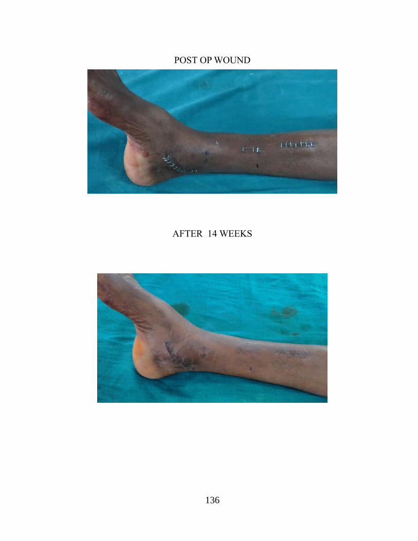
136
POST OP WOUND
AFTER 14 WEEKS

137
FRACTURE UNION

138
CASE 2 :
PRE OP X RAYS
POST OP X RAYS

139
POST OP RANGE OF MOVEMENTS
AFTER 16 WEEKS

140
CASE 3 :
PRE OP XRAY
POST OP X RAY

141
AFTER 14 WEEKS
HEALED SCAR

142
22. PROFORMA
TOPIC : A CASE STUDY OF DISTAL TIBIAL
FRACTURES MANAGED WITH LOCKING
COMPRESSION PLATE USING MIPO TECHNIQUE
Dr. P.VANAJ KUMAR
NAME:
I.P.NO:
ADDRESS :
AGE / SEX :
D.O.A :
D.O.S :
D.O.D :
Presenting complaints :
I f female whether Pre- / Post-menopausal:
Examination:
X-Ray :
Diagnosis :
Treatment:
Post op protocol:

143
Advice on discharge:
FOLLOW-UP NOTES
Time since
surgery
X-Ray
Findings
Range of
Movements
Functional
Assessment
Any
complications
Special Points:

144
23. MASTER CHART
S.
NO
NAME AGE/
SEX
I.P.
NO
DIAGNOSIS ASS.
INJURY
TREATMENT OTHER
PROCEDU
RE
TIME
FOR
UNION
ANKLE
MOVEMEN
TS
COMPLICA
TIONS
RESULT
DF PF
1 Joseph
raj
38/M 37630 Simple
pilon # rt.
Rt
Head
Injury
MIPO with LCP
ORIF fibula
- 22
weeks
30 40
- Excellent
2 Kaveri
samy
32/M 28621 CompGr.II
Distal tibia # lt.
Rt
- MIPO with LCP - 28
weeks
20 40 Wound
infection
Delayed
union
Good
3 Ganesan 34/M 51085 CompGr.I
Distal tibia # lt.
Lt
Verti cal
patella #
MIPO with LCP
OR & IF
with 4mm
malleolar
screw for
patella
16
weeks
20 40
Skin
necrosis
Excellent
4 Uthaminat
han
21/M 39610 Comp.Gr.II
tibial pilon # rt.
Comp.Gr.
IIIB SC #
femur
MIPO with LCP
ORIF fibula
OR & IF of
SC # with
LCP
24
weeks
20 40
Wound
infection
Good
5 Antony
raj
33/M 29781 Simple distal
BB leg # lt.
L ¼ left
- MIPO with LCP - 14
weeks
20 40 - Excellent
6 Ranjitham
36/F 38210 Simple pilon #
lt.
BB leg # L ¼
left
- MIPO with LCP
ORIF fibula
- 16
weeks
20 40 - Excellent

145
7 David
rajan
27/M 22655 Simple
tibial pilon # rt.
Communi
ted
patella #
MIPO with LCP Patellectom
y
14
weeks
20 40 - Excellent
8 Kadarkar
ai
45/M 23782 CompGr.I distal
tibia# rt.
Distal
radius #
MIPO with LCP CR & IF
RADIUS
16
weeks
20 40 Skin
necrosis
Good
9 Gomathin
ayagam
40/M 31274 Simple
tibial pilon # lt.
Communi
ted
patella #
MIPO with LCP OR & IF of
patella
20
weeks
20 40 - Excellent
10 Muthuraj 36/M 29241 Simple distal
BB leg # lt.
L 1/3 Rt
-
MIPO with LCP
- 14
weeks
20 30 - Excellent
11 Antony 22/M 30081 Simple distal BB
leg # lt.
- MIPO with LCP
ORIF fibula
- 16
weeks
20 40
5 degree
Valgus
Fair
12 Marudhu 45/M 32817 Simple leg
# distal BB leg
rt.
- K wire for fibula
MIPO with LCP
- -
Death
13 Saravana
n
38/M 36281 CompGr.I
Pilon # lt.
- MIPO with LCP
ORIF fibula
-
32
weeks
10 20 Ankle
Stiffness
delayed
union
Fair
14 mariappan
hameed
35/M 31181 Comp.GrI distal
BB leg rt.t
- MIPO with LCP - 18
weeks
10 40 Skin
necrosis
Good
15 Kannan 36/M 28121 Simple tibial
Pilon # rt.
Type VI
- MIPO with LCP
ORIF fibula
- 14
weeks
20 40 Shortening
0.5 cm
Good

146
16 Vembu 42/M 30012 Simple tibial
pilon # rt.
Type VI
- MIPO with LCP
ORIF fibula
- 14
weeks
20 40 Skin
necrosis
Excellent
17 Paulraj 40/M 27181 Comp.Gr.I
tibial pilon # rt.
Commu
nited
patella #
MIPO with LCP OR & IF of
patella
with TBW
14
weeks
10 30 5 0 valgus
Shortening
1 cm
Fair
18 manikand
an
51/M 21261 CompGr.II
BB leg # rt.
- MIPO with LCP
ORIF fibula
14
weeks
20 40 - Excellent