a case report on oral subcutaneous dirofilariasis
TRANSCRIPT

Case ReportA Case Report on Oral Subcutaneous Dirofilariasis
R. D. Jayasinghe,1 S. R. Gunawardane,2 M. A. M. Sitheeque,1 and S. Wickramasinghe3
1Department of Oral Medicine and Periodontology, Faculty of Dental Sciences, University of Peradeniya, 20400 Peradeniya, Sri Lanka2Department of Oral and Maxillofacial Surgery, Faculty of Dental Sciences, University of Peradeniya, 20400 Peradeniya, Sri Lanka3Department of Parasitology, Faculty of Medicine, University of Peradeniya, 20400 Peradeniya, Sri Lanka
Correspondence should be addressed to R. D. Jayasinghe; [email protected]
Received 11 November 2015; Revised 4 December 2015; Accepted 6 December 2015
Academic Editor: Gernot Walder
Copyright © 2015 R. D. Jayasinghe et al.This is an open access article distributed under theCreative CommonsAttribution License,which permits unrestricted use, distribution, and reproduction in any medium, provided the original work is properly cited.
Dirofilariasis is an uncommon zoonotic parasitic infection affecting human.The natural hosts for this nematode are animals such asdogs, cats, foxes, jackals, and raccoons. This disease is endemic in South Eastern United States, Australia, Europe, and Central andSouthern Asia. Dirofilaria immitis and D. repens are the common mosquito borne filarial nematodes that cause infection. Severalspecies of mosquitos includingMansonia uniformis,M. annulifera, and Aedes aegypti are the potential vectors for this disease in SriLanka. Two rare cases of dirofilariasis presenting as facial and intraoral lumps are presented.
1. Introduction
Dirofilariasis is an emerging zoonotic parasitic infectioncaused by a habitual parasite of canineswhich rarely can causeaccidental infections in human beings. In man, dirofilarialinfections usually present as pulmonary, peritoneal, ocular, orsubcutaneous lesions [1]. Among the 40 species recognized,Dirofilaria repens, Dirofilaria ursi, Dirofilaria tenuis, andDirofilaria striata are found in the subcutaneous tissues whileDirofilaria immitis and Dirofilaria spectrum are found in theheart and blood vessels of man [2]. It is a vector borne diseaseand transmission to man occurs through the bite of potentialmosquito vectors. Exposed part of the body including thehead and neck region and the lower extremities form thecommon subcutaneous site of involvement with the majorityof the cases occurring in the ocular and periocular region[3]. We present a rare case of oral dirofilariasis and discussthe various differential diagnosis, clinical, radiologic, andhistopathological features and the management.
2. Case Report
The following cases presented to the Oral Medicine Clinic oftheUniversity Dental Hospital, Peradeniya, Sri Lanka, duringa span of four years from July 2011.
2.1. Case 1. A 21-year-old male patient presented with apainless nodular swelling on the left cheek of several monthsduration in July 2011. The lesion measured approximately1.5 cm in diameter. There was no significant mobility of thenodule under the skin/oral mucosa although it was palpableintraorally as well. Neither the skin nor the mucosa overly-ing the nodule showed any erythematous appearance. Thepatient’s medical history was nonremarkable and the patientwas unable to recall any injury to the face or any insect bite.No larger swelling of the face prior to the development of thenodule was reported. No regional lymph node enlargementwas detected.
A differential diagnosis of adenoma arising from minorsalivary glands, a fibrosed/calcified lymph node, an inspis-sated submucosal abscess, and an infected inspissated seba-ceous cyst was made. Routine haematological examinationrevealed no abnormality.
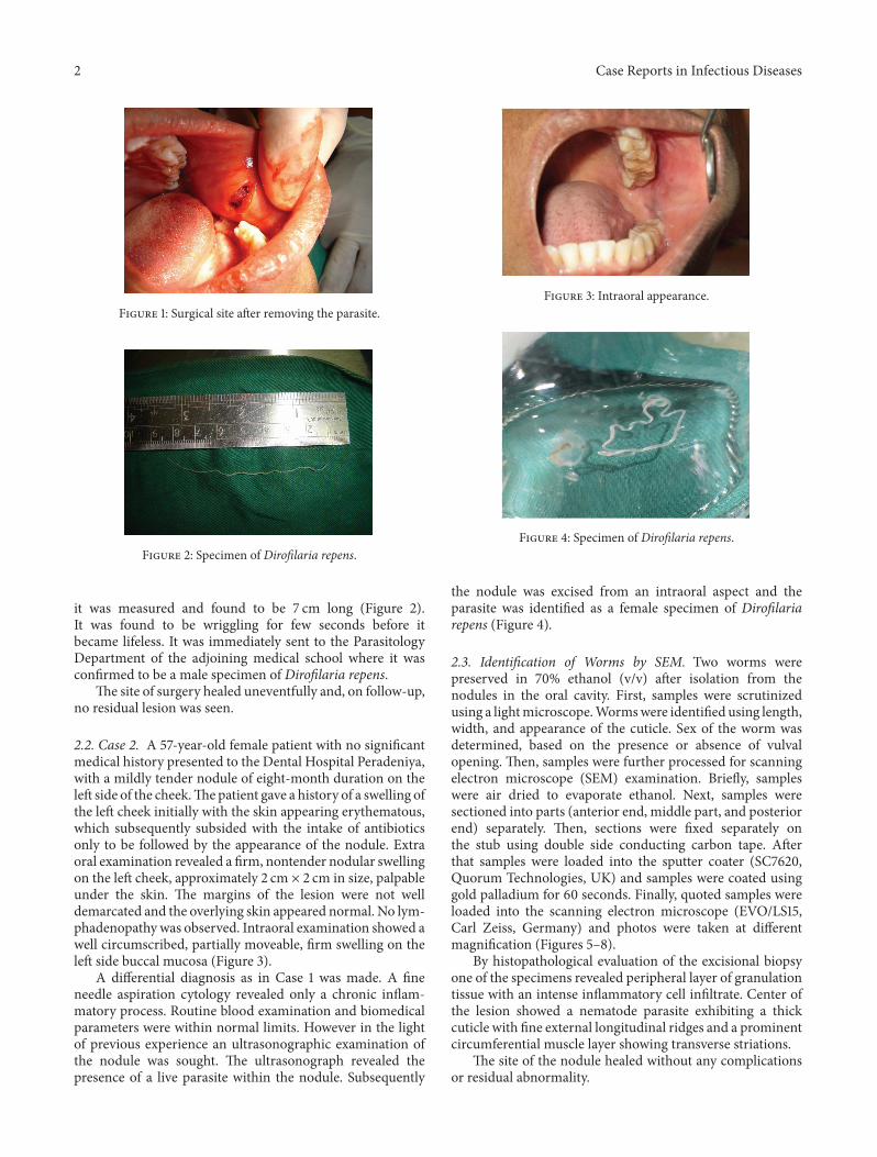
A decision was taken to enucleate the swelling froman intraoral aspect. During the surgery the surface of thenodule was accidentally punctured and a pus-like fluid wasfound to be oozing from the lesion. It was then decided toopen the nodule to drain the remainder of the fluid whena thin ribbon-like object was observed to emerge from thenodule. The remainder of the object was gently evacuated(Figure 1). On becoming apparent the object was a worm
Hindawi Publishing CorporationCase Reports in Infectious DiseasesVolume 2015, Article ID 648278, 4 pageshttp://dx.doi.org/10.1155/2015/648278
2 Case Reports in Infectious Diseases
Figure 1: Surgical site after removing the parasite.
Figure 2: Specimen of Dirofilaria repens.
it was measured and found to be 7 cm long (Figure 2).It was found to be wriggling for few seconds before itbecame lifeless. It was immediately sent to the ParasitologyDepartment of the adjoining medical school where it wasconfirmed to be a male specimen of Dirofilaria repens.
The site of surgery healed uneventfully and, on follow-up,no residual lesion was seen.
2.2. Case 2. A 57-year-old female patient with no significantmedical history presented to the Dental Hospital Peradeniya,with a mildly tender nodule of eight-month duration on theleft side of the cheek.Thepatient gave a history of a swelling ofthe left cheek initially with the skin appearing erythematous,which subsequently subsided with the intake of antibioticsonly to be followed by the appearance of the nodule. Extraoral examination revealed a firm, nontender nodular swellingon the left cheek, approximately 2 cm × 2 cm in size, palpableunder the skin. The margins of the lesion were not welldemarcated and the overlying skin appeared normal. No lym-phadenopathy was observed. Intraoral examination showed awell circumscribed, partially moveable, firm swelling on theleft side buccal mucosa (Figure 3).
A differential diagnosis as in Case 1 was made. A fineneedle aspiration cytology revealed only a chronic inflam-matory process. Routine blood examination and biomedicalparameters were within normal limits. However in the lightof previous experience an ultrasonographic examination ofthe nodule was sought. The ultrasonograph revealed thepresence of a live parasite within the nodule. Subsequently
Figure 3: Intraoral appearance.
Figure 4: Specimen of Dirofilaria repens.
the nodule was excised from an intraoral aspect and theparasite was identified as a female specimen of Dirofilariarepens (Figure 4).
2.3. Identification of Worms by SEM. Two worms werepreserved in 70% ethanol (v/v) after isolation from thenodules in the oral cavity. First, samples were scrutinizedusing a lightmicroscope.Wormswere identified using length,width, and appearance of the cuticle. Sex of the worm wasdetermined, based on the presence or absence of vulvalopening. Then, samples were further processed for scanningelectron microscope (SEM) examination. Briefly, sampleswere air dried to evaporate ethanol. Next, samples weresectioned into parts (anterior end, middle part, and posteriorend) separately. Then, sections were fixed separately onthe stub using double side conducting carbon tape. Afterthat samples were loaded into the sputter coater (SC7620,Quorum Technologies, UK) and samples were coated usinggold palladium for 60 seconds. Finally, quoted samples wereloaded into the scanning electron microscope (EVO/LS15,Carl Zeiss, Germany) and photos were taken at differentmagnification (Figures 5–8).
By histopathological evaluation of the excisional biopsyone of the specimens revealed peripheral layer of granulationtissue with an intense inflammatory cell infiltrate. Center ofthe lesion showed a nematode parasite exhibiting a thickcuticle with fine external longitudinal ridges and a prominentcircumferential muscle layer showing transverse striations.
The site of the nodule healed without any complicationsor residual abnormality.

Case Reports in Infectious Diseases 3
Figure 5: Anterior end of the worm showing thick longitudinalridges and oral cavity.
Figure 6: Posterior end of the worm showing thick longitudinalridges.
3. Discussion
Dirofilariasis is a parasite infection of animals and rarelyhuman with nematodes of the genus Dirofilaria. Dogs,monkeys, and cats are the primary host and mosquitoessuch as Mansonia uniformis, Mansonia annulifera, and Adesaegypti are considered potential vectors in Sri Lanka[4]. In humans, two types of diseases, namely, pulmonarydirofilariasis, primarily caused by Dirofilaria immitis, andsubcutaneous dirofilariasis, primarily caused by Dirofilariatenuis and Dirofilaria repens, can be seen. In Sri Lanka, theonly species that is responsible for human dirofilariasis isDirofilaria repens [1, 4].
The parasite was first recognized by Railliet and Henryin 1911 from a dog and Skrjabin (1917) had described ahuman case under the name of Loa extraocularis. Skrjabinet al. (1930) attributed a second human case to Dirofilariarepens and Skrjabin and Schikhobalova (1948) recognizedLoa extraocularis as a synonym of Dirofilaria repens [5, 6].
Prevalence of this disease is increasing and emerging as asignificant health problem in different areas of the world.Thedisease is said to be commonly encountered in the 4th to 5thdecade of life while showing significant female predilection.Our first case was found in a young male. Significantgeographical variation was observed as an endemic in Indiaand Sri Lanka other than Africa, Asia, Australia, Europe, andAmerica [7]. A literature search showed that Tilakaratne and
Figure 7: Cuticle of the worm (middle part) showing transversestriations and longitudinal ridges.
Figure 8: A cross section of the worm showing cuticular ridges andthick musculature.
Pitakotuwage (2003) had published a series of seven cases ofD. repens infection in Sri Lankan patients [8].
The parasite Dirofilaria repens is composed of thickexternal cuticula with longitudinal ridges (microruffling ofthe surface), leading to a cogwheel-like appearance in atransverse section. Large lateral chords with tall, slendercoelomian muscle layer and a single gut tube is also presentin this group of parasites. In humans, nodules can occurin any part of the body especially those that are likely sitesfor mosquito bite. A study of the aetiopathogenesis showsthat the adult nematode lives in subcutaneous tissues of theirnatural hosts and attain full size and deposit microfilaria inblood. Vector arthropods ingest the first stage of larva (L1)while feeding on an infected host and L1 microfilaria developto infective third stage (L3) and migrate to the proboscis.Transmission occurs when the mosquito vector carrying theinfective third stage lava (L3) penetrates a new host, whichcan be either a human or a natural host. Since the humansare a dead-end host, the nematode in humans does notreach sexual maturity and remains nonfertile. Consequentlymicrofilaria are not released into the peripheral blood inhumans and, due to the presence of an adult nematode inthe subcutaneous tissue, chronic inflammatory infiltrationcan occur in the surrounding tissue forming a parasiticgranuloma [1, 2, 7, 9].
Clinical features can be seen as single nontender subcu-taneous nodule. Mostly the patients are asymptomatic and

4 Case Reports in Infectious Diseases
no particular sensation is attributable to the insect bite.Subsequent formation of subcutaneous or subconjunctivalsuppurative nodules after 2–12 months can be encountered,or very rarely satellite lymphadenopathies with hyperpyrexiacan occur. In the eyes it can cause detached retina, crystallineopacity, glaucoma, uveitis, episcleritis and limited loss ofvision. Subcutaneous migration of parasite in tissues of headcan cause trigeminal neuralgia [1, 7, 9].
Hematological investigations such as FBC can showperipheral hypereosinophilia as this process has a chronicinflammatory process. Radiological investigations such ascomputed tomography scan andmagnetic resonance imagingmay be useful. In this case ultrasonographic imaging showeda worm-like structure that displayed movement inside thecyst and contributed to the diagnosis very significantly. ColorDoppler imaging also may be useful in the diagnosis ofthe disease. Serological investigations such as measuring thereduction in the level of anti-D. repens serous antibodiesby immunoenzymatic means for 3–6 months from the timeof surgery can be used in the follow-up care of patients.Investigations like Giemsa stain and fine needle aspirationcytology followed by excisional biopsy are useful in confirm-ing the diagnosis. Once the excision of the lesion is performedparasitological evaluation can be performed for the wormspecimen [1, 9].
As humans are the end hosts, surgical excision of theworm is the appropriate management. Human dirofilaria-sis is usually regarded as an infection by a single worm.Oral therapy with diethylcarbamazine (DEC) has also beenrecommended to destroy occult worms [10]. The diagnosticdilemma with subcutaneous/submucosal nodular swellingsof the oral cavity is evident from previous reports [11].Tilakaratne and Pitakotuwage in their series of seven casesgave a variety of differential diagnosis for submucosal noduleswithout inflammatory signs. The various differential diag-noses given were fibroepithelial polyp, adenoma, salivarygland hyperplasia, lipoma, calcified lymph node, organizedmucocele, and tuberculosis [8].
4. Conclusion
Even though subcutaneous dirofilariasis is rare, it can beincluded in the differential diagnosis, when patient presentswith a firm submucosal nodule without overlying inflam-matory signs which does not completely respond to routinetherapy, especially in patients from endemic areas like SriLanka.
Conflict of Interests
The authors declare that there is no conflict of interestsregarding the publication of this paper.
References
[1] S. Pampiglione and F. Rivasi, “Human dirofilariasis due toDirofilaria (Nochtiella) repens: an update of world literaturefrom 1995 to 2000,” Parassitologia, vol. 42, no. 3-4, pp. 231–254,2000.
[2] A. Muro, C. Genchi, M. Cordero, and F. Simon, “Human dirofi-lariasis in the European union,” Parasitology Today, vol. 15, no.9, pp. 386–389, 1999.
[3] S. Pampiglione, F. Rivasi, G. Angeli et al., “Dirofilariasis due toDirofilaria repens in Italy, an emergent zoonosis: report of 60new cases,” Histopathology, vol. 38, no. 4, pp. 344–354, 2001.
[4] A. S. Dissanaike, W. Abeyewickreme, M. D. Wijesundera, M. V.Weerasooriya, and M. M. Ismail, “Human dirofilariasis causedby Dirofilaria (Nochtiella) repens in Sri Lanka,” Parassitologia,vol. 39, no. 4, pp. 375–382, 1997.
[5] D. Otranto, E. Brianti, G. Gaglio, F. Dantas-Torres, S. Azzaro,and S. Giannetto, “Short report: human ocular infection withDirofilaria repens (Railliet and Henry, 1911) in an area endemicfor canine dirofilariasis,” The American Journal of TropicalMedicine and Hygiene, vol. 84, no. 6, pp. 1002–1004, 2011.
[6] S. Phantana, C. Shutidamrong, and W. Chusattayanend, “Diro-filaria immitis (Leidy, 1856) Railliet and Henry, 1911,” in FelineClinical Parasitology, vol. 8, p. 331, John Wiley & Sons, 2008.
[7] T. Jelinek, J. Schulte-Hillen, and T. Loscher, “Human dirofilar-iasis,” International Journal of Dermatology, vol. 35, no. 12, pp.872–875, 1996.
[8] W. M. Tilakaratne and T. N. Pitakotuwage, “Intra−oral Diro-filariarepens infection: report of seven cases,” Journal of OralPathology and Medicine, vol. 32, no. 8, pp. 502–505, 2003.
[9] L. L. Pereira, R. D. Coletta, L. C. Monteiro, V. Y. N. Ferreira, J.E. Leon, and P. R. F. Bonan, “Dirofilariasis involving the oralcavity: report of the first case from South America,” Revista daSociedade Brasileira deMedicina Tropical, vol. 48, no. 3, pp. 361–363, 2015.
[10] J.-J. Gicquel, J. Berthonneau, L. Curutchet, B. Hue, and P.Dighiero, “Management of subconjunctival Dirofilaria repens,”Archives of Ophthalmology, vol. 122, no. 3, p. 416, 2004.
[11] E.W. H. To,W.M. Tsang, and K. F. Chan, “Human dirofilariasisof the buccal mucosa: a case report,” International Journal ofOral and Maxillofacial Surgery, vol. 32, no. 1, pp. 104–106, 2003.