a case report of intracholecystic papillary neoplasm of
TRANSCRIPT

CASE REPORT Open Access
A case report of intracholecystic papillaryneoplasm of the gallbladder resembling asubmucosal tumorRyo Muranushi1*, Hideyuki Saito2, Asuka Matsumoto2, Toshihide Kato2, Naritaka Tanaka2, Kenji Nakazato2,Nobuhiro Morinaga2, Yoshinori Shitara2, Masatoshi Ishizaki2, Takatomo Yoshida3, Shinichi Aishima4
and Ken Shirabe1
Abstract
Background: Intracholecystic papillary neoplasm (ICPN) is defined as papillary tumors detected macroscopically inthe gallbladder. We report a case of ICPN which exhibited the atypical form like a submucosal tumor.
Case presentation: A 70-year-old man was admitted to our hospital because of hepatic disorder. Computedtomography and magnetic resonance imaging showed irregular thickening of the wall within the gallbladderfundus. Because the lesion might have been malignant, we performed laparoscopic cholecystectomy and liverbed resection. Macroscopic findings showed the mucosal surface of the tumor was smooth, and its form wassimilar to that of a submucosal tumor. Histopathological examination revealed papillary tumors within themass with low-grade dysplasia; therefore, we diagnosed ICPN.
Conclusion: In the present case, ICPN was resembling a submucosal tumor macroscopically because the tumorsarose into the Rokitansky-Aschoff sinus and the adenomyomatous hyperplasia was merged with the ICPN. It isnecessary to consider the possibility of tumor lesions within adenomyomatous hyperplasia.
Keywords: Intracholecystic papillary neoplasm, Adenomyomatous hyperplasia, Laparoscopic cholecystectomy
BackgroundIntracholecystic papillary neoplasm (ICPN) is preinva-sive neoplastic lesions characterized by papillary growthin the gallbladder. ICPN is defined as gallbladder lesionsof intraductal papillary neoplasm of the bile duct (IPNB).IPNB is a premalignant lesion of the biliary tract and iscounterpart of intraductal papillary-mucinous neoplasm(IPMN) in the pancreatic duct epithelium [1]. ICPN is apapillary tumor generally detected macroscopically andis sometimes diagnosed by imaging findings. Herein, wereport a case of ICPN which exhibited atypical form andwhich was distinguished difficultly from gallbladderadenocarcinoma.Through this case, we consider clinicopathological
characteristics and therapeutic strategies of ICPN.
Case presentationThe patient was a 70-year-old man. He was admitted toour hospital because of a hepatic disorder that was dis-covered during a routine health examination. Blood testsshowed aspartate aminotransferase 48 U/L (normalrange, 13 to 33 U/L), alanine phosphatase 66 U/L (nor-mal range, 8.0 to 42 U/L), alkaline phosphatase 263 U/L(normal range, 115 to 359 U/L), gamma-glutamyl trans-peptidase 100 (normal range, 10 to 47 IU/L), total biliru-bin 0.5 mg/dL (normal range, 0.2 to 1.2 mg/dL),carcinoembryonic antigen 4.4 ng/mL (normal range, <5.0 ng/ml), and carbohydrate antigen 19-9 10.4 U/mL(normal range, < 15 U/mL). Abdominal ultrasonographyshowed an 8 × 7-mm solid mass at the gallbladder fun-dus and several stones in the gallbladder (Fig. 1a). En-hanced computed tomography (CT) showed thatirregular wall thickening at the gallbladder fundus andthe boundary between tumor and the liver was indistinct(Fig. 1b). T2-weighted magnetic resonance imaging
* Correspondence: [email protected] of Hepatobiliary and Pancreatic Surgery, Gunma UniversityGraduate School of Medicine, Gunma University, 3-39-15 Showa-Machi,Maebashi, Gunma 371-8511, JapanFull list of author information is available at the end of the article
© The Author(s). 2018 Open Access This article is distributed under the terms of the Creative Commons Attribution 4.0International License (http://creativecommons.org/licenses/by/4.0/), which permits unrestricted use, distribution, andreproduction in any medium, provided you give appropriate credit to the original author(s) and the source, provide a link tothe Creative Commons license, and indicate if changes were made.
Muranushi et al. Surgical Case Reports (2018) 4:124 https://doi.org/10.1186/s40792-018-0524-2

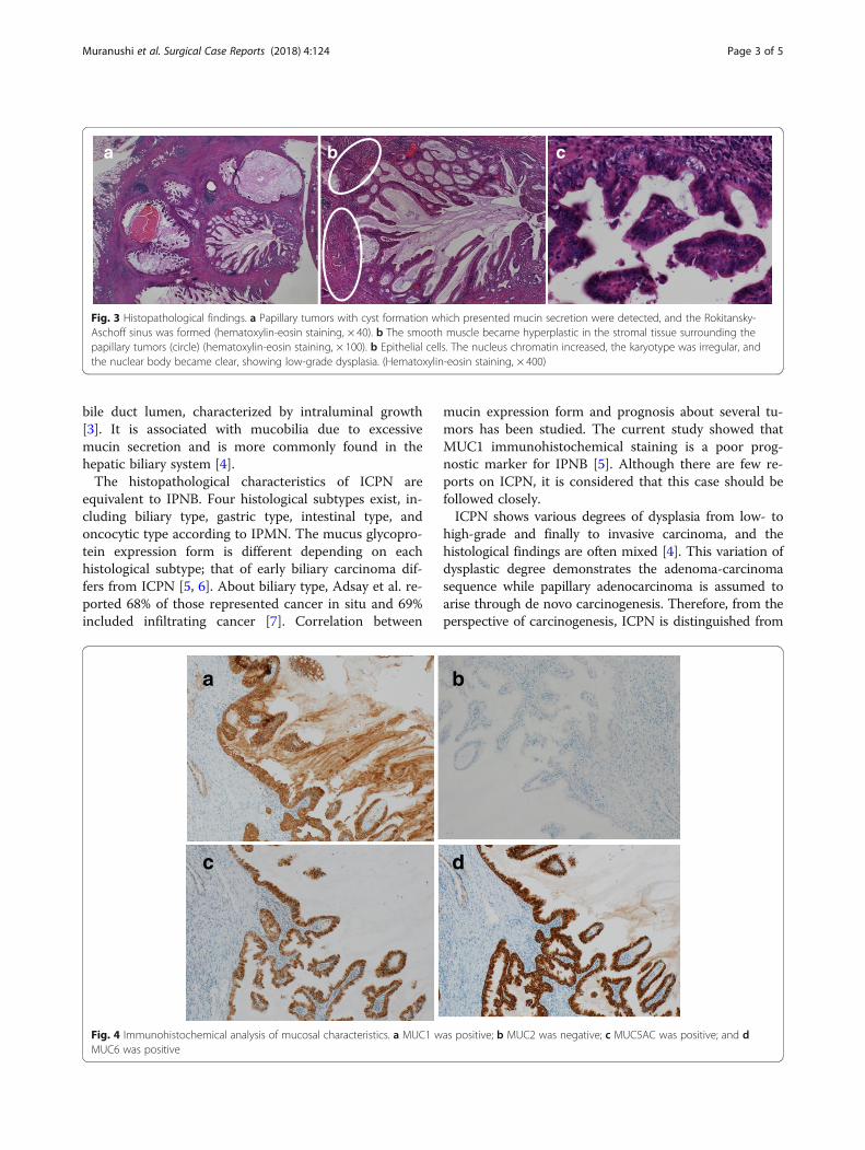
(MRI) showed a high-intensity nodule inside the thick-ened wall at the gallbladder fundus (Fig. 1c). Accordingto these findings, we diagnosed the lesion as suspiciousof malignancy and decided to perform surgery. Duringsurgery, a tumor of approximately10 mm was found atthe gallbladder fundus and color change of the liver bedfloor adjacent to the tumor was detected. We performedlaparoscopic cholecystectomy and liver bed resection.The macroscopic findings of the resected specimenshowed a 15 × 10-mm milky yellow mass at the gallblad-der fundus, and its cut surface showed papillary lesions(Fig. 2). The tumor mucosal surface was smooth, and itsform was similar to that of a submucosal tumor. Histo-pathological findings showed papillary tumors with cystformation, and the tumors represented mucin secretion(Fig. 3a). Additionally, the Rokitansky-Aschoff sinus(RAS) was formed, and the smooth muscle becamehyperplasia in the stromal tissue surrounding the papil-lary tumors (Fig. 3a, b). Within the epithelial cells, nu-cleus chromatin increased, karyotype was irregular, andnuclear body became clear. There was no invasion intothe stromal tissue. These findings demonstrated ICPN
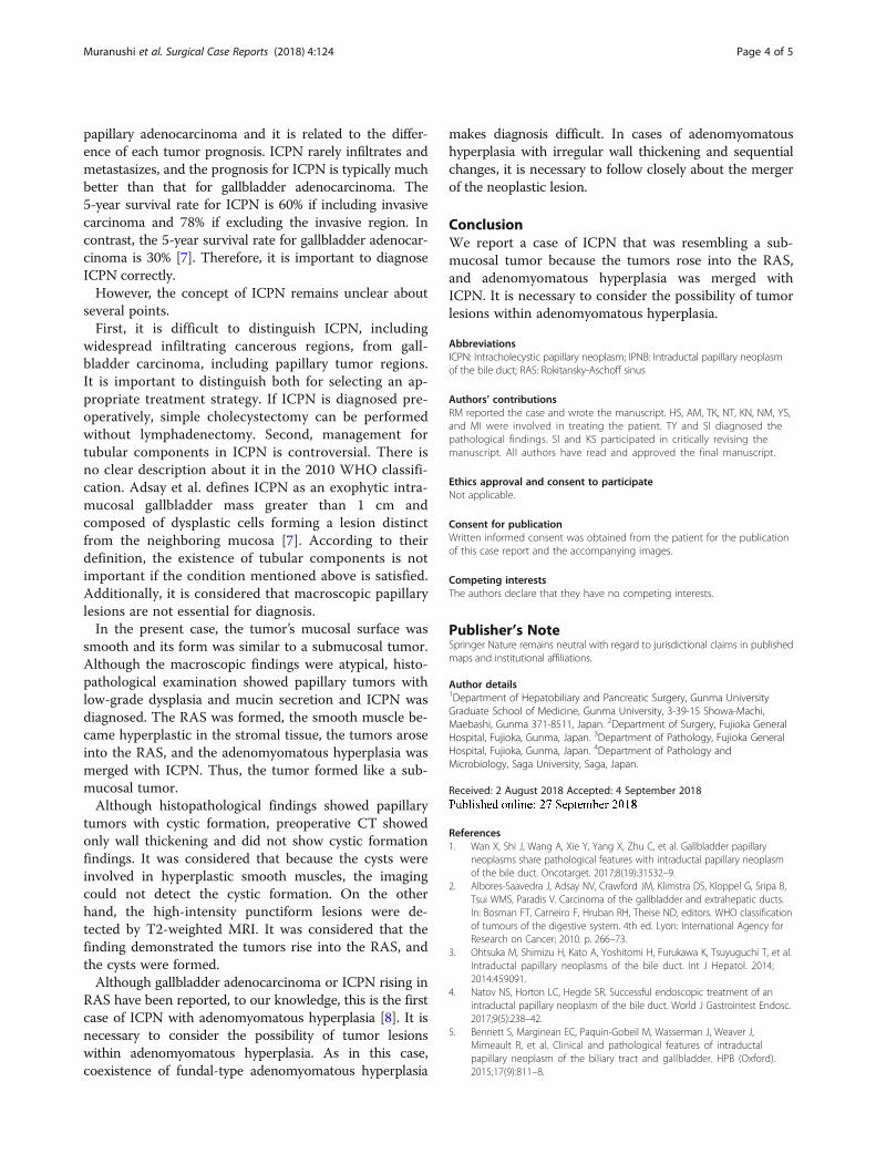
with low-grade dysplasia (Fig. 3c). There was neitherdysplasia nor biliary intraepithelial neoplasia on thebackground mucosa. Immunohistochemical analysis ofthe mucosal characteristics showed that MUC1,MUC5AC, and MUC6 were positive, whereas MUC2was negative (Fig. 4). According to the predominant pat-tern on morphology and the mucin expression form, itwas diagnosed as biliary type. Additionally, because Ki67index was a little less than 10%, it was denied that thetumor was malignant. The postoperative course wasgood, and the patient was discharged 9 days after the op-eration. A recurrence has not been detected for3.5 years.
DiscussionICPN was first described as gallbladder lesions of IPNBin the 2010 WHO classification and was classified aspremalignant lesions of biliary system in the same cat-egory as adenoma, biliary intraepithelial neoplasia, andmucinous cystic neoplasm [2]. IPNB is defined as biliarytumors with an exophytic nature exhibiting papillarymass which can be detected macroscopically within the
a b c
Fig. 1 Preoperative imaging findings. a Abdominal ultrasonography showed an 8 × 7-mm solid mass at the gallbladder fundus. b Enhanced CTshowed that irregular wall thickening at the gallbladder fundus, and the boundary between the tumor and liver was indistinct. c T2-weightedMRI showed high-intensity nodules inside the thickened wall of the gallbladder
Fig. 2 Photograph of resected specimen. It shows the gallbladder fundus on the left side and the cystic duct on the right side. A 15 × 10-mmmass like a submucosal tumor is visible within the gallbladder fundus (arrow), and its cut surface shows the papillary lesions
Muranushi et al. Surgical Case Reports (2018) 4:124 Page 2 of 5
bile duct lumen, characterized by intraluminal growth[3]. It is associated with mucobilia due to excessivemucin secretion and is more commonly found in thehepatic biliary system [4].The histopathological characteristics of ICPN are
equivalent to IPNB. Four histological subtypes exist, in-cluding biliary type, gastric type, intestinal type, andoncocytic type according to IPMN. The mucus glycopro-tein expression form is different depending on eachhistological subtype; that of early biliary carcinoma dif-fers from ICPN [5, 6]. About biliary type, Adsay et al. re-ported 68% of those represented cancer in situ and 69%included infiltrating cancer [7]. Correlation between
mucin expression form and prognosis about several tu-mors has been studied. The current study showed thatMUC1 immunohistochemical staining is a poor prog-nostic marker for IPNB [5]. Although there are few re-ports on ICPN, it is considered that this case should befollowed closely.ICPN shows various degrees of dysplasia from low- to
high-grade and finally to invasive carcinoma, and thehistological findings are often mixed [4]. This variation ofdysplastic degree demonstrates the adenoma-carcinomasequence while papillary adenocarcinoma is assumed toarise through de novo carcinogenesis. Therefore, from theperspective of carcinogenesis, ICPN is distinguished from
a b c
Fig. 3 Histopathological findings. a Papillary tumors with cyst formation which presented mucin secretion were detected, and the Rokitansky-Aschoff sinus was formed (hematoxylin-eosin staining, × 40). b The smooth muscle became hyperplastic in the stromal tissue surrounding thepapillary tumors (circle) (hematoxylin-eosin staining, × 100). b Epithelial cells. The nucleus chromatin increased, the karyotype was irregular, andthe nuclear body became clear, showing low-grade dysplasia. (Hematoxylin-eosin staining, × 400)
a b
c d
Fig. 4 Immunohistochemical analysis of mucosal characteristics. a MUC1 was positive; b MUC2 was negative; c MUC5AC was positive; and dMUC6 was positive
Muranushi et al. Surgical Case Reports (2018) 4:124 Page 3 of 5
papillary adenocarcinoma and it is related to the differ-ence of each tumor prognosis. ICPN rarely infiltrates andmetastasizes, and the prognosis for ICPN is typically muchbetter than that for gallbladder adenocarcinoma. The5-year survival rate for ICPN is 60% if including invasivecarcinoma and 78% if excluding the invasive region. Incontrast, the 5-year survival rate for gallbladder adenocar-cinoma is 30% [7]. Therefore, it is important to diagnoseICPN correctly.However, the concept of ICPN remains unclear about
several points.First, it is difficult to distinguish ICPN, including
widespread infiltrating cancerous regions, from gall-bladder carcinoma, including papillary tumor regions.It is important to distinguish both for selecting an ap-propriate treatment strategy. If ICPN is diagnosed pre-operatively, simple cholecystectomy can be performedwithout lymphadenectomy. Second, management fortubular components in ICPN is controversial. There isno clear description about it in the 2010 WHO classifi-cation. Adsay et al. defines ICPN as an exophytic intra-mucosal gallbladder mass greater than 1 cm andcomposed of dysplastic cells forming a lesion distinctfrom the neighboring mucosa [7]. According to theirdefinition, the existence of tubular components is notimportant if the condition mentioned above is satisfied.Additionally, it is considered that macroscopic papillarylesions are not essential for diagnosis.In the present case, the tumor’s mucosal surface was
smooth and its form was similar to a submucosal tumor.Although the macroscopic findings were atypical, histo-pathological examination showed papillary tumors withlow-grade dysplasia and mucin secretion and ICPN wasdiagnosed. The RAS was formed, the smooth muscle be-came hyperplastic in the stromal tissue, the tumors aroseinto the RAS, and the adenomyomatous hyperplasia wasmerged with ICPN. Thus, the tumor formed like a sub-mucosal tumor.Although histopathological findings showed papillary
tumors with cystic formation, preoperative CT showedonly wall thickening and did not show cystic formationfindings. It was considered that because the cysts wereinvolved in hyperplastic smooth muscles, the imagingcould not detect the cystic formation. On the otherhand, the high-intensity punctiform lesions were de-tected by T2-weighted MRI. It was considered that thefinding demonstrated the tumors rise into the RAS, andthe cysts were formed.Although gallbladder adenocarcinoma or ICPN rising in
RAS have been reported, to our knowledge, this is the firstcase of ICPN with adenomyomatous hyperplasia [8]. It isnecessary to consider the possibility of tumor lesionswithin adenomyomatous hyperplasia. As in this case,coexistence of fundal-type adenomyomatous hyperplasia
makes diagnosis difficult. In cases of adenomyomatoushyperplasia with irregular wall thickening and sequentialchanges, it is necessary to follow closely about the mergerof the neoplastic lesion.
ConclusionWe report a case of ICPN that was resembling a sub-mucosal tumor because the tumors rose into the RAS,and adenomyomatous hyperplasia was merged withICPN. It is necessary to consider the possibility of tumorlesions within adenomyomatous hyperplasia.
AbbreviationsICPN: Intracholecystic papillary neoplasm; IPNB: Intraductal papillary neoplasmof the bile duct; RAS: Rokitansky-Aschoff sinus
Authors’ contributionsRM reported the case and wrote the manuscript. HS, AM, TK, NT, KN, NM, YS,and MI were involved in treating the patient. TY and SI diagnosed thepathological findings. SI and KS participated in critically revising themanuscript. All authors have read and approved the final manuscript.
Ethics approval and consent to participateNot applicable.
Consent for publicationWritten informed consent was obtained from the patient for the publicationof this case report and the accompanying images.
Competing interestsThe authors declare that they have no competing interests.
Publisher’s NoteSpringer Nature remains neutral with regard to jurisdictional claims in publishedmaps and institutional affiliations.
Author details1Department of Hepatobiliary and Pancreatic Surgery, Gunma UniversityGraduate School of Medicine, Gunma University, 3-39-15 Showa-Machi,Maebashi, Gunma 371-8511, Japan. 2Department of Surgery, Fujioka GeneralHospital, Fujioka, Gunma, Japan. 3Department of Pathology, Fujioka GeneralHospital, Fujioka, Gunma, Japan. 4Department of Pathology andMicrobiology, Saga University, Saga, Japan.
Received: 2 August 2018 Accepted: 4 September 2018
References1. Wan X, Shi J, Wang A, Xie Y, Yang X, Zhu C, et al. Gallbladder papillary
neoplasms share pathological features with intraductal papillary neoplasmof the bile duct. Oncotarget. 2017;8(19):31532–9.
2. Albores-Saavedra J, Adsay NV, Crawford JM, Klimstra DS, Kloppel G, Sripa B,Tsui WMS, Paradis V. Carcinoma of the gallbladder and extrahepatic ducts.In: Bosman FT, Carneiro F, Hruban RH, Theise ND, editors. WHO classificationof tumours of the digestive system. 4th ed. Lyon: International Agency forResearch on Cancer; 2010. p. 266–73.
3. Ohtsuka M, Shimizu H, Kato A, Yoshitomi H, Furukawa K, Tsuyuguchi T, et al.Intraductal papillary neoplasms of the bile duct. Int J Hepatol. 2014;2014:459091.
4. Natov NS, Horton LC, Hegde SR. Successful endoscopic treatment of anintraductal papillary neoplasm of the bile duct. World J Gastrointest Endosc.2017;9(5):238–42.
5. Bennett S, Marginean EC, Paquin-Gobeil M, Wasserman J, Weaver J,Mimeault R, et al. Clinical and pathological features of intraductalpapillary neoplasm of the biliary tract and gallbladder. HPB (Oxford).2015;17(9):811–8.
Muranushi et al. Surgical Case Reports (2018) 4:124 Page 4 of 5
6. Park SY, Roh SJ, Kim YN, Kim SZ, Park HS, Jang KY, et al. Expression of MUC1,MUC2, MUC5AC and MUC6 in cholangiocarcinoma: prognostic impact.Oncol Rep. 2009;22(3):649–57.
7. Adsay V, Jang KT, Roa JC, Dursun N, Ohike N, Bagci P, et al. Intracholecysticpapillary-tubular neoplasms (ICPN) of the gallbladder (neoplastic polyps,adenomas, and papillary neoplasms that are ≥1.0 cm): clinicopathologicand immunohistochemical analysis of 123 cases. Am J Surg Pathol. 2012;36(9):1279–301.
8. Sato R, Ando T, Tateno H, Rikiyama T, Furukawa T, Ebina N. Intracysticpapillary neoplasm with an associated mucinous adenocarcinoma arising inRokitansky-Aschoff sinus of the gallbladder. Surg Case Rep. 2016;2(1):62.
Muranushi et al. Surgical Case Reports (2018) 4:124 Page 5 of 5