a case of variant creutzfeldt-jakob disease in romania · variant creutzfeld-jakob disease (vcjd)...
TRANSCRIPT
Rom J Leg Med [23] 157-161 [2015]DOI: 10.4323/rjlm.2015.157© 2015 Romanian Society of Legal Medicine
157
A case of variant Creutzfeldt-Jakob disease in Romania
Mihai Ceauşu1, Corneliu Octavian Capatina2, Sorin Hostiuc3,*, Dan Dermengiu3
_________________________________________________________________________________________ Abstract: In humans prion diseases can occur sporadically, through genetic mutations, or can be transmitted from animal, human (kuru disease), or iatrogenic sources. Even though transmissible forms are the most well-known, the sporadic and heritable forms are much more frequent, accounting for about 85% of all cases. The purpose of this case report is to present an atypical variant CJD in a 26 years-old woman. The patient died secondary to infectious complications caused by an acute overdose with alcohol, beta-blockers and oral anti-diabetic drugs, and had an atypical neuropathology pattern, with absent amyloid plaques, but present focal, perivascular deposits of amyloid precursor protein and a positive immunohistochemical reaction for prp. Key Words: variant Creutzfeldt-Jakob disease, Romania, amyloid precursor protein.
1) (a) “Carol Davila” University of Medicine and Pharmacy, Dept. of Pathology; (b) “Mina Minovici” National Institute of Legal Medicine, Dept. of Pathology, Bucharest, Romania2) “Mina Minovici” National Institute of Legal Medicine, Dept. of Forensic Pathology, Bucharest, Romania3) (a) “Carol Davila” University of Medicine and Pharmacy, Dept. of Legal Medicine and Bioethics; (b) “Mina Minovici” National Institute of Legal Medicine, Dept. of Forensic Pathology, Bucharest, Romania* Corresponding author: Sos. Vitan Barzesti 9, 042122, Sector 4, Bucharest, Romania, Tel.: 40-723-791072, Email: [email protected]; [email protected]
In humans prion diseases can occur sporadically, through genetic mutations, or can transmitted
from animal, human (kuru disease), or iatrogenic sources. Even though transmissible forms are the most well-known, the sporadic and heritable forms are much more frequent, accounting for about 85% of all cases [1]. Variant Creutzfeld-Jakob disease (vCJD) appears in younger patients compared to hereditary or sporadic forms (16-39 years), has a longer median course of the disease (13 months compared to four in sporadic CJD) and has a predominance of psychiatric symptoms instead of cerebellar ataxia or progressive dementia [2]. Until 2010 have been identified 219 cases of vCJD, most of them in UK [3]. The triad of microscopic features that characterize the prion disease is composed of: spongiform degeneration of neurons, severe astrocytic gliosis and amyloid plaque formation. The purpose of this case report is to present a
case of CJD in a 26 years old woman.
CAse RepoRt
A 26 years-old woman was admitted about four months before her death in a psychiatry hospital for psychotic behaviors caused by the use of psychoactive drugs (mainly ketamine). She was diagnosed with the code F23.8 (Other psychotic disorders, acute and transitory), F60.8 (Other specific personality disorders – personality with unstable, dysthymic features), and F.19.2 (Past history of substance abuse). About one month before death she was admitted in the toxicology department for toxic encephalopathy caused by an acute intoxication with alcohol, beta-blockers and oral anti-diabetic drugs. The toxicology analysis conducted on gastric lavage was positive. In the ICU the patient was aware and cooperative, stable hemodynamically with a

158
Ceauşu M. et al. Report of a vCJD case in Romania
tendency to bradycardia and hypotension. The second day after admission she entered in a deep coma, with significant opisthotonus, polypnea, but still stable hemodynamically. CT scan did not show anything significant. Neurological examination revealed no signs of meningitis, eyeballs with a left shift, mydriatic pupils, moderately reactive, left hemi-facial myoclonus, poor response to nociceptive stimuli. A lumbar puncture was done, but showed negative results (bacteria or viruses). Tetanus was suspected and was initiated antitetanic therapy and vaccination. After four days a new CT scan was conducted, that showed severe generalized edema, without pathological doses of contrast. Is was recommended a MRI scan, done after three weeks, that showed extensive, symmetric, bilaterally extended lesions in the white mater around the lateral ventricles, and basal nuclei (caudate, putamen, claustrum, globus pallidus), which appeared as hyper intense T2 signal, FLAIR, hypo intense T1 signal, and small areas of hyper intense T1 signal bilaterally in the head of the caudate. The pattern is specific for leukoencephalopathy with necrotico-hemorrhagic lesions. Was suspected a multifocal polymorph leukoencephalopathy, but the CSF, tested for John Cunningham (JC) virus, gave a negative result. It was than raised the suspicion for a prion disease; the ELISA test for 14.3.3 protein was positive on CSF samples. The patient died after another week with iatrogenic pneumonia as the direct cause of dead (she tested positive for MDR Pseudomonas aeruginosa). Autopsy findings Brain had an obvious limit between the white and grey matter. The white substance was smooth, with frequent red spots that disappeared at washing. The brain had a spongy appearance, with a decreased consistency, especially at the occipital (where was also noted an ischemic area), temporal and basal ganglia levels (Fig. 1). The grey mater had an overall decreased thickness (a mean of about 0.3 cm). Lungs showed signs of condensation. Other organs did now show significant lesions.
MAteRiAl And Methods
Histopathology investigation Tissue sampling and stains: Tissue specimens from different parts of the brain and cerebellum were taken for histopathology investigation. These fragments were harvested from the temporal lobes, occipital lobes and basal nuclei. The selected tissue samples were fixed in 10% neutral buffered formalin (pH - 7) for 24–48 hours and paraffin embedded. Sections were cut at 5 μm and stained with standard HE and van Gieson. In addition, special stains, such as Nissl and PAS have been done. Immunohistochemistry: Immunohistochemical analysis (IHC) was done using sections displayed on slides treated first with poly-L-lysine. IHC was performed on 3 μm thick sections from formalin-fixed paraffin-embedded specimens. The method used was an indirect tristadial Avidin-Biotin-Complex technique, with a NovoLink Polymer detection system which utilizes a novel control polymerization technology to prepare polymeric HRP-linker antibody conjugates, according to the manufacturer’s specifications (Novocastra, UK). Briefly, the procedure comprised: deparaffination in toluene and rehydration in alcohol series, washing in phosphate buffer saline (PBS), blocking with normal serum, for 5 min, incubation with primary antibody 60 min, incubation with post-primary block 30 min, then with NovoLink Polymer 30 min. Sections are further incubated with the substrate / chromogen 3,3'-DAB and counterstained with Meyers’ hematoxylin. The antibodies used for IHC were: PrPSc (clone: F89/160.1.5, for research use only, conc: 5 μg/ml, Abcam), Ubiquitin (clone: FPM1, 1:50, Novocastra), APP (clone 3G12, 1:50, Novocastra), GFAP (clone NCL-GA5, 1:100, Novocastra). Antigen retrieval techniques for the antibodies (thermal or enzymatic pretreatment) were done,
Figure 1. Cut-section of the brain showing spongiform degeneration and edema, gross.
Figure 2. Micro-cysts of various shapes and sizes, on a background of edema and congestion, HE, 200x (right).
Romanian Journal of Legal Medicine Vol. XXIII, No 3(2015)
159
according to the producer’s specifications. Both positive and negative controls were used. To ensure the reliability of the experimental study, internal quality control of histopathologic and IHC techniques were performed as a part of an implemented and certified quality assurance system (ISO 9001/2008).All slides were examined and photographed on a Zeiss Axio Imager microscope (Gottingen, Germany). Digital images acquired with Zeiss Axio Vision program have been processed and analyzed with ACDSee Pro Photo Manager (Washington DC), running under Windows Vista.
Results
On multiple serial sections from brain samples we found numerous micro-cysts disseminated in the gray matter, located predominantly in subcortical areas, accompanied by neuronal vacuolar degeneration and gliosis, on a background of congestion and edema (Fig. 2). No inflammatory reaction has been noticed.
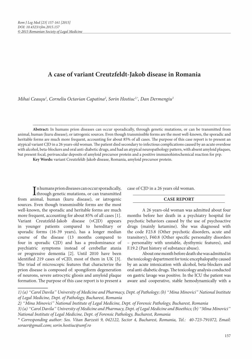
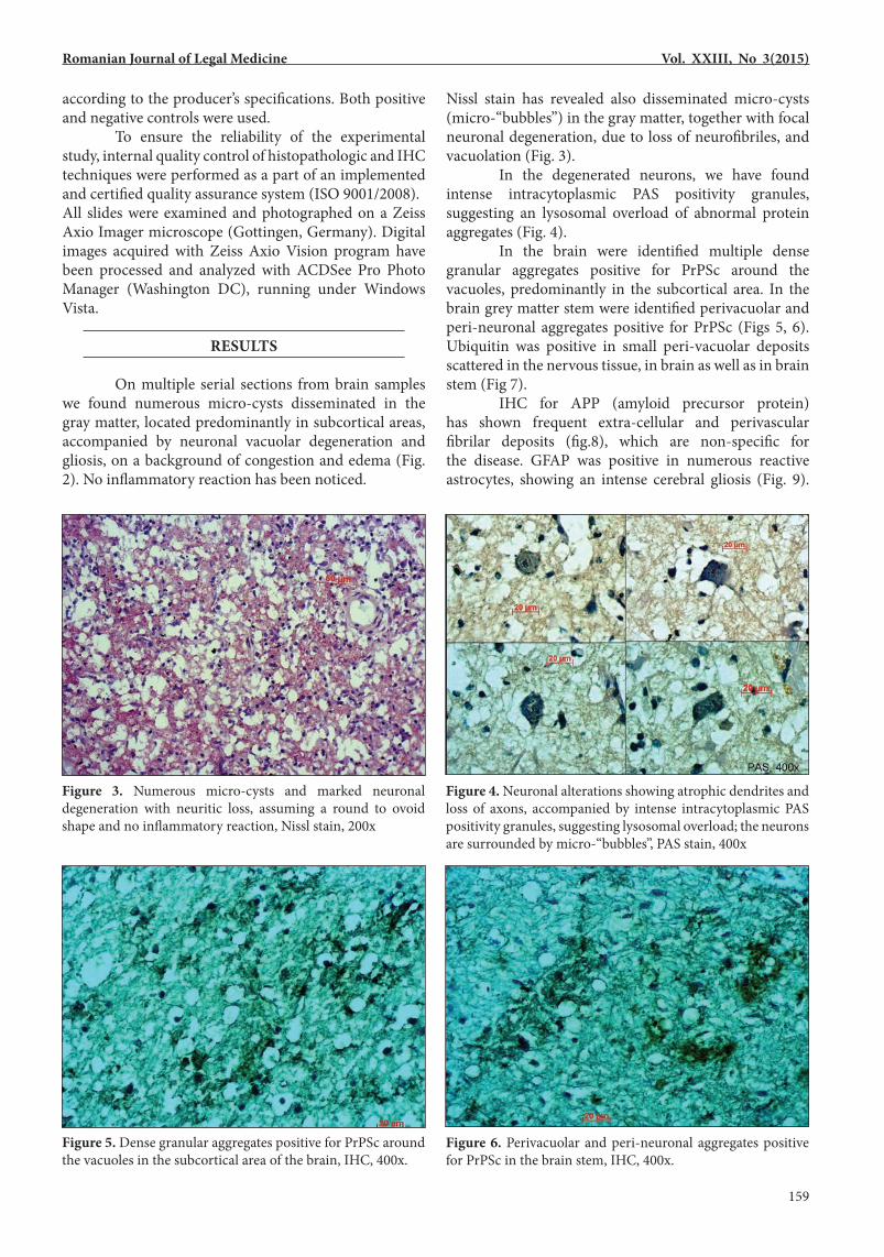
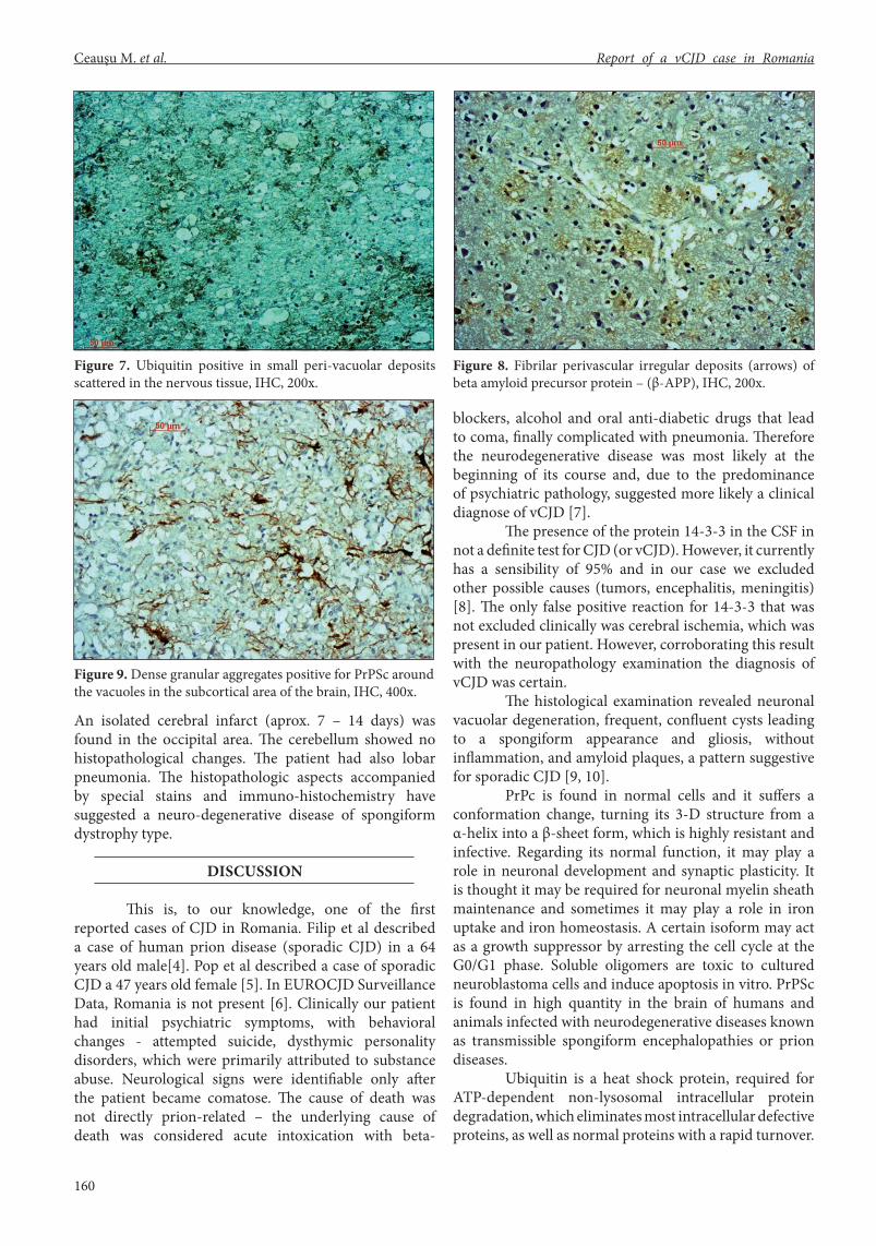
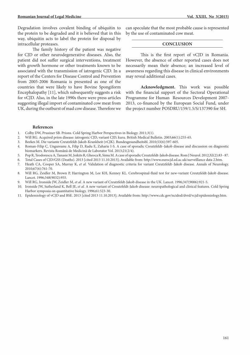
Nissl stain has revealed also disseminated micro-cysts (micro-“bubbles”) in the gray matter, together with focal neuronal degeneration, due to loss of neurofibriles, and vacuolation (Fig. 3). In the degenerated neurons, we have found intense intracytoplasmic PAS positivity granules, suggesting an lysosomal overload of abnormal protein aggregates (Fig. 4). In the brain were identified multiple dense granular aggregates positive for PrPSc around the vacuoles, predominantly in the subcortical area. In the brain grey matter stem were identified perivacuolar and peri-neuronal aggregates positive for PrPSc (Figs 5, 6). Ubiquitin was positive in small peri-vacuolar deposits scattered in the nervous tissue, in brain as well as in brain stem (Fig 7). IHC for APP (amyloid precursor protein) has shown frequent extra-cellular and perivascular fibrilar deposits (fig.8), which are non-specific for the disease. GFAP was positive in numerous reactive astrocytes, showing an intense cerebral gliosis (Fig. 9).
Figure 5. Dense granular aggregates positive for PrPSc around the vacuoles in the subcortical area of the brain, IHC, 400x.
Figure 3. Numerous micro-cysts and marked neuronal degeneration with neuritic loss, assuming a round to ovoid shape and no inflammatory reaction, Nissl stain, 200x
Figure 6. Perivacuolar and peri-neuronal aggregates positive for PrPSc in the brain stem, IHC, 400x.
Figure 4. Neuronal alterations showing atrophic dendrites and loss of axons, accompanied by intense intracytoplasmic PAS positivity granules, suggesting lysosomal overload; the neurons are surrounded by micro-“bubbles”, PAS stain, 400x
160
Ceauşu M. et al. Report of a vCJD case in Romania
An isolated cerebral infarct (aprox. 7 – 14 days) was found in the occipital area. The cerebellum showed no histopathological changes. The patient had also lobar pneumonia. The histopathologic aspects accompanied by special stains and immuno-histochemistry have suggested a neuro-degenerative disease of spongiform dystrophy type.
disCussion
This is, to our knowledge, one of the first reported cases of CJD in Romania. Filip et al described a case of human prion disease (sporadic CJD) in a 64 years old male[4]. Pop et al described a case of sporadic CJD a 47 years old female [5]. In EUROCJD Surveillance Data, Romania is not present [6]. Clinically our patient had initial psychiatric symptoms, with behavioral changes - attempted suicide, dysthymic personality disorders, which were primarily attributed to substance abuse. Neurological signs were identifiable only after the patient became comatose. The cause of death was not directly prion-related – the underlying cause of death was considered acute intoxication with beta-
blockers, alcohol and oral anti-diabetic drugs that lead to coma, finally complicated with pneumonia. Therefore the neurodegenerative disease was most likely at the beginning of its course and, due to the predominance of psychiatric pathology, suggested more likely a clinical diagnose of vCJD [7]. The presence of the protein 14-3-3 in the CSF in not a definite test for CJD (or vCJD). However, it currently has a sensibility of 95% and in our case we excluded other possible causes (tumors, encephalitis, meningitis)[8]. The only false positive reaction for 14-3-3 that was not excluded clinically was cerebral ischemia, which was present in our patient. However, corroborating this result with the neuropathology examination the diagnosis of vCJD was certain. The histological examination revealed neuronal vacuolar degeneration, frequent, confluent cysts leading to a spongiform appearance and gliosis, without inflammation, and amyloid plaques, a pattern suggestive for sporadic CJD [9, 10]. PrPc is found in normal cells and it suffers a conformation change, turning its 3-D structure from a α-helix into a β-sheet form, which is highly resistant and infective. Regarding its normal function, it may play a role in neuronal development and synaptic plasticity. It is thought it may be required for neuronal myelin sheath maintenance and sometimes it may play a role in iron uptake and iron homeostasis. A certain isoform may act as a growth suppressor by arresting the cell cycle at the G0/G1 phase. Soluble oligomers are toxic to cultured neuroblastoma cells and induce apoptosis in vitro. PrPSc is found in high quantity in the brain of humans and animals infected with neurodegenerative diseases known as transmissible spongiform encephalopathies or prion diseases. Ubiquitin is a heat shock protein, required for ATP-dependent non-lysosomal intracellular protein degradation, which eliminates most intracellular defective proteins, as well as normal proteins with a rapid turnover.
Figure 9. Dense granular aggregates positive for PrPSc around the vacuoles in the subcortical area of the brain, IHC, 400x.
Figure 7. Ubiquitin positive in small peri-vacuolar deposits scattered in the nervous tissue, IHC, 200x.
Figure 8. Fibrilar perivascular irregular deposits (arrows) of beta amyloid precursor protein – (β-APP), IHC, 200x.
Romanian Journal of Legal Medicine Vol. XXIII, No 3(2015)
161
Degradation involves covalent binding of ubiquitin to the protein to be degraded and it is believed that in this way, ubiquitin acts to label the protein for disposal by intracellular proteases. The family history of the patient was negative for CJD or other neurodegenerative diseases. Also, the patient did not suffer surgical interventions, treatment with growth hormone or other treatments known to be associated with the transmission of iatrogenic CJD. In a report of the Centers for Disease Control and Prevention from 2005-2006 Romania is presented as one of the countries that were likely to have Bovine Spongiform Encephalopathy [11], which subsequently suggests a risk for vCJD. Also, in the late 1990s there were press articles suggesting illegal import of contaminated cow meat from UK, during the outburst of mad cow disease. Therefore we
can speculate that the most probable cause is represented by the use of contaminated cow meat.
ConClusion
This is the first report of vCJD in Romania. However, the absence of other reported cases does not necessarily mean their absence; an increased level of awareness regarding this disease in clinical environments may reveal additional cases.
Acknowledgment. This work was possible with the financial support of the Sectoral Operational Programme for Human Resources Development 2007-2013, co-financed by the European Social Fund, under the project number POSDRU/159/1.5/S/137390 for SH.
References1. Colby DW, Prusiner SB. Prions. Cold Spring Harbor Perspectives in Biology. 2011;3(1).2. Will RG. Acquired prion disease: iatrogenic CJD, variant CJD, kuru. British Medical Bulletin. 2003;66(1):255-65.3. Beekes M. Die variante Creutzfeldt-Jakob-Krankheit (vCJK). Bundesgesundheitsbl. 2010;53(6):597-605.4. Roman-Filip C, Ungureanu A, Filip D, Radu E, Zaharie I-S. A case of sporadic Creutzfeldt–Jakob disease and discussion on diagnostic
biomarkers. Revista Română de Medicină de Laborator Vol. 2013;21(2/4).5. Pop R, Teodorescu A, Tanasie M, Joikits R, Gheoca R, Simu M. A case of sporadic Creutzfeldt-Jakob disease. Rom J Neurol. 2012;XI(2):83 - 87.6. Total Cases of CJD/GSS (Deaths). 2013 [cited 2013 11.10.2013]; Available from: http://www.eurocjd.ed.ac.uk/surveillance data 2.htm.7. Heath CA, Cooper SA, Murray K, et al. Validation of diagnostic criteria for variant Creutzfeldt–Jakob disease. Annals of Neurology.
2010;67(6):761-70.8. Will RG, Zeidler M, Brown P, Harrington M, Lee KH, Kenney KL. Cerebrospinal-fluid test for new-variant Creutzfeldt-Jakob disease.
Lancet. 1996;348(9032):955.9. Will RG, Ironside JW, Zeidler M, et al. A new variant of Creutzfeldt-Jakob disease in the UK. Lancet. 1996;347(9006):921-5.10. Ironside JW, Sutherland K, Bell JE, et al. A new variant of Creutzfeldt-Jakob disease: neuropathological and clinical features. Cold Spring
Harbor symposia on quantitative biology. 1996;61:523-30.11. Epidemiology of vCJD and BSE. 2013 [cited 2013 11.10.2013]; Available from: http://www.cdc.gov/ncidod/dvrd/vcjd/epidemiology.htm.